Embed Size (px)
Citation preview
Case ReportInternuclear Ophthalmoplegia Secondary to Cocaine Abuse
Richard L. Rabin,1 Azeem Wasay,2 Nicolas Biro,3 and Marcelle Morcos1
1Department of Ophthalmology, Nassau University Medical Center, 2201 Hempstead Turnpike, East Meadow, NY 11554, USA2Lincoln Medical and Mental Health Center, 234 E. 149th St., Bronx, NY 10451, USA3Neuro-Ophthalmology, Department of Ophthalmology, Nassau University Medical Center, 2201 Hempstead Turnpike,East Meadow, NY 11554, USA
Correspondence should be addressed to Richard L. Rabin; [email protected]
Received 28 July 2016; Revised 27 October 2016; Accepted 20 November 2016; Published 6 February 2017
Academic Editor: Winfried M. Amoaku
Copyright © 2017 Richard L. Rabin et al. This is an open access article distributed under the Creative Commons AttributionLicense, which permits unrestricted use, distribution, and reproduction in any medium, provided the original work is properlycited.
Purpose. To report a case of internuclear ophthalmoplegia (INO) caused by cocaine.Method.We report a case of a 54-year-old femalewho presentedwith a left INO three days after snorting cocaine, andwe review the literature.Results.MRI of the brain demonstratedseveral small abnormal foci in the pons on FLAIR and diffusion weighted imaging consistent with ischemic infarction.The patient’ssymptoms remained stable throughout her hospitalization. She was sent to a rehabilitation facility and was lost to follow-up.Conclusion. In cases of extraocular movement abnormalities, it is important to inquire about recreational drug use.
1. Introduction
Internuclear ophthalmoplegia (INO) secondary to cocaineinduced ischemic stroke is a rare event with few reportedcases in the literature. Diaz-Calderon et al. and Strupp et al.report two similar cases; however, neither is identical to ourpatient [1, 2]. The pathogenesis of cocaine induced stroke iscontroversial but likely is secondary to acute hypertension,loss of cerebral vascular autoregulation, cerebral vasospasm,increased platelet activation, and in some cases cardiacthromboembolism [3–6].
2. Case Presentation
A 54-year-old heroin user with a history of hypertensionpresented to the emergency department three days aftersnorting cocaine. She reported leg weakness, unsteadiness,and diplopia that began one day after using cocaine. Shedenied using any other substances at that time and deniedtobacco use.
Her blood pressure on presentation was 117/77 with aheart rate of 77 beats per minute. Visual acuity was 20/60OD and OS. Pinhole acuity was 20/40 OD and 20/60 OS.Autorefraction showed mild myopia. Vision did not improve
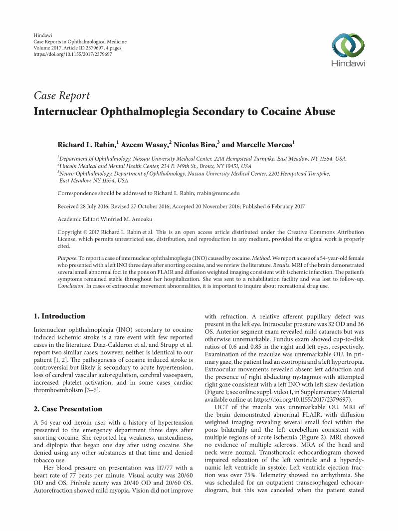
with refraction. A relative afferent pupillary defect waspresent in the left eye. Intraocular pressure was 32 OD and 36OS. Anterior segment exam revealed mild cataracts but wasotherwise unremarkable. Fundus exam showed cup-to-diskratios of 0.6 and 0.85 in the right and left eyes, respectively.Examination of the maculae was unremarkable OU. In pri-mary gaze, the patient had an exotropia and a left hypertropia.Extraocular movements revealed absent left adduction andthe presence of right abducting nystagmus with attemptedright gaze consistent with a left INO with left skew deviation(Figure 1; see online suppl. video 1, in SupplementaryMaterialavailable online at https://doi.org/10.1155/2017/2379697).
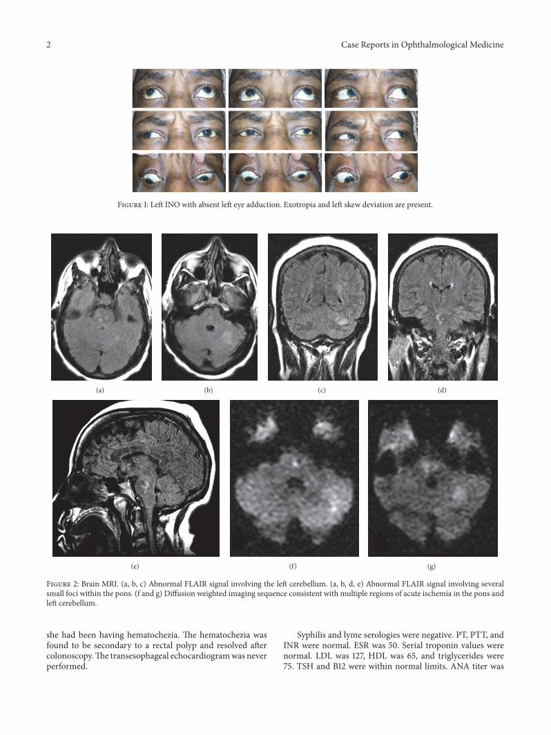
OCT of the macula was unremarkable OU. MRI ofthe brain demonstrated abnormal FLAIR, with diffusionweighted imaging revealing several small foci within thepons bilaterally and the left cerebellum consistent withmultiple regions of acute ischemia (Figure 2). MRI showedno evidence of multiple sclerosis. MRA of the head andneck were normal. Transthoracic echocardiogram showedimpaired relaxation of the left ventricle and a hyperdy-namic left ventricle in systole. Left ventricle ejection frac-tion was over 75%. Telemetry showed no arrhythmia. Shewas scheduled for an outpatient transesophageal echocar-diogram, but this was canceled when the patient stated
HindawiCase Reports in Ophthalmological MedicineVolume 2017, Article ID 2379697, 4 pageshttps://doi.org/10.1155/2017/2379697
2 Case Reports in Ophthalmological Medicine
Figure 1: Left INO with absent left eye adduction. Exotropia and left skew deviation are present.
(a) (b) (c) (d)
(e) (f) (g)
Figure 2: Brain MRI. (a, b, c) Abnormal FLAIR signal involving the left cerebellum. (a, b, d, e) Abnormal FLAIR signal involving severalsmall foci within the pons. (f and g) Diffusion weighted imaging sequence consistent with multiple regions of acute ischemia in the pons andleft cerebellum.
she had been having hematochezia. The hematochezia wasfound to be secondary to a rectal polyp and resolved aftercolonoscopy.The transesophageal echocardiogramwas neverperformed.
Syphilis and lyme serologies were negative. PT, PTT, andINR were normal. ESR was 50. Serial troponin values werenormal. LDL was 127, HDL was 65, and triglycerides were75. TSH and B12 were within normal limits. ANA titer was
Case Reports in Ophthalmological Medicine 3
positive at 40 with a diffuse pattern, and the anti-DS DNAtest was negative.
Neurology consultation was obtained. The patient wasdiagnosed with a left INO with left skew deviation secondaryto cocaine induced stroke. She was also diagnosed withglaucoma and started on glaucoma medications. Symptomsremained stable during her hospitalization. She was sent to arehabilitation facility but was lost to follow-up.
3. Discussion
Internuclear ophthalmoplegia (INO) is a disorder of conju-gate gaze due to a defect in the medial longitudinal fasciculus(MLF). In a literature search for cocaine induced INO, onlytwo case reports and one poster presentation were found.Diaz-Calderon et al. reported a case of cocaine inducedhemorrhagic stroke of the midbrain tegmentum, resulting inan INO [1]. Strupp et al. reported a case of an ischemic strokecausing an INO, but this was following coadministrationof amphetamine and cocaine [2]. Their case demonstrateda single unilateral T2 intense area near the midline of themesencephalon. In contrast, our patient suffered multipleischemic infarctions of the pontine microvasculature as wellas an ischemic infarction of the left cerebellum. We believethat one of the pontine lesions was responsible for the INO.
The MLF is necessary for horizontal conjugate gaze. Forconjugate horizontal eye movements to occur, motor neuronaxons from the abducens nucleus innervate the ipsilaterallateral rectus muscle, causing abduction. The internuclearneurons from the abducens nucleus (the MLF) cross themidline and ascend to the contralateral CN III medialrectus subnucleus activating the medial rectus on that side,causing adduction. InMLF injury, the affected eye (ipsilateralto the site of injury) has an adduction deficit while thecontralateral eye demonstrates abducting nystagmus withfast phase towards the abducting direction [7]. In our case,the left hypertropia was due to a skew deviation. Skewdeviation frequently occurs as a hypertropia on the side ofthe INO. Skew deviation is known to occur with strokes ofthe brainstem or cerebellum [8]. Exotropia in primary gazeis more typical of bilateral INO; however, it may occur inunilateral INO as was seen in our patient [8, 9].
Multiple factors likely contribute to cocaine inducedstroke. Cocaine is a sympathomimetic drug that prevents thereuptake of norepinephrine, dopamine, and serotonin. Thesurge of these neurotransmitters causes tachycardia, vaso-constriction, hypertension, and cerebral vasospasm. Cocainemay also induce a vasculitis [3–6, 10]. The cause of cocaineinduced vasculitis is unknown, but cocaine has been shownto induce apoptosis in cerebrovascular smooth muscle cellsand to increase leukocyte migration across cerebral bloodvessel walls [10]. Because cocaine has a half-life of only onehour, one may expect its effects to wear off quickly. However,its breakdown products can induce vasoconstriction andvasospasm for days [4]. These effects last longer in chronicabusers [11, 12].
There is conflicting evidence as to whether or not cocaineinduces or inhibits platelet aggregation. Togna et al. showedthat platelet responsiveness varied depending on cocaine
concentration and whether platelets were exposed to arachi-donic acid, collagen, or ADP and collagen. Some combi-nations were activating while others were inhibitory [13].Kugelmass et al. concluded that, in vitro, cocaine increasedplatelet activation, but only to significant levels in fewer than50 percent of blood donor samples [14]. In a second article,Kugelmass et al. demonstrated in vivo that platelet activationincreased in dogs upon exposure to cocaine [15]. Rinder etal. measured levels of activated platelets in current cocaineusers and showed that only a small portion of samples hadincreased platelet activation. They also showed that in vitroexposure of blood to concentrations of cocaine documentedas achievable in vivo had no effect on platelet aggregation[16]. Jennings et al. exposed blood in vitro to cocaine andconcluded that cocaine negatively effects platelet aggregationand dissociates preformed platelet aggregates [17].There is noclear explanation for these disparate findings.
Our patient’s presentation of cocaine induced INO dueto ischemic stroke is an infrequent occurrence. A similarcase may present to a neurologist, an ophthalmologist, oran emergency medicine physician. Common causes of INOinclude stroke, multiple sclerosis, and tumor; however, aswith any oculomotor disturbance, it is important to inquireabout recreational drug use [18, 19].
Competing Interests
The authors declare that they have no competing interests.
References
[1] E. Diaz-Calderon, O. H. Del Brutto, R. Aguirre, and T. A.Alarcon, “Bilateral internuclear ophthalmoplegia after smoking”crack” cocaine,” Journal of Clinical Neuro-Ophthalmology, vol.11, no. 4, pp. 297–299, 1991.
[2] M. Strupp, G. F. Hamann, and T. Brandt, “Combined ampheta-mine and cocaine abuse causedmesencephalic ischemia in a 16-year-old boy—due to vasospasm?” European Neurology, vol. 43,no. 3, pp. 181–182, 2000.
[3] N. M. Dabbouseh and A. Ardelt, “Cocaine mediated apoptosisof vascular cells as a mechanism for carotid artery dissectionleading to ischemic stroke,” Medical Hypotheses, vol. 77, no. 2,pp. 201–203, 2011.
[4] A. C. Fonseca and J. M. Ferro, “Drug abuse and stroke,” Currentneurology and neuroscience reports, vol. 13, no. 2, p. 325, 2013.
[5] S. R. Levine, J. C. M. Brust, N. Futrell et al., “Cerebrovascularcomplications of the use of the ‘crack’ form of alkaloidalcocaine,”The New England Journal of Medicine, vol. 323, no. 11,pp. 699–704, 1990.
[6] S. Martin-Schild, K. C. Albright, H. Hallevi et al., “Intracerebralhemorrhage in cocaine users,” Stroke, vol. 41, no. 4, pp. 680–684,2010.
[7] U. Schiefer and H. Wilhelm, Clinical Neuro-Ophthalmology: APractical Guide, Springer, Berlin, Germany, 2007.
[8] R. J. Leigh and D. S. Zee, The Neurology of Eye Movements,Oxford University Press, New York, NY, USA, 3rd edition, 1999.
[9] K. Johkura, Y. Kudo, Y. Amano et al., “Gaze palsy and exotropiain internuclear ophthalmoplegia,” Journal of the NeurologicalSciences, vol. 353, no. 1-2, pp. 158–160, 2015.
4 Case Reports in Ophthalmological Medicine
[10] J. S. Han, D. M. Mandell, J. Poublanc et al., “BOLD-MRI cere-brovascular reactivity findings in cocaine-induced cerebralvasculitis,” Nature Clinical Practice Neurology, vol. 4, no. 11, pp.628–632, 2008.
[11] T. P. Enevoldson, “Recreational drugs and their neurologicalconsequences,” Journal of Neurology, Neurosurgery, and Psychi-atry, vol. 75, supplement 3, pp. iii9–iii15, 2004.
[12] S. D. Treadwell and T. G. Robinson, “Cocaine use and stroke,”Postgraduate Medical Journal, vol. 83, no. 980, pp. 389–394,2007.
[13] G. Togna, E. Tempesta, A. R. Togna, N. Dolci, B. Cebo,and L. Caprino, “Platelet responsiveness and biosynthesis ofthromboxane and prostacyclin in response to in vitro cocainetreatment,” Haemostasis, vol. 15, no. 2, pp. 100–107, 1985.
[14] A. D. Kugelmass, A. Oda, K. Monahan, C. Cabral, and J. A.Ware, “Activation of human platelets by cocaine,” Circulation,vol. 88, no. 3, pp. 876–883, 1993.
[15] A. D. Kugelmass, R. P. Shannon, E. L. Yeo, and J. A. Ware,“Intravenous cocaine induces platelet activation in the con-scious dog,” Circulation, vol. 91, no. 5, pp. 1336–1340, 1995.
[16] H. M. Rinder, K. A. Ault, P. I. Jatlow, T. R. Kosten, and B.R. Smith, “Platelet alpha-granule release in cocaine users,”Circulation, vol. 90, no. 3, pp. 1162–1167, 1994.
[17] L. K. Jennings, M. M. White, C. M. Sauer, A. M. Mauer, and J.T. Robertson, “Cocaine-induced platelet defects,” Stroke, vol. 24,no. 9, pp. 1352–1359, 1993.
[18] R. L. Rabin, “A case report of nystagmus with acute comitantesotropia secondary to heroin withdrawal: a novel presenta-tion,” Case Reports in Ophthalmology, vol. 6, no. 3, pp. 333–338,2015.
[19] A. Y. Firth, “Ocular sequelae from the illicit use of classAdrugs,”British and Irish Orthoptic Journal, vol. 1, pp. 10–18, 2004.

Submit your manuscripts athttps://www.hindawi.com
Stem CellsInternational
Hindawi Publishing Corporationhttp://www.hindawi.com Volume 2014
Hindawi Publishing Corporationhttp://www.hindawi.com Volume 2014
MEDIATORSINFLAMMATION
of
Hindawi Publishing Corporationhttp://www.hindawi.com Volume 2014
Behavioural Neurology
EndocrinologyInternational Journal of
Hindawi Publishing Corporationhttp://www.hindawi.com Volume 2014
Hindawi Publishing Corporationhttp://www.hindawi.com Volume 2014
Disease Markers
Hindawi Publishing Corporationhttp://www.hindawi.com Volume 2014
BioMed Research International
OncologyJournal of
Hindawi Publishing Corporationhttp://www.hindawi.com Volume 2014
Hindawi Publishing Corporationhttp://www.hindawi.com Volume 2014
Oxidative Medicine and Cellular Longevity
Hindawi Publishing Corporationhttp://www.hindawi.com Volume 2014
PPAR Research
The Scientific World JournalHindawi Publishing Corporation http://www.hindawi.com Volume 2014
Immunology ResearchHindawi Publishing Corporationhttp://www.hindawi.com Volume 2014
Journal of
ObesityJournal of
Hindawi Publishing Corporationhttp://www.hindawi.com Volume 2014
Hindawi Publishing Corporationhttp://www.hindawi.com Volume 2014
Computational and Mathematical Methods in Medicine
OphthalmologyJournal of
Hindawi Publishing Corporationhttp://www.hindawi.com Volume 2014
Diabetes ResearchJournal of
Hindawi Publishing Corporationhttp://www.hindawi.com Volume 2014
Hindawi Publishing Corporationhttp://www.hindawi.com Volume 2014
Research and TreatmentAIDS
Hindawi Publishing Corporationhttp://www.hindawi.com Volume 2014
Gastroenterology Research and Practice
Hindawi Publishing Corporationhttp://www.hindawi.com Volume 2014
Parkinson’s Disease
Evidence-Based Complementary and Alternative Medicine
Volume 2014Hindawi Publishing Corporationhttp://www.hindawi.com