Embed Size (px)
Citation preview
a SpringerOpen Journal
Medhi et al. SpringerPlus 2014, 3:151http://www.springerplus.com/content/3/1/151
CASE STUDY Open Access
Lithopedion diagnosed during infertility workup:a case reportRobin Medhi1, Banashree Nath1* and Mangal Prasad Mallick2
IntroductionLithopedion is an exceedingly rare entity in the mod-ern era of medicine. Since the earliest case discoveredin 1582 in France (Bondeson 1996), less than 300 casesof lithopedion have been reported (Irick et al. 1970;Frayer and Hibbert 1999; Spiritos et al. 1987). Howeverin places with limited access to health care facilitiesand poor health awareness, lithopedion on rare occa-sions can baffle physicians with its appearance. Herewe report a case of lithopedion in a young woman of20 years resulting from ruptured ectopic pregnancywho attended our hospital for infertility.
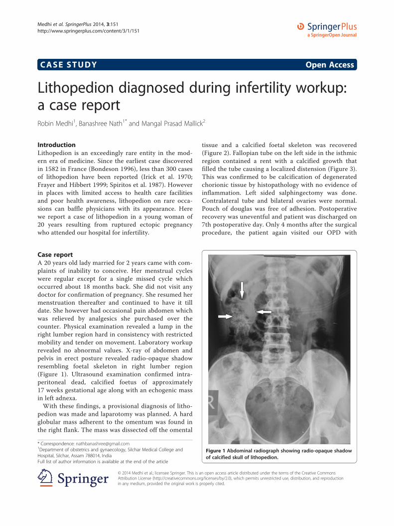
Case reportA 20 years old lady married for 2 years came with com-plaints of inability to conceive. Her menstrual cycleswere regular except for a single missed cycle whichoccurred about 18 months back. She did not visit anydoctor for confirmation of pregnancy. She resumed hermenstruation thereafter and continued to have it tilldate. She however had occasional pain abdomen whichwas relieved by analgesics she purchased over thecounter. Physical examination revealed a lump in theright lumber region hard in consistency with restrictedmobility and tender on movement. Laboratory workuprevealed no abnormal values. X-ray of abdomen andpelvis in erect posture revealed radio-opaque shadowresembling foetal skeleton in right lumber region(Figure 1). Ultrasound examination confirmed intra-peritoneal dead, calcified foetus of approximately17 weeks gestational age along with an echogenic massin left adnexa.With these findings, a provisional diagnosis of litho-
pedion was made and laparotomy was planned. A hardglobular mass adherent to the omentum was found inthe right flank. The mass was dissected off the omental
* Correspondence: [email protected] of obstetrics and gynaecology, Silchar Medical College andHospital, Silchar, Assam 788014, IndiaFull list of author information is available at the end of the article
© 2014 Medhi et al.; licensee Springer. This is aAttribution License (http://creativecommons.orin any medium, provided the original work is p
tissue and a calcified foetal skeleton was recovered(Figure 2). Fallopian tube on the left side in the isthmicregion contained a rent with a calcified growth thatfilled the tube causing a localized distension (Figure 3).This was confirmed to be calcification of degeneratedchorionic tissue by histopathology with no evidence ofinflammation. Left sided salphingectomy was done.Contralateral tube and bilateral ovaries were normal.Pouch of douglas was free of adhesion. Postoperativerecovery was uneventful and patient was discharged on7th postoperative day. Only 4 months after the surgicalprocedure, the patient again visited our OPD with
Figure 1 Abdominal radiograph showing radio-opaque shadowof calcified skull of lithopedion.
n open access article distributed under the terms of the Creative Commonsg/licenses/by/2.0), which permits unrestricted use, distribution, and reproductionroperly cited.

Figure 2 Lithopedion in the process of extraction from abdominal cavity showing adherent omental tissue to calcified mass.
Medhi et al. SpringerPlus 2014, 3:151 Page 2 of 4http://www.springerplus.com/content/3/1/151
complaints of cessation of menstruation for 2 months.Intrauterine gestation was confirmed. Patient attendedantenatal clinic regularly. She subsequently deliveredat 38 weeks a healthy female baby weighing 2.8 kgspontaneously.
Figure 3 Rent in the left fallopian tube filled with growth of calcified cho
DiscussionLithopedion is a greek word which means ‘stonechild’.This rare event occurs in 0.0054% of all gestations (Edeet al. 2011). Incidence of secondary abdominal preg-nancy is 1 in 11,000 pregnancies. Lithopedion occurs
rionic tissue showing the site of rupture of tubal ectopic pregnancy.

Figure 4 Lithopedion after extraction.
Medhi et al. SpringerPlus 2014, 3:151 Page 3 of 4http://www.springerplus.com/content/3/1/151
in 1.5 to 1.8% of these cases (Costa et al. 1991; Frayerand Hibbert 1999).Lithopedion describes an intraabdominal calcified
dead fetus. A lithopedion can result from a primaryabdominal pregnancy, or from a secondary abdominalimplantation following tubal abortion or rupture oftubal or intrauterine pregnancy. It occurs when a ster-ile extrauterine fetus survives for more than 3 monthsin abdominal cavity and escapes medical discoveryalong with minimal and sluggish circulation invitingcalcium deposition (Irick et al. 1970; Frayer andHibbert 1999; Costa et al. 1991). Secondary abdominalimplantation is one of rarest consequence of rupturedtubal pregnancy and the formation of lithopedion outof it is even rarer.Age of the patients in various case reports ranged
from 23 to 100 years at the time of diagnosis (Lachmanet al. 2001). The occurrence of this rare condition in awoman of 20 years in our case is quite unusual. Pre-operative diagnosis of lithopedion was made with sim-ple diagnostic tools averting the need for expensive,sophisticated gadgets. This is specially rewarding inareas with scarce diagnostic facilities where from theserare cases of lithopedion are reported. The formationand diagnosis of lithopedion in our case (Figure 4)occured in less than 18 months duration since the ges-tational age of the recovered stonechild far exceeds anestimated period of 8 weeks when the tubal rupture isassumed to occur. This therefore is the earliest periodof diagnosis in literature with various case reportsciting the period of retention to be 4 to 60 years (Ede
et al. 2011). The tubal rupture which resulted insecondary abdominal pregnancy is evident from therent in the tube that was filled with calcified growth ofdegenerated chorionic tissue. This synchronous evi-dence of cause and effect is unique in itself renderingthis the first of its kind. In view of the absence ofsalphingitis or adhesion, the obvious cause of ectopicpregnancy could not be elicited. However factors caus-ing infertility could probably be imputed to lithopedionon the right side resulting in distorsion of pelvic anat-omy hindering ovum pickup. Removal of lithopedionrestored the tubo-ovarian relationship resulting in con-ception within 2 months of surgical intervention. Sal-phingectomy was adopted as the procedure of choiceas the tube was grossly damaged. Surgical interventionis hence well justified in this young lady in contrast toconservative approach in view of the long survivalyears that can ensue her to various complications.A rare entity though, lithopedion is not exinct and its
diagnosis should not be missed in young infertilepatients where period of retention may be smallwith minimal symptoms and vague obstetrical history.Appropriate history and keen suspicion in such casesfrom areas with limited access to healthcare facilitiesnot only helps in diagnosis but can avert the dreadfulcomplications it can accrue in course of time.
ConsentWritten informed consent was obtained from thepatient for the publication of this report and anyaccompanying images.

Medhi et al. SpringerPlus 2014, 3:151 Page 4 of 4http://www.springerplus.com/content/3/1/151
Competing interestsThe authors declare that they have no competing interests.
Authors’ contributionsRM attended the case, made preoperative diagnosis of the rare condition,performed the surgical intervention to extract the stonechild and also didthe postoperative followup. BN assisted in the surgical interventionprocedure and drafted the manuscript. MPM assisted in the surgicalintervention procedure and did postoperative followup. All authors readand approved the final manuscript.
Author details1Department of obstetrics and gynaecology, Silchar Medical College andHospital, Silchar, Assam 788014, India. 2Department of obstetrics andgynaecology, Ramakrishna Mission Seva Pratisthan, 99 Sarat Bose Road,Kolkata, West Bengal 700026, India.
Received: 7 June 2013 Accepted: 18 November 2013Published: 19 March 2014
ReferencesBondeson J (1996) The earliest known case of a lithopedion. J R Soc Med 89:13–18Costa SD, Presley J, Bastert G (1991) Advanced abdominal pregnancy. Obstet
Gynecol Surv 46:515–525Ede J, Sobnach S, Castillo F, Bhyat A, Corbett JH (2011) The lithopedion – an
unusual cause of an abdominal mass. S Afr J Surg 49:140–141Frayer CA, Hibbert ML (1999) Abdominal pregnancy in a 67 year old woman
undetected for 37 years: a case report. J Reprod Med 44:633–635Irick MB, Kitsos CN, O’ Leary JA (1970) Therapeutic aspects in the management of
a lithopaedion. Am Surg 36:232–234Lachman N, Satyapal KS, Kalideen JM, Moodley TR (2001) Lithopedion: a case
report. Clin Anat 14:52–54Spiritos NM, Eisenkop SM, Mishell DR (1987) Lithokelyphos: a case report and
literature review. J Reprod Med 32:43–46
doi:10.1186/2193-1801-3-151Cite this article as: Medhi et al.: Lithopedion diagnosed during infertilityworkup: a case report. SpringerPlus 2014 3:151.
Submit your manuscript to a journal and benefi t from:
7 Convenient online submission
7 Rigorous peer review
7 Immediate publication on acceptance
7 Open access: articles freely available online
7 High visibility within the fi eld
7 Retaining the copyright to your article
Submit your next manuscript at 7 springeropen.com


