Embed Size (px)
Citation preview
Case ReportTwo Cases of Heerfordt’s Syndrome: A RareManifestation of Sarcoidosis
Keishi Fujiwara,1 Yasushi Furuta,2 and Satoshi Fukuda1
1Department of Otolaryngology, Head and Neck Surgery, Graduate School of Medicine, Hokkaido University, N15W7,Kita-ku, Sapporo 0608638, Japan2Department of Otolaryngology, Head and Neck Surgery, Teine-Keijinkai Hospital, 12-1-40 Maeda 1-Jo, Teine-Ku,Sapporo 0068555, Japan
Correspondence should be addressed to Keishi Fujiwara; [email protected]
Received 16 September 2015; Accepted 21 December 2015
Academic Editor: Abrao Rapoport
Copyright © 2016 Keishi Fujiwara et al.This is an open access article distributed under the Creative Commons Attribution License,which permits unrestricted use, distribution, and reproduction in any medium, provided the original work is properly cited.
Heerfordt’s syndrome is a rare manifestation of sarcoidosis characterized by the presence of facial nerve palsy, parotid glandenlargement, anterior uveitis, and low grade fever. Two cases of Heerfordt’s syndrome and a literature review are presented. Case 1.A 53-year-old man presented with swelling of his right eyelid, right facial nerve palsy, and swelling of his right parotid gland. Abiopsy specimen from the swollen eyelid indicated sarcoidosis and he was diagnosed with incomplete Heerfordt’s syndrome basedon the absence of uveitis. His symptoms were improved by corticosteroid therapy. Case 2. A 55-year-old woman presented with leftfacial nerve palsy, bilateral hearing loss, and swelling of her bilateral parotid glands. She had been previously diagnosed with uveitisand bilateral hilar lymphadenopathy. Although no histological confirmation was performed, she was diagnosed with completeHeerfordt’s syndrome on the basis of her clinical symptoms. Swelling of the bilateral parotid glands and left facial nerve palsy wereimproved immediately by corticosteroid therapy. Sarcoidosis is a relatively uncommon disease for the otolaryngologist. However,the otolaryngologist may encounter Heerfordt’s syndrome as this syndrome presents with facial nerve palsy and swelling of theparotid gland. Therefore, we otolaryngologists should diagnose and treat Heerfordt’s syndrome appropriately in cooperation withpneumologists and ophthalmologists.
1. Introduction
Sarcoidosis is a systemic granulomatous disease of unknownetiology. Although it usually affects the lung, any organ maybe involved. Heerfordt’s syndrome is a rare manifestation ofsarcoidosis characterized by the presence of facial nerve palsy,parotid gland enlargement, anterior uveitis, and low gradefever [1]. Heerfordt described three patients with uveitis,parotid swelling, cranial nerve palsy, and fever in 1909 [1] andWaldenstrom classified it as a distinct manifestation of sar-coidosis in 1937 [2]. A diagnosis of Heerfordt’s syndrome canusually bemade with confidence on the basis of characteristicclinical features. The simultaneous presence of all symptomsrepresents the complete form of this syndrome, with thecomplete form constituting 0.3%of all cases of sarcoidosis [3].Because of its rarity, there are few case reports of Heerfordt’ssyndrome in the English literature [4–7]. Here, we report twocases of Heerfordt’s syndrome.
2. Case Presentation
2.1. Case 1. A 53-year-old man presented with swelling of hisright eyelid in March and was referred to the Departmentof Dermatology in a local hospital. A biopsy specimenfrom his eyelid revealed granulomatous blepharitis, and agranulomatous disease such as sarcoidosis was suspected.In spite of detailed examination, no uveitis or bilateralhilar lymphadenopathy (BHL) was detected and he wasfollowed up closely without treatment. He was admitted tothe Department of Dermatology in our hospital in August,because of worsening swelling of his right eyelid. As he alsopresented with a 3-month history of swelling of the rightparotid gland and a 1-month history of right facial palsy, hewas referred to the Department of Otolaryngology.
Physical examination revealed a swollen right eyelid andright parotid gland. He presented with a regular generalstatus. Right facial nerve palsy especially in the forehead was
Hindawi Publishing CorporationCase Reports in OtolaryngologyVolume 2016, Article ID 3642735, 4 pageshttp://dx.doi.org/10.1155/2016/3642735
2 Case Reports in Otolaryngology
Figure 1: Ultrasound image for Case 1, showing the enlarged rightparotid gland and interspersed hypoechoic areas (arrow).
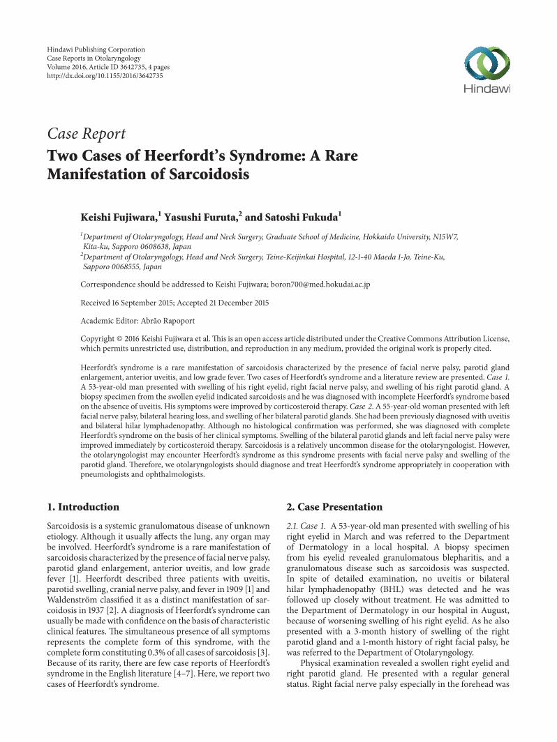
Figure 2: Positron emission tomographic image for Case 1, showinghypermetabolic activity in the right parotid gland (circles), righteyelid, and anterior mediastinal lymph nodes.
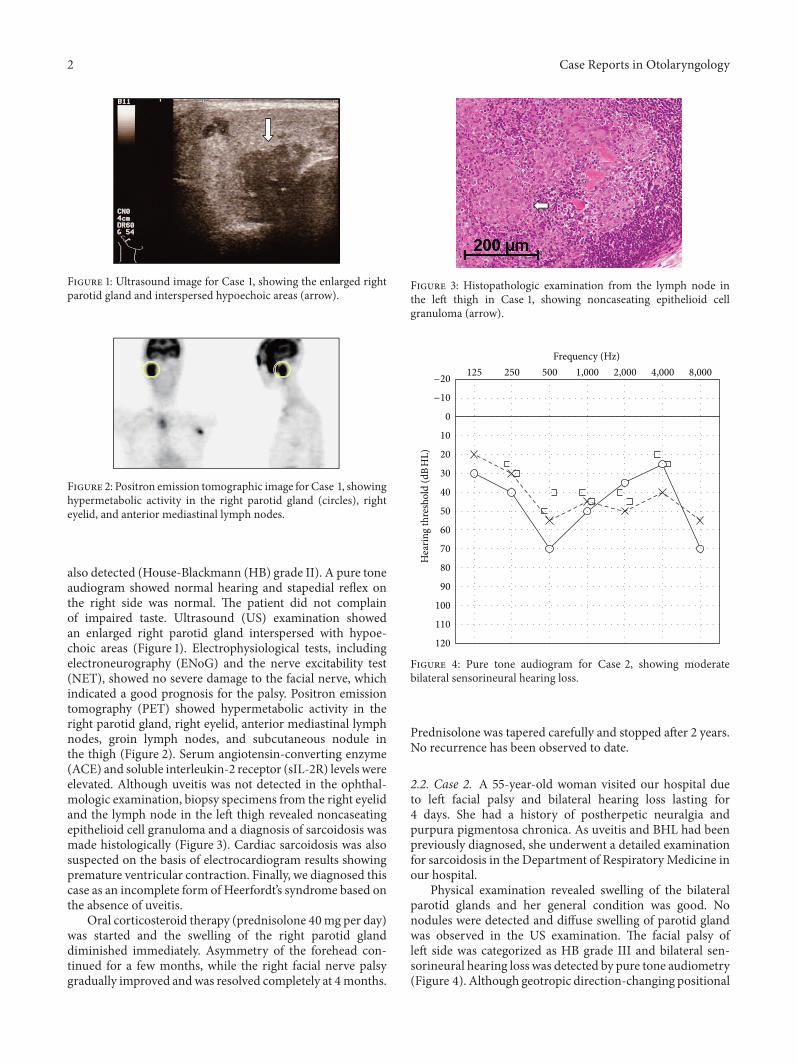
also detected (House-Blackmann (HB) grade II). A pure toneaudiogram showed normal hearing and stapedial reflex onthe right side was normal. The patient did not complainof impaired taste. Ultrasound (US) examination showedan enlarged right parotid gland interspersed with hypoe-choic areas (Figure 1). Electrophysiological tests, includingelectroneurography (ENoG) and the nerve excitability test(NET), showed no severe damage to the facial nerve, whichindicated a good prognosis for the palsy. Positron emissiontomography (PET) showed hypermetabolic activity in theright parotid gland, right eyelid, anterior mediastinal lymphnodes, groin lymph nodes, and subcutaneous nodule inthe thigh (Figure 2). Serum angiotensin-converting enzyme(ACE) and soluble interleukin-2 receptor (sIL-2R) levels wereelevated. Although uveitis was not detected in the ophthal-mologic examination, biopsy specimens from the right eyelidand the lymph node in the left thigh revealed noncaseatingepithelioid cell granuloma and a diagnosis of sarcoidosis wasmade histologically (Figure 3). Cardiac sarcoidosis was alsosuspected on the basis of electrocardiogram results showingpremature ventricular contraction. Finally, we diagnosed thiscase as an incomplete form of Heerfordt’s syndrome based onthe absence of uveitis.
Oral corticosteroid therapy (prednisolone 40mg per day)was started and the swelling of the right parotid glanddiminished immediately. Asymmetry of the forehead con-tinued for a few months, while the right facial nerve palsygradually improved andwas resolved completely at 4months.
Figure 3: Histopathologic examination from the lymph node inthe left thigh in Case 1, showing noncaseating epithelioid cellgranuloma (arrow).
Frequency (Hz)8,0004,0002,0001,000500250125
Hea
ring
thre
shol
d (d
BH
L)
120
110
100
90
80
70
60
50
40
30
20
10
0
−10
−20
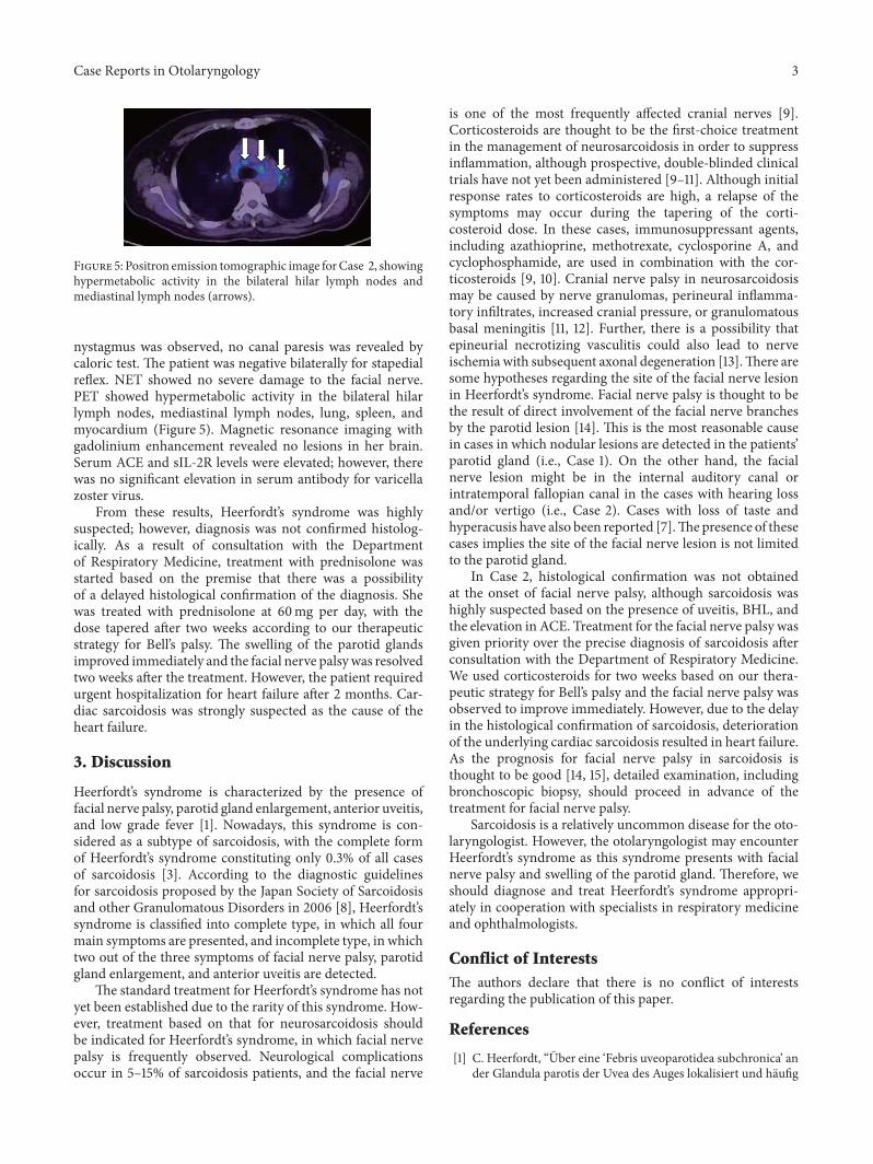
Figure 4: Pure tone audiogram for Case 2, showing moderatebilateral sensorineural hearing loss.
Prednisolone was tapered carefully and stopped after 2 years.No recurrence has been observed to date.
2.2. Case 2. A 55-year-old woman visited our hospital dueto left facial palsy and bilateral hearing loss lasting for4 days. She had a history of postherpetic neuralgia andpurpura pigmentosa chronica. As uveitis and BHL had beenpreviously diagnosed, she underwent a detailed examinationfor sarcoidosis in the Department of Respiratory Medicine inour hospital.
Physical examination revealed swelling of the bilateralparotid glands and her general condition was good. Nonodules were detected and diffuse swelling of parotid glandwas observed in the US examination. The facial palsy ofleft side was categorized as HB grade III and bilateral sen-sorineural hearing loss was detected by pure tone audiometry(Figure 4). Although geotropic direction-changing positional
Case Reports in Otolaryngology 3
Figure 5: Positron emission tomographic image forCase 2, showinghypermetabolic activity in the bilateral hilar lymph nodes andmediastinal lymph nodes (arrows).
nystagmus was observed, no canal paresis was revealed bycaloric test. The patient was negative bilaterally for stapedialreflex. NET showed no severe damage to the facial nerve.PET showed hypermetabolic activity in the bilateral hilarlymph nodes, mediastinal lymph nodes, lung, spleen, andmyocardium (Figure 5). Magnetic resonance imaging withgadolinium enhancement revealed no lesions in her brain.Serum ACE and sIL-2R levels were elevated; however, therewas no significant elevation in serum antibody for varicellazoster virus.
From these results, Heerfordt’s syndrome was highlysuspected; however, diagnosis was not confirmed histolog-ically. As a result of consultation with the Departmentof Respiratory Medicine, treatment with prednisolone wasstarted based on the premise that there was a possibilityof a delayed histological confirmation of the diagnosis. Shewas treated with prednisolone at 60mg per day, with thedose tapered after two weeks according to our therapeuticstrategy for Bell’s palsy. The swelling of the parotid glandsimproved immediately and the facial nerve palsywas resolvedtwo weeks after the treatment. However, the patient requiredurgent hospitalization for heart failure after 2 months. Car-diac sarcoidosis was strongly suspected as the cause of theheart failure.
3. Discussion
Heerfordt’s syndrome is characterized by the presence offacial nerve palsy, parotid gland enlargement, anterior uveitis,and low grade fever [1]. Nowadays, this syndrome is con-sidered as a subtype of sarcoidosis, with the complete formof Heerfordt’s syndrome constituting only 0.3% of all casesof sarcoidosis [3]. According to the diagnostic guidelinesfor sarcoidosis proposed by the Japan Society of Sarcoidosisand other Granulomatous Disorders in 2006 [8], Heerfordt’ssyndrome is classified into complete type, in which all fourmain symptoms are presented, and incomplete type, in whichtwo out of the three symptoms of facial nerve palsy, parotidgland enlargement, and anterior uveitis are detected.
The standard treatment for Heerfordt’s syndrome has notyet been established due to the rarity of this syndrome. How-ever, treatment based on that for neurosarcoidosis shouldbe indicated for Heerfordt’s syndrome, in which facial nervepalsy is frequently observed. Neurological complicationsoccur in 5–15% of sarcoidosis patients, and the facial nerve
is one of the most frequently affected cranial nerves [9].Corticosteroids are thought to be the first-choice treatmentin the management of neurosarcoidosis in order to suppressinflammation, although prospective, double-blinded clinicaltrials have not yet been administered [9–11]. Although initialresponse rates to corticosteroids are high, a relapse of thesymptoms may occur during the tapering of the corti-costeroid dose. In these cases, immunosuppressant agents,including azathioprine, methotrexate, cyclosporine A, andcyclophosphamide, are used in combination with the cor-ticosteroids [9, 10]. Cranial nerve palsy in neurosarcoidosismay be caused by nerve granulomas, perineural inflamma-tory infiltrates, increased cranial pressure, or granulomatousbasal meningitis [11, 12]. Further, there is a possibility thatepineurial necrotizing vasculitis could also lead to nerveischemia with subsequent axonal degeneration [13].There aresome hypotheses regarding the site of the facial nerve lesionin Heerfordt’s syndrome. Facial nerve palsy is thought to bethe result of direct involvement of the facial nerve branchesby the parotid lesion [14]. This is the most reasonable causein cases in which nodular lesions are detected in the patients’parotid gland (i.e., Case 1). On the other hand, the facialnerve lesion might be in the internal auditory canal orintratemporal fallopian canal in the cases with hearing lossand/or vertigo (i.e., Case 2). Cases with loss of taste andhyperacusis have also been reported [7].Thepresence of thesecases implies the site of the facial nerve lesion is not limitedto the parotid gland.
In Case 2, histological confirmation was not obtainedat the onset of facial nerve palsy, although sarcoidosis washighly suspected based on the presence of uveitis, BHL, andthe elevation in ACE. Treatment for the facial nerve palsy wasgiven priority over the precise diagnosis of sarcoidosis afterconsultation with the Department of Respiratory Medicine.We used corticosteroids for two weeks based on our thera-peutic strategy for Bell’s palsy and the facial nerve palsy wasobserved to improve immediately. However, due to the delayin the histological confirmation of sarcoidosis, deteriorationof the underlying cardiac sarcoidosis resulted in heart failure.As the prognosis for facial nerve palsy in sarcoidosis isthought to be good [14, 15], detailed examination, includingbronchoscopic biopsy, should proceed in advance of thetreatment for facial nerve palsy.
Sarcoidosis is a relatively uncommon disease for the oto-laryngologist. However, the otolaryngologist may encounterHeerfordt’s syndrome as this syndrome presents with facialnerve palsy and swelling of the parotid gland. Therefore, weshould diagnose and treat Heerfordt’s syndrome appropri-ately in cooperation with specialists in respiratory medicineand ophthalmologists.
Conflict of InterestsThe authors declare that there is no conflict of interestsregarding the publication of this paper.
References
[1] C. Heerfordt, “Uber eine ‘Febris uveoparotidea subchronica’ ander Glandula parotis der Uvea des Auges lokalisiert und haufig
4 Case Reports in Otolaryngology
mit Paresen cerebrospinaler Nerver kompliziert,” Archives ofOphthalmology, vol. 70, pp. 254–273, 1909.
[2] J.Waldenstrom, “Some observations on uveoparotitis and alliedconditions with special reference to the symptoms from thenervous system,” Acta Medica Scandinavica, vol. 91, no. 1-2, pp.53–68, 1937.
[3] P. Darlington, L. Tallstedt, L. Padyukov et al., “HLA-DRB1∗alleles and symptoms associated with Heerfordt’s syndrome insarcoidosis,” European Respiratory Journal, vol. 38, no. 5, pp.1151–1157, 2011.
[4] A. Dua and A. Manadan, “Images in clinical medicine. Heer-fordt’s syndrome, or uveoparotid fever,” The New EnglandJournal of Medicine, vol. 369, no. 5, article 458, 2013.
[5] M. C. Denny and A. D. Fotino, “The Heerfordt-Waldenstromsyndrome as an initial presentation of sarcoidosis,” Proceedings(Baylor University Medical Center), vol. 26, no. 4, pp. 390–392,2013.
[6] T. Fischer, S. Filimonow, J. Petersein, C. Zimmer,D. Beyersdorff,and H. Guski, “Diagnosis of Heerfordt’s syndrome by state-of-the-art ultrasound in combination with parotid biopsy: a casereport,” European Radiology, vol. 12, no. 1, pp. 134–137, 2002.
[7] V. Lambert and S. H. Richards, “Facial palsy in Heerfordt’ssyndrome,” The Journal of Laryngology & Otology, vol. 78, pp.684–693, 1964.
[8] Task Group in Japan Society of Sarcoidosis and Other Gran-ulomatous Disorders, “Diagnostic standard and guideline forsarcoidosis-2006,”The Japanese Journal of Sarcoidosis and OtherGranulomatous Disorders, vol. 27, no. 1, pp. 89–102, 2006.
[9] B. M. Segal, “Neurosarcoidosis: diagnostic approaches andtherapeutic strategies,” Current Opinion in Neurology, vol. 26,no. 3, pp. 307–313, 2013.
[10] F. G. Joseph and N. J. Scolding, “Neurosarcoidosis: a study of 30new cases,” Journal of Neurology, Neurosurgery and Psychiatry,vol. 80, no. 3, pp. 297–304, 2009.
[11] E.Hoitsma, C.G. Faber,M.Drent, andO. P. Sharma, “Neurosar-coidosis: a clinical dilemma,”The Lancet Neurology, vol. 3, no. 7,pp. 397–407, 2004.
[12] R. W. Babin, C. Liu, and C. Aschenbrener, “Histopathologyof neurosensory deafness in sarcoidosis,” Annals of Otology,Rhinology and Laryngology, vol. 93, no. 4, pp. 389–393, 1984.
[13] G. Said, C. Lacroix, V. Plante-Bordeneuve et al., “Nerve gran-ulomas and vasculitis in sarcoid peripheral neuropathy: aclinicopathological study of 11 patients,” Brain, vol. 125, no. 2,pp. 264–275, 2002.
[14] E. E. Lower, J. P. Broderick, T. G. Brott, and R. P. Baugh-man, “Diagnosis and management of neurological sarcoidosis,”Archives of Internal Medicine, vol. 157, no. 16, pp. 1864–1868,1997.
[15] D. G. James, “Differential diagnosis of facial nerve palsy,”Sarcoidosis Vasculitis and Diffuse Lung Disease, vol. 14, no. 2, pp.115–120, 1997.

Submit your manuscripts athttp://www.hindawi.com
Stem CellsInternational
Hindawi Publishing Corporationhttp://www.hindawi.com Volume 2014
Hindawi Publishing Corporationhttp://www.hindawi.com Volume 2014
MEDIATORSINFLAMMATION
of
Hindawi Publishing Corporationhttp://www.hindawi.com Volume 2014
Behavioural Neurology
EndocrinologyInternational Journal of
Hindawi Publishing Corporationhttp://www.hindawi.com Volume 2014
Hindawi Publishing Corporationhttp://www.hindawi.com Volume 2014
Disease Markers
Hindawi Publishing Corporationhttp://www.hindawi.com Volume 2014
BioMed Research International
OncologyJournal of
Hindawi Publishing Corporationhttp://www.hindawi.com Volume 2014
Hindawi Publishing Corporationhttp://www.hindawi.com Volume 2014
Oxidative Medicine and Cellular Longevity
Hindawi Publishing Corporationhttp://www.hindawi.com Volume 2014
PPAR Research
The Scientific World JournalHindawi Publishing Corporation http://www.hindawi.com Volume 2014
Immunology ResearchHindawi Publishing Corporationhttp://www.hindawi.com Volume 2014
Journal of
ObesityJournal of
Hindawi Publishing Corporationhttp://www.hindawi.com Volume 2014
Hindawi Publishing Corporationhttp://www.hindawi.com Volume 2014
Computational and Mathematical Methods in Medicine
OphthalmologyJournal of
Hindawi Publishing Corporationhttp://www.hindawi.com Volume 2014
Diabetes ResearchJournal of
Hindawi Publishing Corporationhttp://www.hindawi.com Volume 2014
Hindawi Publishing Corporationhttp://www.hindawi.com Volume 2014
Research and TreatmentAIDS
Hindawi Publishing Corporationhttp://www.hindawi.com Volume 2014
Gastroenterology Research and Practice
Hindawi Publishing Corporationhttp://www.hindawi.com Volume 2014
Parkinson’s Disease
Evidence-Based Complementary and Alternative Medicine
Volume 2014Hindawi Publishing Corporationhttp://www.hindawi.com