Embed Size (px)
Citation preview

Case ReportSuprapubic Catheter Migration: A Review of a Rare Complication
Amr Elmoheen ,1,2 Mahmoud Saqr,1 Waleed Salem ,1 Khalid Bashir ,1,2
and Ayman Hagras 3
1Emergency Department, Hamad Medical Corporation, Qatar2QU Health, College of Medicine, Qatar University, Qatar3Urology Department, College of Medicine, Tanta University, Egypt
Correspondence should be addressed to Amr Elmoheen; [email protected]
Received 5 September 2020; Revised 9 November 2020; Accepted 30 December 2020; Published 5 January 2021
Academic Editor: Tun-Chieh Chen
Copyright © 2021 Amr Elmoheen et al. This is an open access article distributed under the Creative Commons AttributionLicense, which permits unrestricted use, distribution, and reproduction in any medium, provided the original work isproperly cited.
Background. Suprapubic catheter migration to the vesicoureteral junction is an unusual complication, causing an obstruction thatled to hydronephrosis and dilation of the pelvicalyceal system. Case presentation. A 30-year-old man with a suprapubic catheter(SPC) that was inserted one month before this current Emergency Department (ED) visit had severe left flank pain for 48 hours.The SPC was inserted in the context of urethral injury after falling astride. Point-of-Care Ultrasound (POCUS) showed asemifilled urinary bladder and moderate hydronephrosis on the left side. A computed tomography scan (CT scan) of theabdomen was performed and showed migration of the suprapubic catheter’s tip into the left vesicoureteral junction, causingureteral obstruction dilation of the ipsilateral pelvicalyceal system. The suprapubic catheter was changed in the ED, causingrelief of symptoms, and the patient was referred to the urology department for follow-up. It was uneventful on the follow-upfrom the SPC clinic. Conclusions. This case report describes a rare complication of migration of the suprapubic catheter to thevesicoureteral junction causing acute ureteral obstruction and hydronephrosis.
1. Introduction
Suprapubic catheter drainage is a widely used urologicalprocedure in patients with neurogenic bladder and acuteretention of urine with failed urethral catheterization orin patients in whom the placement of a urethral catheteris contraindicated as urethral trauma [1]. The complica-tion rate associated with cystotomy varies from 1.6 to2.4% [2]. A poorly reported complication but with a sig-nificant impact on the quality of life of patients is cathetermigration, which should be considered in patients withacute urine retention, obstructive urinary symptoms, lowback pain, or impaired renal function [3]. This reviewdescribes an unusual complication of migration of thesuprapubic catheter to the vesicoureteral junction, causing
an obstruction that led to hydronephrosis and dilation ofthe left pelvicalyceal system.
2. Patient Information
A 30-year-old man with a suprapubic catheter (SPC) that wasinserted one month before this current Emergency Depart-ment (ED) visit had severe left flank pain for 48 hours. TheSPC was inserted in the context of urethral injury after fallingastride. He did not report any fever, changes in urine colora-tion, nausea, vomiting, or diarrhea. The suprapubic catheterwas changed five days before this current ED presentation,and the patient reported some resistance and difficulty duringthe exchange. Physical examination reveals only granuloma-tous tissue with pus in the suprapubic catheter port; however,
HindawiCase Reports in UrologyVolume 2021, Article ID 8816213, 4 pageshttps://doi.org/10.1155/2021/8816213

there is no pus in the catheter track. The rest of the general andsystemic examination was within normal parameters.
3. Investigations
In order to rule out the presence of a urinary tract infection, aurine test was performed that reveals a high white blood cellcount (28μL). Laboratory tests reported leukocytosis(13900/μL), elevated absolute neutrophil count (10300/μL),hyponatremia (132mmol/L), hypochloremia (93.0mmol/L),and normal kidney function (creatinine, 77μmol/L).
Bedside Point-of-Care Ultrasound (POCUS) showed acollapsed urinary bladder and moderate to severe hydrone-phrosis on the left side, and the balloon of the catheter inthe bladder cannot be observed.
Computed tomography (CT) abdomen was performed,showing the tip of the suprapubic catheter inserted at the leftureterovesical junction causing obstruction the ureter(Figures 1 and 2) and moderate dilatation of the left pelvica-lyceal system (Figure 3). This finding confirms the diagnosisof catheter migration.
4. Treatment
The emergency physician replaced the old suprapubiccatheter with a new 16Fr catheter. Its proper placementwas confirmed by ultrasound, and a sample was taken forurine culture. After replacing the catheter, the patientdrained 1200mL of clear urine over 3 hours. At the time ofhome discharge, the patient was in good general conditionand pain-free, and the catheter was draining clear urine. Out-patient treatment with ciprofloxacin 500mg orally every 12hours for seven days, paracetamol 1 gram orally every 8hours for seven days, and outpatient follow-up with urologyservice were arranged.
5. Outcome and Follow-Up
After performing a combined cystourethrogram, thatrevealed short urethral stricture (less than 5mm), a visualizedinternal urethrotomy was done after two weeks. During the
cystoscopy, the left ureteral orifice was slightly dilated thanthe right one. The suprapubic catheter was removed, andan 18 Fr silicone urethral catheter was fixed. The patienthad a regular follow-up with the urology outpatient servicesfor three months. He did not report any new complicationslater on.
6. Discussion
The placement of a suprapubic cystotomy catheter is a fre-quent procedure used in urology, especially in patients withneurogenic bladder who require long-term bladder drainage.Suprapubic cystotomy is also indicated in cases of trauma orurethral pathology that prevent the placement of a transure-thral catheter. The most frequent complications associatedwith cystotomy are bacteriuria, bleeding, and bladder stones[4]. Other complications such as intestinal perforation,enterocutaneous fistulas (ECF), neoplastic changes in theurinary bladder, and migration of the catheter into the ureterhave also been reported [5, 6]. However, multiple meta-analyses have shown that bladder drainage using a suprapu-bic catheter is associated with a lower rate of complicationscompared to transurethral catheter drainage.
The migration of the suprapubic catheter to the vesicour-eteral junction is a rare complication. The patients at greatestrisk are those who have a permanent catheter and/or requirelong-term bladder drainage. This is because the bladder ofthese patients tends to contract, modifying the anatomicalrelationship between the bladder neck and the ureterovesicaljunction, which makes it easier for the catheter to enter theureter. Furthermore, these patients require a regular changeof the catheter; therefore, there is a greater probability thatthe catheter will inadvertently be inserted into the ureter.Patients with neurogenic bladder usually present ureterovesi-cal reflux, which also predisposes to this complication [7].
In 1987, Borrero et al. reported a 35-year-old paraplegicman with suprapubic catheter migration diagnosed by intra-venous venogram [8]. In 2010, Dangle Pankaj et al. describedanother case of suprapubic catheter migration to the left ure-ter that led to obstructive uropathy and hydronephrosis in apatient with a neurogenic bladder [9]. In 2011, Singh et al.
Figure 1: Computed tomography (CT) abdomen, axial view showing the tip of the suprapubic catheter inserted at the left ureterovesicaljunction (red arrow).
2 Case Reports in Urology

defined a case of suprapubic migration due to a gapingureteric orifice in a 27-year-old man with traumaticbulbomembranous urethral stricture [10]. In 2016, Luoet al. reported three cases of accidental suprapubic catheterplacement at the vesicoureteral junction in patients with neu-rogenic bladder and/or voiding dysfunction [7]. In the sameyear, Shuaibin et al. reported on the case of an elderly malepatient with senile dementia whose suprapubic catheter hadmigrated into a previously normal nongaping ureter [11].
This paper presents the case of a male patient, 30-year-old, with a history of urethral trauma (falling astride) that ismanaged by suprapubic catheter insertion one month beforehis current ED presentation to be prepared later for definitivemanagement presented suprapubic heaviness and left flankpain for 48 hours. The diagnosis of catheter migration wasconfirmed by performing a computed tomography showingthe tip of the catheter at the left ureterovesical junction, caus-ing urinary flow obstruction, hydroureter, and ipsilateralhydronephrosis.
According to the cases reported so far, the diagnosis ofcatheter migration should be considered in a patient withlong-term bladder drainage and/or urinary dysfunctionwho presents resistance when inflating the catheter balloon,
urine leakage around the catheter or obstruction of thecatheter, flank pain, urinary retention, and/or obstructiveurinary symptoms.
Although most cases are reported in patients with a long-term catheter, we must bear in mind that inadvertentplacement of the catheter in the ureter may occur at the firstinsertion.
Abdominal ultrasonography is a very useful tool to iden-tify the catheter’s location and its balloon. The computedtomography with contrast allows confirming the diagnosisof catheter migration and also allows evaluating the presenceof complications such as ureteral rupture, hydronephrosis,and pyelonephritis. Patients who do not present with compli-cations can be managed conservatively, repositioning orremoving the catheter and treating urinary tract infectionwith antibiotics if present, as was done in this case.
7. Conclusion
In a patient with suprapubic cystotomy, urinary retention,hydronephrosis, flank pain, obstructive urinary symptoms,pericatheter urine leakage, or catheter blockage should makethe diagnosis of catheter migration suspect. Point-of-Care
Figure 2: Computed tomography (CT) abdomen, sagittal view showing the tip of the suprapubic catheter inserted at the left ureterovesicaljunction (red arrow).
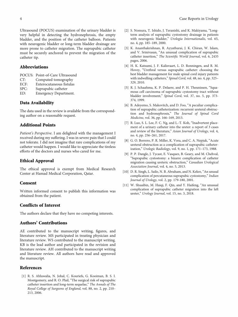
Figure 3: Computed tomography (CT) abdomen, coronal view showing moderate dilatation of the left pelvicalyceal system (red arrow).
3Case Reports in Urology
Ultrasound (POCUS) examination of the urinary bladder isvery helpful in detecting the hydronephrosis, the emptybladder, and the position of the catheter balloon. Patientswith neurogenic bladder or long-term bladder drainage aremore prone to catheter migration. The suprapubic cathetermust be securely anchored to prevent the migration of thecatheter tip.
Abbreviations
POCUS: Point-of-Care UltrasoundCT: Computed tomographyECF: Enterocutaneous fistulasSPC: Suprapubic catheterED: Emergency Department.
Data Availability
The data used in the review is available from the correspond-ing author on a reasonable request.
Additional Points
Patient’s Perspective. I am delighted with the management Ireceived during my suffering. I was in severe pain that I couldnot tolerate. I did not imagine that rare complications of mycatheter would happen. I would like to appreciate the tirelessefforts of the doctors and nurses who cared for me.
Ethical Approval
The ethical approval is exempt from Medical ResearchCenter at Hamad Medical Corporation, Qatar.
Consent
Written informed consent to publish this information wasobtained from the patient.
Conflicts of Interest
The authors declare that they have no competing interests.
Authors’ Contributions
AE contributed to the manuscript writing, figures, andliterature review. MS participated in treating physician andliterature review. WS contributed to the manuscript writing.KB is the lead author and participated in the revision andliterature review. AH contributed to the manuscript writingand literature review. All authors have read and approvedthe manuscript.
References
[1] R. S. Ahluwalia, N. Johal, C. Kouriefs, G. Kooiman, B. S. I.Montgomery, and R. O. Plail, “The surgical risk of suprapubiccatheter insertion and long-term sequelae,” The Annals of TheRoyal College of Surgeons of England, vol. 88, no. 2, pp. 210–213, 2006.
[2] S. Nomura, T. Ishido, J. Teranishi, and K. Makiyama, “Long-term analysis of suprapubic cystostomy drainage in patientswith neurogenic bladder,” Urologia Internationalis, vol. 65,no. 4, pp. 185–189, 2000.
[3] K. Ananthakrishnan, R. Ayyathurai, J. K. Chiran, W. Islam,and V. Srinivasan, “An unusual complication of suprapubiccatheter insertion,” The Scientific World Journal, vol. 6, 2435pages, 2006.
[4] H. K. Katsumi, J. F. Kalisvaart, L. D. Ronningen, and R. M.Hovey, “Urethral versus suprapubic catheter: choosing thebest bladder management for male spinal cord injury patientswith indwelling catheters,” Spinal Cord, vol. 48, no. 4, pp. 325–329, 2010.
[5] R. J. Schaafsma, K. P. Delaere, and P. H. Theunissen, “Squa-mous cell carcinoma of suprapubic cystostomy tract withoutbladder involvement,” Spinal Cord, vol. 37, no. 5, pp. 373-374, 1999.
[6] B. Adeyemo, S. Makovitch, and D. Foo, “A peculiar complica-tion of suprapubic catheterization: recurrent ureteral obstruc-tion and hydronephrosis,” The Journal of Spinal CordMedicine, vol. 36, pp. 166–169, 2013.
[7] R. Luo, S. L. Lee, F. C. Ng, and L.-T. Koh, “Inadvertent place-ment of a urinary catheter into the ureter: a report of 3 casesand review of the literature,” Asian Journal of Urology, vol. 4,no. 4, pp. 256–261, 2017.
[8] G. O. Borrero, P. R. Miller, K. Vora, and C. A. Nepjuk, “Acuteureteral obstruction as a complication of suprapubic catheter-ization,” Urologic Radiology, vol. 9, no. 1, pp. 171–173, 1988.
[9] P. P. Dangle, J. Tycast, E. Vasquez, B. Geary, and M. Chehval,“Suprapubic cystostomy: a bizarre complication of cathetermigration causing ureteric obstruction,” Canadian UrologicalAssociation Journal, vol. 4, no. 5, 2013.
[10] D. R. Singh, L. Sailo, N. B. Abraham, and N. Kekre, “An unusalcomplication of percutaneous suprapubic cystostomy,” IndianJournal of Urology, vol. 2, pp. 179-180, 2001.
[11] W. Shuaibin, M. Haiqi, F. Qin, and Y. Haifeng, “An unusualcomplication of suprapubic catheter migration into the leftureter,” Urology Journal, vol. 15, no. 3, 2018.
4 Case Reports in Urology