Embed Size (px)
Citation preview
Hindawi Publishing CorporationCase Reports in Gastrointestinal MedicineVolume 2012, Article ID 328474, 4 pagesdoi:10.1155/2012/328474
Case Report
Successful Nonoperative Management of Spontaneous SplenicHematoma and Hemoperitoneum due to CMV Infection
Georgios Lianos, Eleftheria Ignatiadou, Christina Bali,Haralampos Harissis, and Christos Katsios
Department of Surgery, University Hospital of Ioannina, St. Niarchou Avenue, 45110 Ioannina, Greece
Correspondence should be addressed to Georgios Lianos, [email protected]
Received 21 October 2012; Accepted 19 November 2012
Academic Editors: O. I. Giouleme and T. Hirata
Copyright © 2012 Georgios Lianos et al. This is an open access article distributed under the Creative Commons AttributionLicense, which permits unrestricted use, distribution, and reproduction in any medium, provided the original work is properlycited.
Introduction. Spontaneous splenic hematoma or splenic rupture due to CMV infection in immunocompetent adults is rare andlife-threatening. Case Report. Herein we report a rare case of spontaneous splenic hematoma and hemoperitoneum due to CMVinfection in a 23-year-old Caucasian male in whom conservative management was successful. Conclusion. Spontaneous splenichematoma and spontaneous splenic rupture are extremely rare conditions during primary CMV infection. Though rare, theymust be always considered by the operating surgeon, because any misinterpretation may result in unfavorable outcomes.
1. Introduction
Human cytomegalovirus is a member of the herpes familyof viruses and undergoes latency after primary infection[1, 2]. The primary infection is diagnosed by a stronglypositive CMV IgM antibody test result or CMV IgGseroconversion. Spontaneous splenic rupture or subcapsularsplenic hematoma is really an uncommon condition inprimary CMV infection [3]. The management of thesecomplications has been a matter of debate during the lastyears [4]. Although splenectomy is the appropriate treat-ment for hemodynamically unstable patients, it seems thatnonoperative management in selected patients is nowadaysconsidered the gold standard of care [5, 6].
2. Case Report
A 23-year-old Caucasian male was admitted to the emer-gency department of our hospital due to severe left upperquadrant abdominal pain. His medical history was free,and no recent trauma was reported. Clinical examinationrevealed no pyrexia, heart rate at 90 per minute, and normalblood pressure. Upon physical examination, upper abdom-inal tenderness was revealed. On auscultation, abdominal
sounds were present. Rectal examination showed an emptyrectum.
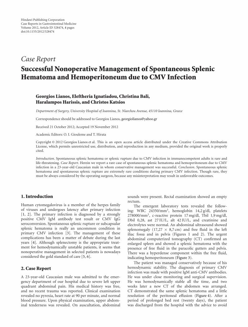
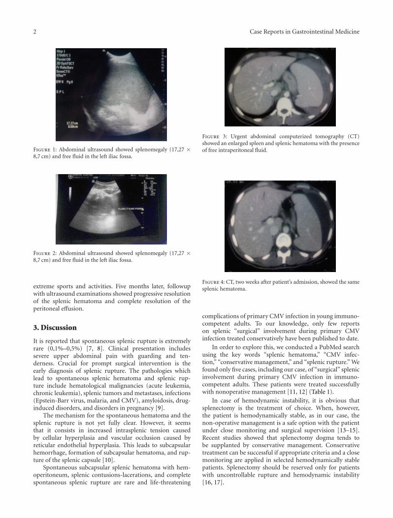
The emergent laboratory tests revealed the follow-ing: WBC 24350/mm3, hemoglobin 14,2 g/dl, platelets278000/mm3, c-reactive protein 17 mg/dl, Tbil 1,9 mg/dl,Dbil 0,28, ast 27 IU/L, alt 42 IU/L, and creatinine andelectrolytes were normal. An abdominal ultrasound showedsplenomegaly (17,27 × 8,7 cm) and free fluid in the leftiliac fossa and in pelvis (Figures 1 and 2). The urgentabdominal computerized tomography (CT) confirmed anenlarged spleen and showed a splenic hematoma with thepresence of free fluid in the paracolic gutters and pelvis.There was a hyperdense component within the free fluid,indicating hemoperitoneum (Figure 3).
The patient was managed conservatively because of hishemodynamic stability. The diagnosis of primary CMVinfection was made with positive IgM anti-CMV antibodies.He was under close monitoring and surgical supervision.He was hemodynamically stable all the time, and twoweeks later a new CT of the abdomen was arranged.CT demonstrated the same splenic hematoma and a littleresolution of the peritoneal effusion (Figure 4). After aperiod of prolonged bed rest (twenty days), the patientwas discharged from the hospital with the advice to avoid
2 Case Reports in Gastrointestinal Medicine
Figure 1: Abdominal ultrasound showed splenomegaly (17,27 ×8,7 cm) and free fluid in the left iliac fossa.
Figure 2: Abdominal ultrasound showed splenomegaly (17,27 ×8,7 cm) and free fluid in the left iliac fossa.
extreme sports and activities. Five months later, followupwith ultrasound examinations showed progressive resolutionof the splenic hematoma and complete resolution of theperitoneal effusion.
3. Discussion
It is reported that spontaneous splenic rupture is extremelyrare (0,1%–0,5%) [7, 8]. Clinical presentation includessevere upper abdominal pain with guarding and ten-derness. Crucial for prompt surgical intervention is theearly diagnosis of splenic rupture. The pathologies whichlead to spontaneous splenic hematoma and splenic rup-ture include hematological malignancies (acute leukemia,chronic leukemia), splenic tumors and metastases, infections(Epstein-Barr virus, malaria, and CMV), amyloidosis, drug-induced disorders, and disorders in pregnancy [9].
The mechanism for the spontaneous hematoma and thesplenic rupture is not yet fully clear. However, it seemsthat it consists in increased intrasplenic tension causedby cellular hyperplasia and vascular occlusion caused byreticular endothelial hyperplasia. This leads to subcapsularhemorrhage, formation of subcapsular hematoma, and rup-ture of the splenic capsule [10].
Spontaneous subcapsular splenic hematoma with hem-operitoneum, splenic contusions-lacerations, and completespontaneous splenic rupture are rare and life-threatening
Figure 3: Urgent abdominal computerized tomography (CT)showed an enlarged spleen and splenic hematoma with the presenceof free intraperitoneal fluid.
Figure 4: CT, two weeks after patient’s admission, showed the samesplenic hematoma.
complications of primary CMV infection in young immuno-competent adults. To our knowledge, only few reportson splenic “surgical” involvement during primary CMVinfection treated conservatively have been published to date.
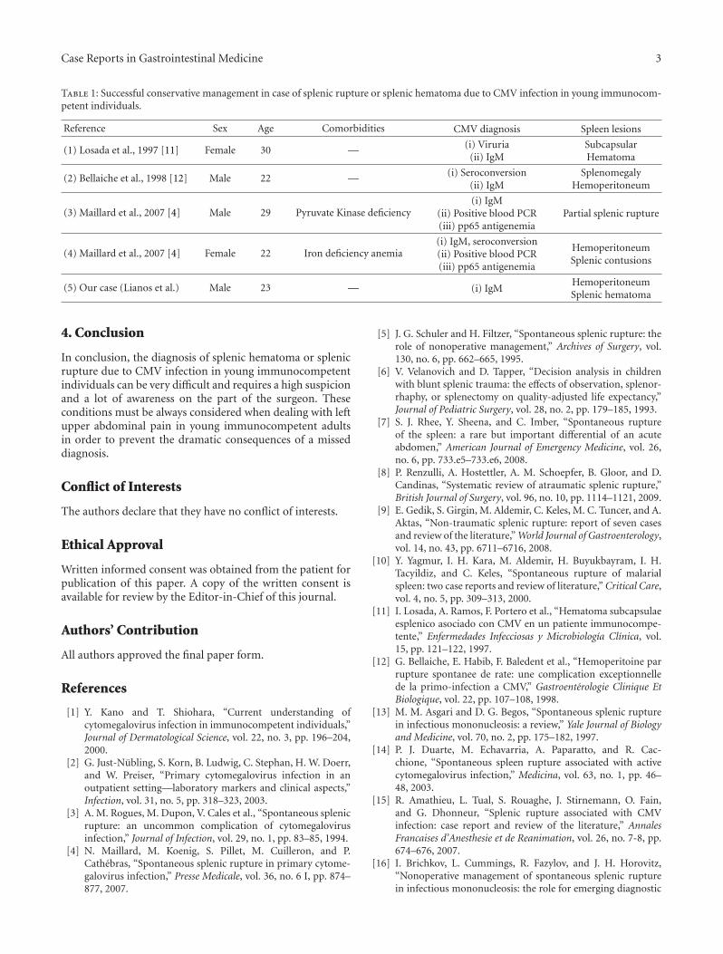
In order to explore this, we conducted a PubMed searchusing the key words “splenic hematoma,” “CMV infec-tion,” “conservative management,” and “splenic rupture.” Wefound only five cases, including our case, of “surgical” splenicinvolvement during primary CMV infection in immuno-competent adults. These patients were treated successfullywith nonoperative management [11, 12] (Table 1).
In case of hemodynamic instability, it is obvious thatsplenectomy is the treatment of choice. When, however,the patient is hemodynamically stable, as in our case, thenon-operative management is a safe option with the patientunder close monitoring and surgical supervision [13–15].Recent studies showed that splenectomy dogma tends tobe supplanted by conservative management. Conservativetreatment can be successful if appropriate criteria and a closemonitoring are applied in selected hemodynamically stablepatients. Splenectomy should be reserved only for patientswith uncontrollable rupture and hemodynamic instability[16, 17].
Case Reports in Gastrointestinal Medicine 3
Table 1: Successful conservative management in case of splenic rupture or splenic hematoma due to CMV infection in young immunocom-petent individuals.
Reference Sex Age Comorbidities CMV diagnosis Spleen lesions
(1) Losada et al., 1997 [11] Female 30 — (i) Viruria(ii) IgM
SubcapsularHematoma
(2) Bellaiche et al., 1998 [12] Male 22 — (i) Seroconversion(ii) IgM
SplenomegalyHemoperitoneum
(3) Maillard et al., 2007 [4] Male 29 Pyruvate Kinase deficiency(i) IgM
(ii) Positive blood PCR(iii) pp65 antigenemia
Partial splenic rupture
(4) Maillard et al., 2007 [4] Female 22 Iron deficiency anemia(i) IgM, seroconversion(ii) Positive blood PCR(iii) pp65 antigenemia
HemoperitoneumSplenic contusions
(5) Our case (Lianos et al.) Male 23 — (i) IgMHemoperitoneumSplenic hematoma
4. Conclusion
In conclusion, the diagnosis of splenic hematoma or splenicrupture due to CMV infection in young immunocompetentindividuals can be very difficult and requires a high suspicionand a lot of awareness on the part of the surgeon. Theseconditions must be always considered when dealing with leftupper abdominal pain in young immunocompetent adultsin order to prevent the dramatic consequences of a misseddiagnosis.
Conflict of Interests
The authors declare that they have no conflict of interests.
Ethical Approval
Written informed consent was obtained from the patient forpublication of this paper. A copy of the written consent isavailable for review by the Editor-in-Chief of this journal.
Authors’ Contribution
All authors approved the final paper form.
References
[1] Y. Kano and T. Shiohara, “Current understanding ofcytomegalovirus infection in immunocompetent individuals,”Journal of Dermatological Science, vol. 22, no. 3, pp. 196–204,2000.
[2] G. Just-Nubling, S. Korn, B. Ludwig, C. Stephan, H. W. Doerr,and W. Preiser, “Primary cytomegalovirus infection in anoutpatient setting—laboratory markers and clinical aspects,”Infection, vol. 31, no. 5, pp. 318–323, 2003.
[3] A. M. Rogues, M. Dupon, V. Cales et al., “Spontaneous splenicrupture: an uncommon complication of cytomegalovirusinfection,” Journal of Infection, vol. 29, no. 1, pp. 83–85, 1994.
[4] N. Maillard, M. Koenig, S. Pillet, M. Cuilleron, and P.Cathebras, “Spontaneous splenic rupture in primary cytome-galovirus infection,” Presse Medicale, vol. 36, no. 6 I, pp. 874–877, 2007.
[5] J. G. Schuler and H. Filtzer, “Spontaneous splenic rupture: therole of nonoperative management,” Archives of Surgery, vol.130, no. 6, pp. 662–665, 1995.
[6] V. Velanovich and D. Tapper, “Decision analysis in childrenwith blunt splenic trauma: the effects of observation, splenor-rhaphy, or splenectomy on quality-adjusted life expectancy,”Journal of Pediatric Surgery, vol. 28, no. 2, pp. 179–185, 1993.
[7] S. J. Rhee, Y. Sheena, and C. Imber, “Spontaneous ruptureof the spleen: a rare but important differential of an acuteabdomen,” American Journal of Emergency Medicine, vol. 26,no. 6, pp. 733.e5–733.e6, 2008.
[8] P. Renzulli, A. Hostettler, A. M. Schoepfer, B. Gloor, and D.Candinas, “Systematic review of atraumatic splenic rupture,”British Journal of Surgery, vol. 96, no. 10, pp. 1114–1121, 2009.
[9] E. Gedik, S. Girgin, M. Aldemir, C. Keles, M. C. Tuncer, and A.Aktas, “Non-traumatic splenic rupture: report of seven casesand review of the literature,” World Journal of Gastroenterology,vol. 14, no. 43, pp. 6711–6716, 2008.
[10] Y. Yagmur, I. H. Kara, M. Aldemir, H. Buyukbayram, I. H.Tacyildiz, and C. Keles, “Spontaneous rupture of malarialspleen: two case reports and review of literature,” Critical Care,vol. 4, no. 5, pp. 309–313, 2000.
[11] I. Losada, A. Ramos, F. Portero et al., “Hematoma subcapsulaeesplenico asociado con CMV en un patiente immunocompe-tente,” Enfermedades Infecciosas y Microbiologıa Clınica, vol.15, pp. 121–122, 1997.
[12] G. Bellaiche, E. Habib, F. Baledent et al., “Hemoperitoine parrupture spontanee de rate: une complication exceptionnellede la primo-infection a CMV,” Gastroenterologie Clinique EtBiologique, vol. 22, pp. 107–108, 1998.
[13] M. M. Asgari and D. G. Begos, “Spontaneous splenic rupturein infectious mononucleosis: a review,” Yale Journal of Biologyand Medicine, vol. 70, no. 2, pp. 175–182, 1997.
[14] P. J. Duarte, M. Echavarria, A. Paparatto, and R. Cac-chione, “Spontaneous spleen rupture associated with activecytomegalovirus infection,” Medicina, vol. 63, no. 1, pp. 46–48, 2003.
[15] R. Amathieu, L. Tual, S. Rouaghe, J. Stirnemann, O. Fain,and G. Dhonneur, “Splenic rupture associated with CMVinfection: case report and review of the literature,” AnnalesFrancaises d’Anesthesie et de Reanimation, vol. 26, no. 7-8, pp.674–676, 2007.
[16] I. Brichkov, L. Cummings, R. Fazylov, and J. H. Horovitz,“Nonoperative management of spontaneous splenic rupturein infectious mononucleosis: the role for emerging diagnostic

4 Case Reports in Gastrointestinal Medicine
and treatment modalities,” American Surgeon, vol. 72, no. 5,pp. 401–404, 2006.
[17] C. Rapp, T. Debord, P. Imbert, O. Lambotte, and R. Roue,“Spontaneous splenic rupture in infectious diseases: splenec-tomy or conservative treatment? Report of three cases,” Revuede Medecine Interne, vol. 23, no. 1, pp. 85–91, 2002.

Submit your manuscripts athttp://www.hindawi.com
Stem CellsInternational
Hindawi Publishing Corporationhttp://www.hindawi.com Volume 2014
Hindawi Publishing Corporationhttp://www.hindawi.com Volume 2014
MEDIATORSINFLAMMATION
of
Hindawi Publishing Corporationhttp://www.hindawi.com Volume 2014
Behavioural Neurology
EndocrinologyInternational Journal of
Hindawi Publishing Corporationhttp://www.hindawi.com Volume 2014
Hindawi Publishing Corporationhttp://www.hindawi.com Volume 2014
Disease Markers
Hindawi Publishing Corporationhttp://www.hindawi.com Volume 2014
BioMed Research International
OncologyJournal of
Hindawi Publishing Corporationhttp://www.hindawi.com Volume 2014
Hindawi Publishing Corporationhttp://www.hindawi.com Volume 2014
Oxidative Medicine and Cellular Longevity
Hindawi Publishing Corporationhttp://www.hindawi.com Volume 2014
PPAR Research
The Scientific World JournalHindawi Publishing Corporation http://www.hindawi.com Volume 2014
Immunology ResearchHindawi Publishing Corporationhttp://www.hindawi.com Volume 2014
Journal of
ObesityJournal of
Hindawi Publishing Corporationhttp://www.hindawi.com Volume 2014
Hindawi Publishing Corporationhttp://www.hindawi.com Volume 2014
Computational and Mathematical Methods in Medicine
OphthalmologyJournal of
Hindawi Publishing Corporationhttp://www.hindawi.com Volume 2014
Diabetes ResearchJournal of
Hindawi Publishing Corporationhttp://www.hindawi.com Volume 2014
Hindawi Publishing Corporationhttp://www.hindawi.com Volume 2014
Research and TreatmentAIDS
Hindawi Publishing Corporationhttp://www.hindawi.com Volume 2014
Gastroenterology Research and Practice
Hindawi Publishing Corporationhttp://www.hindawi.com Volume 2014
Parkinson’s Disease
Evidence-Based Complementary and Alternative Medicine
Volume 2014Hindawi Publishing Corporationhttp://www.hindawi.com