Embed Size (px)
Citation preview
Int J Clin Exp Med 2017;10(1):1345-1352www.ijcem.com /ISSN:1940-5901/IJCEM0040490
Case Report Primary paraganglioma of the thoracic spine: a rare case report and review of literature
Wen Xue1, Yuxin Song1, Xiaoli Guan2, Zengping Wang3, Zhongren Kang4, Lin Liu1, Yaowen Qian1
1Department of Orthopedics, The People’s Hospital of Gansu Province, Lanzhou, China; 2Department of Orthopedics, The Second Hospital of Lanzhou University, Lanzhou, China; 3Clinical College of Gansu University of Chinese Medicine, Lanzhou, China; 4Department of Orthopedics, Huining County People’s Hospital, Huining, China
Received September 22, 2016; Accepted October 28, 2016; Epub January 15, 2017; Published January 30, 2017
Abstract: Background: Paragangliomas (PGLs) are neuroendocrine tumors that arise from the neuroepithelial cell group called paraganglia. Spinal paragangliomas (SPs) are extremely rare, most of which are observed as intradural tumors in the cauda equine, the filum terminale, and the lumbosacral region, but rarely in the thoracic region. We aimed to study the clinical, radiographic and pathologic characteristics of spinal paragangliomas and review related literatures. Methods: This report presented the case of a 46-year-old male patient who was suffered with pathologi-cal fracture of the thoracic spine (T4) causing incomplete spinal cord injury. With no other lesions observed, the patient was diagnosed as primary paraganglioma of the thoracic spine. Results: Under general anesthesia, total en bloc spondylectomy (TES) via a single posterior approach was performed followed by bone graft fusion using titanium mesh and pedicle screw fixation system. Conclusion: This rare case report might be helpful for the future study of the clinical, radiographic and pathologic characteristics of spinal paragangliomas.
Keywords: Paraganglioma, thoracic spine, spinal reconstruction, total en bloc spondylectomy
Introduction
Paragangliomas are originate fromneural crest cells, arising from sympathetic or parasympa-thetic neural paraganglia. The rare neuroen-docrine tumors could locate at various sites in the body. Paragangliomas of spine mainly loca-te in the lumbosacral region, cauda equina and filum terminale area within the spinal canal. Malignant paragangliomas frequently combine with bone metastasis, but spinal bone meta-stasis is rare. In the spine, the most affected segment is the lumbar region. Thoracic spinal gangliomas are distinctly unusual, and so far only six cases have been described [1-4]. Here, we reported a case of primary thoracic spinal paraganglioma, obtaining satisfactory curative effect with en bloc tumor resection and recon-struction. The aim of this study was to describe a rare case of paraganglioma in the thoracic spine, and also to review the literature on this topic.
Case description
The patient was a 42-year-old man who repor-ted that he had suffered from painfully swollen
in the thoracodorsal region for five years, with progressively weakness of both lower extremi-ties for three months. Five years prior to hospi-tal admission, the patient felt swelling pain around the chest and back, with unclarified etiology. The pain is more obvious after long standing and holding heavy object with upper limbs. Nearly three months ago, the swelling pain of chest and back is further exacerbated, causing burning sensation and progressively loss of muscle strength in both of lower extre-mities. Since the onset of the disease, the pati-ent has showed normal cauda equina function with no body weight changes.
Physical examination was carried out. No super-ficial lymph nodes swelling around the body was found, and the examinations of heart, lung, and abdomen were normal. The spine showed no kyphotic-scoliotic deformity. Localized tender-ness and percussion pain were positive around the spinous process of T4. Pain, touch, and temperature sensation below the nipple flat on both sides were decreased. Muscular tone was strengthened in both of the lower limbs. The strength of bilateral quadriceps, tibialis anterior
Primary paraganglioma of the thoracic spine
1346 Int J Clin Exp Med 2017;10(1):1345-1352
muscle, thumb long extensor, and extensor digitorum longus were level III. The strength of triceps surae was level VI. The reflection of abdominal wall on both sides and cremasteric reflex were diminished. Patellar tendon reflex and Achilles tendon reflex were increased. Bilateral Babinski sign is positive. The VAS pain score is 7. No special family history, such as
and flattened. The lesion was hypointense on T1-weighted images and hyperintense on T2-weighted images. Centrum posterior kypho-sis and placeholder in spinal canal compres-sing the spinal cord were observed. No obvious abnormal signal was found in the spinal cord (Figure 4). ECT showed a sheet of condensed radioactive accumulation in T4 vertebra, wit-hout any abnormal radioactive density collecti-ve focus and defect inother bone area (Figure 5). Based on the above examination results, a presumptive diagnosis of spinal tumor was made.
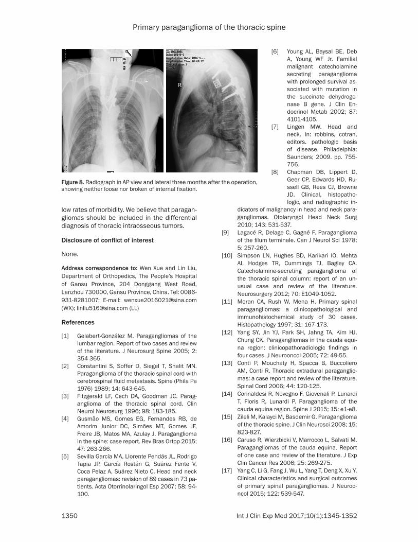
Before surgery, vertebral pedicle biopsy was carried out. Grossly, pathological results sug-gested a soft cellular neoplasm with taupe color comprised of nests and tubular adenoid tumor cells (Figure 6), divided by small vascular interstitium that was rich and dilated into blood sinus. Immunohistochemically, the tumor cells were positive for CgA, Syn and CD56, and nega-tive for S-100, GFAP, CKp, EMA and Vimentin. The proportion of Ki-67-positive cells was low (index: 1-3%) (Figure 7). On the basis of histolo-gic and immunohistochemical features, a dia-gnosis of primary paraganglioma in the the fourth thoracic spine was made.
Total en-bloc spondylectomy (TES) and bone graft fusion using titanium mesh and pedicle
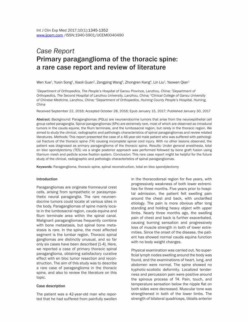
Figure 1. Radiograph of the thoracolumbar spine, showing osteolysis of T4 vertebral body.
schwannomas, neurofibrom- as, or other brain and spinal cord tumors were reported.
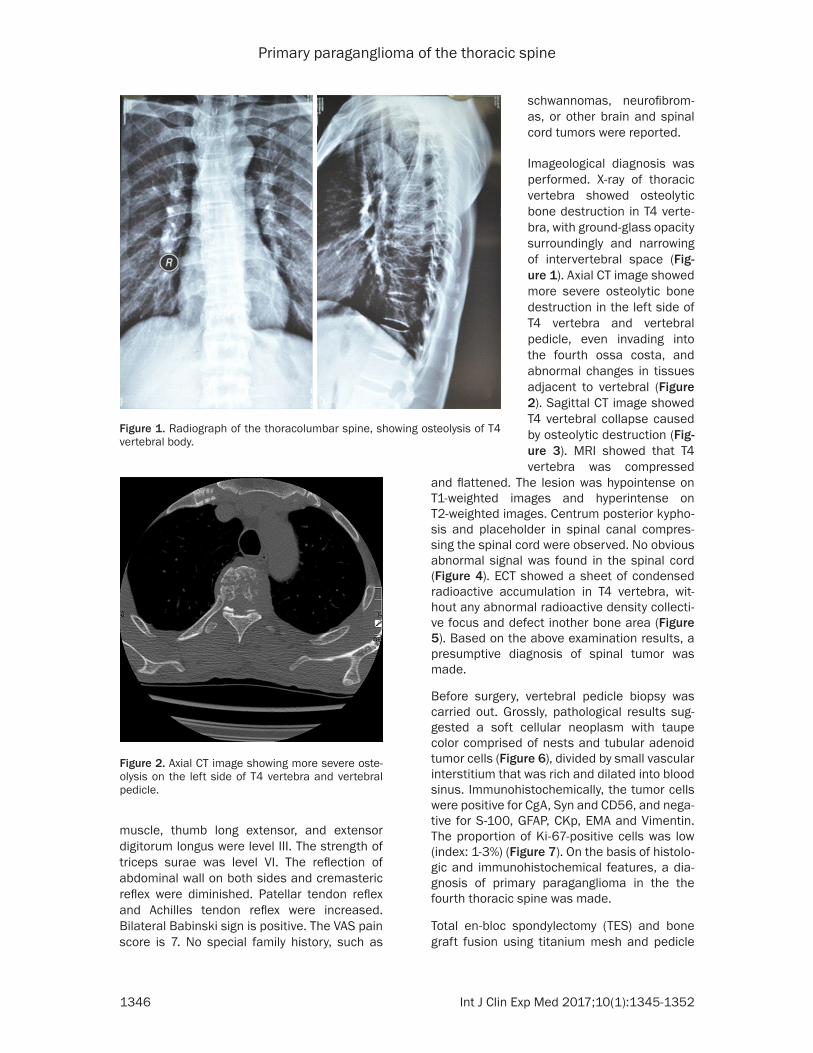
Imageological diagnosis was performed. X-ray of thoracic vertebra showed osteolytic bone destruction in T4 verte-bra, with ground-glass opacity surroundingly and narrowing of intervertebral space (Fig- ure 1). Axial CT image showed more severe osteolytic bone destruction in the left side of T4 vertebra and vertebral pedicle, even invading into the fourth ossa costa, and abnormal changes in tissues adjacent to vertebral (Figure 2). Sagittal CT image showed T4 vertebral collapse caused by osteolytic destruction (Fig- ure 3). MRI showed that T4 vertebra was compressed
Figure 2. Axial CT image showing more severe oste-olysis on the left side of T4 vertebra and vertebral pedicle.
Primary paraganglioma of the thoracic spine
1347 Int J Clin Exp Med 2017;10(1):1345-1352
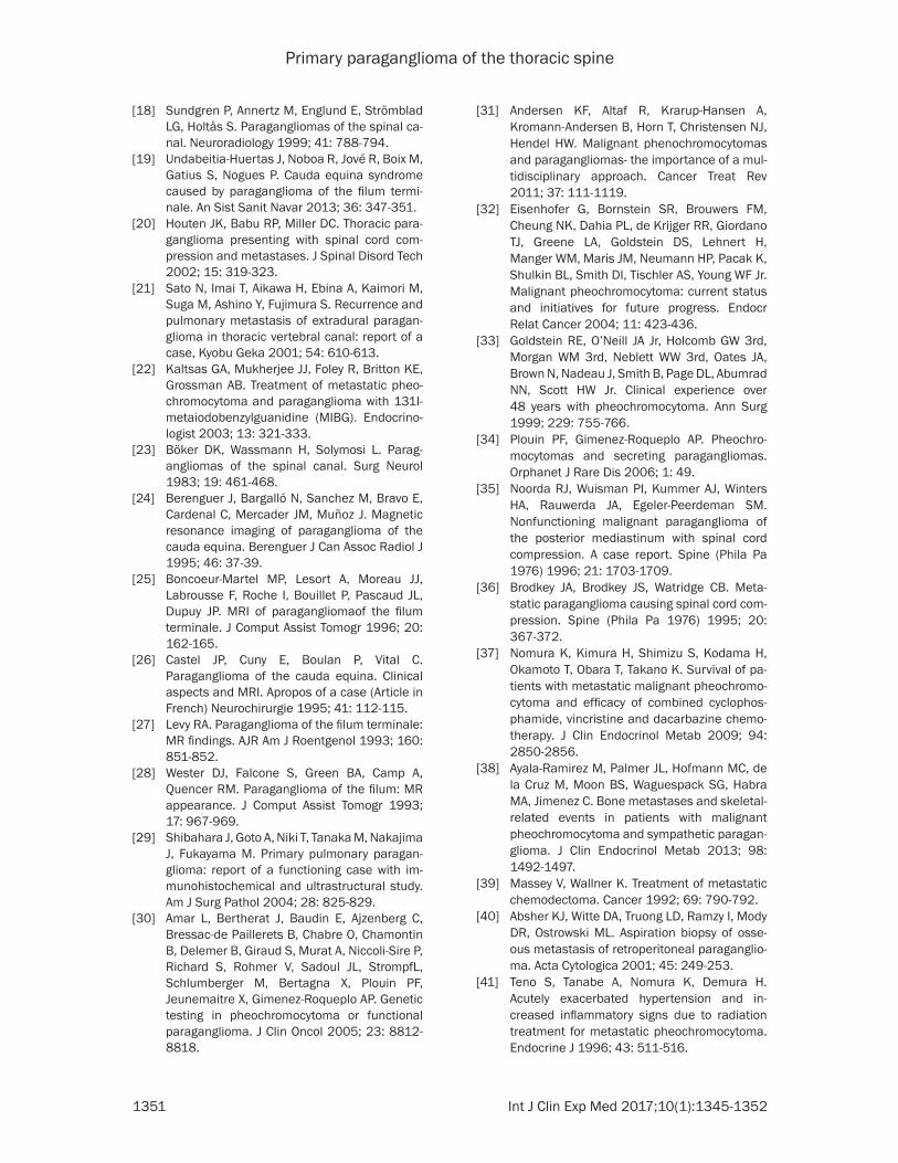
screw fixation system (4.0 mm diameter and 40 mm in length implanted at T2, T3, T5, and T6) were performed. On the left side of the separated vertebral body, tumor was found to erode the cortex, but still not penetrate into adjacent soft tissues. Anterior and middle spine column reconstruction was then achieved using titanium meshes filling with allogeneic bone. The postoperative pathological examina-tion was consistent with that of preoperative. The chest and back pain of the patient was greately relieved postoperatively. Two weeks after the operation, the VAS score is 3. Com- bined with 3 courses of hyperbaric oxygen treatment, the muscular strength of double lower limbs recovered to Level IV one month after the operation. Three months after the operation, the imaging test showed neither loose nor broken of internal fixation (Figure 8). Unfortunately, the patient died in a car accident when followed up to 5 months.
Discussion
Paraganglioma is an uncommon vascularized extra-adrenal tumor of neuroectodermal origin representing 0.012% of all tumors of the body [5]. Although most paragangliomas occur spo-radically, some are associated with familial syn-dromes such as Von Hippel-Lindau disease,
multiple endocrine neoplasia type 2, neurofi-bromatosis type 1, or Carney’s syndrome [6]. Paragangliomas are generally considered slow-growing and typically present in the fifth and sixth decades of life [7]. 90% of paraganglioma cases occur in the carotid body and bulbus venae jugular, and only about 90 cases has been reported involving the spine in the current literature [8, 10]. Retrospective analysis of recent ten years literature shows that the inci-dence of primary spinal paraganglioma is not increased, and the male/female ratio is 1.7:1 [16, 17].
Although most patients with spinal paragangli-omas are asymptomatic, spinal paraganglio-mas could lead to pathologic fracture of verte-bral body and compression of spinal cord and cauda equine, involving manifestations, such as low back pain, hypokinesia of lower extremi-ties, and dysfunction of intestine and bladder [17-21]. According to the tumor’s ability to secrete catecholamines or other hormonal sub-stances, paragangliomas can be subdivided into functional or non-functional categories. Most cases drop into the former category [22]. Only one case of functional spinal paraganglio-ma that could secrete catecholamines was reported up to now, and besides symptoms of spinal nerve compression, this type of spinal paraganglioma also presents with elevated blood pressure, facial flush, tachycardia, dia-phoresis, tremor, weight loss, nausea and eme-sis due to the release of high concentrations of catecholamines [23]. The present case mainly presents with destruction of T4 vertebral body caused by tumor growth, local pain due to microfracture, symptoms of spinal compres-sion caused by tumor invasion into correspond-ing vertebral canal, decreased sensation below the nipple flat on both sides, and lower limb dysfunction.
The CT features of spinal paragangliomas revealed homogeneous masses, and homoge-neously enhanced hypervascular tumor in con-trast enhanced CT scan, sometimes including calcification, bone destruction, pathological fracture, and enlargement of foramen interver-tebrale. The MRI characteristics of the present case are quite typical for paragangliomas including low/intermediate signal intensity on T1-weighted scansand intermediate/high sig-nal intensity on T2-weighted scans. A classic
Figure 3. Sagittal CT image showing T4 vertebral col-lapse.

Primary paraganglioma of the thoracic spine
1348 Int J Clin Exp Med 2017;10(1):1345-1352
characteristic of paragangliomas is “salt and pepper” sign, which is used to refer to a speck-led appearance of tissue in T2WI resulting from a rich vascular nature of paragangliomas [24-28].
posed of nests (Zellballen) of round-to-oval chief cells surrounded by delicate septae com-posed of sustentacular cells, with minimal exhi-bition of pleomorphism and mitotic activity [7]. However, myxopapillaryependymoma and schwannoma have the same histopathological manifestations with paragangliomas, but show different immunohistochemical characteris-tics. Thus, for further identification and confir-mation of paraganglioma, immunological stains for chromogranin A, synaptophysin, or S100 protein are necessary and critical [29]. Most paragangliomas are defined as benign in nature, only 10-20% possessing metastatic potential [30, 31]. The accepted criteria for determining the malignancy of paraganliomas preoperatively are based on whether there is tumor spread to regional lymph nodes or dis-tant metastasis [32, 33].
In the treatment of paragangliomas, chemo-therapy is ineffective [37], and radiotherapy is only feasible in terms of palliative for pain or prevention of fracture [38, 39], thus completely resection of tumor is the preferred treatment method for spinal paragangliomas [10, 35, 36]. If the tumor could be eradicated after one-stage operation, most patients could get relief and have good life quality with low tumor recur-rence [33]. Surgical resection is also suggested for the treatment of malignant paragangliomas, but the prognosis depends primarily on wheth-er the tumor metastasis [40, 41]. For patients with functional paragangliomas, preoperative treatment is critical, which requires administra-tion of α-anti-adrenergic agent at least 2 weeks before the surgery to allow the chronically con-
Figure 4. MRI showing T4 compressed and flattened vertebra. A: T2 sagittal MRI showing hypersignal and destruction of the vertebral body; B: T1 sagittal MRI showing vertebral compression in the spinal cord and destruction of the vertebral body, while sparing adjacent discs; C: Axial MRI at the T4 level showing a left paraspinal soft tissue mass.
Considering the lack of char-acteristic clinical manifesta-tions of spinal paraganglioma and its low incidence, the pre-operative diagnosis is diffi-cult. As MRI manifestation of paraganglioma is not typical, other tumors that are rich in blood vessels, such as vascu-lar tumor, hemangioma, and etc. should also be consid-ered. The confirmed diagno-sis can only be made depend-ing on the histopathological examination, in which para-gangliomas are mainly com-
Figure 5. ECT showing a sheet of condensed radioac-tive shadow in T4 vertebra.
Primary paraganglioma of the thoracic spine
1349 Int J Clin Exp Med 2017;10(1):1345-1352
tracted extravascular space to expandand achieve management of hypertension [42, 43]. In our surgical regimen, the tumor was com-pletely resected and at the same time decom-pression of the spinal cord was obtained. Finally, reconstruction of spinal stability was carried out using titanium mesh and pedicle screw fixation system. Follow-up examination 4 months after the operation revealed satisfac-tory curative effect. Another problem that we should pay special attention to is the character-istic of paragangliomas which are abundant with blood supply and should apply tumor vas-cular embolization preoperatively according to existing knowledge [44]. However, in the pres-
ent case, instead of following the conventional method [45], we did ligation of part of blood vessels in the region of diseased vertebral intraoperatively, effectively reducing the amo- unt of intraoperative bleeding and blood trans-fusion, and avoiding postoperative complica-tions, assuring the successful completion of operation.
To a very limited literature on thoracic intraos-seous paragangliomas, we add the sixth case report. With the introduction of new technolo-gies and techniques, en bloc resection of the tumor is an excellent technique when applied to certain indications. It can be performed with
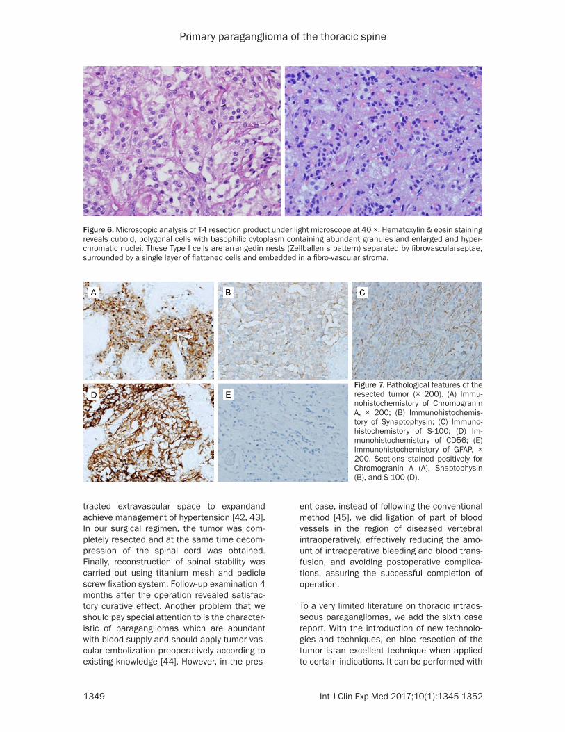
Figure 6. Microscopic analysis of T4 resection product under light microscope at 40 ×. Hematoxylin & eosin staining reveals cuboid, polygonal cells with basophilic cytoplasm containing abundant granules and enlarged and hyper-chromatic nuclei. These Type I cells are arrangedin nests (Zellballen s pattern) separated by fibrovascularseptae, surrounded by a single layer of flattened cells and embedded in a fibro-vascular stroma.
Figure 7. Pathological features of the resected tumor (× 200). (A) Immu-nohistochemistory of Chromogranin A, × 200; (B) Immunohistochemis-tory of Synaptophysin; (C) Immuno-histochemistory of S-100; (D) Im-munohistochemistory of CD56; (E) Immunohistochemistory of GFAP, × 200. Sections stained positively for Chromogranin A (A), Snaptophysin (B), and S-100 (D).
Primary paraganglioma of the thoracic spine
1350 Int J Clin Exp Med 2017;10(1):1345-1352
low rates of morbidity. We believe that paragan-gliomas should be included in the differential diagnosis of thoracic intraosseous tumors.
Disclosure of conflict of interest
None.
Address correspondence to: Wen Xue and Lin Liu, Department of Orthopedics, The People’s Hospital of Gansu Province, 204 Donggang West Road, Lanzhou 730000, Gansu Province, China. Tel: 0086-931-8281007; E-mail: [email protected] (WX); [email protected] (LL)
References
[1] Gelabert-González M. Paragangliomas of the lumbar region. Report of two cases and review of the literature. J Neurosurg Spine 2005; 2: 354-365.
[2] Constantini S, Soffer D, Siegel T, Shalit MN. Paraganglioma of the thoracic spinal cord with cerebrospinal fluid metastasis. Spine (Phila Pa 1976) 1989; 14: 643-645.
[3] Fitzgerald LF, Cech DA, Goodman JC. Parag- anglioma of the thoracic spinal cord. Clin Neurol Neurosurg 1996; 98: 183-185.
[4] Gusmão MS, Gomes EG, Fernandes RB, de Amorim Junior DC, Simōes MT, Gomes JF, Freire JB, Matos MA, Azulay J. Paraganglioma in the spine: case report. Rev Bras Ortop 2015; 47: 263-266.
[5] Sevilla García MA, Llorente Pendás JL, Rodrigo Tapia JP, García Rostán G, Suárez Fente V, Coca Pelaz A, Suárez Nieto C. Head and neck paragangliomas: revision of 89 cases in 73 pa-tients. Acta Otorrinolaringol Esp 2007; 58: 94-100.
dicators of malignancy in head and neck para-gangliomas. Otolaryngol Head Neck Surg 2010; 143: 531-537.
[9] Lagacé R, Delage C, Gagné F. Paraganglioma of the filum terminale. Can J Neurol Sci 1978; 5: 257-260.
[10] Simpson LN, Hughes BD, Karikari IO, Mehta AI, Hodges TR, Cummings TJ, Bagley CA. Catecholamine-secreting paraganglioma of the thoracic spinal column: report of an un-usual case and review of the literature. Neurosurgery 2012; 70: E1049-1052.
[11] Moran CA, Rush W, Mena H. Primary spinal paragangliomas: a clinicopathological and immunohistochemical study of 30 cases. Histopathology 1997; 31: 167-173.
[12] Yang SY, Jin YJ, Park SH, Jahng TA, Kim HJ, Chung CK. Paragangliomas in the cauda equi-na region: clinicopathoradiologic findings in four cases. J Neurooncol 2005; 72: 49-55.
[13] Conti P, Mouchaty H, Spacca B, Buccoliero AM, Conti R. Thoracic extradural paraganglio-mas: a case report and review of the literature. Spinal Cord 2006; 44: 120-125.
[14] Corinaldesi R, Novegno F, Giovenali P, Lunardi T, Floris R, Lunardi P. Paraganglioma of the cauda equina region. Spine J 2015; 15: e1-e8.
[15] Zileli M, Kalayci M, Basdemir G. Paraganglioma of the thoracic spine. J Clin Neurosci 2008; 15: 823-827.
[16] Caruso R, Wierzbicki V, Marrocco L, Salvati M. Paragangliomas of the cauda equina. Report of one case and review of the literature. J Exp Clin Cancer Res 2006; 25: 269-275.
[17] Yang C, Li G, Fang J, Wu L, Yang T, Deng X, Xu Y. Clinical characteristics and surgical outcomes of primary spinal paragangliomas. J Neuroo- ncol 2015; 122: 539-547.
Figure 8. Radiograph in AP view and lateral three months after the operation, showing neither loose nor broken of internal fixation.
[6] Young AL, Baysal BE, Deb A, Young WF Jr. Familial malignant catecholamine secreting paraganglioma with prolonged survival as-sociated with mutation in the succinate dehydroge-nase B gene. J Clin En- docrinol Metab 2002; 87: 4101-4105.
[7] Lingen MW. Head and neck. In: robbins, cotran, editors. pathologic basis of disease. Philadelphia: Saunders; 2009. pp. 755-756.
[8] Chapman DB, Lippert D, Geer CP, Edwards HD, Ru- ssell GB, Rees CJ, Browne JD. Clinical, histopatho- logic, and radiographic in-
Primary paraganglioma of the thoracic spine
1351 Int J Clin Exp Med 2017;10(1):1345-1352
[18] Sundgren P, Annertz M, Englund E, Strömblad LG, Holtås S. Paragangliomas of the spinal ca-nal. Neuroradiology 1999; 41: 788-794.
[19] Undabeitia-Huertas J, Noboa R, Jové R, Boix M, Gatius S, Nogues P. Cauda equina syndrome caused by paraganglioma of the filum termi-nale. An Sist Sanit Navar 2013; 36: 347-351.
[20] Houten JK, Babu RP, Miller DC. Thoracic para-ganglioma presenting with spinal cord com-pression and metastases. J Spinal Disord Tech 2002; 15: 319-323.
[21] Sato N, Imai T, Aikawa H, Ebina A, Kaimori M, Suga M, Ashino Y, Fujimura S. Recurrence and pulmonary metastasis of extradural paragan-glioma in thoracic vertebral canal: report of a case, Kyobu Geka 2001; 54: 610-613.
[22] Kaltsas GA, Mukherjee JJ, Foley R, Britton KE, Grossman AB. Treatment of metastatic pheo-chromocytoma and paraganglioma with 131I- metaiodobenzylguanidine (MIBG). Endocrino- logist 2003; 13: 321-333.
[23] Böker DK, Wassmann H, Solymosi L. Parag- angliomas of the spinal canal. Surg Neurol 1983; 19: 461-468.
[24] Berenguer J, Bargalló N, Sanchez M, Bravo E, Cardenal C, Mercader JM, Muñoz J. Magnetic resonance imaging of paraganglioma of the cauda equina. Berenguer J Can Assoc Radiol J 1995; 46: 37-39.
[25] Boncoeur-Martel MP, Lesort A, Moreau JJ, Labrousse F, Roche I, Bouillet P, Pascaud JL, Dupuy JP. MRI of paragangliomaof the filum terminale. J Comput Assist Tomogr 1996; 20: 162-165.
[26] Castel JP, Cuny E, Boulan P, Vital C. Paraganglioma of the cauda equina. Clinical aspects and MRI. Apropos of a case (Article in French) Neurochirurgie 1995; 41: 112-115.
[27] Levy RA. Paraganglioma of the filum terminale: MR findings. AJR Am J Roentgenol 1993; 160: 851-852.
[28] Wester DJ, Falcone S, Green BA, Camp A, Quencer RM. Paraganglioma of the filum: MR appearance. J Comput Assist Tomogr 1993; 17: 967-969.
[29] Shibahara J, Goto A, Niki T, Tanaka M, Nakajima J, Fukayama M. Primary pulmonary paragan-glioma: report of a functioning case with im-munohistochemical and ultrastructural study. Am J Surg Pathol 2004; 28: 825-829.
[30] Amar L, Bertherat J, Baudin E, Ajzenberg C, Bressac-de Paillerets B, Chabre O, Chamontin B, Delemer B, Giraud S, Murat A, Niccoli-Sire P, Richard S, Rohmer V, Sadoul JL, StrompfL, Schlumberger M, Bertagna X, Plouin PF, Jeunemaitre X, Gimenez-Roqueplo AP. Genetic testing in pheochromocytoma or functional paraganglioma. J Clin Oncol 2005; 23: 8812-8818.
[31] Andersen KF, Altaf R, Krarup-Hansen A, Kromann-Andersen B, Horn T, Christensen NJ, Hendel HW. Malignant phenochromocytomas and paragangliomas- the importance of a mul-tidisciplinary approach. Cancer Treat Rev 2011; 37: 111-1119.
[32] Eisenhofer G, Bornstein SR, Brouwers FM, Cheung NK, Dahia PL, de Krijger RR, Giordano TJ, Greene LA, Goldstein DS, Lehnert H, Manger WM, Maris JM, Neumann HP, Pacak K, Shulkin BL, Smith DI, Tischler AS, Young WF Jr. Malignant pheochromocytoma: current status and initiatives for future progress. Endocr Relat Cancer 2004; 11: 423-436.
[33] Goldstein RE, O’Neill JA Jr, Holcomb GW 3rd, Morgan WM 3rd, Neblett WW 3rd, Oates JA, Brown N, Nadeau J, Smith B, Page DL, Abumrad NN, Scott HW Jr. Clinical experience over 48 years with pheochromocytoma. Ann Surg 1999; 229: 755-766.
[34] Plouin PF, Gimenez-Roqueplo AP. Pheochro- mocytomas and secreting paragangliomas. Orphanet J Rare Dis 2006; 1: 49.
[35] Noorda RJ, Wuisman PI, Kummer AJ, Winters HA, Rauwerda JA, Egeler-Peerdeman SM. Nonfunctioning malignant paraganglioma of the posterior mediastinum with spinal cord compression. A case report. Spine (Phila Pa 1976) 1996; 21: 1703-1709.
[36] Brodkey JA, Brodkey JS, Watridge CB. Meta- static paraganglioma causing spinal cord com-pression. Spine (Phila Pa 1976) 1995; 20: 367-372.
[37] Nomura K, Kimura H, Shimizu S, Kodama H, Okamoto T, Obara T, Takano K. Survival of pa-tients with metastatic malignant pheochromo-cytoma and efficacy of combined cyclophos-phamide, vincristine and dacarbazine chemo-therapy. J Clin Endocrinol Metab 2009; 94: 2850-2856.
[38] Ayala-Ramirez M, Palmer JL, Hofmann MC, de la Cruz M, Moon BS, Waguespack SG, Habra MA, Jimenez C. Bone metastases and skeletal-related events in patients with malignant pheochromocytoma and sympathetic paragan-glioma. J Clin Endocrinol Metab 2013; 98: 1492-1497.
[39] Massey V, Wallner K. Treatment of metastatic chemodectoma. Cancer 1992; 69: 790-792.
[40] Absher KJ, Witte DA, Truong LD, Ramzy I, Mody DR, Ostrowski ML. Aspiration biopsy of osse-ous metastasis of retroperitoneal paraganglio-ma. Acta Cytologica 2001; 45: 249-253.
[41] Teno S, Tanabe A, Nomura K, Demura H. Acutely exacerbated hypertension and in-creased inflammatory signs due to radiation treatment for metastatic pheochromocytoma. Endocrine J 1996; 43: 511-516.

Primary paraganglioma of the thoracic spine
1352 Int J Clin Exp Med 2017;10(1):1345-1352
[42] Jeffs GJ, Lee GY, Wong GT. Functioning para-ganglioma of the thoracic spine: case report. Neurosurgery 2003; 53: 992-995.
[43] Spector JA, Willis DN, Ginsburg HB. Para- ganglioma (pheochromo-cytoma) of the poste-rior mediastinum: a case report and review of the literature. J Pediatr Surg 2003; 38: 1114-1116.
[44] Kwan RB, Erasmus AM, Hunn AW, Dubey A, Waites P, Jessup PJ, Burgess JR, Beasley A. Pre-operative embolisation of metastatic para-ganglioma of the thoracic spine. J Clin Neurosci 2010; 17: 394-396.
[45] Kitagawa R, Murakami H, Kato S, Nakada M, Demura S, Tsuchiya H. En bloc resection and reconstruction using a frozen tumor-bearing bone for metastases of the spine and cranium from retroperitoneal paraganglioma. World Neurosurg 2016; 90: 698, e1-5.