Embed Size (px)
Citation preview

J Ayub Med Coll Abbottabad 2013;25(1-2)
http://www.ayubmed.edu.pk/JAMC/25-1/Qasim.pdf 215
CASE REPORT NON-MOYAMOYA BILATERAL MIDDLE CEREBRAL ARTERY
AGENESIS MIMICKING MULTIPLE SCLEROSIS Qasim Bashir, Aamir Badruddin*, Saeed Arif†, Tamir Hersonsky††
Department of Clinical & Interventional Neurology, CMH Lahore Medical College, Lahore, *Department of Stroke Neurology and Neuroendovascular Surgery, Neuroscience Institute, Provena St. Joseph Medical Centre, Joliet, IL, †Department of Medicine, CMH
Lahore Medical College Lahore, ††Department of Neurosurgery, Neuroscience Institute, Provena St. Joseph Medical Centre, Joliet, IL
We present a case of a 41-year-old woman who carried the misdiagnosis of multiple sclerosis for 13 years while on disease modifying therapy [DMT]. Her neurologic work-up showed an extremely rare type of bilateral middle cerebral artery occlusive disease, a basilar apex aneurysm and paroxysmal atrial fibrillation. A thorough search for alternative neurologic diagnosis is recommended before patients are given a definitive diagnosis and committed to DMT.
J Ayub Med Coll Abbottabad 2013;25(1-2):215–6
INTRODUCTION The problem of misdiagnosing multiple sclerosis (MS) persists despite major advances in its understanding of pathophysiology, diagnosis and treatment. Once misdiagnosed, patients are maintained on expensive and potentially harmful disease modifying therapy (DMT). Non-specific MRI changes are frequently used to make the diagnosis.
CASE REPORT A 41-year-old woman, smoker, with a 13-year diagnosis of MS was transferred with an un-ruptured basilar apex aneurysm. She at age 28 years was diagnosed with MS at a tertiary hospital. At that time she presented with progressive symptoms of left-sided weakness, dexterity difficulty and numbness. Based on a single MRI brain study and clinical symptomatology, she was given a diagnosis of MS and started on DMT with no-follow-up imaging. Patient refused a diagnostic lumbar puncture. A few weeks prior to admission she started complaining of ‘spells’ that occurred on average once a month where she experienced arm stiffness and eyes rolling back, speech difficulty but with preserved comprehend. These spells were considered as potential seizures and she was started on antiepileptic therapy. The symptoms continued to persist and now were associated with progressive headaches and chest palpitations. An MRI brain showed mild peri-ventricular and left parietal lobe T2/Flair
hyper-intense foci and a large flow void at the basilar apex that was suspicious for a large aneurysm. The MRI brain T2/Flair findings were more compatible with small vessel ischemic changes versus a demyelinating process.
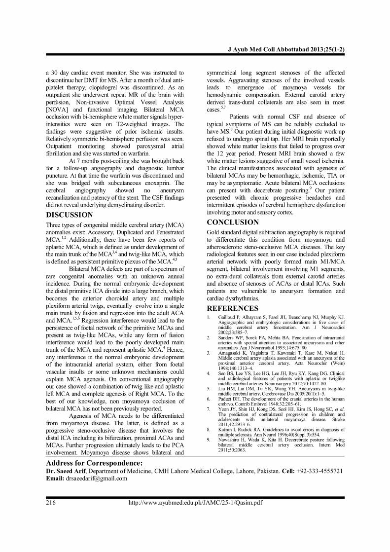
Upon admission, her neurological examination was completely normal. She underwent a diagnostic cerebral angiography that showed bilateral Middle Cerebral Artery (MCA) occlusive disease with robust collaterals from Anterior, and Posterior Cerebral Arteries (ACA, and PCA) but no obvious moyamoya changes. Numerous tiny angiomatous/twig-like vessels were seen supplying the basal ganglia bilaterally [Figure-1A, 1B, 1C]. On Left ICA Oblique Views [Figure-1D, 1E], a faint streak distal Left M1 trunk and its trifurcation branches supplying the temporal and parietal lobes were visualised. A large saccular basilar apex aneurysm measuring 11 mm deep9 mm wide was identified. On 3D Rotational Angiography the proximal left P1/PCA was noticed to be incorporated within the neck of the aneurysm.
Case was reviewed with the neurosurgery service and joint consensus developed to treat the aneurysm with neuroendovascular approach. She underwent uneventfully stent [Codman Enterprise VRD system 4.5×22 mm] assist coiling of the basilar apex aneurysm via the right posterior communicating artery and left vertebral artery approach.
Figure-1A,1B,1C: Right ICA Anteroposterior and oblique view projections showing twig-like vessels to basal ganglia during the early arterial phase. 1C: Right ICA Oblique Late Arterial phase shows collateral vessels from ACA and PCA to MCA. 1D,1E: Left ICA Anteroposterior/oblique view show twig-
like MCA branches fusing to form distal left M1 trunk/trifurcation and ACA/ PCA collaterals to MCA territory.
Pre- and post-procedure, her cardiac tele-monitoring showed paroxymal supra-ventricular tachycardia,
frequent PACs and atrial bigeminy. She was started on low dose beta blocker therapy and discharged home with
J Ayub Med Coll Abbottabad 2013;25(1-2)
http://www.ayubmed.edu.pk/JAMC/25-1/Qasim.pdf 216
a 30 day cardiac event monitor. She was instructed to discontinue her DMT for MS. After a month of dual anti-platelet therapy, clopidogrel was discontinued. As an outpatient she underwent repeat MR of the brain with perfusion, Non-invasive Optimal Vessel Analysis [NOVA] and functional imaging. Bilateral MCA occlusion with bi-hemisphere white matter signals hyper-intensities were seen on T2-weighted images. The findings were suggestive of prior ischemic insults. Relatively symmetric bi-hemisphere perfusion was seen. Outpatient monitoring showed paroxysmal atrial fibrillation and she was started on warfarin.
At 7 months post-coiling she was brought back for a follow-up angiography and diagnostic lumbar puncture. At that time the warfarin was discontinued and she was bridged with subcutaneous enoxaprin. The cerebral angiography showed no aneurysm recanalization and patency of the stent. The CSF findings did not reveal underlying demyelinating disorder. DISCUSSION Three types of congenital middle cerebral artery (MCA) anomalies exist: Accessory, Duplicated and Fenestrated MCA.1,2 Additionally, there have been few reports of aplastic MCA, which is defined as under development of the main trunk of the MCA3,4 and twig-like MCA, which is defined as persistent primitive plexus of the MCA.4,5
Bilateral MCA defects are part of a spectrum of rare congenital anomalies with an unknown annual incidence. During the normal embryonic development the distal primitive ICA divide into a large branch, which becomes the anterior choroidal artery and multiple plexiform arterial twigs, eventually evolve into a single main trunk by fusion and regression into the adult ACA and MCA.1,5,6 Regression interference would lead to the persistence of foetal network of the primitive MCAs and present as twig-like MCAs, while any form of fusion interference would lead to the poorly developed main trunk of the MCA and represent aplastic MCA.4 Hence, any interference in the normal embryonic development of the intracranial arterial system, either from foetal vascular insults or some unknown mechanisms could explain MCA agenesis. On conventional angiography our case showed a combination of twig-like and aplastic left MCA and complete agenesis of Right MCA. To the best of our knowledge, non moyamoya occlusion of bilateral MCA has not been previously reported.
Agenesis of MCA needs to be differentiated from moyamoya disease. The latter, is defined as a progressive steno-occlusive disease that involves the distal ICA including its bifurcation, proximal ACAs and MCAs. Further progression ultimately leads to the PCA involvement. Moyamoya disease shows bilateral and
symmetrical long segment stenoses of the affected vessels. Aggravating stenoses of the involved vessels leads to emergence of moymoya vessels for hemodynamic compensation. External carotid artery derived trans-dural collaterals are also seen in most cases.5,7
Patients with normal CSF and absence of typical symptoms of MS can be reliably excluded to have MS.8 Our patient during initial diagnostic work-up refused to undergo spinal tap. Her MRI brain reportedly showed white matter lesions that failed to progress over the 12 year period. Present MRI brain showed a few white matter lesions suggestive of small vessel ischemia. The clinical manifestations associated with agenesis of bilateral MCAs may be hemorrhagic, ischemic, TIA or may be asymptomatic. Acute bilateral MCA occlusions can present with decerebrate posturing.9 Our patient presented with chronic progressive headaches and intermittent episodes of cerebral hemisphere dysfunction involving motor and sensory cortex. CONCLUSION Gold standard digital subtraction angiography is required to differentiate this condition from moyamoya and atherosclerotic steno-occlusive MCA diseases. The key radiological features seen in our case included plexiform arterial network with poorly formed main M1/MCA segment, bilateral involvement involving M1 segments, no extra-dural collaterals from external carotid arteries and absence of stenoses of ACAs or distal ICAs. Such patients are vulnerable to aneurysm formation and cardiac dysrhythmias. REFERENCES 1. Gailloud P, Albayram S, Fasel JH, Beauchamp NJ, Murphy KJ.
Angiographic and embryologic considerations in five cases of middle cerebral artery fenestration. Am J Neuroradiol 2002;23:585–7.
2. Sanders WP, Sorek PA, Mehta BA. Fenestration of intracranial arteries with special attention to associated aneurysms and other anomalies. Am J Neuroradiol 1993;14:675–80.
3. Amagasaki K, Yagishita T, Kawataki T, Kase M, Nukui H. Middle cerebral artery aplasia associated with an aneurysm of the proximal anterior cerebral artery. Acta Neurochir (Wein) 1998;140:1313–4.
4. Seo BS, Lee YS, Lee HG, Lee JH, Ryu KY, Kang DG. Clinical and radiological features of patients with aplastic or twiglike middle cerebral arteries. Neurosurgery 2012;70:1472–80.
5. Liu HM, Lai DM, Tu YK, Wang YH. Aneurysms in twig-like middle cerebral artery. Cerebrovasc Dis 2005;20(1):1–5.
6. Padget DH. The development of the cranial arteries in the human embryo. Contrib Embryol 1948;32:205–61.
7. Yeon JY, Shin HJ, Kong DS, Seol HJ, Kim JS, Hong SC, et al. The prediction of contralateral progression in children and adolescents with unilateral moyamoya disease. Stroke 2011;42:2973–6.
8. Katzan I, Rudick RA. Guidelines to avoid errors in diagnosis of multiple sclerosis. Ann Neurol 1996;40(Suppl 3):554.
9. Nawashiro H, Wada K, Kita H. Decerebrate posture following bilateral middle cerebral artery occlusion. Intern Med 2011;50:2063.
Address for Correspondence: Dr. Saeed Arif, Department of Medicine, CMH Lahore Medical College, Lahore, Pakistan. Cell: +92-333-4555721 Email: [email protected]