Embed Size (px)
Citation preview

Hindawi Publishing CorporationCase Reports in SurgeryVolume 2013, Article ID 131962, 4 pageshttp://dx.doi.org/10.1155/2013/131962
Case ReportMultiple Intracranial Meningiomas:A Review of the Literature and a Case Report
F. Koech,1 J. Orege,2 F. Ndiangui,3 B. Macharia,2 and N. Mbaruku4
1 Department of Neurosurgery, Moi University School of Medicine, P.O. Box 4606-30100, Eldoret 30100, Kenya2Department of Pathology, Moi University School of Medicine, P.O. Box 4606-30100, Eldoret 30100, Kenya3Department of Radiology, Moi University School of Medicine, P.O. Box 4606-30100, Eldoret 30100, Kenya4Department of Orthopedics, Moi University School of Medicine, P.O. Box 4606-30100, Eldoret 30100, Kenya
Correspondence should be addressed to F. Koech; [email protected]
Received 27 May 2013; Accepted 29 July 2013
Academic Editors: C. Foroulis and T. Sorimachi
Copyright © 2013 F. Koech et al.This is an open access article distributed under the Creative Commons Attribution License, whichpermits unrestricted use, distribution, and reproduction in any medium, provided the original work is properly cited.
Multiple intracranial meningiomas are a condition where there is more than one meningioma in several intracranial locationsin the same patient without signs of neurofibromatosis. Incidence varies from 1 to 10%. The prognosis of multiple intracranialmeningioma does not differ from benign solitary meningiomas despite the multiplicity. However, the simultaneous occurrence ofdifferent grades of malignancy is observed in one-third of multiple meningiomas. Surgery remains the best option for treatment ofsymptomatic lesions. Our case review aims to present and discuss a 75-year-old female patient diagnosed withmultiple intracranialmeningiomas, describing their clinical, radiological, histological characteristics. It also highlights the fact that the patient had twotumours, underwent surgery, and so far has a good quality of life.
1. Introduction
Cushing and Eisenhardt [1] in 1938 were the first to coin thetermmeningioma.They came upwith what closely resemblesour contemporary understanding of the frequency of menin-giomas by location. They defined multiple meningiomas as“at least two spatially separated meningiomas in a patientwithout signs of neurofibromatosis” [1]. The incidence ofmultiplemeningiomas, defined byCushing andEisenhardt, isabout 1 to 2 percent of all meningioma cases, and these resultsare comparable to those obtained by other authors [1].
2. Case Report
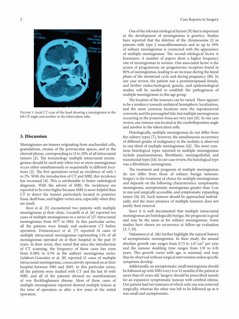
A 75-year-old female patient presented with a six-monthhistory of global headaches, loss of balance, and a changein facial sensation. She had no stigmata of neurofibromato-sis. Central nervous system examination revealed reducedsensation on the left side, a GCS of 15/15, and all othercranial nerves were otherwise normal. Contrast enhancedCT scans (Figure 1) revealed two intracranial lesions, one
in the left cerebellopontine angle (CPA) and another inthe tuberculum sella area. T1W, T2W, and FLAIR magneticresonance imaging scans (MRI) sequences showed hyperin-tense well circumscribed lesions in the left CPA and tuber-culum sella (Figures 2(a) and 2(b)). The left infratentorial-CPA tumour showed significant mass effect. The MRI fea-tures of the two lesions were highly suggestive of multiplemeningiomas. The patient was seen in the neurosurgeryclinic and was scheduled for suboccipital craniotomy of theCPA tumour. This was removed achieving Simpsons Grade2. The tuberculum sella lesion was not excised based onthe age of the patient, small size of the tumour, and itslack of symptoms. Histological examination of the cere-bellopontine lesion revealed a lesion composed of manysheets of fibroblastic cells separated by collagen bundles(Figures 3(a) and 3(b)). After surgery, she showed symp-tomatic improvement. Postoperatively, the patient improvedremarkably with no headaches. The patient’s first followupafter surgery was unremarkable and is now scheduled for asecond followup with a repeat MRI at 6 months and remainsasymptomatic.
2 Case Reports in Surgery
Figure 1: Axial CT scan of the head showing a meningioma in theleft CP angle and another in the tuberculum sella.
3. Discussion
Meningiomas are tumors originating from arachnoidal cells,granulations, stroma of the perivascular spaces, and in thechoroid plexus, corresponding to 13 to 20% of all intracranialtumors [2]. The terminology multiple intracranial menin-giomas should be used only when two or more meningiomasoccur either simultaneously or sequentially in different loca-tions [3]. The first quotations reveal an incidence of only 1to 2%. With the introduction of CT and MRI, this incidencehas increased [4]. This is attributable to better radiologicaldiagnosis. With the advent of MRI, the incidences arereported to be even higher because MRI is more helpful thanCT to detect the tumors particularly located in posteriorfossa, skull base, and higher vertex area, especially when theyare small.
Kyoi et al. [5] encountered two patients with multiplemeningiomas at their clinic. Locatelli et al. [6] reported tencases of multiple meningiomas in a series of 227 intracranialmeningiomas from 1977 to 1984. In this particular series,all the patients were female and underwent CT beforeoperation. Domenicucci et al. [7] reported 14 cases ofmultiple intracranial meningiomas representing 1.1% of allmeningiomas operated on at their hospital in the past 35years. In their series, they noted that since the introductionof CT scanning, the frequency of these cases has risenfrom 0.58% to 4.5% in the authors’ meningioma series.Gelabert-Gonzalez et al. [8] reported 13 cases of multipleintracranial meningiomas, consecutively operated on at theirhospital between 1983 and 2003. In this particular series,all the patients were studied with CT and the last 10 withMRI, and all of the patients showed no manifestationsof von Recklinghausen disease. Most of these cases ofmultiple meningiomas reported showed multiple lesions atthe time of operation or after a few years of the initialoperation.
One of the relevant etiological factors [9] that is importantin the development of meningiomas is genetics. Studieshave reported that the deletion of the chromosome 22 inpatients with type 2 neurofibromatosis and in up to 50%of solitary meningiomas is connected with the appearanceof multiple meningiomas. The second etiological factor ishormones. A number of papers show a higher frequencyrate of meningiomas in women. One associated factor is theaction of progesterone on progesterone receptors found in80% of meningiomas, leading to an increase during the lutealphase of the menstrual cycle and during pregnancy [10]. Inour case review, the patient was a postmenopausal female,and further endocrinological, genetic, and epidemiologicalstudies will be needed to establish the pathogenesis ofmultiple meningiomas in this age group.
The location of the tumours can be varied. There appearsto be a tendency towards unilateral hemispheric localization,and the most common locations were the supratentorialconvexity and the parasagittal falx, butmultiplemeningiomasoccurring in the posterior fossa are very rare [11]. In our casereview, one tumour was located at the cerebellopontine angleand another in the tuberculum sella.
Histologically, multiple meningiomas do not differ fromthe solitary types [7]; however, the simultaneous occurrenceof different grades of malignancy in the nodules is observedin one-third of multiple meningiomas [12]. The most com-mon histological types reported in multiple meningiomasinclude psammomatous, fibroblastic, meningothelial, andtransitional types [13]. In our case review, the histological typewas a fibroblastic meningioma.
The treatment and prognosis of multiple meningiomasdo not differ from those of solitary benign tumours.Surgery is the treatment of choice for multiple meningiomasand depends on the following characteristics: symptomaticmeningioma, asymptomatic meningioma greater than 3 cmin size and surgically accessible, and symptomatic expandingtumor [14, 15]. Each tumour should be approached individ-ually, and the mere presence of multiple tumours does notjustify their removal.
Since it is well documented that multiple intracranialmeningiomas are histologically benign, the prognosis is goodand may be the same as for solitary meningiomas. Somestudies have shown no recurrence at follow-up evaluation[3, 7, 13].
Nakamura et al. [16] further highlight the natural historyof asymptomatic meningiomas. In their study, the annualabsolute growth rate ranges from 0.73 to 1.67 cm3 per yearand the tumour doubling time ranges from 1.19 to 6.81years. This growth varies with age, is minimal, and maythus be observedwithout surgical intervention unless specificsymptoms develop.
Additionally, an asymptomatic small meningioma shouldbe followed up withMRI every 6 or 12months if the patient ismore than 65 years old. Surgery should be prescribed mainlyfor an expansive symptomatic tumour with cerebral edema.Our patient had two tumours of which only one was removedsurgically, whereas the other was left to be followed up as itwas small and asymptomatic.

Case Reports in Surgery 3
(a) (b)
Figure 2: Axial T2W and FLAIR images showing a meningioma in the left CP angle and tuberculum sella (a) and (b).
(a) (b)
Figure 3: Histology results showing a fibroblastic meningioma (a) and (b).
4. Conclusion
Multiple meningiomas are rare. When they do occur theyare more common in females. Their location can be varied,most commonly unihemispheric rarely in the posterior fossa.The histological subtypes are similar to the solitary types withthe most common ones being psammomatous, fibroblastic,meningothelial, and transitional types. Definitive treatmentis surgical removal of expansive tumours with cerebral edemaand subsequent followup of small asymptomatic tumours.
Conflict of Interests
There is no conflict of interests or financial support from anycompany other than the authors’ individual contributions.
References
[1] H. Cushing and L. Eisenhardt, Meningiomas: Their Classifica-tion, Regional Behaviour, Life History and Surgical End Result,Charles CThomas, Springfield, Ill, USA, 1938.
[2] D. S. Russel and L. J. Rubinstein, Pathology of Tumors of theNervous System, Williams and Wilkins, Baltimore, Md, USA,1977.
[3] M. Turgut, S. Palaoglus, O. E. Osmam, and G. Gurcay, “Mul-tiples Meningiomas of the central nervous system without thestigmata of neurofibromatosis: clinical and therapeutic study,”Neurosurgical Review, vol. 20, no. 2, pp. 117–123, 1997.
[4] G. Butti, R. Assietti, R. Casalone, and P. Paoletti, “MultipleMeningiomas: a clinical, surgical, and cytogenetic analysis,”Surgical Neurology, vol. 31, no. 4, pp. 255–260, 1989.

4 Case Reports in Surgery
[5] K. Kyoi, K. Yokoyama, T. Hoshida et al., “Mulitiple Menin-gioma,” No Shinkei Geka, vol. 11, no. 10, pp. 1099–1105, 1983.
[6] D. Locatelli, A. Bottoni, C. Uggetti, and L. Gozzoli, “Multi-ple Meningiomas evaluated by computed tomography,” Neu-rochirurgia, vol. 30, no. 1, pp. 8–10, 1987.
[7] M. Domenicucci, A. Santoro, D. H. D’Osvaldo et al., “Multipleintracranial Meningiomas,” Journal of Neurosurgery, vol. 70, no.1, pp. 41–44, 1989.
[8] M. Gelabert-Gonzalez, R. Leira-Muino, J.M. Fernandez-Villa etal., “Multiple intracranial Meningiomas,” Revista de Neurologia,vol. 37, no. 8, pp. 717–722, 2003.
[9] P. Black, A. Morokoff, J. Zauberman, E. Claus, and R. Carroll,“Meningiomas: science and surgery,”Clinical Neurosurgery, vol.54, pp. 91–99, 2007.
[10] T. Gruber, A. O. Dare, L. L. Balos, S. Lele, and R. A. Fen-stermaker, “Multiple Meningiomas arising during long-termtherapy with the progesterone agonist megestrol acetate: casereport,” Journal of Neurosurgery, vol. 100, no. 2, pp. 328–331,2004.
[11] T. S. Kim, J. K. Park, S. Jung et al., “Multiple intracranialMeningiomas,” Journal of Korean Neurosurgical, vol. 26, no. 12,pp. 1685–1691, 1997.
[12] K. Mocker, H. Holland, P. Ahnert et al., “Multiple meningiomawith different grades of malignancy: case report with geneticanalysis applying single-nucleotide polymorphism array andclassical cytogenetics,” Pathology Research and Practice, vol. 207,no. 1, pp. 67–72, 2011.
[13] M. S. M. Eljamel and P. M. Foy, “Multiple Meningiomas andtheir relation to neurofibromatosis,” Surgical Neurology, vol. 32,no. 2, pp. 131–136, 1989.
[14] M. Salvati, E. Caroli, and L. Ferrante, “Zentrabl,”Neurochir, vol.65, no. 4, pp. 180–184, 2004.
[15] J. P. Sheehy and H. A. Crockard, “Multiple Meningiomas: alongterm review,” Journal of Neurosurgery, vol. 59, no. 1, pp. 1–5,1983.
[16] M. Nakamura, F. Roser, J. Michel, C. Jacobs, andM. Samii, “Thenatural history of incidental Meningiomas,” Neurosurg, vol. 53,no. 1, pp. 62–70, 2003.

Submit your manuscripts athttp://www.hindawi.com
Stem CellsInternational
Hindawi Publishing Corporationhttp://www.hindawi.com Volume 2014
Hindawi Publishing Corporationhttp://www.hindawi.com Volume 2014
MEDIATORSINFLAMMATION
of
Hindawi Publishing Corporationhttp://www.hindawi.com Volume 2014
Behavioural Neurology
EndocrinologyInternational Journal of
Hindawi Publishing Corporationhttp://www.hindawi.com Volume 2014
Hindawi Publishing Corporationhttp://www.hindawi.com Volume 2014
Disease Markers
Hindawi Publishing Corporationhttp://www.hindawi.com Volume 2014
BioMed Research International
OncologyJournal of
Hindawi Publishing Corporationhttp://www.hindawi.com Volume 2014
Hindawi Publishing Corporationhttp://www.hindawi.com Volume 2014
Oxidative Medicine and Cellular Longevity
Hindawi Publishing Corporationhttp://www.hindawi.com Volume 2014
PPAR Research
The Scientific World JournalHindawi Publishing Corporation http://www.hindawi.com Volume 2014
Immunology ResearchHindawi Publishing Corporationhttp://www.hindawi.com Volume 2014
Journal of
ObesityJournal of
Hindawi Publishing Corporationhttp://www.hindawi.com Volume 2014
Hindawi Publishing Corporationhttp://www.hindawi.com Volume 2014
Computational and Mathematical Methods in Medicine
OphthalmologyJournal of
Hindawi Publishing Corporationhttp://www.hindawi.com Volume 2014
Diabetes ResearchJournal of
Hindawi Publishing Corporationhttp://www.hindawi.com Volume 2014
Hindawi Publishing Corporationhttp://www.hindawi.com Volume 2014
Research and TreatmentAIDS
Hindawi Publishing Corporationhttp://www.hindawi.com Volume 2014
Gastroenterology Research and Practice
Hindawi Publishing Corporationhttp://www.hindawi.com Volume 2014
Parkinson’s Disease
Evidence-Based Complementary and Alternative Medicine
Volume 2014Hindawi Publishing Corporationhttp://www.hindawi.com