Embed Size (px)
Citation preview

Int J Clin Exp Pathol (2009) 2, 304-309 www.ijcep.com/IJCEP808004
Case Report Large Cell Neuroendocrine Carcinoma of the Ovary Associated with Serous Carcinoma with Mucin Production: A Case Report and Literature Review Rossitza Anguelova Draganova-Tacheva1, Jasvirs S. Khurana1, Yajue Huang1, Enrique Hernandez2 and Xinmin Zhang1
Departments of Pathology and Laboratory Medicine1 and Gynecologic Oncology2, Temple University Hospital, Philadelphia, Pennsylvania, USA Received 16 August 2008; Accepted in revision 16 October 2008; Available online 8 November 2008 Abstract: Large cell neuroendocrine carcinoma (LCNEC) of the ovary is a rare entity and is frequently associated with ovarian surface epithelial tumors. However, its association with serous carcinoma has only been described recently in one case. We report another case of such with mucin production in a 68-year old woman. Immunohistochemistry and mucicarmine stain confirmed the diagnosis. Its clinicopathologic association is discussed and the literature is reviewed. Keywords: Ovary, neuroendocrine carcinoma, non-small cell type, serous carcinoma, immunohistochemistry
Introduction Large cell neuroendocrine carcinoma (LCNEC) of the ovary is a rare tumor and is now included in the World Health Organization tumor classification [1]. Its prognosis is generally very poor even when the diagnosis is made at an early stage. Since Collins et al [2] described the first case of mixed neuroendocrine and mucinous carcinomas, a few more cases of primary ovarian LCNECs have been reported. The majority of the tumors are associated with other epithelial neoplasms, among which only one case of serous carcinoma has been recently described [3]. Here we present another case of LCNEC of the ovary associated with serous carcinoma with mucin production. Clinical History A 68-year-old G1P0 woman presented with abdominal distension for 5 months. Her past medical history was remarkable only for hypertension. Physical examination was remarkable for ascites, bilateral inguinal lymphadenopathy, 3+ pitting leg edema and an 18 cm firm, fixed mass in the pelvis. A
chest radiograph showed no metastatic disease. There were bilateral small pleural effusions. Computerized tomography of the abdomen and pelvis confirmed the 18 cm pelvic mass with peritoneal and omental nodules and ascites. Inguinal lymphadenopathy measuring up to 4 cm was identified. The patient had shortness of breath at rest due to the massive abdominal distention. A paracentesis was performed and malignant cells consistent with papillary serous adenocarcinoma were identified. Her performance status was extremely poor and she was hypoalbuminemic. She received three courses of neoadjuvant chemotherapy (carboplatin/paclitaxel) with a decrease in serum CA125 from 1235 to 95 U/ml and clinical resolution of ascites. Exploratory laparotomy identified right and left ovarian tumors and multiple tumor nodules on the peritoneum, omentum, bladder and colon. The surfaces of the uterus, bladder peritoneum, sigmoid colon and diaphragm were coated by thick tumor plaques. The patient underwent tumor debulking including omentectomy and bilateral salpingo-oophorectomy. However, optimal debulking could not be achieved and the uterus was not removable. The liver,
Draganova-Tacheva RA et al/Large Cell Neuroendocrine Carcinoma of the Ovary
Figure 1 A. Low-power view of the tumor showing a combination of neuroendocrine carcinoma of the large cell type and serous carcinoma. B. High-power view of the neuroendocrine carcinoma showing solid islands of medium to large cells with a variable amount of amphophilic cytoplasm and large nuclei with stippled evenly distributed chromatin. C. Neuroendocrine component positive for synaptophysin, but not the serous component. D. High-power view of the serous carcinoma showing papillary configuration and glandular differentiation with marked nuclear atypia and abundant psammoma bodies. E. Pan-cytokeratin positive in serous component and negative in neuroendocrine component. F. Focal mucin production within the serous carcinoma component stained by mucicarmine. stomach, bowel, gallbladder and appendix were normal. There was no evidence of intraluminal gastric or intestinal tumors. Postoperatively she received two additional courses of carboplatin and paclitaxel with the
serum CA125 dropping to 51 U/ml. However, by the time she was due for her sixth course of chemotherapy, an upper abdominal mass was noted and the serum CA125 had increased to 474 U/ml. The chemotherapy was switched to
305 Int J Clin Exp Pathol (2009) 2, 304-309
Draganova-Tacheva RA et al/Large Cell Neuroendocrine Carcinoma of the Ovary
Doxil of which she received two courses without response. She died seven months after the diagnosis of ovarian carcinoma.
Pathologic Findings The gross pathology examination revealed that both ovaries were almost completely occupied by a 7 cm tumor on the right side and a 5 cm tumor on the left side. On cut surface the tumors consisted of white to tan solid soft tissue with focal necrosis. The peritoneal and omental tumors ranged in size from 1 to 2.5 cm in the greatest dimensions. Histologically the tumor had two discernable but admixed cellular components: neuroendocrine carcinoma contributing to approximately 60% of the tumor mass, the remaining being ovarian carcinoma of the serous type (Figure 1A). The neuroendocrine
component of the tumor consisted of solid islands and cords of medium to large epithelial cells with a variable amount of amphophilic cytoplasm and large nuclei with stippled evenly distributed chromatin. There were numerous mitotic figures (Figure 1B). The tumor cells were positive for chromogranin and synaptophysin, and negative for CK7, CK20 and EMA (Figure 1C). The ovarian epithelial component of the tumor showed papillary configuration and glandular differentiation with marked nuclear atypia. Abundant psammoma bodies were present (Figure 1D). This component was positive for AE1/3, CK7 and EMA, and negative for chromogranin and synaptophysin (Figure 1E). Focal mucin secretion by the tumor cells was proven by mucicarmine stain (Figure 1F) (Table 1). The peritoneal and omental tumor nodules were histologically identical to the primary ovarian tumor.
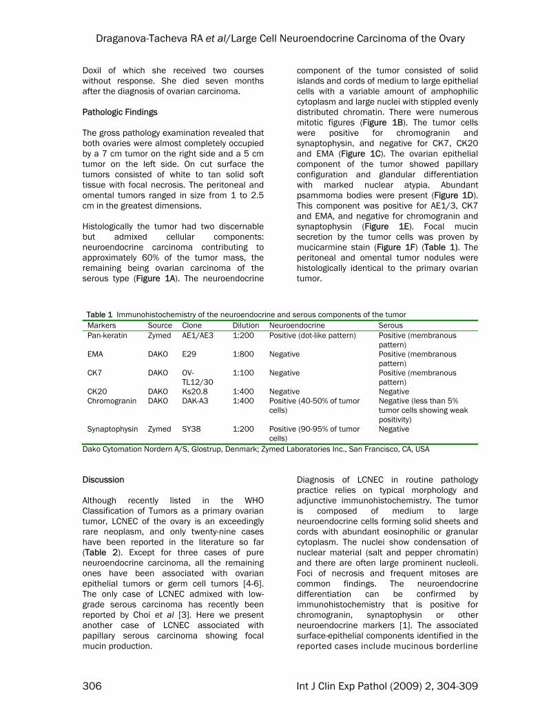
Table 1 Immunohistochemistry of the neuroendocrine and serous components of the tumor
Markers Source Clone Dilution Neuroendocrine Serous Pan-keratin Zymed AE1/AE3 1:200 Positive (dot-like pattern) Positive (membranous
pattern) EMA DAKO E29 1:800 Negative Positive (membranous
pattern) CK7 DAKO OV-
TL12/30 1:100 Negative Positive (membranous
pattern) CK20 DAKO Ks20.8 1:400 Negative Negative Chromogranin DAKO DAK-A3 1:400 Positive (40-50% of tumor
cells) Negative (less than 5% tumor cells showing weak positivity)
Synaptophysin Zymed SY38 1:200 Positive (90-95% of tumor cells)
Negative
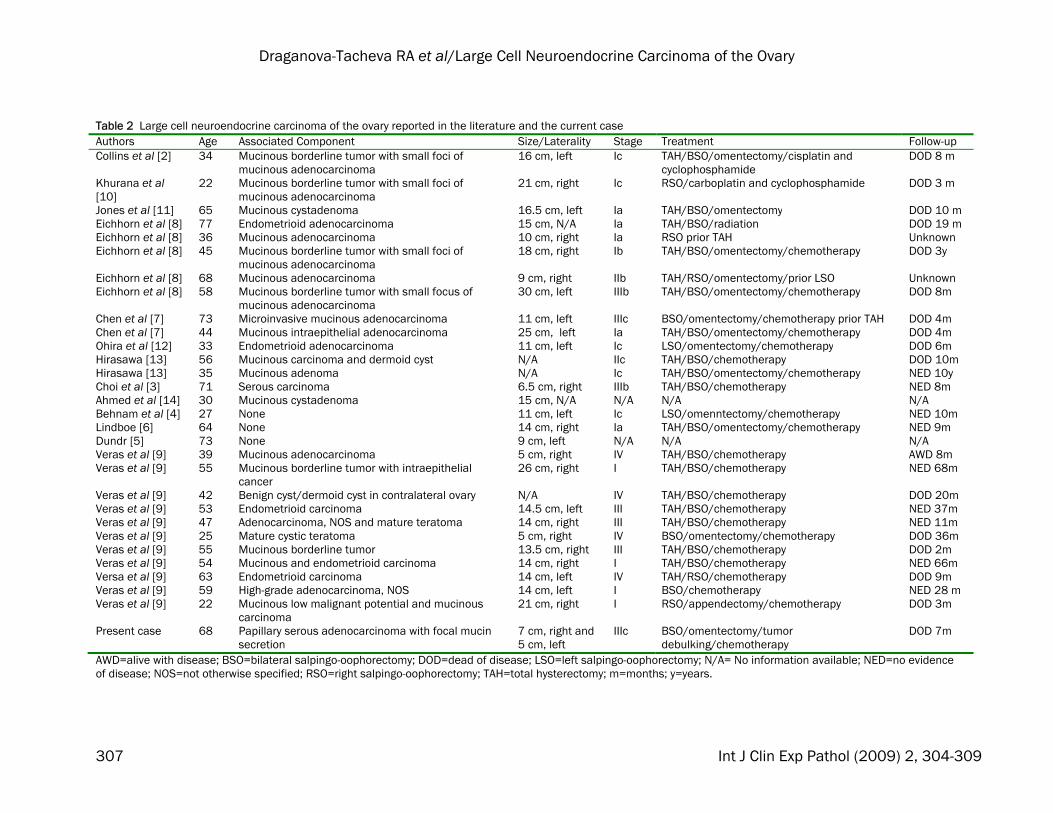
Dako Cytomation Nordern A/S, Glostrup, Denmark; Zymed Laboratories Inc., San Francisco, CA, USA Discussion Although recently listed in the WHO Classification of Tumors as a primary ovarian tumor, LCNEC of the ovary is an exceedingly rare neoplasm, and only twenty-nine cases have been reported in the literature so far (Table 2). Except for three cases of pure neuroendocrine carcinoma, all the remaining ones have been associated with ovarian epithelial tumors or germ cell tumors [4-6]. The only case of LCNEC admixed with low-grade serous carcinoma has recently been reported by Choi et al [3]. Here we present another case of LCNEC associated with papillary serous carcinoma showing focal mucin production.
Diagnosis of LCNEC in routine pathology practice relies on typical morphology and adjunctive immunohistochemistry. The tumor is composed of medium to large neuroendocrine cells forming solid sheets and cords with abundant eosinophilic or granular cytoplasm. The nuclei show condensation of nuclear material (salt and pepper chromatin) and there are often large prominent nucleoli. Foci of necrosis and frequent mitoses are common findings. The neuroendocrine differentiation can be confirmed by immunohistochemistry that is positive for chromogranin, synaptophysin or other neuroendocrine markers [1]. The associated surface-epithelial components identified in the reported cases include mucinous borderline
306 Int J Clin Exp Pathol (2009) 2, 304-309
Draganova-Tacheva RA et al/Large Cell Neuroendocrine Carcinoma of the Ovary
Table 2 Large cell neuroendocrine carcinoma of the ovary reported in the literature and the current case Authors Age Associated Component Size/Laterality Stage Treatment Follow-up Collins et al [2] 34 Mucinous borderline tumor with small foci of
mucinous adenocarcinoma 16 cm, left Ic TAH/BSO/omentectomy/cisplatin and
cyclophosphamide DOD 8 m
Khurana et al [10]
22 Mucinous borderline tumor with small foci of mucinous adenocarcinoma
21 cm, right Ic RSO/carboplatin and cyclophosphamide DOD 3 m
Jones et al [11] 65 Mucinous cystadenoma 16.5 cm, left Ia TAH/BSO/omentectomy DOD 10 m Eichhorn et al [8] 77 Endometrioid adenocarcinoma 15 cm, N/A Ia TAH/BSO/radiation DOD 19 m Eichhorn et al [8] 36 Mucinous adenocarcinoma 10 cm, right Ia RSO prior TAH Unknown Eichhorn et al [8] 45 Mucinous borderline tumor with small foci of
mucinous adenocarcinoma 18 cm, right Ib TAH/BSO/omentectomy/chemotherapy DOD 3y
Eichhorn et al [8] 68 Mucinous adenocarcinoma 9 cm, right IIb TAH/RSO/omentectomy/prior LSO Unknown Eichhorn et al [8] 58 Mucinous borderline tumor with small focus of
mucinous adenocarcinoma 30 cm, left IIIb TAH/BSO/omentectomy/chemotherapy DOD 8m
Chen et al [7] 73 Microinvasive mucinous adenocarcinoma 11 cm, left IIIc BSO/omentectomy/chemotherapy prior TAH DOD 4m Chen et al [7] 44 Mucinous intraepithelial adenocarcinoma 25 cm, left Ia TAH/BSO/omentectomy/chemotherapy DOD 4m Ohira et al [12] 33 Endometrioid adenocarcinoma 11 cm, left Ic LSO/omentectomy/chemotherapy DOD 6m Hirasawa [13] 56 Mucinous carcinoma and dermoid cyst N/A IIc TAH/BSO/chemotherapy DOD 10m Hirasawa [13] 35 Mucinous adenoma N/A Ic TAH/BSO/omentectomy/chemotherapy NED 10y Choi et al [3] 71 Serous carcinoma 6.5 cm, right IIIb TAH/BSO/chemotherapy NED 8m Ahmed et al [14] 30 Mucinous cystadenoma 15 cm, N/A N/A N/A N/A Behnam et al [4] 27 None 11 cm, left Ic LSO/omenntectomy/chemotherapy NED 10m Lindboe [6] 64 None 14 cm, right Ia TAH/BSO/omentectomy/chemotherapy NED 9m Dundr [5] 73 None 9 cm, left N/A N/A N/A Veras et al [9] 39 Mucinous adenocarcinoma 5 cm, right IV TAH/BSO/chemotherapy AWD 8m Veras et al [9] 55 Mucinous borderline tumor with intraepithelial
cancer 26 cm, right I TAH/BSO/chemotherapy NED 68m
Veras et al [9] 42 Benign cyst/dermoid cyst in contralateral ovary N/A IV TAH/BSO/chemotherapy DOD 20m Veras et al [9] 53 Endometrioid carcinoma 14.5 cm, left III TAH/BSO/chemotherapy NED 37m Veras et al [9] 47 Adenocarcinoma, NOS and mature teratoma 14 cm, right III TAH/BSO/chemotherapy NED 11m Veras et al [9] 25 Mature cystic teratoma 5 cm, right IV BSO/omentectomy/chemotherapy DOD 36m Veras et al [9] 55 Mucinous borderline tumor 13.5 cm, right III TAH/BSO/chemotherapy DOD 2m Veras et al [9] 54 Mucinous and endometrioid carcinoma 14 cm, right I TAH/BSO/chemotherapy NED 66m Versa et al [9] 63 Endometrioid carcinoma 14 cm, left IV TAH/RSO/chemotherapy DOD 9m Veras et al [9] 59 High-grade adenocarcinoma, NOS 14 cm, left I BSO/chemotherapy NED 28 m Veras et al [9] 22 Mucinous low malignant potential and mucinous
carcinoma 21 cm, right I RSO/appendectomy/chemotherapy DOD 3m
Present case 68 Papillary serous adenocarcinoma with focal mucin secretion
7 cm, right and 5 cm, left
IIIc BSO/omentectomy/tumor debulking/chemotherapy
DOD 7m
AWD=alive with disease; BSO=bilateral salpingo-oophorectomy; DOD=dead of disease; LSO=left salpingo-oophorectomy; N/A= No information available; NED=no evidence of disease; NOS=not otherwise specified; RSO=right salpingo-oophorectomy; TAH=total hysterectomy; m=months; y=years.
307 Int J Clin Exp Pathol (2009) 2, 304-309

Draganova-Tacheva RA et al/Large Cell Neuroendocrine Carcinoma of the Ovary
tumor, mucinous adenocarcinoma, endometrioid adenocarcinoma, mucinous adenoma/cystadenoma, adenocarcinoma NOS, admixed mucinous and endometrioid carcinoma, and serous carcinoma. The LCNEC component in our case had a morphology typical of neuroendocrine differentiation and an immunoprofile that was positive for neuroendocrine differentiation and negative for epithelial markers. However, the interesting feature of this case was that the intermingled ovarian epithelial tumor consisted of serous carcinoma showing focal mucin production. The high grade nuclear atypia, papillary configuration and glandular formation, and the psammoma bodies formed the classic morphology of a serous carcinoma. The reversed immunoprofile positive for epithelial markers and negative for neuroendocrine differentiation helped establish the diagnosis. Focal mucin secretion has been described in serous carcinomas and its prognostic significance is not established. Whether it modifies their clinical behavior remains to be determined. The differential diagnoses of ovarian LCNEC are other primary and secondary neuroendocrine tumors. Small cell carcinoma has smaller cell size with molding and significant necrosis. Metastatic neuroendocrine carcinoma would almost never admix with ovarian surface epithelial tumors. Most of the reported cases presented with stage I disease, with tumors confined to one ovary. In our case, the tumor was identified in both ovaries with extensive peritoneal disease. It is possible that the tumor started on one side and spread to the other ovary and the peritoneum. However, this assumption cannot be proven in a patient with such widespread disease. It is interesting to note that both Choi’s patient [4] and the current patient with serous carcinoma components presented in advanced stage of disease. Follow-up (2 months to 10 years) was available in 26 of the 30 reported cases (including ours). At the time of publication nine (6 stage I, 3 stage III) of the 30 patients had no evidence of disease. The three long-term survivors (60 months) had stage I disease and received adjuvant chemotherapy. Sixteen patients died of disease 2 months to three
years (median, 8 months) after diagnosis. Nine of these 16 patients were reported to have stage I disease and three of these nine did not receive adjuvant chemotherapy. One patient with stage IV ovarian LCNEC was alive with disease eight months after diagnosis at the time of the report. LCNEC of the ovary appears to be highly aggressive. The presence of LCNEC component in an otherwise usual type epithelial tumor should be reported because of the potential negative prognostic impact of this histologic finding, although there are too few cases in the literature to determine the prognostic impact of the percentage of LCNEC component in an epithelial ovarian tumor. Prior to our report, only the neuroendocrine component of the carcinoma has been described in the metastatic sites when metastasis occurs [7]. Unlike the previously reported cases, our patient’s peritoneal metastases contained both LCNEC and surface-epithelial components identical to those in the primary tumor. Most patients died of disease within 1 year of diagnosis despite extensive surgery and adjuvant chemotherapy [3, 4, 8, 9]. Our patient presented with widespread peritoneal disease and showed an initial response to the chemotherapy followed by rapid progression and death, which is typical of the aggressive behavior of these tumors. In summary, we report a rare case of an ovarian neuroendocrine carcinoma, large cell type, associated with papillary serous carcinoma with mucin production. Please address all correspondences to Xinmin Zhang,, M.D., Department of Pathology and Laboratory Medicine, Temple University, School of Medicine, 3401 N. Broad Street, Philadelphia, PA 19140. Tel: 215-707-8273; Fax: 215-707-8132; Email: [email protected] References [1] Roth LM, Tsubara A, Dietel M and Senzaki H.
Miscellaneous tumors and tumor-like conditions of the ovary. In: Tavassoli FA and Devilee P (eds). Pathology and Genetics of Tumors of the Breast and Female Genital Organs. World Health Organization Classification of Tumors. Lyon, IARC Press, 2003, pp182-190.
[2] Collins RJ, Cheung A, Ngan HY, Wong LC, Chan SY and Ma HK. Primary mixed neuroendocrine
308 Int J Clin Exp Pathol (2009) 2, 304-309

Draganova-Tacheva RA et al/Large Cell Neuroendocrine Carcinoma of the Ovary
309 Int J Clin Exp Pathol (2009) 2, 304-309
and mucinous carcinoma of the ovary. Arch Gynecol Obstet 1991;248:139-143.
[3] Choi YD, Lee JS, Choi C, Park CS and Nam JH. Ovarian neuroendocrine carcinoma, non-small cell type, associated with serous carcinoma. Gynecol Oncol 2007;104:747-752.
[4] Behnam K, Kabus D and Behnam M. Primary ovarian undifferentiated non-small cell carcinoma, neuroendocrine type. Gynecol Oncol 2004;92:372-375.
[5] Dundr P, Fischerova D, Povysil C and Cibula D. Primary pure large-cell neuroendocrine carcinoma of the ovary. Pathol Res Pract 2008; 204:133-137.
[6] Lindboe CF. Large cell neuroendocrine carcinoma of the ovary. APMIS 2007;115:169-176.
[7] Chen KT. Composite large-cell neuroendocrine carcinoma and surface epithelial-stromal neoplasm of the ovary. Int J Surg Pathol 2000; 8:169-174.
[8] Eichhorn JH, Lawrence WD, Young RH and Scully RE. Ovarian neuroendocrine carcinomas of non-small cell type associated with surface epithelial adenocarcinomas. A study of five cases and review of the literature. Int J Gynecol
Pathol 1996;15:303-314. [9] Veras E, Deavers MT, Silva EG and Malpica A.
Ovarian nonsmall cell neuroendocrine carcinoma. A clinicopathologic and immunohistochemical study of 11 cases. Am J Surg Pathol 2007;31:774-782.
[10] Khurana K, Tornos C and Silva E. Ovarian neuroendocrine carcinoma associated with mucinous neoplasm. Arch Pathol Lab Med 1994;118:1032-1034.
[11] Jones K, Diaz J and Donner L. Neuroendocrine carcinoma arising in an ovarian mucinous cystadenoma. Int J Gynecol Pathol 1996;15: 167-170.
[12] Ohira S, Itoh K, Shiozawa T, Horiuchi A, Ono K, Takeuchi H, Hosoda W and Konishi I. Ovarian NSCNEC with paraneoplastic parathyroid hormone-related hypercalcemia. Int J Gynecol Pathol 2004;23:393-397.
[13] Hirasawa T. Ovarian neuroendocrine carcinoma associated with mucinous carcinoma and teratoma. Nippon Rinsho 2004;62:973-978.
[14] Ahmed Z, Aftab K and Kayani N. Ovarian primary neuroendocrine carcinoma of non-small cell type: report of an extremely rare neoplasm. J Pak Med Assoc 2005;55:82-84.


![Neuroendocrine carcinoma of the esophagus: Report of a ...[1,2]. Regarding to small cell (endocrine) carcinoma, small cell lung cancer is a more common disease, ac-counting for up](https://img.dokumen.tips/doc/110x75/5f251236fe328953f826e73a/neuroendocrine-carcinoma-of-the-esophagus-report-of-a-12-regarding-to-small.jpg)