Embed Size (px)
Citation preview
Case ReportFirst Trimester Spontaneous Uterine Rupture in a YoungWoman with Uterine Anomaly
Esra Nur Tola
Department of Obstetrics and Gynecology, Faculty of Medicine, Suleyman Demirel University, Isparta, Turkey
Correspondence should be addressed to Esra Nur Tola; [email protected]
Received 3 October 2013; Accepted 12 November 2013; Published 16 January 2014
Academic Editors: R. P. Kauffman and C.-C. Liang
Copyright © 2014 Esra Nur Tola.This is an open access article distributed under the Creative CommonsAttribution License, whichpermits unrestricted use, distribution, and reproduction in any medium, provided the original work is properly cited.
Spontaneous uterine rupture is a life-threatening obstetrical emergency carrying a high risk for the mother and the fetus.Spontaneous uterine rupture in early pregnancy is very rare complication and it occurs usually in scarred uterus. Uterine anomaliesare one of the reasons for spontaneous unscarred uterine rupture in early pregnancy. Obstetricians must consider this diagnosiswhen a pregnant patient presented with acute abdomen in early pregnancy. We present a case of spontaneous uterine ruptureat 12 weeks of gestation in 24-year-old multigravida who had uterine anomaly presenting as an acute abdomen. Our preoperativediagnosis was ectopic pregnancy. Emergency laparotomy confirmed a spontaneous uterine rupture. Uterine anomaly is a risk factorfor spontaneous uterine rupture in the early pregnancy. Clinical signs of uterine rupture in early pregnancy are nonspecific andmustbe distinguished from acute abdominal emergencies.
1. Introduction
Rupture of a pregnant uterus is one of the life-threateningcomplications associated with obstetric practice [1].There areseveral risk factors associated with uterine rupture (UR), butthe most common is a previous Cesarean section. Unscarreduterine rupture (UUR) is a rare event that usually occurs inlate pregnancy or during labour. Risk factors forUUR includehigh parity, placental abnormalities, and uterine anomaly.UUR during pregnancy, especially before the end of thesecond trimester, occurs relatively rarely and is associatedwith high mortality and morbidity for both the fetus andmother.
We here report a case of a spontaneous unscarred uterinerupture (SUUR) in early pregnancy, in a woman with abicornuate uterus.
2. Case Report
A 24-year-old woman was admitted to our departmentwith 3-month amenorrhea and sudden, severe, generalizedabdominal pain and vaginal bleeding of 2-hour duration.Her first pregnancy resulted in abortus at 8 gestationalweeks, but no surgical procedure was performed. On physical
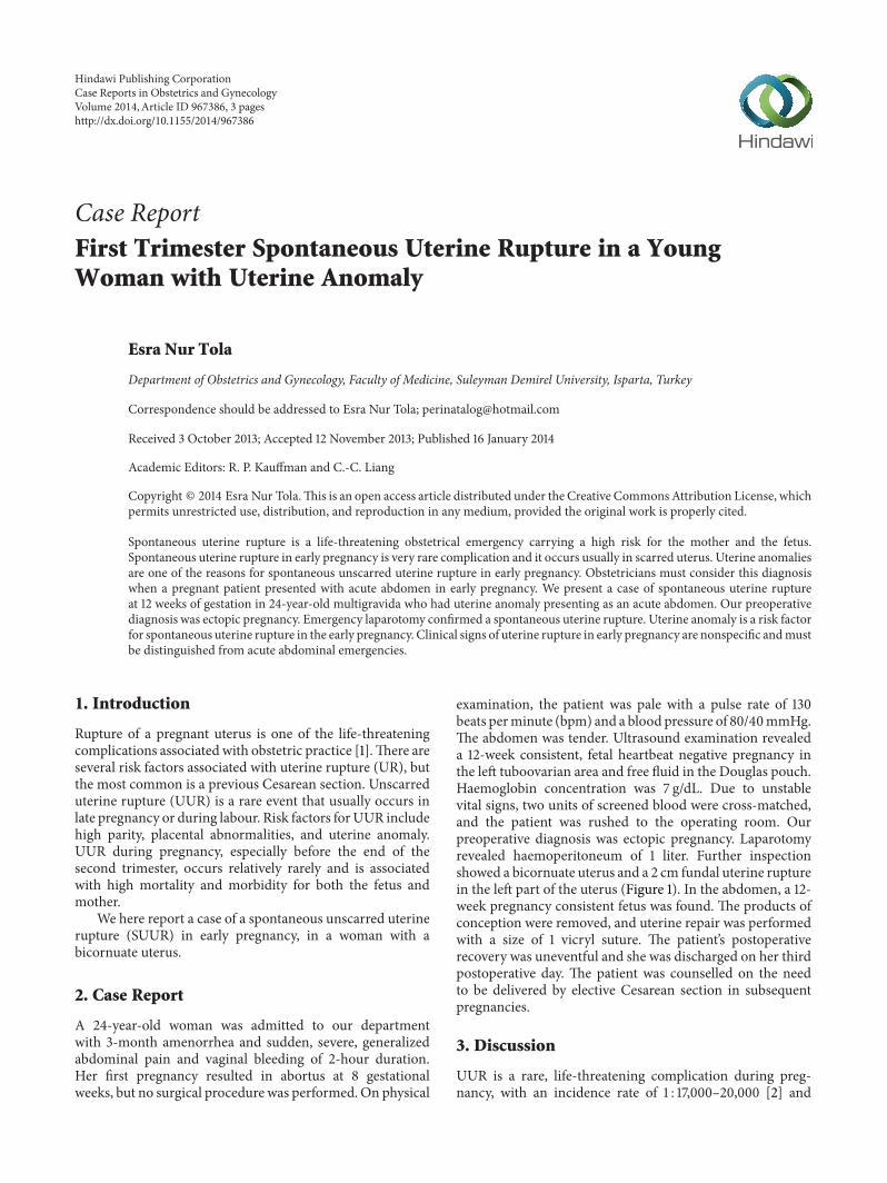
examination, the patient was pale with a pulse rate of 130beats perminute (bpm) and a blood pressure of 80/40mmHg.The abdomen was tender. Ultrasound examination revealeda 12-week consistent, fetal heartbeat negative pregnancy inthe left tuboovarian area and free fluid in the Douglas pouch.Haemoglobin concentration was 7 g/dL. Due to unstablevital signs, two units of screened blood were cross-matched,and the patient was rushed to the operating room. Ourpreoperative diagnosis was ectopic pregnancy. Laparotomyrevealed haemoperitoneum of 1 liter. Further inspectionshowed a bicornuate uterus and a 2 cm fundal uterine rupturein the left part of the uterus (Figure 1). In the abdomen, a 12-week pregnancy consistent fetus was found. The products ofconception were removed, and uterine repair was performedwith a size of 1 vicryl suture. The patient’s postoperativerecovery was uneventful and she was discharged on her thirdpostoperative day. The patient was counselled on the needto be delivered by elective Cesarean section in subsequentpregnancies.
3. Discussion
UUR is a rare, life-threatening complication during preg-nancy, with an incidence rate of 1 : 17,000–20,000 [2] and
Hindawi Publishing CorporationCase Reports in Obstetrics and GynecologyVolume 2014, Article ID 967386, 3 pageshttp://dx.doi.org/10.1155/2014/967386

2 Case Reports in Obstetrics and Gynecology
Figure 1: Fundal uterine rupture in the left part of the bicornuateuterus.
a mortality andmorbidity rate estimated to be between 20.8%and 64.6% [3].
UR is usually observed in association with uterine scar-ring either in late pregnancy or during labour [4]. First andearly second trimester UURs are very rare, and there areonly a few cases in literature describing first and early secondtrimester UURs [4–7]. In our case, UUR occurred in thetwelfth week of pregnancy.
Although a prior Cesarean section is a major risk factorfor UR, in patients with unscarred uteri, high parity (≥4births) is a major risk factor. Other risk factors for UURinclude abnormal placentation, uterine anomalies, obstetricmanoeuvres,malpresentations, excessive uterine expressions,curettage, injudicious use of oxytocin, uterine diverticula [8],and chronic corticosteroid use [3], whereas some cases haveno obvious cause [9]. In our case, uterine anomaly may beimplicated in the UR, because the patient had a bicornuateuterus, and there were no other obvious risk factors. Singhand Jain and Kahyaoglu et al. previously reported UUR withuterine anomaly in the first trimester [6, 10]. A few cases ofUUR in the early trimester with no previous risk factors [5, 7]and as a result of placenta percreta, have been reported [11, 12].
Clinical signs of UR in early pregnancy are nonspecificand must be distinguished from acute abdominal emer-gencies. Abdominal pain, vaginal bleeding, and vomitingare classic findings. Differential diagnoses are bleeding cor-pus luteum, heterotropic or ectopic pregnancy, and molarpregnancy with secondary invasion [4]. The most relevantdifferential diagnosis is ectopic pregnancy [6]. Sometimesultrasound has limited value and urgent surgery is necessaryto prevent catastrophic sequelae. An emergency laparoscopyor laparotomy is needed for the correct diagnosis and toenable the necessary treatment to take place. Early correctdiagnosis and proper management are necessary to decreasethe high maternal and fetal mortality and morbidity ratesassociated with UR. In our case, our initial diagnosis wasectopic pregnancy, and we performed emergency laparotomyafter judging that laparoscopic instrumentation was deficientand because of the unstable vital signs of the patient.
UUR usually occurs in the lower segment (the weakestpart) of uterus [13]. If the rupture part is the fundus,as in our case, the diagnosis is often delayed because
the haemorrhage is not revealed immediately, as bloodcollects in the intraperitoneal space [13].
Early surgical intervention is usually the key to successfultreatment of UR. Treatment will primarily depend on theextent of the lesion, the parity, age and condition of thepatient, and expertise of the surgeon. Although in thepast hysterectomy has been suggested for the therapeuticmanagement, recent studies have shown that the suture canbe performed with bilateral tubal ligation or suture withouttubal ligation in women who wish to preserve fertility with arecurrence risk of UR assessed to be between 4 and 19% at asubsequent pregnancy [14]. For this reason the patient mustbe counselled on the need to undergo aCesarean section in allfuture pregnancies. In our case we performed uterine suturewithout tubal ligation because our patient had no previouschildren.
4. Conclusion
In conclusion, UUR in early pregnancy is a rare and poten-tially catastrophic event. Uterine anomalies are one of thereasons of UUR.The current case highlights uterine anomalyas a risk factor for spontaneous UR in the first trimesterof pregnancy. Clinical signs of UR in early pregnancy arenonspecific and must be distinguished from other acuteabdominal emergencies.
Abbreviations
UUR: Unscarred uterine ruptureSUUR: Spontaneous unscarred uterine ruptureUR: Uterine rupture.
Conflict of Interests
The author declares that there is no conflict of interestsregarding the publication of this paper.
References
[1] M. Gueye, M. Mbaye, M. D. Ndiaye-Gueye et al., “Spontaneousuterine rupture of an unscarred uterus before labour,” CaseReports in Obstetrics and Gynecology, vol. 2012, Article ID598356, 3 pages, 2012.
[2] K. Ofir, E. Sheiner, A. Levy, M. Katz, and M. Mazor, “Uterinerupture: differences between a scarred and an unscarred uterus,”American Journal of Obstetrics and Gynecology, vol. 191, no. 2,pp. 425–429, 2004.
[3] D. C. Schrinsky and R. C. Benson, “Rupture of the pregnantuterus: a review,” Obstetrical and Gynecological Survey, vol. 33,no. 4, pp. 217–232, 1978.
[4] S. Ijaz, A. Mahendru, and D. Sanderson, “Spontaneous uterinerupture during the 1st trimester: a rare but life-threateningemergency,” Journal of Obstetrics and Gynaecology, vol. 31, no.8, p. 772, 2011.
[5] M. M. Biljan, K. Cushing, I. W. McDicken, and A. S. Garden,“Spontaneous uterine rupture in the first trimester of preg-nancy,” Journal of Obstetrics and Gynaecology, vol. 16, no. 3, pp.174–175, 1996.

Case Reports in Obstetrics and Gynecology 3
[6] A. Singh and S. Jain, “Spontaneous rupture of unscarreduterus in early pregnancy—a rare entity,” Acta Obstetricia etGynecologica Scandinavica, vol. 79, no. 5, pp. 431–432, 2000.
[7] K. I. Dibbs, R. H. Ball, and P. C. Huettner, “Spontaneous uterinerupture and hemoperitoneum in the first trimester,” AmericanJournal of Perinatology, vol. 12, no. 6, pp. 439–441, 1995.
[8] I. Uzun, A. Yıldırım, I. Kalelioglu, and R. Has, “Spontaneousrupture of unscarred uterus at 27 weeks of gestation,” Archivesof Gynecology and Obstetrics, vol. 281, no. 6, pp. 999–1001, 2010.
[9] B. Chigbu, S. Onwere, C. Kamanu, C. Aluka, E. Adibe, and C.Onichakwe, “Rupture of the uterus in a primigravida: a casereport,” Nigerian Journal of Clinical Practice, vol. 13, no. 2, pp.233–234, 2010.
[10] S. Kahyaoglu, I. Turgay, and O. Kaymak, “Onyedi haftalıknonkommunike rudimenter uterus horn gebeligi ve uterusrupturu: olgu sunumu,” Journal of Perinatalogy, vol. 13, pp. 179–182, 2005.
[11] N. H. Morken and H. Henriksen, “Plasenta percreata—twocases and reviewof the literature,”European Journal ofObstetrics& Gynecology, vol. 10, pp. 112–115, 2001.
[12] W. J. LeMaire, C. Louisy, K. Dalessandri, and F. Muschenheim,“Placenta percreta with spontaneous rupture of an unscarreduterus in the second trimester,” Obstetrics and Gynecology, vol.98, no. 5, pp. 927–929, 2001.
[13] M. Misra, R. Roychowdhury, and N. C. Sarkar, “The sponta-neous prelabour rupture of an unscarred uterus at 34 weeks ofpregnancy,” Journal of Clinical and Diagnostic Research, vol. 7,pp. 548–549, 2013.
[14] S. Ahmadi, M. Nouira, M. Bibi et al., “Uterine rupture ofthe unscarred uterus. About 28 cases,” Gynecologie ObstetriqueFertilite, vol. 31, no. 9, pp. 713–717, 2003.

Submit your manuscripts athttp://www.hindawi.com
Stem CellsInternational
Hindawi Publishing Corporationhttp://www.hindawi.com Volume 2014
Hindawi Publishing Corporationhttp://www.hindawi.com Volume 2014
MEDIATORSINFLAMMATION
of
Hindawi Publishing Corporationhttp://www.hindawi.com Volume 2014
Behavioural Neurology
EndocrinologyInternational Journal of
Hindawi Publishing Corporationhttp://www.hindawi.com Volume 2014
Hindawi Publishing Corporationhttp://www.hindawi.com Volume 2014
Disease Markers
Hindawi Publishing Corporationhttp://www.hindawi.com Volume 2014
BioMed Research International
OncologyJournal of
Hindawi Publishing Corporationhttp://www.hindawi.com Volume 2014
Hindawi Publishing Corporationhttp://www.hindawi.com Volume 2014
Oxidative Medicine and Cellular Longevity
Hindawi Publishing Corporationhttp://www.hindawi.com Volume 2014
PPAR Research
The Scientific World JournalHindawi Publishing Corporation http://www.hindawi.com Volume 2014
Immunology ResearchHindawi Publishing Corporationhttp://www.hindawi.com Volume 2014
Journal of
ObesityJournal of
Hindawi Publishing Corporationhttp://www.hindawi.com Volume 2014
Hindawi Publishing Corporationhttp://www.hindawi.com Volume 2014
Computational and Mathematical Methods in Medicine
OphthalmologyJournal of
Hindawi Publishing Corporationhttp://www.hindawi.com Volume 2014
Diabetes ResearchJournal of
Hindawi Publishing Corporationhttp://www.hindawi.com Volume 2014
Hindawi Publishing Corporationhttp://www.hindawi.com Volume 2014
Research and TreatmentAIDS
Hindawi Publishing Corporationhttp://www.hindawi.com Volume 2014
Gastroenterology Research and Practice
Hindawi Publishing Corporationhttp://www.hindawi.com Volume 2014
Parkinson’s Disease
Evidence-Based Complementary and Alternative Medicine
Volume 2014Hindawi Publishing Corporationhttp://www.hindawi.com