Embed Size (px)
Citation preview

IP International Journal of Ocular Oncology and Oculoplasty 2021;7(2):204–206
Content available at: https://www.ipinnovative.com/open-access-journals
IP International Journal of Ocular Oncology andOculoplasty
Journal homepage: https://ijooo.org/
Case Report
Diffuse infiltrating retinoblastoma: A diagnostic conundrum
Aditi Mehta1,*, Dipankar Das2, Kasturi Bhattacharjee1, Manabjyoti Barman1,Ganesh Chandra Kuri1, Hemlata Deka1, Harsha Bhattacharjee1,*, Nilutparna Deori1,Vatsalya Venkatraman1, Apurba Deka2
1Dept. of Ophthalmology, Sri Sanakaradeva Nethralaya, Sri Sanakaradeva Nethralaya, Guwahati, Assam, India2Dept. of Pathology, Uveitis and Neuro-ophthalmology services, Sri Sanakaradeva Nethralaya, Guwahati, Assam, India
A R T I C L E I N F O
Article history:Received 06-06-2021Accepted 18-06-2021Available online xx xx xxxx
Keywords:RetinoblastomaDiffuse infiltratingLeukocoriaTNM stagingHereditary retinoblastoma
A B S T R A C T
Purpose: Diffuse infiltrating retinoblastoma (DIR) is characterized by absence of intraocular mass, lack ofcalcification. It may mimic inflammatory uveitis or exudative retinopathy.Observations: An eight-years-old boy presented with progressive loss of vision in left eye. Clinicalevaluation revealed neovascular glaucoma with a yellow–gray fundal glow, exudative retinal detachment,subretinal exudation and telangiectatic vessels. The presentation was consistent with exudative retinopathy(Coat’s disease) but for the presence of a family history of retinoblastoma in the younger sibling. Despitethe absence of an intraocular mass or calcification on multimodal imaging, the enucleation was done on thebasis of clinical suspicion of retinoblastoma. Histopathology confirmed a diagnosis of DIR.Conclusions: DIR can pose a diagnostic challenge due to its non-characteristic clinical and imagingfeatures and atypical presentation. A high index of suspicion along with a positive family history waskey to diagnosis in our case; histopathology was confirmatory.
© This is an open access article distributed under the terms of the Creative Commons AttributionLicense (https://creativecommons.org/licenses/by/4.0/) which permits unrestricted use, distribution, andreproduction in any medium, provided the original author and source are credited.
1. Introduction
Retinoblastoma is the commonest intraocular childhoodmalignancy with an incidence of 1 in 14,000 to 20,000live births. Mean age of diagnosis depends on laterality andheredity; 90% of are diagnosed by three years of age.1 Thepresenting symptoms are determined by the size, extent aswell as location of the tumour, which can be exophytic (intosubretinal space), endophytic (into vitreous cavity), mixed,or diffusely infiltrating (flat lesion along ocular coats).Leukocoria is often the first symptom, along with decreasedvision or squinting.2,3 The differential diagnoses includeCoat’s disease, persistent fetal vasculature, congenitalcataract, coloboma, familial exudative vitreoretinopathy,retinopathy of prematurity, toxocariasis, endophthalmitis,and rare tumours like medulloepithelioma, astrocytic
* Corresponding authors.E-mail address: [email protected] (A. Mehta).
hamartoma. Though they all great morbidity, malignancieshave the gravest prognosis, especially if diagnosis isdelayed.
Retinoblastoma diagnosis involves clinical evaluationsupplemented with ultrasonography and radiologicalimaging. Presence of calcification within the lesion,demonstrated as high spikes on ultrasonography B-scan,high density on computed tomography (CT) and areasof hypo-intensity on magnetic resonance imaging (MRI)is a diagnostic indicator.4 Other causes of calcificationinclude phthisis bulbi (in setting of trauma and/orchronic inflammation), hyperparathyroidism (systemicfeatures), choroidal osteoma, drusen or idiopathic; these aredistinguished by both demographics and morphologyof calcification. In retinoblastoma, calcification ispresent within the mass or rarely, along sclerochoroidalcoats, conforming the globe contour (diffuse infiltratingretinoblastoma, DIR, late stage).5
https://doi.org/10.18231/j.ijooo.2021.0402581-5024/© 2021 Innovative Publication, All rights reserved. 204
Mehta et al. / IP International Journal of Ocular Oncology and Oculoplasty 2021;7(2):204–206 205
DIR is uncommon, occurring in about 1-2% of tumours.This plaque like lesion grows slowly towards the anteriorsegment, does not develop calcification till late stage,and manifests with pseudo-inflammatory complications.6,7
We present a challenging case of DIR and its clinicaland histopathologic correlation. This report adheres to theDeclaration of Helsinki and written informed consent of thelegal guardians was taken for photographs.
2. Case Description
An eight-years-old boy presented with graduallyprogressive, painless diminution of vision of left eye(OS) for the past two months. The visual acuity was 6/6in right eye (OD) and perception of light in OS. Ocularmotility range was complete and there was no manifestsquint. Slit lamp examination of OS showed ciliarycongestion, relative afferent pupillary defect, ectropionuveae, clear lens, and a yellow-gray fundal glow (Fig.1a). Fundus examination revealed an exudative retinaldetachment with a retinal cyst, vitreous haemorrhage,dilated telangiectatic vessels in the temporal periphery andmarked subretinal exudation (Fig. 1b). Intraocular pressures(IOP) were 14 and 26 mmHg in OD and OS respectively.Right eye examination was unremarkable. A clinicaldiagnosis of Coat’s disease was formulated. The B-scanconfirmed exudative retinal detachment with intraretinalcyst (Fig. 1c). Mild thickening of ocular coats was notedbut calcification was not discernible. The optic nerveshadow was unremarkable. While the parents were beingcounselled, a significant family history was revealed. Thechild had a deceased younger sibling, who was diagnosedwith bilateral retinoblastoma with intracranial extension atthe age of two years, four years prior. Our case had notbeen screened till now. With this significant history, we keptretinoblastoma as a likely diagnosis, even though clinicalfindings favoured an exudative retinopathy.
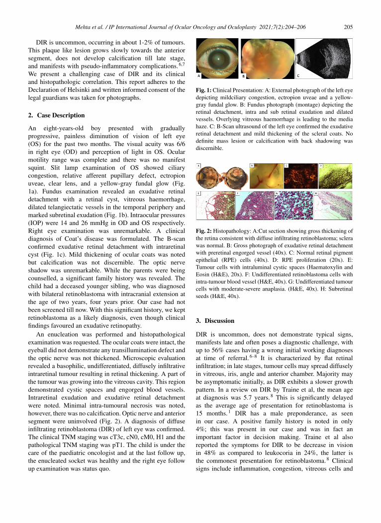
An enucleation was performed and histopathologicalexamination was requested. The ocular coats were intact, theeyeball did not demonstrate any transillumination defect andthe optic nerve was not thickened. Microscopic evaluationrevealed a basophilic, undifferentiated, diffusely infiltrativeintraretinal tumour resulting in retinal thickening. A part ofthe tumour was growing into the vitreous cavity. This regiondemonstrated cystic spaces and engorged blood vessels.Intraretinal exudation and exudative retinal detachmentwere noted. Minimal intra-tumoural necrosis was noted,however, there was no calcification. Optic nerve and anteriorsegment were uninvolved (Fig. 2). A diagnosis of diffuseinfiltrating retinoblastoma (DIR) of left eye was confirmed.The clinical TNM staging was cT3c, cN0, cM0, H1 and thepathological TNM staging was pT1. The child is under thecare of the paediatric oncologist and at the last follow up,the enucleated socket was healthy and the right eye followup examination was status quo.
Fig. 1: Clinical Presentation: A: External photograph of the left eyedepicting mildciliary congestion, ectropion uveae and a yellow-gray fundal glow. B: Fundus photograph (montage) depicting theretinal detachment, intra and sub retinal exudation and dilatedvessels. Overlying vitreous haemorrhage is leading to the mediahaze. C: B-Scan ultrasound of the left eye confirmed the exudativeretinal detachment and mild thickening of the scleral coats. Nodefinite mass lesion or calcification with back shadowing wasdiscernible.
Fig. 2: Histopathology: A:Cut section showing gross thickening ofthe retina consistent with diffuse infiltrating retinoblastoma; sclerawas normal. B: Gross photograph of exudative retinal detachmentwith preretinal engorged vessel (40x). C: Normal retinal pigmentepithelial (RPE) cells (40x). D: RPE proliferation (20x). E:Tumour cells with intraluminal cystic spaces (Haematoxylin andEosin (H&E), 20x). F: Undifferentiated retinoblastoma cells withintra-tumour blood vessel (H&E, 40x). G: Undifferentiated tumourcells with moderate-severe anaplasia. (H&E, 40x). H: Subretinalseeds (H&E, 40x).
3. Discussion
DIR is uncommon, does not demonstrate typical signs,manifests late and often poses a diagnostic challenge, withup to 56% cases having a wrong initial working diagnosesat time of referral.6–8 It is characterized by flat retinalinfiltration; in late stages, tumour cells may spread diffuselyin vitreous, iris, angle and anterior chamber. Majority maybe asymptomatic initially, as DIR exhibits a slower growthpattern. In a review on DIR by Traine et al, the mean ageat diagnosis was 5.7 years.8 This is significantly delayedas the average age of presentation for retinoblastoma is15 months.1 DIR has a male preponderance, as seenin our case. A positive family history is noted in only4%; this was present in our case and was in fact animportant factor in decision making. Traine et al alsoreported the symptoms for DIR to be decrease in visionin 48% as compared to leukocoria in 24%, the latter isthe commonest presentation for retinoblastoma.8 Clinicalsigns include inflammation, congestion, vitreous cells and

206 Mehta et al. / IP International Journal of Ocular Oncology and Oculoplasty 2021;7(2):204–206
raised IOP; our case had congestion and raised IOP.7,8
Several mechanisms regarding tumour antigen presentation,adhesion, heterotopic precursors and variable immuneresponse have been proposed for the special growth patternseen in DIR, and research is ongoing.9 Calcification, whichis considered a hallmark of retinoblastoma, is conspicuouslyabsent and thus imaging may not be conclusive.10 In thepresence of uveitis-like picture with anterior chamber cells,a diagnostic paracentesis may be confirmatory. In our case,the suspicion for malignancy was heightened with a positivefamily history. An enucleation served as a diagnostic andtherapeutic procedure. Our case also highlights a missedopportunity. With parental counselling and education aboutretinoblastoma and its hereditary nature, this child couldhave been screened at least four years prior, when hisyounger sibling was detected with retinoblastoma.
4. Conclusion
A high index of suspicion, thorough clinical examinationas well as detailed history is important in all paediatriceye diseases. DIR remains a clinical diagnostic challengeand histopathology is confirmatory, and guides furthermanagement.
5. Source of Funding
None.
6. Conflicts of Interest
All contributing authors declare no conflict of interest.
References1. Darwich R, Ghazawi F, Rahme E, Alghazawi N, Burnier JV, Sasseville
D, et al. Retinoblastoma Incidence Trends in Canada: A NationalComprehensive Population-Based Study. J Pediatr OphthalmolStrabismus. 2019;56(2):124–30. doi:10.3928/01913913-20190128-02.
2. Balmer A, Munier F. Differential diagnosis of leukocoria andstrabismus, first presenting signs of retinoblastoma. Clin Ophthalmol.2007;1(4):431–9.
3. Abramson DH, Beaverson K, Sangani P, Vora RA, Lee TC,Hochberg HM, et al. Screening for Retinoblastoma: PresentingSigns as Prognosticators of Patient and Ocular Survival. Pediatrics.2003;112(6):1248–55. doi:10.1542/peds.112.6.1248.
4. Apushkin MA, Apushkin MA, Shapiro MJ, MafeeMF. Retinoblastoma and Simulating Lesions: Role of
Imaging. Neuroimaging Clin N Am. 2005;15(1):49–67.doi:10.1016/j.nic.2005.02.003.
5. Levy J, Frenkel S, Neufeld M, Pe’er J. Calcifications inRetinoblastoma: Histologic Findings and Statistical Analysis of 302Cases. Invest Ophthalmol Vis Sci. 2010;95(8):2074.
6. Morgan G. Diffuse infiltrating retinoblastoma. Br J Ophthalmol.1971;55(9):600–6. doi:10.1136/bjo.55.9.600.
7. Jhaj G, Damarjian T, Kopplin L. Diffuse Infiltrating Retinoblastoma.Retina. 2018;38(10):e82–3. doi:10.1097/iae.0000000000002344.
8. Traine PG, Schedler KJ, Rodrigues EB. Clinical Presentation andGenetic Paradigm of Diffuse Infiltrating Retinoblastoma: A Review.Ocul Oncol Pathol. 2015;2(3):128–32. doi:10.1159/000441528.
9. Schedler KJE, Traine PG, Lohmann DR, Haritoglou C,Metz KA, Rodrigues EB, et al. Hereditary DiffuseInfiltrating Retinoblastoma. Ophthal- mic Genet . 2014;3:1–3.doi:10.3109/13816810.2014.921315.
10. Brisse HJ, Lumbroso L, Fréneaux PC. Sonographic, CT, andMR imaging findings in diffuse infiltrative retinoblastoma: report oftwo cases with histologic comparison. AJNR Am J Neuroradiol.2001;22(3):499–504.
Author biography
Aditi Mehta, Fellow
Dipankar Das, Consultant
Kasturi Bhattacharjee, Consultant
Manabjyoti Barman, Consultant
Ganesh Chandra Kuri, Consultant
Hemlata Deka, Consultant
Harsha Bhattacharjee, Consultant
Nilutparna Deori, Consultant
Vatsalya Venkatraman, Fellow
Apurba Deka, Laboratory Technician
Cite this article: Mehta A, Das D, Bhattacharjee K, Barman M, KuriGC, Deka H, Bhattacharjee H, Deori N, Venkatraman V, Deka A.Diffuse infiltrating retinoblastoma: A diagnostic conundrum. IP Int JOcul Oncol Oculoplasty 2021;7(2):204-206.