Embed Size (px)
Citation preview

Case ReportAn Endoscopic Endonasal Approach forEarly-Stage Olfactory Neuroblastoma: An Evaluation of2 Cases with Minireview of Literature
Hidenori Yokoi,1 Satoru Kodama,2 Yasunao Kogashiwa,1 Yuma Matsumoto,1
Yasuo Ohkura,3 Takayuki Nakagawa,4 and Naoyuki Kohno1
1Department of Otolaryngology, Head and Neck Surgery, Kyorin University School of Medicine, 6-20-2 Shinkawa,Mitaka, Tokyo 181-8611, Japan2Department of Otolaryngology, Faculty of Medicine, Oita University, 1-1 Idaigaoka, Hazama-cho, Yufu, Oita 879-5593, Japan3Department of Pathology, Kyorin University School of Medicine, 6-20-2 Shinkawa, Mitaka, Tokyo 181-8611, Japan4Department of Otolaryngology, Head and Neck Surgery, Graduate School of Medicine, Kyoto University,54 Shogoin Kawahara-cho, Sakyo-ku, Kyoto 606-8507, Japan
Correspondence should be addressed to Hidenori Yokoi; [email protected]
Received 31 October 2014; Revised 21 December 2014; Accepted 24 December 2014
Academic Editor: Rong-San Jiang
Copyright © 2015 Hidenori Yokoi et al.This is an open access article distributed under the Creative Commons Attribution License,which permits unrestricted use, distribution, and reproduction in any medium, provided the original work is properly cited.
We describe the clinical findings in two patients with pathologically diagnosed olfactory neuroblastoma (ONB) of the sinonasalarea and the surgical methods used for its treatment. Using an endoscopic endonasal approach (EEA) without dura resection,along with radiotherapy, we successfully treated ONB at the Kadish stage A. One of our patients, however, experienced tumorrecurrence 24 years after open surgery with radiotherapy that was conducted at another hospital. This patient was no longereligible for radiotherapy, and the tumor was therefore resected with dura resection using an EEA combined with duraplasty. Thedura resection with duraplasty using fascia lata and a pedicled nasal septal flap was minimally invasive. As with surgery withoutduraplasty, a postoperative computed tomography (CT) examination revealed that EEA with duraplasty led to quick improvementof the postoperative inflammatory response as well as pneumocranium. Here, we investigated whether to modify the methodof surgery depending upon the primary site of early-stage ONB. We suggest that, in early-stage ONB, an endoscopic endonasalapproach is an effective and less invasive method. It is also advisable to perform dura mater resection of the lesion site despite theabsence of obvious intracranial invasions in image findings.
1. Introduction
Recently, much progress has been made in the field of oto-laryngology with regard to the use of endoscopic endonasalsurgery. In recent years, this type of surgery has been suc-cessfully performed on patients with skull base tumors. It hasalso been reported that an extended endoscopic endonasalapproach (EEA) is effective in the treatment of malignantsinonasal tumors such as olfactory neuroblastoma (ONB),chondrosarcoma, chordoma, early-stage squamous cell car-cinoma, and adenocarcinoma [1]. Here, we investigated thebenefits of modifying themethod of surgery depending uponthe primary site of early-stage ONB.
2. Cases
Case 1. A 42-year-old man presented with a chief complaintof right-sided epistaxis and nasal stuffiness. He had nonotable medical history. An anterior rhinoscopy revealed adark-reddish tumour occupying the right nasal cavity (Figure1(a)). A biopsy of the nasal cavity (performed under localanesthesia) led to a diagnosis of ONB.
A nonenhanced coronal computed tomography (CT)image showed a mass with a polyp-like appearance in theright nasal cavity, arising from the cribriform plate. Nobone destruction was found. A contrast-enhanced coronalCT image detected a strongly enhanced mass. A magnetic
Hindawi Publishing CorporationCase Reports in OtolaryngologyVolume 2015, Article ID 541026, 7 pageshttp://dx.doi.org/10.1155/2015/541026

2 Case Reports in Otolaryngology
Nasalseptum
(a) (b)
(c) (d)
Figure 1: Preoperative local finding andMRI in case 1. (a) Tumor in the right nasal cavity (black arrow). (b) T1-weighted axial image showinglow-intensity mass (white arrow). (c) T2-weighted coronal image showing a heterogeneous-intensity mass (black arrow). (d) T1-weightedcoronal image (gadolinium+) showing contrast enhancement (black arrow).
resonance image (MRI) T1-weighted axial image revealeda low-intensity signal (Figure 1(b)), whereas a T2-weightedcoronal image showed a clear heterogeneous mass and aT1-weighted coronal image provided a high-contrast image(Figures 1(c) and 1(d)). There was no evidence of the tumorhaving invaded the dura or intracranial space. No metastasiswas evident from the examination of the positron emissiontomography-CT (PET-CT). Therefore, the mass was diag-nosed as an ONB in Kadish stage A.
Extirpation of the lesion by EEA was performed undergeneral anesthesia.The tumor was resected from the base, theright ethmoid sinus was opened, and themiddle nasal conchaand superior nasal concha were removed. Intraoperativerapid diagnostic tests were performed as needed.Themucosaaround the base of the tumor was then abraded from the baseof the nose and the cribriform plate was removed. Followingthis, the dura mater was exposed (Figure 2(a)) and thefila olfactoria and surrounding mucosa were assessed usingrapid diagnostic tests (Figure 2(b)).There was no evidence ofresidual tumor. Surgery was completed after a pedicled nasalseptal flap was created to cover the dura mater (Figure 2(c)).Hematoxylin-eosin stains revealed a nest-like tumor mass
under the mucosa. Tumor cells showed monotonous growth.Based on the infrequency of both anisokaryosis and mitoticfigures, the tumor was diagnosed as an ONB, Hyams’s gradeI-II (Figure 3). Gamma knife irradiation at a dose of 18Gy (abiological effective dose of 54Gy) was performed 10 weeksafter surgery. An MRI with contrast enhancement on theT1-weighted coronal image (Figure 4(a)), PET-CT scanning,and localized findings (Figure 4(b)) 50 months after surgeryshowed no evidence of tumor recurrence.
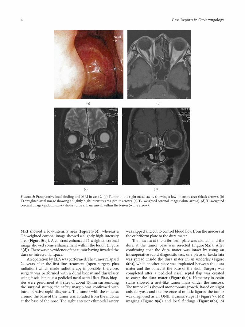
Case 2. A 67-year-old man was diagnosed with an ONB(Kadish grade A-B) in 1989. He underwent lateral rhinotomyfollowed by irradiation and regularly completed follow-upvisits at another hospital. At the end of August 2012, or 24years after the initial tumor, the patient was referred to usfor nosebleeds resulting from a mass in his right nasal cavity(Figure 5(a)).The tumor was biopsied under local anesthesia,and a diagnosis of recurrent ONB was made.
A coronal CT image showed themass occupying the rightanterior ethmoid sinus without a middle nasal turbinate. Acontrast-enhanced axial CT scan showed patchy heteroge-neous enhancement within the mass. A T1-weighted axial

Case Reports in Otolaryngology 3
(a) (b) (c)
Figure 2: Intraoperative findings in case 1. (a) Only the cribriform plate (white arrow) was removed; biopsy of the dura mater (whitearrowhead) was not performed. (b) It was confirmed through rapid intraoperative diagnosis that there was no obvious tumor infiltrationin the cribriform plate mucosa, including a part of the olfactory glomeruli (white arrow), after tumor resection. (c) The dura mater wascovered using a pedicled nasal septal flap (white arrow).
H-E ×10
Figure 3: Pathological findings in case 1. Hematoxylin-eosin staining revealed a nest-like tumor mass under the mucosa. The tumor cellsshowedmonotonous growth. Based on infrequent anisokaryosis and mitotic figures, the tumor was diagnosed as an ONB, Hyams’s stage I-II.
(a) (b)
Figure 4: Postoperative MRI and local finding in case 1. (a) A T1-weighted coronal image (gadolinium+) showing no recurring mass. (b)Thewhite arrow shows the pedicled nasal septal flap in the right nasal cavity.
4 Case Reports in Otolaryngology
Nasalseptum
(a) (b)
(c) (d)
Figure 5: Preoperative local finding and MRI in case 2. (a) Tumor in the right nasal cavity showing a low-intensity area (black arrow). (b)T1-weighted axial image showing a slightly high-intensity area (white arrow). (c) T2-weighted coronal image (white arrow). (d) T1-weightedcoronal image (gadolinium+) shows some enhancement within the lesion (white arrow).
MRI showed a low-intensity area (Figure 5(b)), whereas aT2-weighted coronal image showed a slightly high-intensityarea (Figure 5(c)). A contrast enhanced T1-weighted coronalimage showed some enhancement within the lesion (Figure5(d)).There was no evidence of the tumor having invaded thedura or intracranial space.
An operation by EEAwas performed.The tumor relapsed24 years after the first-line treatment (open surgery plusradiation) which made radiotherapy impossible; therefore,surgery was performed with a dural biopsy and duraplastyusing fascia lata plus a pedicled nasal septal flap. First, biop-sies were performed at 4 sites of about 15mm surroundingthe surgical stump; the safety margin was confirmed withintraoperative rapid diagnosis. The tumor with the mucosaaround the base of the tumor was abraded from the mucosaat the base of the nose. The right anterior ethmoidal artery
was clipped and cut to control blood flow from the mucosa atthe cribriform plate to the dura mater.
The mucosa at the cribriform plate was ablated, and thedura at the tumor base was resected (Figure 6(a)). Afterconfirming that the dura mater was intact by using anintraoperative rapid diagnostic test, one piece of fascia latawas spread inside the dura mater in an underlay (Figure6(b)), while another piece was implanted between the duramater and the bones at the base of the skull. Surgery wascompleted after a pedicled nasal septal flap was createdto cover the dura mater (Figure 6(c)). Hematoxylin-eosinstains showed a nest-like tumor mass under the mucosa.The tumor cells showedmonotonous growth. Based on slightanisokaryosis and the presence of mitotic figures, the tumorwas diagnosed as an ONB, Hyams’s stage II (Figure 7). MRimaging (Figure 8(a)) and local findings (Figure 8(b)) 24

Case Reports in Otolaryngology 5
(a) (b) (c)
Figure 6: Intraoperative findings in case 2. Surgery was performed by dura resection ((a) white arrow) and duraplasty using fascia lata ((b)white arrow) plus a pedicled nasal septal flap ((c) white arrow).
H-E ×10
Figure 7: Pathological findings in case 2. Hematoxylin-eosin stains showed a nest-like tumormass under themucosa.The tumor cells showedmonotonous growth. Based on slight anisokaryosis and mitotic figures, the tumor was diagnosed as an ONB, Hyams’s stage II.
months after surgery showed no evidence of tumor recur-rence.
3. Discussion
ONBs are thought to arise from the specialized sensoryneuroepithelial olfactory cells that are normally found inthe upper part of the nasal cavity, including the superiornasal concha, the upper part of septum, the roof of nose,and the cribriform plate of ethmoid [2]. Even though thetumor arises from the olfactory neuroepithelium, anosmiais not a common complaint (5% of cases). Due to thenonspecific nature of the initial presentation and slow growthof tumors, patients often have a long history before diagnosis[2]. The exact cell of origin of ONB is controversial [3];proposed sources include Jacobson’s vomeronasal organ, thesphenopalatine ganglion, the ectodermal olfactory placode,Loci’s ganglion, autonomic ganglia in the nasal mucosa, andthe olfactory epithelium [4].
ONB shows varying biological activity, ranging fromindolent growth to a highly aggressive neoplasm [5]. Thistumor constitutes 3% of all intranasal neoplasms [6] andoccurs over a wide age range (from 3 to 90 years) with abimodal peak in the second and sixth decades of life [7]. BoththeKadish surgical stage [7, 8] andHyams’s pathological stage[9] are predictive of survival.
The current recommended treatment strategy is surgeryand, in selected Kadish stage A cases, this is combinedwith radiotherapy. Radiotherapy is conducted before or aftersurgery, at the primary site and cervical lymph nodes. Adjun-ctive chemotherapy may be added to this treatment depend-ing on the degree of differentiation of the tumor for Kadishstage B. As for Kadish stages C andD, the suggested treatmentstrategy is preoperative chemotherapy and/or radiotherapyfollowed by surgery [10].
Recent findings have shown that sequential resectionwithnegative margins is equivalent to open en bloc resection(Table 1) [11–16]. A comparison of survival results between2002 and 2008 showed that the endoscopic surgery groupmaintained better survival rates than the open surgery groupdid (𝑃 = 0.0018) [17]. Although endoscopic methods havebeen associated with better survival outcomes, the overall fol-low-up was shorter after endoscopic surgery than after opensurgery [17]. Furthermore, most tumors resected via opensurgery were at Kadish stages C and D, whereas endoscopyand endoscopy-assisted techniques were more commonlyused to removeKadish stageA andB tumors. Endoscopic sur-gery is therefore still reserved for patients with less invasivelesions. Further studies are needed to differentiate betweenthe outcomes of these surgical options [17].
It has been reported that olfactory neuroblastoma mostcommonly recurs within the first 4 years but can recur

6 Case Reports in Otolaryngology
(a) (b)
Figure 8: Postoperative MRI and local finding in case 2. (a) T1-weighted coronal image (gadolinium+) showing no recurring mass. (b) Thewhite arrow shows the pedicled nasal septal flap in the right nasal cavity.
Table 1: A sequential resection with negative margins via endoscopic endonasal approach versus open en bloc resection.
Number of cases (stages) Resection Period of observation (m) Recurrence Prognosis (%)Unger et al. (2005) [11] 14 (B5, C9) Piecemeal 2/en bloc 12 13–128 (58.0) 3/14 100Castelnuovo et al. (2007) [12] 10 (A3, B4, C3) Piecemeal 15–79 (37.0) 0/10 100Suriano et al. (2007) [13] 9 (A3, B6) Piecemeal/en bloc 26–60 (42.8) 0/9 100Dave et al. (2007) [14] 9 (A5, B2, C2) Piecemeal 3/en bloc 6 4–105 (36.7) 0/9 100Zafereo et al. (2008) [15] 3 (A2, B1) Piecemeal/en bloc 21–147 (67.3) 1/3 100Folbe et al. (2009) [16] 17 (A2, B11, C4) Piecemeal/en bloc 11–152 (45.2) 0/17 100
very late, for example, after 19.4 years in one case. Thereis currently no universally accepted follow-up regime, buteven late recurrence of the disease is eminently treatable.Therefore, a protocol for lifelong follow-up with both clinicalexamination and serial imaging including the neck and entireintracranial compartment has been proposed [18].
In the 2 cases included in the present report, we per-formed first- and second-line EEA treatment for early-stageONB. In the first case, dura biopsy plus duraplasty was notperformed because residual mucosa in the cribriform platearea was intact in the intraoperative quick diagnostic test,and the current benign course was achieved, with gammaknife treatment for 50 months. In contrast, the second caseshowed tumor recurrence 24 years after craniofacial surgeryplus radiotherapy. This patient was no longer eligible forradiotherapy, and the tumor was therefore resected includingdura biopsy using an EEA combined with duraplasty to avoidmicroinvasion of the skull. To prevent a postoperative leak,we chose a dual repair procedure [19] and used a pediclenasal septal flap [20]. The dura biopsy with duraplasty usingfascia lata as well as a pedicled nasal septal flap wasminimallyinvasive. As was the case with surgery without duraplasty, apostoperative CT examination revealed that these methodsled to quick improvement of the postoperative inflammatoryresponse as well as pneumocranium.This patient has had norecurrence for 24 months.
In this minireview, we examined whether the dura materadjacent to the base of a tumor should be resected in EEA
surgery for early-stage ONB. During surgery, it is mostimportant to first determine a clear resection range and thento confirm this range in an intraoperative margin study. Webelieve that, during operations to treat recurrent ONB, thedura mater adjacent to the base of minimally invasive tumorsshould be resected.
4. Conclusion
We believe that, in early-stage ONB, EEA is an effectiveoption. Additionally, it is advisable to perform dura materbiopsy of the lesion site despite the absence of obviousintracranial invasion in imaging findings. We propose aprotocol for lifelong follow-upwith both clinical examinationand serial imaging.
Conflict of Interests
The authors have no funding, financial relationships, orconflict of interests to disclose.
References
[1] P. Nicolai, P. Battaglia, M. Bignami et al., “Endoscopic surgeryfor malignant tumors of the sinonasal tract and adjacent skullbase: a 10-year experience,”The American Journal of Rhinology,vol. 22, no. 3, pp. 308–316, 2008.
[2] L. D. R.Thompson, “Olfactory Neuroblastoma,”Head and NeckPathology, vol. 3, no. 3, pp. 252–259, 2009.

Case Reports in Otolaryngology 7
[3] P. Dulguerov, A. S. Allal, and T. C. Calcaterra, “Esthesioneurob-lastoma: a meta-analysis and review,”The Lancet Oncology, vol.2, no. 11, pp. 683–690, 2001.
[4] G. Broich, A. Pagliari, and F. Ottaviani, “Esthesioneuroblas-toma: a general review of the cases published since the discoveryof the tumour in 1924,” Anticancer Research, vol. 17, no. 4A, pp.2683–2706, 1997.
[5] P. Dulguerov and T. Calcaterra, “Esthesioneuroblastoma: theUCLA experience 1970–1990,” The Laryngoscope, vol. 102, no.8, pp. 843–849, 1992.
[6] L. J. McCormack and H. E. Harris, “Neurogenic tumors of thenasal fossa,” Journal of the American Medical Association, vol.157, no. 4, pp. 318–321, 1955.
[7] S. Kadish,M.Goodman, andC. C.Wang, “Olfactory neuroblas-toma. A clinical analysis of 17 cases,” Cancer, vol. 37, no. 3, pp.1571–1576, 1976.
[8] A. Morita, M. J. Ebersold, K. D. Olsen et al., “Esthesioneurob-lastoma: prognosis andmanagement,”Neurosurgery, vol. 32, no.5, pp. 706–715, 1993.
[9] V. J. Hyams, “Olfactory neuroblastoma,” in Tumours of theUpper Respiratory Tract and Ear, V. Hyams, J. Batsakis, andL. Michaels, Eds., pp. 240–248, Armed Forces Institute ofPathology, Washington, DC, USA, 1988.
[10] V. J. Lund, H. Stammberger, P. Nicolai et al., “European positionpaper on endoscopic management of tumours of the nose,paranasal sinuses and skull base,” Rhinology, Supplement, vol.22, p. 50, 2010.
[11] F. Unger, K. Haselsberger, C. Walch, H. Stammberger, and G.Papaefthymiou, “Combined endoscopic surgery and radiosur-gery as treatment modality for olfactory neuroblastoma (esthe-sioneuroblastoma),” Acta Neurochirurgica, vol. 147, no. 6, pp.595–601, 2005.
[12] P. Castelnuovo, M. Bignami, G. Delu, P. Battaglia, M. Bignardi,and I. Dallan, “Endonasal endoscopic resection and radiother-apy in olfactory neuroblastoma: our experience,” Head andNeck, vol. 29, no. 9, pp. 845–850, 2007.
[13] M. Suriano, M. de Vincentiis, A. Colli, G. Benfari, A. Mascelli,and A. Gallo, “Endoscopic treatment of esthesioneuroblastoma:a minimally invasive approach combined with radiation ther-apy,”Otolaryngology—Head andNeck Surgery, vol. 136, no. 1, pp.104–107, 2007.
[14] S. P. Dave, A. Bared, and R. R. Casiano, “Surgical outcomesand safety of transnasal endoscopic resection for anterior skulltumors,” Otolaryngology—Head and Neck Surgery, vol. 136, no.6, pp. 920–927, 2007.
[15] M. E. Zafereo, S. Fakhri, R. Prayson et al., “Esthesioneuroblas-toma: 25-year experience at a single institution,” Otolaryngol-ogy: Head and Neck Surgery, vol. 138, no. 4, pp. 452–458, 2008.
[16] A. Folbe, I. Herzallah, U. Duvvuri et al., “Endoscopic endonasalresection of esthesioneuroblastoma: amulticenter study,”Amer-ican Journal of Rhinology and Allergy, vol. 23, no. 1, pp. 91–94,2009.
[17] A. K.Devaiah andM.T. Andreoli, “Treatment of esthesioneuro-blastoma: a 16-yearmeta-analysis of 361 patients,”Laryngoscope,vol. 119, no. 7, pp. 1412–1416, 2009.
[18] J. Rimmer, V. J. Lund, T. Beale,W. I.Wei, andD.Howard, “Olfac-tory neuroblastoma: a 35-year experience and suggested follow-up protocol,” The Laryngoscope, vol. 124, no. 7, pp. 1542–1549,2014.
[19] R. J. Schlosser andW. E. Bolger, “Nasal cerebrospinal fluid leaks:critical review and surgical considerations,” The Laryngoscope,vol. 114, no. 2, pp. 255–265, 2004.
[20] G. Hadad, L. Bassagasteguy, R. L. Carrau et al., “A novelreconstructive technique after endoscopic expanded endonasalapproaches: vascular pedicle nasoseptal flap,”TheLaryngoscope,vol. 116, no. 10, pp. 1882–1886, 2006.

Submit your manuscripts athttp://www.hindawi.com
Stem CellsInternational
Hindawi Publishing Corporationhttp://www.hindawi.com Volume 2014
Hindawi Publishing Corporationhttp://www.hindawi.com Volume 2014
MEDIATORSINFLAMMATION
of
Hindawi Publishing Corporationhttp://www.hindawi.com Volume 2014
Behavioural Neurology
EndocrinologyInternational Journal of
Hindawi Publishing Corporationhttp://www.hindawi.com Volume 2014
Hindawi Publishing Corporationhttp://www.hindawi.com Volume 2014
Disease Markers
Hindawi Publishing Corporationhttp://www.hindawi.com Volume 2014
BioMed Research International
OncologyJournal of
Hindawi Publishing Corporationhttp://www.hindawi.com Volume 2014
Hindawi Publishing Corporationhttp://www.hindawi.com Volume 2014
Oxidative Medicine and Cellular Longevity
Hindawi Publishing Corporationhttp://www.hindawi.com Volume 2014
PPAR Research
The Scientific World JournalHindawi Publishing Corporation http://www.hindawi.com Volume 2014
Immunology ResearchHindawi Publishing Corporationhttp://www.hindawi.com Volume 2014
Journal of
ObesityJournal of
Hindawi Publishing Corporationhttp://www.hindawi.com Volume 2014
Hindawi Publishing Corporationhttp://www.hindawi.com Volume 2014
Computational and Mathematical Methods in Medicine
OphthalmologyJournal of
Hindawi Publishing Corporationhttp://www.hindawi.com Volume 2014
Diabetes ResearchJournal of
Hindawi Publishing Corporationhttp://www.hindawi.com Volume 2014
Hindawi Publishing Corporationhttp://www.hindawi.com Volume 2014
Research and TreatmentAIDS
Hindawi Publishing Corporationhttp://www.hindawi.com Volume 2014
Gastroenterology Research and Practice
Hindawi Publishing Corporationhttp://www.hindawi.com Volume 2014
Parkinson’s Disease
Evidence-Based Complementary and Alternative Medicine
Volume 2014Hindawi Publishing Corporationhttp://www.hindawi.com