Embed Size (px)
Citation preview

Bulgarian Journal of Veterinary Medicine, 2017 ONLINE FIRST ISSN 1311-1477; DOI: 10.15547/bjvm.2016
Case report
A CASE OF PULMONARY BLASTOMYCOSIS IN A COMMON ELAND (TAUROTRAGUS ORYX)
O. O. ALAKA, T. A. JARIKRE, B. N. OGUNRO, Y. G. GURUMYEN, A. C. MARK, T. O. OMADEVUAYE, B. O. EMIKPE, G. A. ADENIRAN,
V. O. TAIWO & O. B. KASALI
Department of Veterinary Pathology, Faculty of Veterinary Medicine, Uni-versity of Ibadan, Nigeria
Summary
Alaka, O. O., T. A. Jarikre, B. N. Ogunro, Y. G. Gurumyen, A. C. Mark, T. O. Omade-vuaye, B. O. Emikpe, G. A. Adeniran, V. O. Taiwo & O. B. Kasali, 2017. A case of pulmo-nary blastomycosis in a common eland (Taurotragus oryx). Bulg. J. Vet. Med. (online first). This case report describes for the first time pulmonary blastomycosis in a captive common eland (Taurotragus oryx). The animal has been in captivity for over 14 years and the clinical signs observed before death were non-specific. The carcass was examined grossly and histologically using special stains. There were yellowish, firm and gritty nodules of varying sizes (0.5–2 cm in diameter) on the pleura, in the lung, mediastinal lymph nodes and pericardium. The nodules showed pyogranuloma-tous inflammation and broad based budding yeast (PAS, Giemsa positive and ZN negative) consistent with Blastomyces dermatitidis. Regular screening of soil and environment including animals in cap-tivity should be encouraged to avert possible spread of the mold in favourable conditions. Public awareness should be improved on fungal diseases.
Key words: blastomycosis, eland, Nigeria, pulmonary
Blastomycosis is an infectious nonconta-gious disease, common to dogs and hu-mans, and seen occasionally in cats, horses and other species (Saccente & Woods, 2010). Blastomycosis is caused by the dimorphic fungus Blastomyces (Ajellomyces) dermatitidis, which exists as yeast (asexual) in tissues and as mould in nature and at room temperature. The transition from the mould to the yeast phase has been associated with a heat-
related partial uncoupling of oxidative phosphorylation (Medoff et al., 1987). It is a chronic pyogranulomatous disease and was first reported by Gilchrist (1898).
Blastomycosis is more prevalent in certain regions of United States, Canada and has been reported in Europe, Africa, and the Middle East (Rodríguez-Tovar et al., 2015). It occurs predominantly among humans who live in watershed areas (Baumgardner et al., 2006). This condi-

A case of pulmonary blastomycosis in a common eland (Taurotragus oryx)
BJVM, ××, No × 2
tion has been reported in aquatic mam-mals including the sea lion (Zalophus californianus and Eumetopias jubatus) (Zwick et al., 2000) and dolphins (Tur-siops truncatus) (Cates et al., 1986). It has also been reported in wild animals, includ-ing the Indian fruit bat (Pteropus gigan-teus), ferret (Mustela putorius furo), Afri-can lion (Panthera leo), American black bear (Ursus americanus) (Storms et al., 2003), wolves (Canis lupus) (Thiel et al., 1987) and coyotes (Canis latrans) (Rodríguez-Tovar et al., 2015). However, it has been rarely observed in ruminant species. This case report describes for the first time pulmonary blastomycosis in common eland (Taurotragus oryx) in a University Zoo, in Nigeria.
Case description. A female eland, (Taurotragus oryx) – one of two, that had been in the collection of the University of Ibadan Zoological Gardens for over 14 years had been losing and regaining body condition in the last one year although regular administration of multivitamins and screening for parasites were usually employed. A few non-specific signs of dullness and inappetence were observed. A day prior to death, the animal was alert with no signs of ill health. The eland was found dead in its pen the following day on sternal recumbency with left lateral flex-ion of the head and neck with a mucoid discharge from the nasal orifices.
Grossly, the carcass was moderately dehydrated (showing loss of skin turgor, sunken eyes) and emaciated (bony promi-nences). The ocular and oral mucous membranes were markedly pale. The lower incisors were uneven and severely worn (senile). The lungs were non-collapsed. The pleura were markedly thickened and opaque especially caudo-dorsally. The pleural surface of the right lung was adherent to the rib cage. There
were fibrous tags on the right lung which loosely bound the various lung lobes to-gether. Both lungs were slightly oedema-tous and moderately congested caudo-ventrally. There were numerous pale yel-lowish, firm and mineralised nodules of varying sizes (0.5–2 cm in diameter) on the pleura, in the lung parenchyma, medi-astinal lymph nodes and pericardium. The nodules were gritty, whitish on incision with a chalky consistency (Fig. 1A). There was serous atrophy of the pericardial, peri renal and coronary fat. The kidneys were pale brown with localised areas of ecchy-mosis on the cortical surface. Numerous cysts of varying sizes (3–10 mm) were also observed in the renal medulla. The rumen was filled with partly digested grains and grasses. Within the ruminal content a thick fairly long (15 cm) ropy twine (foreign body) made of polythene and cotton material was embedded. The abomasal wall was moderately oedema-tous and the mucosa was diffusely hyper-aemic. The meningeal blood vessels were moderately and diffusely congested. No gross lesions were seen in the spleen, liver and intestines.
Cytological examination of Giemsa stained impression smears from mediasti-nal lymph nodes and lung nodules re-vealed a few broad based budding yeasts with a suppurative to pyogranulomatous exudate.
Histologically, the lung showed marked thickening of the pleura due to fibrosis and mononuclear cellular infil-trates. There was also marked obliteration of the affected lung parenchyma with dif-fuse alveolar damage, honey combing of alveoli due to fibrosis of inter-alveolar septae and intra-alveolar fibrous connec-tive tissue proliferation. There were nu-merous intra-alveolar macrophages and Langhan’s giant cells (Fig. 1B) in the al-

O. O. Alaka, T. A. Jarikre, B. N. Ogunro, Y. G. Gurumyen, A. C. Mark, T. O. Omadevuaye ...
BJVM, ××, No × 3
A B
C D
E F
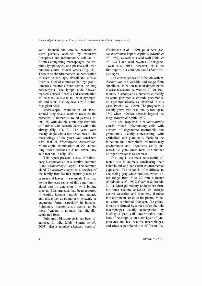
Fig. 1. A. Gritty nodules (arrow) on the pulmonary lobe. B. Granulomatous inflammation with giant cells (arrow) in air spaces (H/E, scale bar= 25 µm); C. Peribronchiolar cellular infiltrates with intra lesional yeasts (arrow) in airway lumen (H/E, scale bar= 25 µm); D. Intensely PAS positive bron-chial exudate (scale bar= 25 µm);. E. PAS-stained yeasts (arrows) in bronchial exudate (scale bar= 25 µm); F. The same lung section negative for ZN stain (scale bar= 25 µm).
A case of pulmonary blastomycosis in a common eland (Taurotragus oryx)
BJVM, ××, No × 4
veoli. Bronchi and terminal bronchioles were partially occluded by extensive fibroplasia and inflammatory cellular in-filtrates comprising macrophages, neutro-phils, lymphocytes, and plasma cells with numerous intra-lesional yeasts (Fig. 1C). There was chondromalacia, mineralisation of necrotic cartilage, alveoli and diffuse fibrosis. Foci of circumscribed pyogranu-lomatous reactions were within the lung parenchyma. The lymph node showed marked cortical fibrosis and accentuation of the medulla due to follicular hyperpla-sia and sinus histiocytocysis with nume-rous giant cells.
Microscopic examination of PAS-stained lung tissue sections revealed the presence of numerous round yeasts (10–20 µm) with double contoured retractile wall mixed with necrotic debris within the airway (Fig. 1D, E). The yeast were mostly single with a few broad based. The morphology of the yeast was consistent with that of Blastomyces dermatitidis. Microscopic examination of ZN-stained lung tissue sections did not reveal any acid fast bacilli (Fig. 1F).
This report presents a case of pulmo-nary blastomycosis in a captive common Eland (Taurotragus oryx). The common eland (Taurotragus oryx) is a species of the family Bovidae that primarily feed on
grasses and leaves in savannah. This may be the first case report of this condition in eland and by extension in wild bovine species. Blastomycosis has been reported in canids, humans, equids and aquatic animals; either as pulmonary, systemic or cutaneous forms especially in humans. Pulmonary blastomycosis seems to be more frequent in animals than the dis-seminated form.
Pulmonary blastomycosis has been di-agnosed in wild felids (Storms et al., 2003), rhesus monkey (Macaca mulatta)
(Wilkinson et al., 1999), polar bear (Ur-sus maritimus) kept in captivity (Morris et al., 1989), as well as a wild wolf (Thiel et al., 1987) and wild coyotes (Rodríguez-Tovar et al., 2015), however, this is the first report in a common eland (Taurotra-gus oryx).
The consequences of infection with B. dermatitidis are variable and range from subclinical infection to fatal disseminated disease (Saccente & Woods, 2010). Pul-monary blastomycosis presents clinically as acute pneumonia, chronic pneumonia, or asymptomatically as observed in this case (Patel et al., 1999). The prognosis is usually grave with case fatality rate up to 78% when infection spreads beyond the lungs (Martin & Smith, 1939).
The host response to B. dermatitidis consist mixed inflammatory cells with clusters of degenerate neutrophils and granulomas, usually noncaseating, with epithelioid and giant cells. Early in the infection, the neutrophilic infiltrates may predominate and organisms easily de-tected. As granulomas form, the number of organisms tends to decrease.
The lung is the most consistently af-fected site in animals considering their behavioural and consistent environmental exposures. The lesion is of multifocal to coalescing gray-white nodules, which of-ten range from 3 to 20 mm diameter (Goldman et al., 1999, Guarner & Brandt, 2011). Most pulmonary nodules are firm, but some become abscesses or undergo central caseation and then may fistulate into a bronchus or on to the pleura. Mine-ralisation is minimal or absent. The granu-lomas are formed by a mass of epithelioid macrophages usually accompanied by histiocytic giant cells and variable num-bers of neutrophils, an outer layer of lym-phocytes and less reactive macrophages, and often a peripheral rim of fibrous tis-

O. O. Alaka, T. A. Jarikre, B. N. Ogunro, Y. G. Gurumyen, A. C. Mark, T. O. Omadevuaye ...
BJVM, ××, No × 5
sue. In some cases, there is extensive cas-eous necrosis in the centers of the granu-lomas, with merely a thin rim of macro-phages. Yeast bodies are quite variable in number and may be missed on haematoxy-lin and eosin-stained sections (Caswell & William, 2007). The double contour of the fungal wall is well described in cytologic preparations or histologically via special stains as observed in this case.
The skin, soft tissue, bone, genitouri-nary, or central nervous system involve-ment have been reported (Lemos et al., 2000; McKinnell & Pappas, 2009; Bariola et al., 2010). In rare cases, skin lesions without lung involvement have been de-scribed, suggesting direct cutaneous ino-culation. The skin lesions most frequently described are painless ulcers or verrucous lesions.
The diagnostic tools often employed include thoracic radiography which may show alveolar or mass-like infiltrate (Bradsher et al., 1985; Patel et al., 1999; Patel et al., 2010). However, reticulono-dular and miliary opacities and or solitary nodular opacities are also characteristic. This has to be differentiated from tubercu-losis, histoplasmosis, and coccidioidomy-cosis which may present similarly. Special techniques such as polymerase chain reac-tion (PCR), in situ hybridisation (Hayden et al., 2001) and immunohistochemistry (Fukuzawa et al., 1995, Jensen et al., 1996) could also be helpful, but examina-tion of special stains remains the most reliable and useful method to identify or-ganisms in tissue (Katzenstein, 2006, El-Zammar & Katzenstein, 2007). The aura-mine-rhodamine stain is also satisfactory, but requires a fluorescent microscope. Periodic acid–Schiff (PAS) Gridley stain is satisfactory but PAS without a counter-stain should not be used because organ-isms are not adequately differentiated
from background necrotic debris (Ulbright & Katzenstein, 1980). The Gomori methe-namine silver (GMS) stain is the most useful. Other stains that may be useful for fungi include the Fontana–Masson (FM) and the combined FM–alcian blue, FM–mucicarmine, or alcian blue–PAS stains, especially for identifying cryptococci (Lazcano et al., 1991; 1993).
The most significant transmission route for B. dermatitidis is inhalation of the conidia. The conidia are aerosolised through disruption of wet soil or organic matter containing microfoci of B. dermati-tidis mycelia. They are inhaled into the airway of susceptible host for propagation of yeast phase. In dogs, however, in-traocular mycosis is the most frequently reported cause of blastomycosis (Broom et al., 1996).
Less commonly, direct cutaneous in-oculation via a penetrating outdoor injury, a laboratory accident, or even the bite of an infected dog occurs (Gnann et al., 1983). B. dermatitidis is not transmitted from animals to humans however shared environmental exposures could explain the occurrence of disease in humans espe-cially immunocompromised patients (McKinnell & Pappas, 2009) and their canine companions (Sarosi et al., 1979).
The source of infection to this eland may be connected to the inhalation of co-nidia from the contaminated environment. Some environmental factors associated with blastomycosis are sandy acidic soil, bodies of water, and exposure to excava-tion sites (Arceneaux et al., 1988). Inhala-tion of infectious spores is considered the main route of pulmonary infection in dogs and is likely the route of infection in this case given the abundance of pyogranulo-mas and fungal elements in the lungs. Animals often appear to be more suscep-tible to pulmonary blastomycosis than

A case of pulmonary blastomycosis in a common eland (Taurotragus oryx)
BJVM, ××, No × 6
humans because they seem to inhale larger quantities of the fungus spores closer to the ground (Legendre, 2012). Blastomy-cosis is more prevalent in climatic re-gions; however, its diagnosis in many animal species and in zoological collec-tions as in this case underscores its impor-tance in Africa.
In conclusion, this is the first reported case pulmonary blastomycosis in a captive eland in Nigeria, this case showed the need for regular screening of soil and en-vironment including animals in captivity to avert possible spread of the mold in favourable conditions hence public aware-ness on most zoonotic fungal diseases should be improved.
REFERENCES
Arceneaux, K. A., J. Taboada & G. Hosgood, 1998. Blastomycosis in dogs: 115 cases (1980–1995). Journal of the American Ve-terinary Medical Association, 213, 658–664.
Baumgardner, D. J., E. M. Knavel, D. Steber & G. R. Swain, 2006. Geographic distribu-tion of human blastomycosis cases in Mil-waukee, Wisconsin, U.S.A.: Association with urban watersheds. Mycopathologia, 161, 275–282.
Bariola, J. R., P. Perry, P. G. Pappas, L. Proia, W. Shealey, W. P. Wright, J. M. Sizemore, M. Robinson & R.W. Bradsher Jr., 2010. Blastomycosis of the central ner-vous system: A multicenter review of di-agnosis and treatment in the modern era. Clinical Infectious Diseases, 50, 797–804.
Bradsher, R. W., D. C. Rice & R. S. Aber-nathy, 1985. Ketoconazole therapy for en-demic blastomycosis. Annals of Internal Medicine, 103, 872–879
Broom, J. D., R. E. Hamor & P. A. Gerding Jr., 1996. Ocular btastomycosis in dogs: 73 cases, 108 eyes (1985-1993). Journal of the American Veterinary Medical Asso-ciation, 209, 1271–1276.
Caswell, J. L. & K. J. William, 2007. Respira-tory system. In: Pathology of Domestic Animals, vol. 2, 5th edn, eds K. V. F. Jubb, P. C. Kennedy & N. Palmer, Academic Press, Inc., San Diego, pp. 524–650.
Cates, M. B., L., Kaufman, J. H. Grabau, J. M. Pletcher & J. P. Schroeder, 1986. Blasto-mycosis in an Atlantic bottlenose dolphin. Journal of the American VeterinaryMedi-cal Association, 189, 1148–1150.
El-Zammar, O. A. & A. L. A. Katzenstein, 2007. Pathological diagnosis of granulo-matous lung disease: A review. Histopa-thology, 50, 289–310.
Fukuzawa, M., H. Inaba, M. Hayama, N. Sakaguchi, K. Sano, M. Ito & M. Hotchi, 1995. Improved detection of medically important fungi by immunoperoxidase staining with polyclonal antibodies. Vir-chows Archiv, 427, 407–414.
Gilchrist, T. C. & W. R. Stokes, 1898. A case of pseudolupus caused by Blastomyces. The Journal of Experimental Medicine, 3, 53–78.
Gnann, J. W. Jr., G. S. Bressler, C. A. Bodet & C. K. Avent, 1983. Human blastomycosis after a dog bite. Annals of Internal Medi-cine, 98, 48–49.
Goldman, M., P.C. Johnson & G.A. Sarosi, 1999. Fungal pneumonias. The endemic mycoses. Clinics in Chest Medicine, 20, 507-519.
Hayden, R. T., X. Qian, G. D. Roberts, & R. V. Lloyd, 2001. In situ hybridization for the identification of yeastlike organisms in tissue section. Diagnostic Molecular Pa-thology, 10, 15–23.
Guarner, J. & M. E. Brandt, 2011. Histopa-thologic diagnosis of fungal infections in the 21st century. Clinical Microbiology Reviews, 24, 247–280.
Jensen, H. E., H. C. Schonheyder, M. Hotchi & L. Kaufman, 1996. Diagnosis of sys-temic mycoses by specific immunohisto-chemical tests. Acta Pathologica, Mic-robiologica et Immunologica Scandi-navica, 104, 241–258.

O. O. Alaka, T. A. Jarikre, B. N. Ogunro, Y. G. Gurumyen, A. C. Mark, T. O. Omadevuaye ...
BJVM, ××, No × 7
Katzenstein, A. L. A., 2006. Katzenstein and Askin’s Surgical Pathology of Non-Neoplastic Lung Disease, 4th edn, Elsevier, Philadelphia.
Lazcano, O., V. O. Speights, J. Bilbao Jr,, J. Becker & J. Diaz, 1991. Combined Fon-tana–Masson–mucin staining of Crypto-coccus neoformans. Archives of Pathology and Laboratory Medicine, 115, 1145–1149.
Lazcano, O., V. O. Speights Jr, J. Strickler, J. Bilbao, J. Becker & J. Diaz, 1993. Com-bined histochemical stains in the differen-tial diagnosis of Cryptococcus neofor-mans. Modern Pathology, 6, 80–84.
Legendre, A. M., 2012. Chapter 57. Blastomy-cosis. In: Greene Infectious Diseases of the Dog and Cat, 4th edn, ed C. E. Green, Saunders, St. Louis, Mo, USA, pp. 606–614.
Lemos, L. B., M. Guo & M. Baliga, 2000. Blastomycosis: Organ involvement and etiologic diagnosis. A review of 123 pa-tients from Mississippi. Annals of Diag-nostic Pathology, 4, 391–406.
Martin, D. S. & D. T. Smith, 1939. Blastomy-cosis. I. A review of the literature. The American Review on Tuberculosis, 39, 275–304.
McKinnell, J. A. & P. G. Pappas, 2009. Blas-tomycosis: New insights into diagnosis, prevention, and treatment. Clinics in Chest Medicine, 30, 227–239.
Medoff, G., A. Painter, & G. S. Kobayashi, 1987. Mycelial to yeast-phase transitions of the dimorphic fungi, Blastomyces der-matitidis and Paracoccidioides brasilien-sis. Journal of Bacteriology, 169, 4055–4060.
Morris, P. J., A. M. Legendre, T. L. Bower-sock, D. E. Brooks, D. J. Krahwinkel, G. M. H. Shires & M. A. Walker, 1989. Di-agnosis and treatment of systemic blasto-mycosis in a polar bear (Ursus maritimus) with itraconazole. Journal of Zoo and Wildlife Medicine, 20, 336–345
Patel, R. G., B. Patel, M. F. Petrini, R. R. Carter & J. Griffith, 1999. Clinical presen-
tation, radiographic findings, and diagnos-tic methods of pulmonary blastomycosis: A review of 100 consecutive cases. South-ern Medical Journal, 92, 289–295.
Patel, A. J., P. Gattuso & V. B. Reddy, 2010. Diagnosis of blastomycosis in surgical pa-thology and cytopathology: Correlation with microbiologic culture. The American Journal of Surgical Pathology, 34, 256–261.
Rodríguez-Tovar, L., A. M. Nevárez-Garza, R. V. Barajas-Juárez, J. J. Zarate-Ramos, R. A. Ledezma-Torres & A. Trejo-Chávez, 2015. Probable pulmonary blastomycosis in a wild coyote (Canis latrans). Case Re-ports in Veterinary Medicine, DOI 10.1155/2015/564610.
Saccente, M. & L. Woods, 2010. Clinical and laboratory update on blastomycosis. Clini-cal Microbiology Reviews, 23, 367–381.
Sarosi, G. A., M. R. Eckman, S. F. Davies & W. K. Laskey, 1979. Canine blastomycosis as a harbinger of human disease. Annals of Internal Medicine, 91, 733–735.
Storms, T. N., V. L. Clyde, L. Munson, & E. C. Ramsay, 2003. Blastomycosis in non-domestic felids. Journal of Zoo and Wild-life Medicine, 34, 231–238.
Thiel, R. P., L. D. Mech, G. R. Ruth, J. R. Archer & L. Kaufman, 19870. Blastomy-cosis in wild wolves. Journal of Wildlife Diseases, 23, 321–323.
Ulbright, T. & A. Katzenstein, 1980. Solitary necrotizing granulomas of the lung. Dif-ferentiating features and etiology. The American Journal of Surgical Pathology, 4, 13–28.
Wilkinson, L. M., J. M. Wallace & J. M. Cline, 1999. “Disseminated blastomycosis in a rhesus monkey (Macaca mulatta), Veterinary Pathology, 36, 460–462.
Zwick, L. S., M. B. Briggs, S. S. Tunev, C. A. Lichtensteiger & R. D. Murnane, 2000. Disseminated blastomycosis in two Cali-fornia sea lions (Zalophus californianus). Journal of Zoo and Wildlife Medicine, 31, 211–214.

A case of pulmonary blastomycosis in a common eland (Taurotragus oryx)
BJVM, ××, No × 8
Paper received 16.01.2017; accepted for publication 07.04.2017
Correspondence: B. O. Emikpe Department of Veterinary Pathology, Faculty of Veterinary Medicine, University of Ibadan, Nigeria tel: +2348066486080 e-mail:[email protected]