Embed Size (px)
Citation preview

Case 5 History
• 63 year old woman
• Indeterminate breast lesion
• Upper inner/outer quadrant
• Ultrasound core biopsy


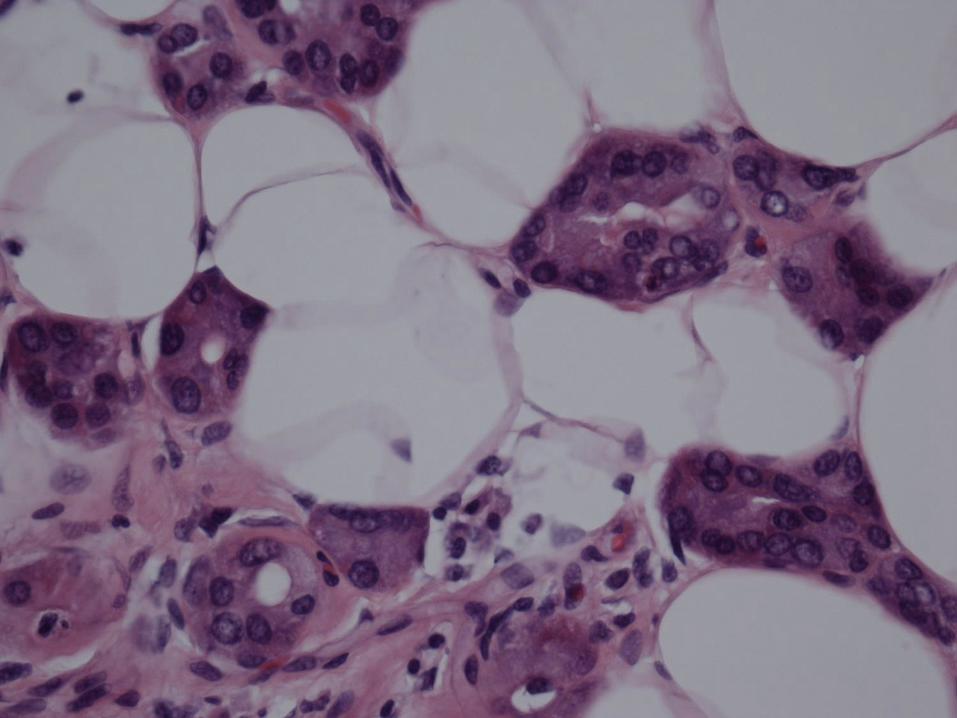
Histological features
• Tubules
• Not a lobular architecture
• Extending into adipose tissue
• No tissue reaction
• Bland cytology

ResponsesMicroglandular adenosis 15
Atypical microglandular adenosis 2
Invasive carcinoma 4
Invasive NST carcinoma 4
Tubular carcinoma 1
Adenoid cystic carcinoma 1
MGA v adenocarcinoma 3
Sclerosing/tubular adenosis v invasive 2
Carcinoma in MGA 2
Involution v adenocarcinoma 1
Sclerosing adenosis + FEA 1
Unclear 3

SM myosin

Collagen IV

S100

ER

CK14

GCDFP15

EMA

Microglandular adenosis• Clue is low power – not lobular
architecture
• Small tubules
• Single layer, cuboidal/flat
• Eosinophilic PAS+ luminal secretion
• Occasionally solid islands
• No stromal reaction
• Bland cytology
• Absent myoepithelial layer
• Basement membrane present
• S100 positive
• EMA, ER, PR and HER2 negative

Microglandular adenosis
• First described in AFIP fascicle 1968
• Clement PB, Azzopardi JG.
Microglandular adenosis of the breast –
a lesion simulating tubular carcinoma.
Histopathology 1983;7:169-80.


Differential diagnosis
Tubular carcinoma
• Tear drop shaped tubules
• Columnar morphology
• Apocrine snouts
• Fibroelastotic stroma
• Oestrogen receptor positive

Follow up
• Underwent wide local excision and
re-excision with clear margins
• 4 years later
• Upper inner/lower inner quadrant
mass in the same breast


ER

HER2

Invasive carcinoma NST
Grade 2 (T3, P2, M1)
17mm
ER positive
HER2 negative
Node negative
No residual microglandular adenosis

Microglandular adenosis
• Previously considered as benign
• But sometimes merges with carcinoma
• Often triple negative
• NST
• Adenoid cystic
• Spindle cell carcinoma
• Matrix-producing carcinoma
• Atypical microglandular adenosis

Shin SJ, al. Molecular evidence for progression
of microglandular adenosis (MGA) to invasive
carcinoma. Am J Surg Pathol 2009; 33; 496–504.• Comparative genomic hybridization
• 5 MGA and 3 AMGA – no alterations
• 7 MGA and 9 AMGA – copy number
changes (some widespread)
• Frequent concordance in genomic
profiles between MGA and carcinoma
arising in MGA
• 3 pure MGA – 2 no copy number changes
and 1 with numerous gains and losses

Geyer et al. Molecular evidence in support of
the neoplastic and precursor nature of
microglandular adenosis. Histopathology
2012, 60, E115–E130
• 10 carcinomas associated with MGA
• NST, matrix producing, acinic cell-like,
adenoid cystic
• All triple negative
• All S100+, CK8/18+
• Focal HMWCK or EGFR

Geyer et al Histopathology 2012
• Microarray comparative genomic
hybridization
• MGA genetically heterogeneous
• Some had complex alterations
• Some had no copy number changes
• Similar changes in MGA and adjacent
carcinoma

Guerini-Rocco et al. J Pathol 2016
236 genes analysed
MGA and AMGA with associated carcinoma
Non-synonymous somatic mutations
MGA (n = 7) median 5 (range 3-14)
AMGA (n = 3) median 3 (range 1-10)
TP53 mutations
MGA 6/7
AMGA 1/3
Identical mutation in adjacent carcinoma
Also mutations in PI3K pathway

Guerini-Rocco et al. J Pathol 2016
MGA without associated carcinoma (n = 2)
No mutations of TP53 or PI3K pathway
• TNBCs associated with MGA /AMGA
have molecular characteristics
consistent with those of unselected
TNBCs
• MGA likely constitutes a non-obligate
precursor of TNBC

Guerini-Rocco et al. J Pathol 2016
MGA with DCIS and invasive carcinoma
• MGA and TNBC clonally related
• DCIS not clonally related to TNBC or
MGA
AMGA with DCIS
• AMGA and DCIS did not share mutations
• But did share some chromosomal gains
and losses
• May have had a clonal origin, but
diverged in tumour progression


Management of
microglandular adenosis
• Core biopsy: categorise as B3 with
epithelial atypia
• Complete surgical excision
• Not VAB excision
• May be associated triple negative
carcinoma


![Sclerosing adenosis and risk of breast cancer...Other studies, however, have found no increased risk in women with SA [3, 4]. To our knowledge, the underlying biology of SA is undefined](https://img.dokumen.tips/doc/110x75/5ff0ca12796afe127e0e0e53/sclerosing-adenosis-and-risk-of-breast-cancer-other-studies-however-have-found.jpg)
![363lo lectura] [Modo de compatibilidad]) · riesgo de cáncer de mama (RR 1.3-2.0) : Adenosis esclerosante, lesiones esclerosantes radiales y complejas, hiperplasia epitelial ductal](https://img.dokumen.tips/doc/110x75/6051fdac53981928930682ef/363lo-lectura-modo-de-compatibilidad-riesgo-de-cncer-de-mama-rr-13-20.jpg)


