Embed Size (px)
Citation preview

Case
• 17 yo AA female
• Fatigue x 1 mo
• Weight loss x 5 #
• Flank pain
• Urine is brown

Nephritis in SLE Epidemiology
– Adults: 25-50%
– Pediatrics: 82%
– Silent disease
3/27 Minimal lesion
12/27 Focal lesion
12/27 Diffuse lesion
(Mahajan, Medicine 56:493-510, 1977)

II
IV III
V
Light Microscopic Patterns of Lupus Nephritis
Diffuse Proliferative Focal Proliferative
Membranous Mesangioproliferative

Systemic Lupus ErythematosisPatient Survival
• w/o Nephritis 95 % at 5 years
• w/ Nephritis
– Untreated proliferative nephritis: 50% at 14 months
– Treated 92 % at 5 years
– Age disparity
– Racial disparity(Pollak J Lab Clin Med 57:495, 1961; Cervera, Medicine 78:167-75, 1999)

Diffuse Proliferative Nephritis (IV)

Diffuse GN (WHO IV)
Austin, Sem Nephrol 19:2, 1999

Racial Disparity
Boumpas, Lancet 340:741, 1992; Dooley, KI, 1997
0
20
40
60
80
100
Black White

Rapidly progressive glomerulonephritis (GN)
Serologic analysis
Anti-neutrophil cytoplasmic autoantibodies (ANCA)
Anti-GBM autoantibodies
Immune complex constituents
No extra-renal disease
“Idiopathic” crescentic GN
Lung hemorrhage
Goodpasture’ssyndrome
Anti-DNAautoantibodies
LupusGN
+ + +No lung
hemorrhageSystemic
necrotizing arteritis
Pulmonary necrotizing granulomas
Anti-strepantibodies
OtherCryo-globulins
Polyarteritis nodosa
Wegener’s granulomatosis
Anti-GBMGN
Post-strep GN
Cryoglob-ulinemic
GN
Other immune complex
GN
ANCA-associated GN Anti-GBM antibody-mediated GN
Immune complex-mediated GN
Modified from: Jennette JC et al. Med Clin North Am 1990; 74:893.

Invitation: Nephrology Elective for …. the rest of the
story.

Crescentic Glomerulonephritis Immune
ComplexAnti-GBMPauci-
Immune
>80%ANCA+
Crescentic Glomerulonephritis is Categorized as Anti-GBM Mediated, Immune Complex Mediated, or Pauci-Immune
(i.e., with a paucity of staining for immunoglobulin)

Anti-glomerular basement membrane disease
• Goodpastures syndrome• Linear staining of GBM• With therapy: 70% risk of ESRD or
Death• Control with Cytoxan, Steroids, and
Plasmapharesis• Does not tend to be a relapsing
disease

Aorta
Arteries
ArterioleCapillary
Venule
Vein
MicroscopicPolyangiitis
Wegener’sGranulomatosis
Churg-Strauss
Syndrome
Nogranulomatousinflammation
or asthma
Granulomatousinflammationbut no asthma
Granulomatousinflammation,asthma, and eosinophilia
ANCA Small Vessel Vasculitis
Organ-limitedPauci-Immune
Vasculitis
Organ-limitedPauci-Immune
Vasculitis
vascular predilection

Anti-Neutrophil Cytoplasmic Autoantibodies (ANCA)
P-ANCA Perinuclear PatternAnti-myeloperoxidase
MPO-ANCA
C-ANCA Cytoplasmic PatternAnti-Proteinase 3
PR3-ANCA

% ANCA-Positive By EIA and IFA
90
80
70
60
50
40
30
20
10
0
C-ANCA &
PR3-ANCA
P-ANCA &
MPO-ANCA
PIC
GN
AG
BM
ICC
GN
ICG
N
MP
GN
SL
EG
N
IGA
N
ME
M
FS
GS
MC
G


Microscopic Polyangiitis
Necrotizing vasculitis with few or no immune deposits affecting small vessels, i.e. capillaries, venules and arterioles. Necrotizing arteritis involving small and medium-sized arteries may be present. Necrotizing
Glomerulonephritis is verycommon. Pulmonarycapillaritis often occurs.

Wegener’s Granulomatosis
Granulomatous inflammation involving the respiratory tract, and necrotizing vasculitis affecting small to medium-sized vessels, e.g. capillaries, venules, arterioles, and arteries. Necrotizing glomerulonephritis is common.

Churg-Strauss Syndrome
Eosinophil-rich and granulomatous inflammation involving the respiratory tract, and necrotizing vasculitis affecting small to medium-sized vessels, associated with asthma and blood eosinophilia.

Pauci-Immune (ANCA) Crescentic Glomerulonephritis

Acute ANCA Glomerulonephritis
Segmental fibrinoid necrosis and apoptosis
Segmental fibrinoid necrosis with GBM lysis, apoptosis, and early
crescent formation

Acute ANCA Renal Vasculitis
Necrotizing Glomerulonephritis
Necrotizing Medullary Angiitis
Necrotizing Arteritis
Segmental Fibrinoid Necrosis

JCJ
Pathogenesis of ANCA Necrotizing Vasculitis
1. If ANCA cause vasculitis, they must induce the following sequence of events:
2. Leukocyte margination, adherence, and diapedesis
3. Leukocyte activation with degranulation and generation of toxic oxygen metabolites 4. Vascular necrosis with karyorrhexis and fibrinous insudation

ANCAANCA AntigenCytokineCytokine ReceptorFc ReceptorAdhesion MoleculeAdhesion Molecule Receptor
JCJ
Jennette & Falk: Nephrol Dial Trans 1998; 13 [Suppl 1]: 16-20

Priming
• Infection
• Environment– Silica– Great Earthquake, Kobe, Japan

Humans with ANCA-GN


Cutaneous Manifestations of Vasculitis
• Purpura
• Petechiae
• Ecchymoses
• Erythematous macules
• Papules
• Nodules
• Urticaria
• Livedo raticularis
• Necrosis
• Ulceration
• Vesicles
• Bullae– Pyoderma gangrenosum-
like lesions
– Erythema nodosum-like lesions
– Sweet’s like lesions


Signs and Symptoms of Necrotizing Small Vessel Vasculitis
• Cutaneous purpura, nodules and ulcerations• Hemoptysis and pulmonary infiltrates or nodules • Peripheral neuropathy (mononeuritis multiplex)• Abdominal pain and blood in stool• Necrotizing (hemorrhagic) sinusitis• Myalgias and arthralgias• Muscle and pancreatic enzymes in blood• Hematuria, proteinuria and renal insufficiency

Treatment of ANCA-GN
IV pulse methylprednisolone 7 mg/kg x 3 days
Prednisone 1 mg/kg X 4 weeks then tapered
with either
IV cyclophosphamide 0.5 g/m2* X 6 months
or
Oral cyclophosphamide 2 mg/kg* X 6 to 12 months
*adjusted based on leukocyte count

Corticosteroids Alone Do Not Work
• Remission rate– cyclophosphamide 85%– corticosteroids 56% (p = 0.003)
• Risk of relapse increased 3-fold in corticosteroids alone group– (RP = 3.2, 95% CI, = 1.2, 8.3*)– *controlling for age, serum creatinine, duration of
treatment, and presence of arteriosclerosis on biopsy

Cyclophosphamide Versus Azathioprine During Remission
Induction with Prednisone and oral Cytoxan
Oral Cytoxan Azathioprine
No difference in creatinine, BVAS score,or vasculitic damage index.
D Jayne. J Am Soc Nephrol 1999, 105A.

Trimethoprim Sulfamethoxazole for Prevention of Relapse of Wegeners

Pediatric ANCA-GN
Source N Age DX (%) CRI (%) ESRD (%) Death (%)
GDCN 23 2-20 MPA 60 10 35 13WG 35NCGN 5
Hattori 31 5-17 MPA 68 19.5 29 3NCGN 32
Valentini 7 11-17 33 14 0
Ellis 3 4-14 33 0
Hall 4 7-13 WG 100 50 0
Orlowski 6 13-20 WG 100 17 50
Total 74 2-20 16.4 29.7 9.5

Pediatric ANCAGDCN Baseline Data
Mean Range
Age 13.4+4.5 2-20
Female 70%
Race
White 84%
Black 16%
Prodrome (w) 11+17 2-72
GFR 52+43 4-124

Pediatric ANCA Organ Involvement
System % Affected
Kidney 100
Pulmonary 70
Sinusitis 25
Gastrointestinal 45
Musculoskeletal 55
Nervous 15
Skin 35
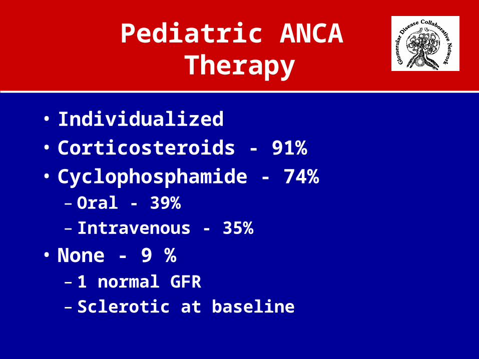
Pediatric ANCA Therapy
• Individualized • Corticosteroids - 91%• Cyclophosphamide - 74%
– Oral - 39%– Intravenous - 35%
• None - 9 %– 1 normal GFR– Sclerotic at baseline

Pediatric ANCAHistopathology
Feature Survivors Non-survivors
p value
Crescents% 39.7+38.1 83.6+42.1 0.01
Glomerular Necrosis
1.5+1.1 2.1+1.5 0.3
Glomerular Sclerosis
1.2+1.3 1.4+0.7 0.5
Tubulointerstitial Disease
1.2+0.9 2.3+0.7 0.01
Activity Score 5.5+3.1 9.4+3.2 0.02
Chronicity Score 3.9+3.1 6.5+2.6 0.09
Injury Score 9.4+4.0 15.3+2.8 <0.01

Pediatric ANCASurvival
Renal and Patient Survival
Time (months)
50403020100-10
Cu
mm
ula
tive
Su
rviv
al
1.2
1.0
.8
.6
.4
.2
0.0
-.2
No Acute Dialysis
Acute Dialysis*
*mean survival 6.8 months
0 0 10 20 30 40 50

Risk Factors for Death and ESRD in Patients with ANCA Peds v Adults
Hogan, JASN, 1996; Gibson and Gipson, JASN, 2001
Adults RR P valueRisk of Death Hemoptysis 8.6 0.0002Risk of ESRD Baseline Cr 2.9 0.0002ChildrenRisk of Death and ESRD Crescents 0.01 Acute dialysis < 0.001 Injury score < 0.01

IV Versus Oral Cyclophosphamide
• No difference in remission or relapse rate
• Higher incidence of leukopenia with the use of oral cyclophosphamide
• Clinically significant higher risk of major side effects

ANCA Resistance and Relapse
• Resistance 23%– Female– Race (AA>CA)– Severe kidney disease at presentation
• Relapse 42%– PR3-ANCA– Lower respiratory tract disease– Upper respiratory tract disease
Hogan, 2005

Recurrence of ANCA-SVV After Renal Transplantation
Nachman PH et al. Kidney Int 1999; 56:1544-50

Alternative Treatment
• Pulse intravenous gamma globulin
• Trimethoprim sulfamethoxazole
• Methotrexate
• Azathioprine
• Mycophenolate mofetil (Cellcept)
• TNF receptor inhibitor