Embed Size (px)
Citation preview

RESEARCH ARTICLE
CAS9 is a genome mutator by directlydisrupting DNA-PK dependent DNA repairpathway
Shuxiang Xu1, Jinchul Kim2,3, Qingshuang Tang1, Qu Chen1,2, Jingfeng Liu1, Yang Xu1,2,3&, Xuemei Fu2,4
1 Cancer Research Institute, Guangdong Provincial Key Laboratory of Cancer Immunotherapy, School of Basic MedicalSciences, Southern Medical University, Guangzhou 510515, China
2 The Eighth Affiliated Hospital, Sun Yat-sen University, Shenzhen 518033, China3 Division of Biological Sciences, University of California, San Diego, 9500 Gilman Drive, La Jolla, CA 92093, USA4 Shenzhen Children’s Hospital, Shenzhen 518026, China& Correspondence: [email protected] (Y. Xu)
Received August 11, 2019 Accepted January 19, 2020
ABSTRACT
With its high efficiency for site-specific genome editingand easy manipulation, the clustered regularly inter-spaced short palindromic repeats (CRISPR)/ CRISPRassociated protein 9 (CAS9) system has become themost widely used gene editing technology in biomedicalresearch. In addition, significant progress has beenmade for the clinical development of CRISPR/CAS9based gene therapies of human diseases, several ofwhich are entering clinical trials. Here we report thatCAS9 protein can function as a genome mutator inde-pendent of any exogenous guide RNA (gRNA) in humancells, promoting genomic DNA double-stranded break(DSB) damage and genomic instability. CAS9 interactswith the KU86 subunit of the DNA-dependent proteinkinase (DNA-PK) complex and disrupts the interactionbetween KU86 and its kinase subunit, leading to defec-tive DNA-PK-dependent repair of DNA DSB damage vianon-homologous end-joining (NHEJ) pathway. XCAS9 isa CAS9 variant with potentially higher fidelity andbroader compatibility, and dCAS9 is a CAS9 variantwithout nuclease activity. We show that XCAS9 anddCAS9 also interact with KU86 and disrupt DNA DSBrepair. Considering the critical roles of DNA-PK inmaintaining genomic stability and the pleiotropic impactof DNA DSB damage responses on cellular proliferationand survival, our findings caution the interpretation of
data involving CRISPR/CAS9-based gene editing andraise serious safety concerns of CRISPR/CAS9 systemin clinical application.
KEYWORDS CAS9, DNA-PK, DNA double-strandedbreaks, genetic instability, DNA repair
INTRODUCTION
CAS9 is a type II DNA endonuclease widely used in genomeediting and epigenetic modification (Jinek et al., 2012; Conget al., 2013; Mali et al., 2013). CAS9 can efficiently target theentire genomic DNA sequence using the Protospacer Adja-cent Motif (PAM) domain and a guide RNA (gRNA) (Komoret al., 2017; Murovec et al., 2017). In addition to its extensiveuse in biomedical research, the clinical application of theCRISPR/CAS9 system in human therapy has been inten-sively investigated (Barrangou and Doudna, 2016; Deveret al., 2016; Urnov, 2018; WareJoncas et al., 2018; Zhuet al., 2019). Variants of CAS9 have also been developed fordistinct biological functions and improved editing fidelity. Forexample, the nuclease dead-CAS9 (dCAS9) has beendeveloped for gene activation (Gilbert et al., 2013; Maederet al., 2013), RNA editing (Cpf1) (Zetsche et al., 2015), andsome smaller editing systems (Harrington et al., 2018).However, a number of problems still challenge the broadutility of CAS9 in research and clinical application (Tan et al.,2015; Haapaniemi et al., 2018; Ihry et al., 2018). Forexample, recent reports indicate that the combinationof CAS9 and gRNA introduces genomic DNA DSBs, leadingto p53-dependent cytotoxicity of human pluripotent stemShuxiang Xu, Jinchul Kim, and Qingshuang Tang have contributed
equally to this work.
© The Author(s) 2020
Protein Cell 2020, 11(5):352–365https://doi.org/10.1007/s13238-020-00699-6 Protein&Cell
Protein
&Cell

cells (Ihry et al., 2018). Using long-read sequencing,researchers recently show that the genome editing mediatedby CRISPR/CAS9 and gRNA could lead to large DNAdeletions and complex rearrangements in the genome (Guoet al., 2018; Kosicki et al., 2018; Lei et al., 2018).
DNA double-stand break (DSB) damage is the mostcommon genomic DNA lesion during cellular proliferationand genome editing (Jackson and Bartek, 2009), whichcould be repaired by two pathways: the direct DNA ligation ofthe broken DNA ends termed NHEJ and homologous DNAtemplate guided homologous directed repair (HDR) (Mlade-nov and Iliakis, 2011). Immediately after the introduction ofDSB, the DNA repair pathways are activated with a largepanel of proteins recruited to the site of DNA DSBs(Mladenov and Iliakis, 2011). In the context of NHEJ, Ku70/86 heterodimer bind to the DNA DSBs to stabilize the DNADSB ends and then recruit DNA dependent protein kinasecatalytic subunit (DNA-PKcs) to the site of DNA DSB (Daviset al., 2014). DNA-PK complex is required for NHEJ byrecruiting and phosphorylating a panel of the DNA repairfactors including XRCC4 to facilitate DNA DSB repair (Ue-matsu et al., 2007; Davis et al., 2014). We demonstrate thatCAS9 can disrupt the formation of DNA-PK complex throughthe interaction with KU86, promoting DNA DSB damage.
RESULTS AND DISCUSSION
CAS9 induces DNA DSB damage in human cellsindependent of gRNA
For the clinical application of any gene editing technology,the genetically modified cells must maintain genomic sta-bility. Because there is no reported study to investigate theimpact of CAS9 on the genomic stability in the absence ofgRNA, we used the doxycycline (Doxy) inducible expressionsystem to induce the expression of CAS9 in human embry-onic stem cells (hESCs). The expression of CAS9 protein inthe absence of any exogenous gRNA activated DNA dam-age responses, including the phosphorylation of p53 atSer15, CHK1 at Ser317, and H2AX at Ser139 (Fig. 1A).Consistent with this finding, the expression of CAS9 proteinalone in hESCs was sufficient to induce the foci formation ofH2AX at Ser139 (γH2AX), a marker for DNA DSBs in thegenome (Fig. 1B). We used comet assay to confirm that theexpression of CAS9 protein alone in hESCs induced DNADSB damage (Fig. 1C). Nuclear fractionation analysis indi-cated that CAS9 is present in both nucleus and cytoplasm(Fig. 1D). Activation of p53 in hESCs can activate apoptosisand differentiation of hESCs as previously shown (Lin et al.,2005; Song et al., 2010). The DNA DSB damage induced byCAS9 activated the expression of p53 target genes suchas PUMA, p21, PERP and NOXA, leading to the cell deathand differentiation of hESCs (Fig. 1E–G).
To confirm this genome mutator function of CAS9 inmammalian cells, we employed the same CAS9 inducibleexpression system to express CAS9 in human inducedpluripotent stem cells (hiPSCs) and human fibroblasts.Consistent with the findings in hESCs, the expression ofCAS9 alone in hiPSCs and human fibroblasts was sufficientto induce DNA DSB damage and activate DNA damageresponses (Figs. 2A and 3A–C). Using decreasing dosages
cFigure 1. The expression of CAS9 alone in hESCs
promotes DNA DSB damage and activates DNA dam-
age response pathway. (A) Inducible expression of CAS9
in hESCs activated DNA damage response. Lentivirus
harboring the inducible CAS9 expression cassette or
control empty vector was used to transduce hESCs. The
expression of CAS9 was induced with 2 µg/mL doxycy-
cline (Doxy) treatment for 0, 24 h and 48 h. The relative
levels of the phosphorylation of p53, H2AX, CHK1 were
indicated at the bottom. n = 3. Data are presented as mean
value ± SD. *P < 0.05, **P < 0.01. (B) The expression of
CAS9 induced the number of the γH2AX foci in hESC.
hESCs with CAS9 inducible expression cassette were
plated on chamber slides and treated with or without 2 µg/
mL doxycycline for 3 days. The γH2AX foci were detected
by a confocal microscope. Scale bar, 10 µm. Unpaired t
test. n = 20. Data are presented as mean value ± SD.
***P < 0.001. (C) Detection of DNA DSB damage by
Comet assay in hESCs after various treatments. CTL,
hESCs with empty expression vector treated with 2 µg/mL
doxycycline (Doxy), doxorubicin (Dox), Doxy + Dox,
hESCs with CAS9 inducible expression vector were
treated with 2 µg/mL doxycycline for three days, 0.5
µmol/L Dox for 2 h, 2 µg/mL doxycycline for three days +
0.5µmol/L Dox for 2 h. Unpaired t test. n = 20. Data are
presented as mean value ± SD. ***P < 0.001. (D) CAS9 is
present in both nucleus and cytoplasm. CTL or CAS9,
hESCs with lentiviral empty vector or CAS9 inducible
expression cassette were treated with 2 µg/mL doxycy-
cline for 3 days. Cells were fractionated into nuclear and
cytoplasmic fractions, and examined for the levels of CAS9
as well as nuclear and cytoplasmic proteins histone H3
and α-tubulin. (E) The expression of p53 target genes in
hESCs after the expression of CAS9. CTL, CAS9, hESCs
with CAS9 inducible expression vector were treated with or
without 2 µg/mL doxycycline for 3 days. n = 3. Data are
presented as mean value ± SD. *P < 0.05, **P < 0.01,
***P < 0.001. (F) Cellular proliferation after the expression
of CAS9 in hESCs. n = 3. Data are presented as mean
value ± SD. ***P < 0.001. (G) The impact of CAS9
expression on the pluripotency of hESCs. The pluripotency
makers TRA1-60 and TRA1-81 were analyzed in hESCs
with or without CAS9 expression induced by 2 µg/mL Doxy
for 4 days.
© The Author(s) 2020 353
Protein
&Cell
CAS9 disrupts non-homologous end-joining DNA repair RESEARCH ARTICLE

γH2A
X
pS15
-p53
CAS
9
0 h
24 h
48 h
Dox
y
p53
H2A
X
CH
K1
pCH
K1
β-Ac
tin
150
kDa
55 k
Da
55 k
Da
15 k
Da
15 k
Da
55 k
Da
55 k
Da
37 k
Da
0 h
24 h
48 h
pp53
/p53
γH2A
X/H
2AX
pCH
K1/C
HK1
Relative protein levels
*
***
**
*
***
Foci number of γH2AX
DAP
I
γH2A
XC
AS9
M
erge
-Doxy +Doxy
-Doxy
+Doxy
B
TRA1
-60
TRA1
-81
IgG
CAS
9Em
pty
vect
or
CTL
C
AS9
C
TL
CAS
9
此图原图无法显示
Nuc
lear
Cyt
opla
sm
150
kDa
CAS
9
α-Tu
bulin
His
tone
H3
55 k
Da
15 k
Da
Cell viability
-D
oxy
+Dox
y
***
Relative mRNA expression
NO
XAp2
1PE
RP
PUM
A
**
**
**
-D
oxy
+Dox
y
*
CTL
Dox
yD
oxD
oxy
+ D
ox
***
Tail length
***
***
DE
FG
A
C
010203040
010203040
CTL
Doxy
Dox Doxy + Dox
0246
051015
0.0
0.2
1 d
2 d
3 d
4 d
5 d
0.4
0.6
0.8
RESEARCH ARTICLE Shuxiang Xu et al.
354 © The Author(s) 2020
Protein
&Cell

of Doxy to induce much lower expression of CAS9, weshowed that, at the expression levels much lower than thoseof standard lenti-viral transduction, CAS9 could still induceDNA DSB damage (Fig. 2B). Therefore, CAS9 can induceDNA DSB damage in mammalian cells independently ofexogenous gRNA.
CAS9 disrupts DNA-PK complex by interactingwith KU86
To elucidate the mechanism how CAS9 induces DNA DSBdamage, we investigated the potential interaction betweenCAS9 and the components of DNA repair pathways. Wediscovered that CAS9 interacted with KU86, a component ofDNA-PK complex essential for NHEJ pathway (Fig. 4A and4B). The interaction between CAS9 and KU86 was furtherconfirmed by proximity ligation assay (Fig. 4C). Using aseries of deletional mutants of CAS9, we found that the PAMdomain of CAS9 was involved in the interaction with KU86and other domains of CAS9 might interfere with the inter-action between the PAM domain and KU86 (Fig. 4D). In
further support of this notion, the expression of the PAMdomain of CAS9 reduced the interaction between CAS9 andKU86 (Fig. 4E). The expression of CAS9 disrupted theinteraction between KU86 with DNA-PKcs or K70, indicatingthat the interaction between CAS9 and KU86 inhibited theformation of DNA-PK complex (Fig. 4F).
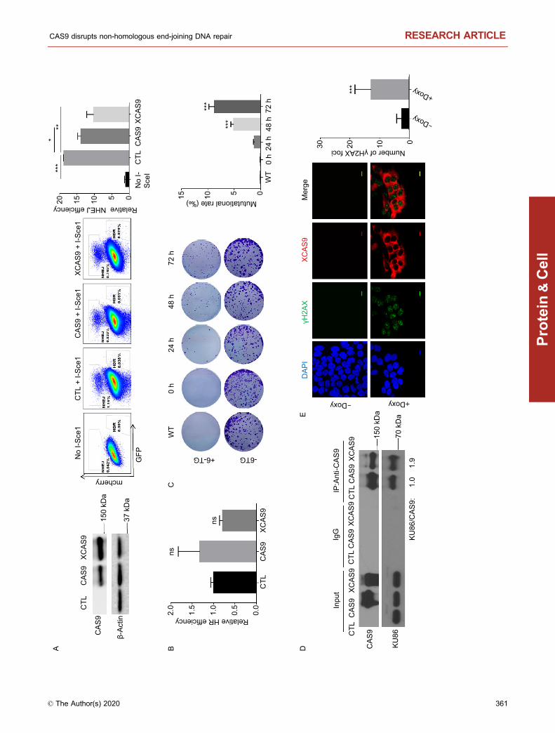
The formation of DNA-PK complex at the site of DNADSB is required for repairing DNA DSB via NHEJ andmaintaining genomic stability (Davis et al., 2014). Therefore,we hypothesized that CAS9 could inhibit DNA DSB repairvia NHEJ pathway and induce genomic instability by dis-rupting the formation of DNA-PK complex. In support of thisnotion, the expression of CAS9 in hESCs reduced the effi-ciency to repair DNA DSB damage via NHEJ pathway(Fig. 5A), but did not affect the efficiency to repair DNA DSBdamage via HDR pathway (Fig. 5B). In addition, theexpression of CAS9 in hESCs significantly increased therate of spontaneous mutation of the HPRT gene in hESCs,indicating that CAS9 induces genomic instability (Fig. 5C).Therefore, the expression of CAS9 induces genomic insta-bility in hESCs.
150 kDa
15 kDa
15 kDa
55 kDa
55 kDa
55 kDa
γH2AX
H2AX
Tubulin
CAS9
Doxy
p53
pp53
γH2AX/H2AX: 1 2.3 2.81 1.8 2.4
1 1.8 2.41 3.5 4.2
2.5 2.53.6 4.5pp53/p53:
pp53/p53:γH2AX/H2AX:
pS15-p53 55 kDa
p53 55 kDa
0 0.5 1 2 2
Tet-CAS9 Lenti-CAS9
CAS9 150 kDa
γH2AX 15 kDa
H2AX 15 kDa
α-Tubulin 55 kDa
A B
0 h 24 h 48 h
Figure 2. The expression of CAS9 in hiPSCs and hESCs promotes DNA DSB damage. (A) The inducible expression of CAS9
promotes DNA DSB damage responses in hiPSCs after 2 µg/mL Doxy treatment. The relative levels of the phosphorylation of p53
and H2AX are indicated at the bottom. Consistent data were obtained from two independent experiments. (B) The impact of
expression levels of CAS9 on DNA DSB damage in hESCs. At the same lentiviral titers, the expression levels of CAS9 in hESCs
transduced by standard lentiviral vector are higher than those transduced by the inducible lentiviral vector after 2 µg/mL Doxy
treatment. Much lower expression levels of CAS9 can also promote DNA DSB damage in hESCs after the treatment with
lower dosages of Doxy. The relative levels of the phosphorylation of p53 and H2AX are indicated at the bottom.
CAS9 disrupts non-homologous end-joining DNA repair RESEARCH ARTICLE
© The Author(s) 2020 355
Protein
&Cell

High fidelity or nuclease-dead CAS9 variants and Cpf1induce DNA DSB damage
In an attempt to improve the fidelity of CAS9 in gene editing,recent studies have described CAS9 variants such asxCAS9 that appear to have higher fidelity (Hu et al., 2018).However, similarly to CAS9, XCAS9 also interacted with
KU86 and induced DNA DSB damage independently of theexogenous gRNA (Fig. 5D and 5E). In addition, XCAS9impaired DNA DSB repair via NHEJ but did not affect theDNA DSB repair via HDR (Fig. 5A). Therefore, XCAS9 isalso a genome mutator that can promote DNA DSB damagein the absence of any gRNAs.
Table 1. Primers used in this study.
Sequence (5′-3′)
Primers for cloning
Fuw-teto-CAS9/XCAS9/dCAS9-pgk-puro Gibson F
TATCGATAAGCTTGATATCGAATTCTCAGGCACCGGGCTTGCGGG
Fuw-teto-CAS9/XCAS9/dCAS9-pgk-puro Gibson R
ATCCAGCCTCCGCGGCCCCGAATTCGCCACCATGGACAAGAAGTACAGCAT
Fuw-teto-control-pgk-puro F AATTCGCCACCATGGATTACAAAGACGATGACGATAAGTAGA
Fuw-teto-control-pgk-puro R CCGGTCTACTTATCGTCATCGTCTTTGTAATCCATGGTGGCG
plenti-CAS9-pgk-puro Gibson F ACCGACTCTAGAGGATCCGCCACCATGGACAAGAAGTACAGCATCGGCC
plenti-CAS9-pgk-puro Gibson R TCCAGAGGTTGATTGTCGACCTACTTATCGTCATCGTCTT
Fuw-teto-95-pgk-puro Gibson F GACCGATCCAGCCTCCGCGGCCCCGAATTCGCCACCATGGACAGCTTCTTCCACAGACTG
Fuw-teto-95-pgk-puro Gibson R TGGAAAAGGCGCAACCCCAAACCGGTCTACTTATCGTCATCGTCTTTGTAATC
plenti-719-pgk-puro Gibson F GACCGATCCAGCCTCCGCGGCCCCGAATTCGCCACCATGAGCCTGCACGAGCACATT
Fuw-teto-719-pgk-puro Gibson R TGGAAAAGGCGCAACCCCAAACCGGTCTACTTATCGTCATCGTCTTTGTAATC
Fuw-teto-910-pgk-puro Gibson F GACCGATCCAGCCTCCGCGGCCCCGAATTCGCCACCATGGAACTGGATAAGGCCGGCTT
Fuw-teto-910-pgk-puro Gibson R TGGAAAAGGCGCAACCCCAAACCGGTCTACTTATCGTCATCGTCTTTGTAATC
Fuw-teto-1100-pgk-puro Gibson F GACCGATCCAGCCTCCGCGGCCCCGAATTCGCCACCATGGTGCAGACAGGCGGCTTCA
Fuw-teto-1100-pgk-puro Gibson R TGGAAAAGGCGCAACCCCAAACCGGTCTACTTATCGTCATCGTCTTTGTAATC
plenti-control-pgk-puro F GATCCGCCACCATGTACCCATACGACGTCCCAGACTACGCTTAGG
plenti-control-pgk-puro R TCGACCTAAGCGTAGTCTGGGACGTCGTATGGGTACATGGTGGCG
plenti-Ku86-pgk-puro Gibson F GACACCGACTCTAGAGGATCCGCCACCATGTACCCATACGACGTCCCAGACTACGCTATGGTGCGGTCGGGGAATAAG
plenti-Ku86-pgk-puro Gibson R TCACAAATTTTGTAATCCAGAGGTTGATTGTCGACGAATTCCTATATCATGTCCAATAAATCGTCC
Fuw-teto-Cpf1-pgk-puro Gibson F AAGCCCTCGAACTGTGTCATGGTGGCGAATTCGGGGCCGCGGAGGCTGGAT
Fuw-teto-Cpf1-pgk-puro Gibson R AGACGATGACGATAAGTAGACCGGTTTGGGGTTGCGCCTTTTCCA
Primers for RT-qPCR
Puma F ACGACCTCAACGCACAGTACGA
Puma R CCTAATTGGGCTCCATCTCGGG
P21 F ACCTGGAGACTCTCAGGGTCG
P21 R TTAGGGCTTCCTCTTGGAGAAGAT
Perp F TCATCCTGTGCATCTGCTTC
Perp R GGGTTATCGTGAAGCCTGAA
Noxa F ACCAAGCCGGATTTGCGATT
Noxa R ACTTGCACTTGTTCCTCGTGG
β-actin F AGCGAGCATCCCCCAAAGTT
β-actin R GGGCACGAAGGCTCATCATT
RESEARCH ARTICLE Shuxiang Xu et al.
356 © The Author(s) 2020
Protein
&Cell

Mammalian cells might express endogenous gRNA-likesmall RNA that could work with CAS9 to induce DNA dam-age. To test this possibility, we examined the impact of theexpression of dCAS9, which is defective in the nucleaseactivity (Gilbert et al., 2013; Maeder et al., 2013), on DNADSB damage. Similar to CAS9 and XCAS9, dCAS9 alsointeracted with KU86 (Fig. 6A). In addition, the expression ofdCAS9 in hESCs also promoted DNA DSB damage andgenetic mutations at HPRT locus (Fig. 6B and 6C). While theimpact of dCAS9 on DNA damage appeared to be lessdramatic than CAS9, the results demonstrate that CAS9 caninduce DNA damage independent of its nuclease activity.Similarly to CAS9, Cpf1 also interacted with KU86 andactivated DNA DSB damage (Fig. 6D and 6E).
Previous studies evaluated the genomic stability of cellsafter the site-specific gene editing induced by CAS9 + gRNA,suggesting that the specificity of gRNA and the deliverymethod of CAS9/gRNA could be optimized to improve thefidelity and safety of this gene editing technology (Haa-paniemi et al., 2018; Ihry et al., 2018; Kosicki et al., 2018).Our data demonstrate that CAS9 and its high-fidelity variantXCAS9 are genome mutators by promoting DNA DSBdamage and genetic mutations independently of gRNA. Thisintrinsic oncogenic activity of CAS9 is achieved by disruptingDNA-PK complex, leading to impaired DNA DSB repair viaNHEJ pathway. In addition, it has been well documented thatdefects in the DNA-PK function will promote DNA DSBdamage and genetic instability (Davis et al., 2014). There-fore, our findings raise concerns for the safety of CRISPR/CAS9 system based human therapy. The development ofCAS9 variants that retain the gene editing activity of wildtype CAS9 but do not disrupt DNA-PK activity might help toimprove the fidelity of gene editing technology and safety ofthe CRISPR/CAS9 system.
MATERIALS AND METHODS
Cell lines, lentivirus production and transduction
Human embryonic stem cell line H9 was obtained fromWiCell Research Institute, Inc. and cultured with mTeSR1medium (STEMCELL, USA). Human fibroblasts were usedbetween passages 3 to 12. The HEK 293 FT and HEK 293Tcell lines were purchased from Thermo Scientific. Humanfibroblasts and HEK 293 FT were cultured in DMEM (Gibco)supplemented with 10% fetal bovine serum (FBS) (Hyclone)and 1% penicillin-streptomycin (Pen/Strep) (Gibco) at 37 °Cwith 5% CO2. The lentivirus production and transductionwere performed as we previously described (Kim et al.,2019). All cell lines were routinely checked for mycoplasmaby a PCR detection kit.
Expression vector construction
To construct vectors that express genes inducibly,CAS9, XCAS9, dCAS9 or Cpf1 cDNA was cloned into TetO-
Fuw-PGK-Puro vector modified from TetO-Fuw-OSKM vec-tor (Addgene plasmid 20321) linearized by EcoRI/AgeIdigestion using Gibson Assembly (2× NEBuilder HiFi DNAAssembly Master Mix) as previously described (Kim et al.,2019). To generate KU86 and CAS9 deletional mutants,pLenti-CMV-GFP vector (Addgene 17448) was linearized byBamHI/SalI digestion and ligated with the PCR products ofKU86 and different domains of CAS9 by Gibson assembly.The PCR primers were provided in Table 1.
Western blot analysis and Co-immunoprecipitation (Co-IP)
Cells were extracted for total proteins using lysis buffercontaining Protease and Phosphatase Inhibitor Cocktail(Cell Signaling Technology CST). For nuclear proteinextraction, nuclear and cytoplasm protein extraction kit(Thermo Fisher Scientific) was used following the instruction.Protein was separated on 6%–15% SDS PAGE and trans-ferred to 0.45 µm nitrocellulose membranes (Merck MilliporeUSA), which were blocked with the blocking buffer (5% skimmilk in TBS with 0.05% Tween 20) and incubated with pri-mary antibodies at 4 °C overnight. The membranes were
Table 2. Antibodies used in this study
Antibody Source and catalog
Anti-γH2AX Cell signaling (9718S, 80312S)
Anti-H2AX Cell signaling (7631S)
Anti-p53 Santa Cruze (sc-126)
Anti-pp53 Cell signaling (9286S)
Anti-CHK1 Cell signaling (2360S)
Anti-pCHK1 Cell signaling (12302S)
Anti-H3 Abcam (ab1971)
Anti-KU86 Santa Cruze (sc-5280), Abcam(ab119935), Cell signaling(2753s)
Anti-KU70 Abcam (ab92450)
Anti-DNA PKcs Abcam (ab70250)
Anti-BRCA1 Cell signaling (14823S)
Anti-p-BRCA1 Cell signaling (9009S)
Anti-CHK2 Cell signaling (6334S)
Anti-pCHK2 Cell signaling (2197S)
Anti-CAS9 Cell signaling (14697S), Abcam(ab189380), Novus Biologicals(NBP2-52717)
(NBP2-52717)
Anti-β-actin Cell signaling (4970S)
Anti-Flag Thermo Fisher (MA1-91878)
Anti-α-tubulin Santa Cruze (sc-73242)
CAS9 disrupts non-homologous end-joining DNA repair RESEARCH ARTICLE
© The Author(s) 2020 357
Protein
&Cell

150 kDa250 kDa
250 kDa
55 kDa
55 kDa
55 kDa
55 kDa55 kDa
55 kDa
15 kDa
15 kDa37 kDa
Foci
num
ber o
f γH
2AX
***
DAPI �H2AX CAS9 Merge
pCHK2(Thr68)
CHK2
H2AX
γH2AX
β-Actin
Doxy
CHK1pCHK1
(Ser317)
pBRCA1(Ser1254)
CAS9
BRCA1
pS15-p53p53
Rat
io o
f int
ensi
ty
* ** ** **
*** *** ***
CTL Doxy
Tail
leng
th
******
***
A B
C
0 h 24 h 48 h
-D
oxy
+D
oxy
-Dox
y
+Dox
y0
10
20
30
40
Dox Doxy + Dox0 h24 h48 h
0
1
2
pBRA
C1/B
RAC1
pCHK
1/CH
K1pC
HK2/
CHK2
γH2A
X/H2
AX
pp53
/p53
3
0
10
20
30
40
CTL
Dox
yDo
xDo
xy +
Dox
Figure 3. The expression of CAS9 in human fibroblasts promotes DNA DSB damage and activates DNA damage response
pathways. (A) The expression of CAS9 in human fibroblasts activates DNA damage responses. The expression of CAS9 was
induced with 2 µg/mL Doxy treatment. The relative levels of phosphorylation of BRCA1, CHK1, CHK2 and p53 are indicated at the
bottom. n = 3. Data are presented as mean value ± SD. *P < 0.05, **P < 0.01, ***P < 0.001. (B) The expression of CAS9 increased the
number of γH2AX foci in human fibroblasts. CTL or CAS9, human fibroblasts with CAS9 inducible expression cassette plated on
chamber slides were treated with or without 2 µg/mL doxycycline for 3 days. The expression of CAS9 and γH2AX foci was revealed
by immunoflourescence analysis. Representative images are shown. Scale bar, 10 µm. Unpaired t test. n = 20. Data are presented as
mean values ± SD. ***P < 0.001. (C) CAS9 induces DNA DSB damage in human fibroblasts. CTL, human fibroblasts with lentiviral
empty vector were treated with 2 µg/mL doxycycline for three days; Doxy, Dox, Doxy + Dox, human fibroblasts with lentiviral CAS9
inducible expression vector were treated with 2 µg/mL doxycycline for 3 days or 0.5 µmol/L Dox for 2 h or 2 µg/mL doxycycline for
three days + 0.5 µmol/L Dox for 2 h, respectively. Representative images are shown. n = 40. Unpaired t test. Data are presented as
mean value ± SD. **P < 0.01.
RESEARCH ARTICLE Shuxiang Xu et al.
358 © The Author(s) 2020
Protein
&Cell

incubated with secondary antibodies at room temperature for1 h and detected with Supersignal West Pico or Duraexposure buffer (Thermo Fisher Scientific). For Co-IP, thecells were collected and lysed with IP lysis buffer (ThermoFisher Scientific) containing protease inhibitor cocktail(Thermo Fisher Scientific) on ice for 30 min. After the cen-trifugation at 12,000 ×g for 20 min at 4 °C, the supernatantswere immunoprecipitated with antibodies followed by incu-bating with magnetic protein A/G beads (Pierce) for 4 h. ForIP analysis of phosphorylated proteins, cells were collectedin the IP lysis buffer containing Protease and PhosphataseInhibitor Cocktail. The antibodies used were provided inTable 2.
Proximity ligation analysis
hESCs with inducible CAS9 expression vector were platedon chamber slides and treated with 2 µg/mL doxycycline for2 days. After the treatment, the cells were washed with PBStwice and then fixed with 4% paraformaldehyde (PFA) for 15min at room temperature. After washing with PBS threetimes, cells were permeabilized with 0.3% Triton X-100 inPBS for 10 min at room temperature. Then Duolink® In SituRed Starter Kit Mouse/Rabbit (Sigma, DUO92101) was usedto reveal the interaction between CAS9 and KU86 asinstructed by the manufacturer. The antibodies of differentspecies (rabbit anti-CAS9 antibody, mouse anti-KU86 anti-body) were used to detect the proteins. DAPI (blue) wasused to stain the nucleus.
Immunofluorescence staining
1 × 10³ H9 or fibroblast cells were seeded onto the chamberslides coated with matrigel (Corning) and treated with 2 µg/mL doxycycline for 2 days. After the treatment, cells werewashed with PBS twice, and then fixed with 4%paraformaldehyde (PFA) for 15 min at room temperature.After washing with PBS three times, cells were permeabi-lized with 0.3% Triton X-100 in PBS for 10 min, blocked with2% bovine serum albumin (BSA) in PBS for 1 h at roomtemperature, stained with anti-CAS9 antibody (Abcam,1:100) and anti-γH2AX antibody (CST, 1:100) at 4 °C over-night, followed by simultaneous incubation with Alexa Fluor568-conjugated anti-mouse secondary antibody and AlexaFluor 488-conjugated anti-rabbit antibody for 1 hr at roomtemperature. Slides were mounted using VECTASHIEDsolution (Vector) with DAPI. The images were acquired aspreviously described (Chen et al., 2018).
Comet assay analysis
Comet assay was carried out as we previously described(Xiong et al., 2015). Briefly, hESCs or fibroblasts plated onthe matrigel-coated 6-well plates were treated with 2 µg/mLdoxycycline for 2 days, followed with or without the treatmentwith 0.5 µmol/L doxorubicin for 4 h. Cells were harvested,
washed with ice-cold PBS twice, resuspended at a density of105 cells/mL, mixed with agarose at a 1:10 ratio, spread onto3-well Comet Assay slides, and kept in dark for 15 min atroom temperature. Slides were immersed in chilled lysissolution for 15 min, washed with chilled TBE buffer threetimes and electrophoresed in chilled TBE (Tris-borate-EDTA)buffer for 15 min at 20 V. Slides were then fixed in 70%ethanol and dried, and DNA was labeled with Vista GreenDNA Dye diluted with TBE buffer. Images were captured andanalyzed using Image J software.
RNA purification and quantitative PCR analysis
The RNA extraction and qPCR were performed as we pre-viously described (Kim et al., 2019). The primers used forqPCR were listed in Table 1.
Cell viability
hESC cells were digested with TripLe (Gibco) to preparesingle cells. 4 × 10³ cells/well were plated onto each well ofthe 96-well plates with triplicate wells per sample. CellCounting Kit (CCK8, DoJindo) was used to evaluate cellviability according to the manufacturer’s instructions.
cFigure 4. CAS9 interacts with KU86 and disrupts the
formation of DNA-PK. (A and B) Reciprocal immunopre-
cipitation shows the interaction between CAS9 and KU86.
Protein extracts from hESCs expressing CAS9 were
immunoprecipitated with anti-KU86 (A) or anti-CAS9 (B),
and immune precipitates were analyzed for the presence
of KU86 and CAS9. (C) The interaction between CAS9
and KU86 was confirmed by the proximity ligation analysis
(PLA). Cell nucleus were revealed by DAPI (Blue) staining
and the CAS9-KU86 interaction indicated by red color.
Scale bar, 25 µm. Unpaired t test. n = 20. Data are
presented as mean value ± SD. ***P < 0.001. (D) Mapping
the domain of CAS9 involved in the interaction with KU86.
The Flag-tagged deletional mutants of CAS9 expressed in
293FT cells were immunoprecipitated with anti-flag anti-
body. Immune precipitates were analyzed for the presence
of CAS9 mutants and KU86. (E) The expression of
the PAM domain (1100) of CAS9 disrupted the interaction
between CAS9 and KU86. The levels of CAS9, KU86,
PAM in the input and immunoprecipitate were analyzed by
Western blot. The ratio of CAS9 versus KU86 in the
immunoprecipitate is shown at the bottom. (F) CAS9
disrupts the formation of DNA-PK complex. Protein
extracts of cells in the presence and absence of CAS9
and DOX treatment were immunoprecipitated with anti-
KU86 antibody. The levels of KU70, DNA-PKcs and CAS9
in the immunoprecipitate were analyzed. The relative
ratios of DNA-PKcs versus KU86 or KU70 versus KU86
are indicated.
CAS9 disrupts non-homologous end-joining DNA repair RESEARCH ARTICLE
© The Author(s) 2020 359
Protein
&Cell

150
kDa
250
kDa
70 k
Da
70 k
Da
150
kDa
70 k
Da
25 k
Da
150
kDa
70 k
Da
150
kDa
70 k
Da
BA
C
D
Ruv
CBr
idge
Rec
IRec
IIR
ecI
Ruv
CH
NH
Ruv
CPA
M
1
6
094
180
308
718
775
909
1099
1368 95 71
991
01,
100
CAS
9
KU70
KU86
CAS
9
DN
A-PK
cs
DN
A PK
cs/K
U86
: 1
0.1
6 1
.3 0
.07
KU70
/KU
86:
1 0
.47
1.4
0.1
3
Dox
y-
+-
+
DO
X-
-+
+
-+
-+
--
++
-+
-+
--
++
Inpu
tIg
GAn
ti-K
U86
Input
IgG
IP: Anti-KU86
CAS
9
KU86
IP: Anti-CAS9
Input
IgG
CAS
9
KU86
Inpu
tIP
KU86
CTL
CAS
995
719
910
1100
CTL
CAS
995
719
910
1100
CAS
9m
utan
ts
70 k
Da
25 k
Da
150
kDa
CAS9-KU86 intensity
CTL
CAS
9
***
DAP
IPL
CAS
9-PL
CAS
9-KU
86M
erge
Blank Anti-CAS9 Anti-KU86 Both
Blank Anti-CAS9 Anti-KU86 Both
CTL
DAP
IKU
86M
erge
CAS9
CAS
9
KU86
Inpu
tIg
GIP
:ant
i-C
AS9
KU86
/CAS
9:
1
0.3
5
CAS
9 C
+P C
AS9
C+P
CAS
9 C
+P
Flag
-PAM
EF
0
500
1,00
0
1,50
0
RESEARCH ARTICLE Shuxiang Xu et al.
360 © The Author(s) 2020
Protein
&Cell
150
kDa
70 k
Da
150
kDa
37 k
Da
WT
0 h
24 h
48 h
72 h
Mututational rate (‰)
WT
0 h
24 h
48 h
72 h
***
***
+6-TG -6TG
CTL
C
AS9
XC
AS9
CAS
9
β-Ac
tin
No
I-Sce
1C
TL +
I-Sc
e1C
AS9
+ I-S
ce1
XCAS
9 +
I-Sce
1
GFP
mcherry
CTL
CAS
9XC
AS9
No
I-Sc
eI
Relative NHEJ efficiency
***
***
Inpu
t
CTL
CAS
9XC
AS9
IP:A
nti-C
AS9
CTL
CAS
9XC
AS9
CAS
9
KU86
IgG
CTL
CAS
9XC
AS9
KU86
/CAS
9:
1.
0
1.9
CTL
CAS
9XC
AS9
ns
ns
Relative HR efficiency
DAP
IγH
2AX
XCAS
9M
erge
Number of γH2AX foci
***
A BC
DE
05101520
0.0
0.5
1.0
1.5
2.0
051015
-Doxy +Doxy
-Doxy
+Doxy
0102030
CAS9 disrupts non-homologous end-joining DNA repair RESEARCH ARTICLE
© The Author(s) 2020 361
Protein
&Cell
Detection of spontaneous mutations of the HPRT genein hESCs
The detection of spontaneous mutations of the HPRT genein hESCs was performed as previously described (Kanget al., 2002). Briefly, hESCs with empty vector or with CAS9inducible expression vector were treated with 1× hypoxan-thine-aminopterin-thymidine (HAT) medium for 5 days toeliminate hESCs with HPRT mutations. After replating, 2 µg/
mL doxycycline was added to induce the expression ofCAS9 for 24 h, 48 h or 72 h. Cells were harvested and platedon triplicate 6 wells at a density of 105 cells/well. Thesecultures were mock treated or treated with 5 µg/mL6-thioguanine (6-TG) for 3 days to select for hESCs lackingfunctional HPRT. Cultures were incubated for 8–10 days andcolonies fixed with 4% paraformaldehyde (PFA) for 10 min atRT. After washing with PBS three times, colonies werevisualized by staining with crystal violet. The number ofcolonies was counted using the Image J software.
Detection of DNA DSB repair via NEHJ and HDRpathways
We used the Traffic Light Reporter system to evaluate theefficiency to repair DNA DSBs via NHEJ and HDR pathwaysas previously described (Gomez-Cabello et al., 2013).Briefly, HEK293FT cells were transduced with Traffic LightReporter virus and 5 μg/mL polybrene. After 48 h, cells wereselected with 1 μg/mL puromycin for 3 days. Surviving clonewas picked, expanded and verified with PCR to confirm theintegration of the reporter harboring an I-sce1 site. Reportercells were transduced with the control or inducible CAS9expression lentivirus. 6 × 10⁴ control or cells with inducibleCAS9 expression vector were plated onto the triplicate wellsof 48-well plates. After the induction of the expression ofCAS9, the cells were transduced with I-SceI-T2A-IFP orcontrol empty lentivirus. Ten days later, cells were collectedand analyzed by flow cytometry. The results were analyzedby Flowjo software.
Statistical analysis
Any statistical method was used to calculate group variationwas not estimated before experiments. Statistical signifi-cance was assayed with GraphPad Prism. For comparingtwo groups, t-test was used. *P < 0.05, **P < 0.01, ***P <0.001, ns means non-significant.
b Figure 5. Both CAS9 and XCAS9 impair NHEJ and induce
genetic mutations. (A and B) Expression of CAS9 and XCAS9
impairs NHEJ. Traffic Light Reporter system was established in
293 cells harboring the CAS9 and XCAS9 inducible expression
vectors. After the induction of CAS9 and XCAS9 expression
with 2 µg/mL doxycycline for 3 days (left panel), the efficiency of
NHEJ (mcherry) and HDR (GFP) was analyzed by flow
cytometry (middle panel). Statistic analysis of the efficiency of
NHEJ (left panel of A) and HDR (B) is presented. n = 3. Data
are presented as mean values ± SD. *P < 0.05, **P < 0.01,
***P < 0.001. ns, non-significant. (C) The expression of CAS9 in
hESCs induces genomic mutations at the endogenous HPRT
locus. After hESCs harboring CAS9 inducible expression vector
were selected with HAT medium for 5 days, they were treated
with 2 µg/mL doxycycline for CAS9 expression for various time
periods, and subsequently, treated with 5 µg/mL 6-TG or mock
treated for 4 days. Mutational rate is calculated as the ratio of
colony number in 6-TG treated samples versus untreated
controls. n = 3. Data are presented as mean values ± SD. ***P <
0.001. (D) XCAS9 interacts with KU86. Protein extracts from
293FT cells expressing Flag-tagged CAS9 or XCAS9 were
immunoprecipitated with anti-Flag antibody. The immune pre-
cipitates were analyzed for the presence of CAS9, XCAS9 and
KU86. The relative ratio of KU86 versus CAS9 or XCAS9 is
indicated. (E) The expression of XCAS9 increases the number
of γH2AX foci in hESCs. hESCs harboring XCAS9 inducible
expression vector were treated with or without 2 µg/mL
doxycycline for 3 days. n = 20. Scale bar, 10 µm. Data are
presented as mean values ± SD. ***P < 0.01.
RESEARCH ARTICLE Shuxiang Xu et al.
362 © The Author(s) 2020
Protein
&Cell

150
kDa
75 k
Da
150
kDa
15 k
Da
15 k
Da
37 k
Da
150
kDa
70 k
Da
WT
CTL
CAS
9dC
AS9
+ 6-TG - 6-TG
Mutuant rate (‰)
***
**
AC
Flag
-CAS
9
KU86
Inpu
tIP
:Ant
i-fla
g
CTL
CAS
9dC
AS9
CTL
CAS
9dC
AS9
KU86
/CAS
9:
1.
0
1.5
Flag
-Cpf
1
H2A
X
γH2A
X
β-Ac
tin
Dox
y
γH2A
X/H
2AX:
1 1.
2 1
.8
3.8
Flag
-Cpf
1
KU86
Input
IgG
IP:Anti-Flag
Tail length
*
**
***
B DE
CTL
Dox
yD
oxD
oxy
+ D
ox
0 h
24 h
48 h
72 h
051020 15
CTL
Doxy
DoxDoxy + Dox
051015
WT
CTL
CAS9
dCAS9
CAS9 disrupts non-homologous end-joining DNA repair RESEARCH ARTICLE
© The Author(s) 2020 363
Protein
&Cell

AUTHOR CONTRIBUTIONS
SX, JK, XF and YX designed the experiments and analyzed the
data. SX, JK, QT performed the majority of the experiments with the
help from QC, JL. XF and YX provided the administrative support.
SX, YX and XF were responsible for the initial draft of the
manuscript, whereas other authors contributed to the final edited
versions.
ACKNOWLEDGMENTS
We thank Dr. Qingjiao Li for help with data analysis. This study was
supported by the a grant from the National High-tech R&D Program
(863 Program No. 2015AA020310), National Natural Science
Foundation of China (Nos. 815300045, 91959204, 81930084,
81871197, U1601222), the leading talents of Guangdong Province
Program (No. 00201516), a grant from the Key Research and
Development Program of Guangdong Province (2019B020235003),
Major basic research developmental project of the Natural Science
Foundation of Guangdong Province (2014A030308018), Develop-
ment and Reform Commission of Shenzhen Municipality
(S2016004730009), and Shenzhen “Sanming” Project of Medicine
(SZSM201602102).
ABBREVIATION
CRISPR, clustered regularly interspaced short palindromic repeats;
CAS9, CRISPR associated protein 9; Cpf1, CRISPR-associated
endonuclease in Prevotella and Francisella 1; DNA-PK, DNA-
dependent protein kinase; DSB, double-stranded break; NHEJ,
non-homologous end joining; DNA-PKcs, DNA dependent protein
kinase catalytic subunit; hiPSC, human induced pluripotent stem
cells; hESC, human embryonic stem cells; HPRT1, hypoxanthine
phosphoribosyltransferase 1
COMPLIANCE WITH ETHICS GUIDELINES
Shuxiang Xu, Jinchul Kim, Qingshuang Tang, Qu Chen, Jingfeng
Liu, Yang Xu and Xuemei Fu declare that they have no conflict of
interest. All institutional and national guidelines for the care and use
of laboratory animals were followed.
OPEN ACCESS
This article is licensed under a Creative Commons Attribution 4.0
International License, which permits use, sharing, adaptation,
distribution and reproduction in any medium or format, as long as
you give appropriate credit to the original author(s) and the source,
provide a link to the Creative Commons licence, and indicate if
changes were made. The images or other third party material in this
article are included in the article's Creative Commons licence, unless
indicated otherwise in a credit line to the material. If material is not
included in the article's Creative Commons licence and your
intended use is not permitted by statutory regulation or exceeds
the permitted use, you will need to obtain permission directly from
the copyright holder. To view a copy of this licence, visit http://
creativecommons.org/licenses/by/4.0/.
REFERENCES
Barrangou R, Doudna JA (2016) Applications of CRISPR technolo-
gies in research and beyond. Nat Biotechnol 34:933–941
Chen J, Li WJ, Cui K, Ji KY, Xu SX, Xu Y (2018) Artemisitene
suppresses tumorigenesis by inducing DNA damage through dereg-
ulating c-Myc-topoisomerase pathway. Oncogene 37:5079–5087
Cong L, Ran FA, Cox D, Lin S, Barretto R, Habib N, Hsu PD, Wu X,
Jiang W, Marraffini LA et al (2013) Multiplex genome engineering
using CRISPR/Cas systems. Science 339:819–823
Davis AJ, Chen BPC, Chen DJ (2014) DNA-PK: a dynamic enzyme
in a versatile DSB repair pathway. DNA Repair 17:21–29
Dever DP, Bak RO, Reinisch A, Camarena J, Washington G, Nicolas
CE, Pavel-Dinu M, Saxena N, Wilkens AB, Mantri S et al (2016)
CRISPR/Cas9 beta-globin gene targeting in human haematopoi-
etic stem cells. Nature 539:384–389
Gilbert Luke A, Larson Matthew H, Morsut L, Liu Z, Brar Gloria A, Torres
Sandra E, Stern-Ginossar N, Brandman O, Whitehead Evan H,
Doudna Jennifer A et al (2013) CRISPR-mediated modular RNA-
guided regulation of transcription in eukaryotes. Cell 154:442–451
Gomez-Cabello D, Jimeno S, Fernández-Ávila MJ, Huertas P (2013)
New tools to study DNA double-strand break repair pathway
choice. PLoS ONE 8:e77206
Guo XG, Chavez A, Tung A, Chan Y, Kaas C, Yin Y, Cecchi R,
Garnier SL, Kelsic ED, Schubert M et al (2018) High-throughput
creation and functional profiling of DNA sequence variant libraries
using CRISPR-Cas9 in yeast. Nat Biotechnol 36:540–546
Figure 6. dCAS9 and Cpf1 impair NHEJ and induce genetic
mutations. (A) Co-immunoprecipitation assay confirmed the
interaction between dCAS9 and KU86. (B) Comet assay
analysis of DNA damage in hESCs expressing dCAS9 or
treated with doxorubicin. CTL, human fibroblasts with lentiviral
empty vector were treated with 2 µg/mL doxycycline for three
days; Doxy, Dox, Doxy + Dox, human fibroblasts with lentiviral
CAS9 inducible expression vector were treated with 2 µg/mL
doxycycline for 3 days or 0.5 µmol/L Dox for 2 h or 2 µg/mL
doxycycline for three days + 0.5 µmol/L Dox for 2 h, respec-
tively. Tail length was analyzed using Image J software. Data
are represented as mean ± SD. *P < 0.05, **P < 0.01, ***P <
0.001. (C) The expression of dCAS9 induces mutation of
endogenous HPRT gene. WT, WT hESCs; CTL, CAS9, dCAS9,
hESCs with empty expression vector, CAS9 inducible expres-
sion vector. Cells with dCAS9 inducible expression vector were
treated with 2 µg/mL doxycycline for 3 days before HAT
treatment. n = 3. Data are presented as mean value ± SD. **P <
0.01, ***P < 0.001. (D) The expression of Cpf1 increased the
levels of γH2AX. (E) Cpf1 interacts with KU86 as confirmed by
Co-immunoprecipitation. Protein extract of Flag-tagged Cpf1
was immunoprecipitated with anti-Flag antibody and the pres-
ence of Cpf1 and KU86 in the immunoprecipitate was examined
by Western blot.
b
RESEARCH ARTICLE Shuxiang Xu et al.
364 © The Author(s) 2020
Protein
&Cell

Haapaniemi E, Botla S, Persson J, Schmierer B, Taipale J (2018)
CRISPR-Cas9 genome editing induces a p53-mediated DNA
damage response. Nat Med 24:927–930
Harrington LB, Burstein D, Chen JS, Paez-Espino D, Ma E, Witte IP,
Cofsky JC, Kyrpides NC, Banfield JF, Doudna JA (2018)
Programmed DNA destruction by miniature CRISPR-Cas14
enzymes. Science 362:839–842
Hu JH, Miller SM, Geurts MH, Tang W, Chen L, Sun N, Zeina CM, Gao
X, Rees HA, Lin Z et al (2018) Evolved Cas9 variants with broad
PAM compatibility and high DNA specificity. Nature 556:57–63
Ihry RJ, Worringer KA, Salick MR, Frias E, Ho D, Theriault K,
Kommineni S, Chen J, Sondey M, Ye CY et al (2018) p53 inhibits
CRISPR-Cas9 engineering in human pluripotent stem cells. Nat
Med 24:939–946
Jackson SP, Bartek J (2009) The DNA-damage response in human
biology and disease. Nature 461:1071–1078
Jinek M, Chylinski K, Fonfara I, Hauer M, Doudna JA, Charpentier E
(2012) A programmable dual-RNA-guided DNA endonuclease in
adaptive bacterial immunity. Science 337:816–821
Kang J, Bronson RT, Xu Y (2002) Targeted disruption of NBS1
reveals its roles in mouse development and DNA repair. EMBO J
21:1447–1455
Kim J, Yu LL, Chen WC, Xu YX, Wu M, Todorova D, Tang QS, Feng
BB, Jiang L, He JJ et al (2019) Wild-type p53 promotes cancer
metabolic switch by inducing PUMA-dependent suppression of
oxidative phosphorylation. Cancer Cell 35:191–203
Komor AC, Badran AH, Liu DR (2017) CRISPR-based technologies
for the manipulation of eukaryotic genomes. Cell 168:20–36
Kosicki M, Tomberg K, Bradley A (2018) Repair of double-strand
breaks induced by CRISPR-Cas9 leads to large deletions and
complex rearrangements. Nat Biotechnol 36:765–771
Lei L, Chen H, Xue W, Yang B, Hu B, Wei J, Wang L, Cui Y, Li W,
Wang J et al (2018) APOBEC3 induces mutations during repair of
CRISPR–Cas9-generated DNA breaks. Nat Struct Mol Biol
25:45–52
Lin T, Chao C, Saito S, Mazur SJ, Murphy ME, Appella E, Xu Y
(2005) p53 induces differentiation of mouse embryonic stem cells
by suppressing Nanog expression. Nat Cell Biol 7:165–171 Epub
2004 Dec 2026
Maeder ML, Linder SJ, Cascio VM, Fu YF, Ho QH, Joung JK (2013)
CRISPR RNA-guided activation of endogenous human genes.
Nat Methods 10:977–979
Mali P, Esvelt KM, Church GM (2013) Cas9 as a versatile tool for
engineering biology. Nat Methods 10:957–963
Mladenov E, Iliakis G (2011) Induction and repair of DNA double
strand breaks: the increasing spectrum of non-homologous end
joining pathways. Mut Res 711:61–72
Murovec J, Pirc Z, Yang B (2017) New variants of CRISPR RNA-
guided genome editing enzymes. Plant Biotechnol J 15:917–926
Song H, Chung SK, Xu Y (2010) Modeling disease in human ESCs
using an efficient BAC-based homologous recombination system.
Cell Stem Cell 6:80–89
Tan EP, Li YL, Velasco-Herrera MD, Yusa K, Bradley A (2015) Off-
target assessment of CRISPR-Cas9 guiding RNAs in human iPS
and mouse ES cells. Genesis 53:225–236
Uematsu N, Weterings E, Yano K, Morotomi-Yano K, Jakob B,
Taucher-Scholz G, Mari PO, van Gent DC, Chen BPC, Chen DJ
(2007) Autophosphorylation of DNA-PKCS regulates its dynam-
ics at DNA double-strand breaks. J Cell Biol 177:219–229
Urnov FD (2018) Ctrl-Alt-inDel: genome editing to reprogram a cell in
the clinic. Curr Opin Genet Dev 52:48–56
WareJoncas Z, Campbell JM, Martínez-Gálvez G, Gendron WAC,
Barry MA, Harris PC, Sussman CR, Ekker SC (2018) Precision
gene editing technology and applications in nephrology. Nat Rev
Nephrol 14:663–677
Xiong J, Todorova D, Su NY, Kim J, Lee PJ, Shen Z, Briggs SP, Xu Y
(2015) Stemness factor Sall4 is required for DNA damage
response in embryonic stem cells. J Cell Biol 208:513–520
Zetsche B, Gootenberg Jonathan S, Abudayyeh Omar O, Slaymaker
Ian M, Makarova Kira S, Essletzbichler P, Volz Sara E, Joung J,
van der Oost J, Regev A et al (2015) Cpf1 Is a single RNA-guided
endonuclease of a class 2 CRISPR-Cas system. Cell 163:759–
771
Zhu J, Ming C, Fu X, Duan YO, Hoang DA, Rutgard J, Zhang RZ,
Wang WQ, Hou R, Zhang D et al (2019) Gene and mutation
independent therapy via CRISPR-Cas9 mediated cellular repro-
gramming in rod photoreceptors (vol 27, pg 830, 2017). Cell Res
29:337–337
CAS9 disrupts non-homologous end-joining DNA repair RESEARCH ARTICLE
© The Author(s) 2020 365
Protein
&Cell