Embed Size (px)
Citation preview

1
Bo
sto
n S
cien
tifi
c (M
aste
r B
ran
d D
FU Te
mp
late
8.2
677i
n x
11.
6929
in A
4, 9
0105
918A
R),
eD
FU, M
B, C
aro
tid
WA
LLS
TE
NT,
EN
, 910
1868
4-01
A
Black (K) ∆E ≤5.0
ONLYCaution: Federal Law (USA) restricts this device to sale by or on the order of a physician.
WARNINGContents supplied STERILE using irradiation process. Do not use if sterile barrier is damaged. If damage is found, call your Boston Scientific representative.For single use only. Do not reuse, reprocess or resterilize. Reuse, reprocessing or resterilization may compromise the structural integrity of the device and/or lead to device failure which, in turn, may result in patient injury, illness or death. Reuse, reprocessing or resterilization may also create a risk of contamination of the device and/or cause patient infection or cross-infection, including, but not limited to, the transmission of infectious disease(s) from one patient to another. Contamination of the device may lead to injury, illness or death of the patient.
After use, dispose of product and packaging in accordance with hospital, administrative and/or local government policy.
Carefully read all instructions prior to use. Observe all warnings and precautions noted throughout these instructions. Failure to do so may result in complications.
1. DEVICE DESCRIPTIONThe Carotid WALLSTENT Monorail Endoprosthesis (Carotid WALLSTENT Endoprosthesis) is a closed cell design self-expanding stent composed of biomedical DFT (Drawn Filled Tubing) alloy monofilament wires braided in a tubular mesh configuration. The wires are manufactured from a biomedical grade cobalt-chromium-iron-nickel-molybdenum alloy (commonly known as Elgiloy® or Conichrome) containing an enhanced radiopaque tantalum core. The device has two components: the stent and the stent delivery system (see Figure 1).
The Monorail delivery system consists of two coaxially arranged shafts: an inner shaft (8) made of stainless steel proximally and thermoplast distally and an outer sheath (5) made of thermoplast. The central lumen (1) within the inner shaft continues to the tip (2) and accepts a 0.014 in (0.36 mm) guidewire, which exits the inner lumen through two guidewire holes (13, 14). To ensure that the inner guidewire lumen remains patent during the shelf life of the product, a packaging stylus (not pictured) is inserted through the tip (2) and out through the inner and outer shaft guidewire holes (13, 14).
91018684-012015-11
Carotid WALLSTENT™
Closed Cell Self-Expanding StentM O N O R A I L ™ E N D O P R O S T H E S I S
TABLE OF CONTENTS
WARNING ..........................................................................................1
1. DEVICE DESCRIPTION ................................................................ 1
Figure 1. Carotid WALLSTENT Monorail Endoprosthesis 2
Table 1. Carotid WALLSTENT Endoprosthesis (0.014 in / 0.36 mm Guidewire Lumen) Stent Sizes and Sizing .........2
1.1 Contents .................................................................................2
2. INDICATIONS ...............................................................................2
3. CONTRAINDICATIONS................................................................ 2
4. WARNINGS ...................................................................................2
4.1 General Warnings .................................................................2
4.2 Patient Selection ...................................................................3
4.2.1 Patient Characteristics .....................................................3
4.2.2 Lesion Characteristics ......................................................3
4.2.3 Access Characteristics .....................................................3
4.3 Device Use .............................................................................3
5. PRECAUTIONS .............................................................................3
5.1 Stent Handling .......................................................................3
5.2 Stent Placement ....................................................................3
5.3 Post Implant ...........................................................................3
5.4 Magnetic Resonance (MRI) Safety Information ..............3
6. ADVERSE EVENTS ....................................................................... 3
6.1 BEACH Observed Adverse Events .....................................3
Table 2. BEACH Trial Major Adverse Events .....................4
Table 3. BEACH Trial Serious Adverse Events ..................5
Table 4. BEACH Trial Causes of Death ...............................6
6.2 CABANA Observed Adverse Events ..................................6
Table 5. CABANA Major Adverse Events, 30 Days, All Enrolled Patients (N=1097) ...................................................6
Table 6. CABANA Rates of Center-Reported Serious Adverse Events, All Enrolled Patients (N=1097) ...............7
Table 7. CABANA Rates of Site-Reported Device-Related Adverse Events, Entire Study Experience, All Enrolled Patients (N=1097) ..................................................................8
Table 8. CABANA Rates of Site-Reported Procedure-Related Adverse Events, Entire Study Experience, All Enrolled Patients (N=1097) ...................................................8
6.3 Potential Adverse Events .....................................................9
7. CLINICAL STUDIES ...................................................................... 9
7.1 BEACH .....................................................................................9
Table 9. Overview of BEACH Trial Study Design ..............9
7.1.1 Eligibility Criteria Summary ..............................................9
7.1.1.1 Anatomic High-Risk Conditions ....................................9
7.1.1.2 Comorbid High-Risk Conditions ....................................9
7.1.1.3 Specific Inclusion Criteria for the Carotid WALLSTENT Monorail Endoprosthesis (Carotid WALLSTENT Endoprosthesis) and FilterWire EZ™ System.............9
7.1.2 Description of Patients Evaluated .................................10
Table 10. BEACH Patient Follow-up ..................................10
Table 11. Baseline Patient Demographics, Lesion Characteristics, and High-Risk Inclusion Criteria ..........10
7.1.3 Results................................................................................10
Table 12. Clinical Results Through 360 Days Follow-up 11
Figure 2. All Pivotal Patients, Freedom from Morbidity and Mortality through 360 Days ......................12
Figure 3. Symptomatic Patients, Freedom from Morbidity and Mortality through 360 Days ......................12
Figure 4. Asymptomatic Patients, Freedom from Morbidity and Mortality through 360 Days ......................12
7.2 CABANA ..............................................................................13
Table 13. Overview of CABANA Trial Study Design .......13
7.2.1 Eligibility Criteria Summary ............................................13
7.2.1.1 Specific Inclusion Criteria for the Carotid WALLSTENT Monorail Endoprosthesis (Carotid WALLSTENTEndoprosthesis) and FilterWire EZ System ..............................13
7.2.2 Description of Patients Evaluated .................................13
Table 14. CABANA Patient Follow-up through the 30-Day Postprocedure Visit ............................................................13
Table 15. CABANA Baseline Demographics and Clinical Characteristics ....................................................................14
Table 16. Additional Data Analysis ...................................14
Table 17. Device Malfunctions ..........................................14
Figure 5. CABANA: Freedom from ST to 30 Days ..........15
Table 18. CEC-Adjudicated Major Adverse Events: Comparison to BEACH ........................................................15
Results:........................................................................................15
8. CLINICAL USE INFORMATION................................................. 16
8.1 Materials Recommended ..................................................16
8.2 Periprocedural Care ..........................................................16
8.3 Pre-procedure .....................................................................16
8.4 Stent Size Determination ...................................................16
8.5 Inspection Prior to Use ......................................................16
8.6 Preparation...........................................................................16
8.6.1 Carotid WALLSTENT Endoprosthesis Preparation .....16
8.6.2 Embolic Protection System Preparation and Delivery .......................................................................................16
8.6.3 Lesion Preparation ...........................................................16
8.7 Delivery Procedure .............................................................16
8.8 Stent Deployment ................................................................17
Figure 6. Stent Deployment ................................................17
Figure 7. Final Stent Deployment ......................................17
Figure 8. Removal of Stent Delivery System ...................17
8.9 Stent Repositioning (Only when absolutely necessary) 17
Figure 9. Stent Repositioning .............................................17
8.10 Post Stent Placement .......................................................17
9. PATIENT INFORMATION........................................................... 17
10. HOW SUPPLIED .......................................................................17
10.1 Storage................................................................................17
11. WARRANTY ..............................................................................17
CV04
2
Bo
sto
n S
cien
tifi
c (M
aste
r B
ran
d D
FU Te
mp
late
8.2
677i
n x
11.
6929
in A
4, 9
0105
918A
R),
eD
FU, M
B, C
aro
tid
WA
LLS
TE
NT,
EN
, 910
1868
4-01
A
Black (K) ∆E ≤5.0
The Carotid WALLSTENT™ Endoprosthesis (6) is pre-loaded on the stent carrier located on the distal segment of the inner shaft. Two radiopaque markers (3a,b) on the inner shaft and one radiopaque marker (4) on the retractable outer sheath are used to facilitate stent placement. The distal end of the outer sheath covers the Carotid WALLSTENT Endoprosthesis and is used to deploy the stent during the interventional procedure. The annular space between the coaxial inner shaft (8) and outer sheath (5) is accessed through the T-connector (9). The proximal end of the Carotid WALLSTENT Endoprosthesis is firmly held on the inner shaft with a holding mechanism (7), which enables a partially deployed Carotid WALLSTENT Endoprosthesis (up to 50%) to be reconstrained and repositioned. However, reconstrainment and repositioning of the Carotid WALLSTENT Endoprosthesis should only be done if absolutely necessary and should be strictly avoided when the partially deployed Carotid WALLSTENT Endoprosthesis is already in contact with the plaque of the stenosis. A black limit marker (11) on the proximal stainless steel tube (10) shows the maximum deployment still allowing reconstrainment of the Carotid WALLSTENT Endoprosthesis. A heart shaped hub (12) located at the end of the stainless steel tube (10) provides product identification.
Figure 1. Carotid WALLSTENT Monorail™ Endoprosthesis
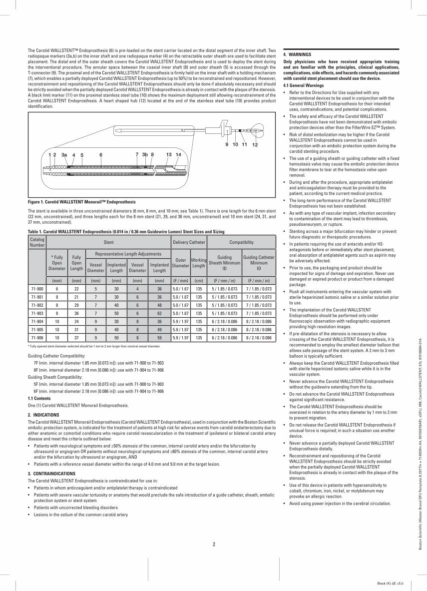
The stent is available in three unconstrained diameters (6 mm, 8 mm, and 10 mm; see Table 1). There is one length for the 6 mm stent (22 mm, unconstrained), and three lengths each for the 8 mm stent (21, 29, and 36 mm, unconstrained) and 10 mm stent (24, 31, and 37 mm, unconstrained).
Table 1. Carotid WALLSTENT Endoprosthesis (0.014 in / 0.36 mm Guidewire Lumen) Stent Sizes and Sizing
Catalog Number Stent Delivery Catheter Compatibility
* Fully Open
Diameter
Fully Open
Length
Representative Length AdjustmentsOuter
DiameterWorking Length
Guiding Sheath Minimum
ID
Guiding Catheter Minimum
IDVessel
DiameterImplanted
LengthVessel
DiameterImplanted
Length
(mm) (mm) (mm) (mm) (mm) (mm) (F / mm) (cm) (F / mm / in) (F / mm / in)
71-900 6 22 5 30 4 36 5.0 / 1.67 135 5 / 1.85 / 0.073 7 / 1.85 / 0.073
71-901 8 21 7 30 6 36 5.0 / 1.67 135 5 / 1.85 / 0.073 7 / 1.85 / 0.073
71-902 8 29 7 40 6 48 5.0 / 1.67 135 5 / 1.85 / 0.073 7 / 1.85 / 0.073
71-903 8 36 7 50 6 62 5.0 / 1.67 135 5 / 1.85 / 0.073 7 / 1.85 / 0.073
71-904 10 24 9 30 8 36 5.9 / 1.97 135 6 / 2.18 / 0.086 8 / 2.18 / 0.086
71-905 10 31 9 40 8 49 5.9 / 1.97 135 6 / 2.18 / 0.086 8 / 2.18 / 0.086
71-906 10 37 9 50 8 59 5.9 / 1.97 135 6 / 2.18 / 0.086 8 / 2.18 / 0.086
* Fully opened stent diameter selected should be 1 mm to 2 mm larger than nominal vessel diameter.
Guiding Catheter Compatibility: 7F (min. internal diameter 1.85 mm [0.073 in]): use with 71-900 to 71-903 8F (min. internal diameter 2.18 mm [0.086 in]): use with 71-904 to 71-906Guiding Sheath Compatibility: 5F (min. internal diameter 1.85 mm [0.073 in]): use with 71-900 to 71-903 6F (min. internal diameter 2.18 mm [0.086 in]): use with 71-904 to 71-9061.1 Contents One (1) Carotid WALLSTENT Monorail Endoprosthesis.
2. INDICATIONSThe Carotid WALLSTENT Monorail Endoprosthesis (Carotid WALLSTENT Endoprosthesis), used in conjunction with the Boston Scientific embolic protection system, is indicated for the treatment of patients at high risk for adverse events from carotid endarterectomy due to either anatomic or comorbid conditions who require carotid revascularization in the treatment of ipsilateral or bilateral carotid artery disease and meet the criteria outlined below:• Patients with neurological symptoms and ≥50% stenosis of the common, internal carotid artery and/or the bifurcation by
ultrasound or angiogram OR patients without neurological symptoms and ≥80% stenosis of the common, internal carotid artery and/or the bifurcation by ultrasound or angiogram, AND
• Patients with a reference vessel diameter within the range of 4.0 mm and 9.0 mm at the target lesion.
3. CONTRAINDICATIONSThe Carotid WALLSTENT Endoprosthesis is contraindicated for use in:• Patients in whom anticoagulant and/or antiplatelet therapy is contraindicated• Patients with severe vascular tortuosity or anatomy that would preclude the safe introduction of a guide catheter, sheath, embolic
protection system or stent system• Patients with uncorrected bleeding disorders• Lesions in the ostium of the common carotid artery
4. WARNINGSOnly physicians who have received appropriate training and are familiar with the principles, clinical applications, complications, side effects, and hazards commonly associated with carotid stent placement should use the device.4.1 General Warnings• Refer to the Directions for Use supplied with any
interventional devices to be used in conjunction with the Carotid WALLSTENT Endoprosthesis for their intended uses, contraindications, and potential complications.
• The safety and efficacy of the Carotid WALLSTENT Endoprosthesis have not been demonstrated with embolic protection devices other than the FilterWire EZ™ System.
• Risk of distal embolization may be higher if the Carotid WALLSTENT Endoprosthesis cannot be used in conjunction with an embolic protection system during the carotid stenting procedure.
• The use of a guiding sheath or guiding catheter with a fixed hemostasis valve may cause the embolic protection device filter membrane to tear at the hemostasis valve upon removal.
• During and after the procedure, appropriate antiplatelet and anticoagulation therapy must be provided to the patient, according to the current medical practice.
• The long-term performance of the Carotid WALLSTENT Endoprosthesis has not been established.
• As with any type of vascular implant, infection secondary to contamination of the stent may lead to thrombosis, pseudoaneurysm, or rupture.
• Stenting across a major bifurcation may hinder or prevent future diagnostic or therapeutic procedures.
• In patients requiring the use of antacids and/or H2-antagonists before or immediately after stent placement, oral absorption of antiplatelet agents such as aspirin may be adversely affected.
• Prior to use, the packaging and product should be inspected for signs of damage and expiration. Never use damaged or expired product or product from a damaged package.
• Flush all instruments entering the vascular system with sterile heparinized isotonic saline or a similar solution prior to use.
• The implantation of the Carotid WALLSTENT Endoprosthesis should be performed only under fluoroscopic observation with radiographic equipment providing high-resolution images.
• If pre-dilatation of the stenosis is necessary to allow crossing of the Carotid WALLSTENT Endoprosthesis, it is recommended to employ the smallest diameter balloon that allows safe passage of the stent system. A 2 mm to 3 mm balloon is typically sufficient.
• Always keep the Carotid WALLSTENT Endoprosthesis filled with sterile heparinized isotonic saline while it is in the vascular system.
• Never advance the Carotid WALLSTENT Endoprosthesis without the guidewire extending from the tip.
• Do not advance the Carotid WALLSTENT Endoprosthesis against significant resistance.
• The Carotid WALLSTENT Endoprosthesis should be oversized in relation to the artery diameter by 1 mm to 2 mm to prevent migration.
• Do not release the Carotid WALLSTENT Endoprosthesis if unusual force is required; in such a situation use another device.
• Never advance a partially deployed Carotid WALLSTENT Endoprosthesis distally.
• Reconstrainment and repositioning of the Carotid WALLSTENT Endoprosthesis should be strictly avoided when the partially deployed Carotid WALLSTENT Endoprosthesis is already in contact with the plaque of the stenosis.
• Use of this device in patients with hypersensitivity to cobalt, chromium, iron, nickel, or molybdenum may provoke an allergic reaction.
• Avoid using power injection in the cerebral circulation.

3
Bo
sto
n S
cien
tifi
c (M
aste
r B
ran
d D
FU Te
mp
late
8.2
677i
n x
11.
6929
in A
4, 9
0105
918A
R),
eD
FU, M
B, C
aro
tid
WA
LLS
TE
NT,
EN
, 910
1868
4-01
A
Black (K) ∆E ≤5.0
4.2 Patient SelectionThe safety and efficacy of the Carotid WALLSTENT™ Endoprosthesis have NOT yet been established in patients with the characteristics noted below.4.2.1 Patient Characteristics• Low to moderate risk for adverse events from carotid
endarterectomy• Experiencing acute ischemic neurologic stroke or having
experienced a stroke within 21 days of the procedure• Intracranial mass lesion (i.e., abscess, tumor, or infection)
or aneurysm >5 mm• Arteriovenous malformations of the territory of the target
carotid artery• Coagulopathies• Presence of fresh unlysed, unorganized thrombus• Patients undergoing laser debulking or electrocoagulation
within the stent• Poor renal function or life threatening allergy which, in the
physician’s opinion, may constitute high risk for a reaction to contrast medium
• Carotid string sign• Aneurysmal dilation immediately proximal or distal to the
lesion• Active infection• Severe dementia• Pregnancy• Under the age of 184.2.2 Lesion Characteristics• Evidence of intraluminal thrombus thought to increase the
risk of plaque fragmentation and distal embolization• Previously placed stent in the target artery• Requirement of more than two stents• Total occlusion of the target vessel• Presence of carotid artery dissection prior to initiation of
the procedure• Highly calcified lesions4.2.3 Access Characteristics• Known peripheral vascular, supra-aortic, or internal carotid
artery tortuosity that would preclude the use of catheter-based techniques
• Femoral access not possible• Inadequate local hemostasis at the access site• Failed guidewire or balloon catheter access4.3 Device Use• This device is intended for single-use only. Do not
reuse. Do not resterilize as this can compromise device performance and increase the risk of cross contamination due to inappropriate reprocessing.
• Do not use the product after the “Use By” date specified on the package.
• Heparinize the patient to achieve and maintain an Activated Clotting Time (ACT) of ≥275 seconds (≥200 seconds if using GP IIb/IIIa inhibitors) to prevent thrombus formation on the devices.
• To minimize the possible introduction of air into the delivery system, it is important to maintain tight catheter connections and to thoroughly flush the delivery system.
• Maintain continuous flush while removing and reinserting devices on the guidewire. Perform all exchanges slowly to prevent air embolism or trauma to the artery.
• Implanting a stent may lead to dissection of the vessel distal and/or proximal to the stent and may cause acute closure of the vessel, requiring additional intervention (carotid endarterectomy, further dilatation, or placement of additional stents).
• The stent may cause a thrombus, distal embolization or may migrate from the site of implant down the arterial lumen. Appropriate sizing of the stent to the vessel is required to reduce the possibility of stent migration. In the event of thrombosis of the expanded stent, thrombolysis and PTA should be attempted.
• In the event of complications such as infection, pseudoaneurysm or fistulization, surgical removal of the stent may be required.
• Overstretching of the artery may result in rupture and life-threatening bleeding.
• If a filter-based embolic protection system is used, allow for and maintain adequate distance between the filter and the stent delivery system or deployed stent to avoid potential entanglement. If filter basket entanglement or basket detachment occurs, surgical conversion or collapsing the basket with a second stent should be considered.
• Balloon angioplasty of the carotid bifurcation may initiate transient hemodynamic instability consisting of bradycardia or hypotension. Appropriate pharmacologic therapy must be immediately available.
5. PRECAUTIONS5.1 Stent Handling• Carefully inspect the Carotid WALLSTENT Endoprosthesis to
verify that the device has not been damaged in shipment. Do not use damaged equipment.
• The delivery system has an internal hypotube. Take care to avoid unnecessary handling, which may kink or damage the delivery system. Do not use if the device is kinked.
• Do not expose the delivery system to organic solvents like alcohol as structural integrity and/or function of the device may be impaired.
• Do not remove the stent from its delivery system as removal may damage the stent. The stent on the delivery system is intended to perform as a system. If removed, the stent cannot be put back on the delivery system.
• Special care must be taken not to handle or in any way disrupt the stent on the delivery system during catheter removal from packaging, stylus removal, placement over the guidewire and advancement through hemostatic valve adapter and guiding catheter or guiding sheath hub.
• Do not hold the sheath or stent during stylus removal. 5.2 Stent Placement• The Carotid WALLSTENT Endoprosthesis is not compatible with
any guidewire larger than 0.014 in (0.36 mm).• The Carotid WALLSTENT Endoprosthesis must be used with a
guiding catheter or guiding sheath to maintain adequate support of the 0.014 in (0.36 mm) guidewire throughout the procedure.
• For best device performance, the guidewire exit notch should remain within the guiding catheter or guiding sheath.
• Ensure the stent system is fully flushed with heparinized saline prior to use. Do not use the delivery system if flush is not observed exiting at the distal end of the sheath.
• Venous access should be available during carotid stenting to manage bradycardia and/or hypotension by either pharmaceutical intervention or placement of a temporary pacemaker, if needed.
• When catheters are in the body, they should be manipulated only under fluoroscopy. Radiographic equipment that provides high quality images is needed.
• The delivery system is not designed for use with power injection. Use of power injection may adversely affect device performance.
• If resistance is met during delivery system introduction, the system should be withdrawn and another system used.
• Prior to stent deployment, remove all slack from the delivery system.
• When more than one stent is required to cover the lesion, or if there are multiple lesions, the distal lesion should be stented first, followed by stenting of the proximal lesion.
• If overlap of sequential stents is necessary, the amount of overlap should be 5 mm. In no instance should more than 2 stents overlap.
5.3 Post Implant• Care must be exercised when crossing a newly deployed stent
with other interventional devices to avoid disrupting stent placement.
• In the event of thrombosis of the expanded stent, thrombolysis and PTA should be attempted.
Magnetic ResonanceConditionalMR
5.4 Magnetic Resonance (MRI) Safety InformationNon-clinical testing has demonstrated the Carotid WALLSTENT System is MR Conditional for single and overlapping lengths up to a total length of 64 mm. It can be scanned safely under the following conditions:• Static magnetic field of 1.5 Tesla or 3.0 Tesla using a
quadrature body coil only• Maximum spatial gradient field of <2500 gauss/cm (<25 T/m)• Maximum MR system reported, whole body averaged
specific absorption rate (SAR) of <1 W/kg for patient landmarks above the umbilicus (patient navel) and 2 W/kg (Normal Operating Mode) for patient landmarks below the umbilicus
MR imaging within these conditions may be performed immediately following the implantation of the stent.Under the scan conditions defined above, the Carotid WALLSTENT System is expected to produce a maximum temperature rise of 4.5ºC after 15 minutes of continuous scanning.Image Artifact InformationThe image artifact extends approximately 7 mm from the perimeter of the device diameter and 4 mm beyond each end of the length of the stent when scanned in non-clinical testing using a Spin Echo sequence. With a Gradient Echo sequence the image artifact extends 10 mm beyond the perimeter of the diameter and 7 mm beyond each end of the length with both sequences partially shielding the lumen in a 3.0 Tesla Intera (Achieva Upgrade), Philips Medical Solutions, software version Release 2.5.3.0 2007-09-28 MR system with a transmit/receive head coil.RecommendationsIt is recommended that patients register the conditions under which the implant can safely be scanned with the MedicAlert Foundation (www.medicalert.org) or an equivalent organization.
6. ADVERSE EVENTS6.1 BEACH Observed Adverse EventsBEACH (Boston Scientific EPI: A Carotid Stenting Trial for High-Risk Surgical Patients) was a prospective, single-arm, multi-center trial to evaluate the safety and efficacy of the Carotid WALLSTENT Endoprosthesis in conjunction with the FilterWire EX®/FilterWire EZ™ Embolic Protection System to treat surgical high risk, symptomatic (≥50% stenosis) and asymptomatic (≥80% stenosis) patients with disease in the carotid artery. The primary objective of the trial was to show non-inferiority between carotid stenting and a historical control representative of outcomes with carotid endarterectomy, based upon the 1-year morbidity and mortality rate including non Q-wave MI to 24 hours; death, stroke, and Q-wave MI through 30 days; and ipsilateral stroke and neurologic death from 31 to 360 days. A total of 747 patients were enrolled in the trial: 189 roll-in patients, 480 pivotal patients and 78 bilateral registry patients.Table 2 and Table 3 present Major Adverse Events (MAE) and Serious Adverse Events (SAE) respectively, as reported in the BEACH pivotal trial patients. A serious adverse event (SAE) may or may not be considered related to the device and was defined as follows:• Death due to any cause• Life-threatening condition (e.g., stroke)• Persistent or significant disability/incapacity• Any event resulting in an unscheduled in-patient
hospitalization or prolongation of existing hospitalization >72 hours post index procedure
• Any event requiring intervention, except for comorbid scheduled events, which are scheduled and planned during the follow-up period
• Congenital abnormality or birth defectSerious adverse events have been coded using the Medical Dictionary for Regulatory Activities (MedDRA™) version 5.0 and are presented by System Organ Class and Preferred Term as follows:• BLOOD AND LYMPHATIC SYSTEM DISORDERS include
events such as anemia.• CARDIAC DISORDERS include events such as angina,
arrhythmias, cardiac failure congestive and myocardial infarction.
• EYE DISORDERS include events such as retinal infarction.

4
Bo
sto
n S
cien
tifi
c (M
aste
r B
ran
d D
FU Te
mp
late
8.2
677i
n x
11.
6929
in A
4, 9
0105
918A
R),
eD
FU, M
B, C
aro
tid
WA
LLS
TE
NT,
EN
, 910
1868
4-01
A
Black (K) ∆E ≤5.0
• GASTROINTESTINAL DISORDERS include events such as gastrointestinal hemorrhage and retroperitoneal hemorrhage.• GENERAL DISORDERS AND ADMINISTRATION SITE CONDITIONS include events such as death, multi-organ failure, and pyrexia.• HEPATOBILIARY DISORDERS include events such as cholelithiasis.• INFECTIONS AND INFESTATIONS include events such as pneumonia, sepsis and urinary tract infection.• INJURY, POISONING AND PROCEDURAL COMPLICATIONS include events such as hip fracture and stent occlusion.• INVESTIGATIONS include events such as blood creatinine increased and neurological examination abnormal.• METABOLISM AND NUTRITION DISORDERS include events such as dehydration and hyperglycemia.• MUSCULOSKELETAL AND CONNECTIVE TISSUE DISORDERS include events such as arthritis and pain.• NEOPLASMS BENIGN, MALIGNANT AND UNSPECIFIED (INCLUDING CYSTS AND POLYPS) include events such as carcinomas,
lung cancer, and neoplasms.• NERVOUS SYSTEM DISORDERS include events such as cerebral hemorrhage, cerebrovascular accident, convulsions, dizziness,
syncope and transient ischemic attack.• PSYCHIATRIC DISORDERS include events such as confusion, depression and mental status changes.• RENAL AND URINARY DISORDERS include events such as renal failure and impairment.• REPRODUCTIVE SYSTEM AND BREAST DISORDERS include events such as vaginal hemorrhage.• RESPIRATORY, THORACIC AND MEDIASTINAL DISORDERS include events such as chronic obstructive airway disease, dyspnea,
pulmonary fibrosis, and respiratory failure.• SKIN AND SUBCUTANEOUS TISSUE DISORDERS include events such as skin ulcer.• SURGICAL AND MEDICAL PROCEDURES include events such as aortic valve replacement, arterial stent insertion, carotid
endarterectomy, coronary artery surgery and revascularization, and hip arthroplasty.• VASCULAR DISORDERS include events such as hematoma, hemorrhage, hypertension, hypotension, peripheral revascularization
and vascular pseudoaneurysm.
Table 2. BEACH Trial Major Adverse Events
Adverse Events ≤ 30 Days 31-360 Days 0-360 Days
Primary Endpoint # of Events
# of Patients
% Patients
# of Events
# of Patients
% Patients
# of Events
# of Patients
% Patients (N=448)
1-Year Morbidity and Mortality1 NA NA NA NA NA NA 50 40 8.9%
Major Adverse Events2 # of Events
# of Patients
% Patients(N=478)
# of Events
# of Patients
% Patients(N=462)
# of Events
# of Patients
% Patients(N=469)
Death 7 7 1.5% 29 29 6.3% 36 36 7.7%
Neurologic 2 2 0.4% 7 7 1.5% 9 9 1.9%
Non-neurologic 5 5 1.0% 22 22 4.8% 27 27 5.8%
Stroke 20 20 4.2% 19 19 4.1% 39 39 8.3%
Ipsilateral Stroke 15 15 3.1% 11 11 2.4% 26 26 5.5%
Major 5 5 1.0% 6 6 1.3% 11 11 2.3%
Minor 9 9 1.9% 2 2 0.4% 11 11 2.3%
Contralateral 5 5 1.0% 8 8 1.7% 13 13 2.8%
Major 0 0 0.0% 3 3 0.6% 3 3 0.6%
Minor 3 3 0.6% 4 4 0.9% 7 7 1.5%
Myocardial Infarction (MI) 5 5 1.0% 8 7 1.5% 13 12 2.6%
Non Q-wave MI 4 4 0.8% 7 6 1.3% 11 10 2.1%
Q-wave MI 1 1 0.2% 1 1 0.2% 2 2 0.4%1The 1-year morbidity and mortality rate is defined as the cumulative incidence of any non Q-wave myocardial infarction within 24 hours, peri-procedural (≤30 days) death, stroke, and Q-wave myocardial infarction, and late ipsilateral stroke or death due to neurologic events from 31 days up to and including 12-month follow-up.
2Major adverse events are defined as any death, stroke, or myocardial infarction.

5
Bo
sto
n S
cien
tifi
c (M
aste
r B
ran
d D
FU Te
mp
late
8.2
677i
n x
11.
6929
in A
4, 9
0105
918A
R),
eD
FU, M
B, C
aro
tid
WA
LLS
TE
NT,
EN
, 910
1868
4-01
A
Black (K) ∆E ≤5.0
Table 3. BEACH Trial Serious Adverse Events
MedDRA™ System Organ Class/ Preferred Term
≤ 30 Days(N=480)
31-360 Days(N=470)
0-360 Days(N=480)
# of Events # of Patients % Patients # of Events # of Patients % Patients # of Events # of Patients % Patients
Any SAE 196 115 24.0% 428 182 38.7% 624 251 52.3%
Blood And Lymphatic System Disorders 9 9 1.9% 14 12 2.6% 23 18 3.8%
Anemia Not Otherwise Specified 9 9 1.9% 14 12 2.6% 23 18 3.8%
Cardiac Disorders 26 21 4.4% 84 54 11.5% 110 69 14.4%
Angina Pectoris 2 2 0.4% 17 12 2.6% 19 13 2.7%
Angina Unstable 0 0 0.0% 4 4 0.9% 4 4 0.8%
Bradycardia Not Otherwise Specified 3 3 0.6% 3 3 0.6% 6 6 1.3%
Cardiac Arrest 2 2 0.4% 3 3 0.6% 5 5 1.0%
Cardiac Failure Congestive 2 2 0.4% 19 15 3.2% 21 16 3.3%
Coronary Artery Disease Not Otherwise Specified 1 1 0.2% 2 2 0.4% 3 3 0.6%
Myocardial Infarction 6 6 1.3% 15 14 3.0% 21 20 4.2%
Other Cardiac Disorders 10 8 1.7% 21 19 4.0% 31 25 5.2%
Eye Disorders 1 1 0.2% 0 0 0.0% 1 1 0.2%
Gastrointestinal Disorders 15 12 2.5% 30 23 4.9% 45 33 6.9%
General Disorders And Administration Site Conditions 6 5 1.0% 8 7 1.5% 14 11 2.3%
Death Not Otherwise Specified 0 0 0.0% 2 2 0.4% 2 2 0.4%
Other General Disorders and Administration Site Conditions 6 5 1.0% 6 5 1.1% 12 9 1.9%
Hepatobiliary Disorders 0 0 0.0% 2 2 0.4% 2 2 0.4%
Infections And Infestations 6 6 1.3% 37 29 6.2% 43 35 7.3%
Injury, Poisoning And Procedural Complications 1 1 0.2% 17 16 3.4% 18 17 3.5%
Stent Occlusion 0 0 0.0% 5 5 1.1% 5 5 1.0%
Other Injury, Poisoning and Procedural Complications 1 1 0.2% 12 11 2.3% 13 12 2.5%
Investigations 5 4 0.8% 5 4 0.9% 10 8 1.7%
Metabolism And Nutrition Disorders 2 2 0.4% 4 4 0.9% 6 5 1.0%
Musculoskeletal And Connective Tissue Disorders 1 1 0.2% 4 4 0.9% 5 4 0.8%
Neoplasms Benign, Malignant And Unspecified (Including Cysts and Polyps) 0 0 0.0% 10 10 2.1% 10 10 2.1%
Nervous System Disorders 53 43 9.0% 48 36 7.7% 101 75 15.6%
Carotid Artery Dissection 3 3 0.6% 0 0 0.0% 3 3 0.6%
Carotid Artery Occlusion 3 3 0.6% 0 0 0.0% 3 3 0.6%
Carotid Artery Stenosis 0 0 0.0% 2 1 0.2% 2 1 0.2%
Cerebral Hemorrhage 2 2 0.4% 2 2 0.4% 4 4 0.8%
Cerebrovascular Accident 14 14 2.9% 19 19 4.0% 33 33 6.9%
Transient Ischemic Attack 17 17 3.5% 8 8 1.7% 25 24 5.0%
Vasovagal Attack 1 1 0.2% 1 1 0.2% 2 2 0.4%
Other Nervous System Disorders 13 11 2.3% 16 12 2.6% 29 22 4.6%
Psychiatric Disorders 2 1 0.2% 7 7 1.5% 9 8 1.7%
Renal And Urinary Disorders 10 10 2.1% 8 8 1.7% 18 18 3.8%
Reproductive System And Breast Disorders 1 1 0.2% 0 0 0.0% 1 1 0.2%
Respiratory, Thoracic And Mediastinal Disorders 8 7 1.5% 27 24 5.1% 35 30 6.3%
Skin And Subcutaneous Tissue Disorders 0 0 0.0% 2 2 0.4% 2 2 0.4%
Surgical And Medical Procedures 16 15 3.1% 62 50 10.6% 78 60 12.5%
Carotid Endarterectomy 0 0 0.0% 2 2 0.4% 2 2 0.4%
Other Surgical and Medical Procedures 16 15 3.1% 60 48 10.2% 76 58 12.1%
Vascular Disorders 34 28 5.8% 59 46 9.8% 93 73 15.2%
Hematoma Not Otherwise Specified 8 8 1.7% 2 2 0.4% 10 10 2.1%
Hemorrhage Not Otherwise Specified 2 2 0.4% 2 2 0.4% 4 4 0.8%
Hypotension Aggravated 1 1 0.2% 0 0 0.0% 1 1 0.2%
Hypotension Not Otherwise Specified 10 10 2.1% 0 0 0.0% 10 10 2.1%
Vascular Pseudoaneurysm 3 3 0.6% 1 1 0.2% 4 4 0.8%
Other Vascular Disorders 10 10 2.1% 54 43 9.1% 64 53 11.0%

6
Bo
sto
n S
cien
tifi
c (M
aste
r B
ran
d D
FU Te
mp
late
8.2
677i
n x
11.
6929
in A
4, 9
0105
918A
R),
eD
FU, M
B, C
aro
tid
WA
LLS
TE
NT,
EN
, 910
1868
4-01
A
Black (K) ∆E ≤5.0
Table 4 presents all deaths, regardless of device or procedure relatedness.
Table 4. BEACH Trial Causes of Death
Death (by type) 0-30 Days 31-360 Days
(N=480) (N=470)
n % n %
Neurologic 2 0.4 7 1.5
Cardiac 3 0.6 8 1.7
General 2 0.4 7 1.5
Respiratory/Pulmonary 0 0.0 5 1.1
Infectious/Inflammatory 0 0.0 2 0.4
6.2 CABANA Observed Adverse EventsCABANA (A Carotid Stenting Boston Scientific Surveillance Program) was a nonrandomized, open-label study intended to: 1) compile early clinical outcomes data for the Carotid WALLSTENT™ and FilterWire EZ™ Embolic Protection System (FilterWire EZ System) in routine clinical practice; 2) evaluate clinical outcomes using a composite rate of death, stroke, and myocardial infarction (MI) rate ≤30 days, in total and by center experience tier; 3) assess the adequacy of the BSC Carotid Stenting Device Training Program.A total of 1097 patients were enrolled in the trial. Investigators were grouped into one of three tiers according to whether they had a high, medium, or low level of previous CAS experience and were also categorized by their CAS-credential-based training requirements for the CABANA study. The endpoint was the composite rate of major adverse events (MAEs), defined as CEC-adjudicated death, stroke, and MI, through 30 days post-index procedure, as well as the rates of these individual events, by physician experience tier, and by physician training tier.Table 5 and Table 6 present the Major Adverse Events (MAE) and Serious Adverse Events (SAE) respectively, as reported in the CABANA patients. A serious adverse event (SAE) may or may not be considered related to the device and was defined as:• Death due to any cause.• Life-threatening condition (e.g., stroke).• Persistent or significant disability/incapacity.• Requires unplanned in-patient hospitalization or
prolongation (> 72 hrs) of existing hospitalization (except for comorbid scheduled events, which are scheduled and planned during the follow-up period).
• Intervention to prevent a permanent impairment of a body function or permanent damage to a body structure.
• Congenital abnormality or birth defect.Serious adverse events have been coded similar to BEACH using the MedDRA™ version 11.1 with the addition of:• IMMUNE SYSTEM DISORDERS include events such as
anaphylactic reaction,There were no REPRODUCTIVE SYSTEM AND BREAST DISORDERS reported in the CABANA study.
Table 5. CABANA Major Adverse Events, 30 Days, All Enrolled Patients (N=1097)
Parameter N=1025 Evaluable Patients 95% Confidence Interval
30-Day MAE 4.6% (47/1025) [3.4%, 6.1%]
Death 1.3% (13/1025) [0.7%, 2.2%]
Neurologic Death 0.5% (5/1025) [0.2%, 1.1%]
Cardiac Death 0.5% (5/1025) [0.2%, 1.1%]
Non-neurologic and Non-cardiac Death 0.3% (3/1025) [0.1%, 0.9%]
Stroke 3.3% (34/1025) [2.3%, 4.6%]
Classification 1: Major or Minor
Major Stroke 2.0% (20/1025) [1.2%, 3.0%]
Minor Stroke 1.4% (14/1025) [0.7%, 2.3%]
Classification 2: Ipsilateral or Contralateral
Ipsilateral Stroke 2.9% (30/1025) [2.0%, 4.2%]
Contralateral Stroke 0.4% (4/1025) [0.1%, 1.0%]
Classification 3: Ischemic or Hemorrhagic
Ischemic Stroke 2.8% (29/1025) [1.9%, 4.0%]
Hemorrhagic Stroke 0.5% (5/1025) [0.2%, 1.1%]
MI 0.5% (5/1025) [0.2%, 1.1%]
Q-wave MI 0.0% (0/1025) [0.0%, 0.4%]
Non-Q-wave MI 0.5% (5/1025) [0.2%, 1.1%]
Death, Stroke, and MI (≤ 24 hours) 2.8% (29/1025) [1.9%, 4.0%]
Death, Stroke, and MI (>24 hours and ≤ 30 days) 2.1% (22/1025) [1.3%, 3.2%]
Numbers are % (count/sample size)

7
Bo
sto
n S
cien
tifi
c (M
aste
r B
ran
d D
FU Te
mp
late
8.2
677i
n x
11.
6929
in A
4, 9
0105
918A
R),
eD
FU, M
B, C
aro
tid
WA
LLS
TE
NT,
EN
, 910
1868
4-01
A
Black (K) ∆E ≤5.0
Table 6. CABANA Rates of Center-Reported Serious Adverse Events, All Enrolled Patients (N=1097)
MedDRA™ System Organ Class/ Preferred Term
≤ 30 Days (N=1097)
# of Events # of Patients % Patients
TOTAL 389 223 20.3%
Blood and lymphatic system disorders 17 17 1.5%
Cardiac disorders 56 55 5.0%
Angina pectoris 7 6 0.5%
Angina unstable 1 1 0.1%
Bradycardia 14 14 1.3%
Cardiac arrest 5 5 0.5%
Cardiac failure congestive 7 7 0.6%
Cardio-respiratory arrest 1 1 0.1%
Coronary artery disease 1 1 0.1%
Myocardial infarction 5 5 0.5%
Sinus bradycardia 1 1 0.1%
Other cardiac disorders 14 14 1.3%
Eye disorders 5 5 0.5%
Gastrointestinal disorders 22 18 1.6%
General disorders and administration site conditions 27 26 2.4%
Death 1 1 0.1%
Other general disorders and administration site conditions 26 25 2.3%
Hepatobiliary disorders 2 2 0.2%
Immune system disorders 1 1 0.1%
Infections and infestations 28 22 2.0%
Injury, poisoning and procedural complications 12 11 1.0%
Investigations 14 12 1.1%
Metabolism and nutrition disorders 6 6 0.5%
Musculoskeletal and connective tissue disorders 6 6 0.5%
Neoplasms benign, malignant and unspecified (incl cysts and polyps) 1 1 0.1%
Nervous system disorders 73 68 6.2%
Carotid artery occlusion 1 1 0.1%
Cerebral haemorrhage 2 1 0.1%
Cerebrovascular accident 25 25 2.3%
Haemorrhagic stroke 4 3 0.3%
Ischaemic stroke 1 1 0.1%
Transient ischaemic attack 9 9 0.8%
Other nervous system disorders 31 28 2.6%
Psychiatric disorders 7 7 0.6%
Renal and urinary disorders 11 10 0.9%
Respiratory, thoracic and mediastinal disorders 31 28 2.6%
Skin and subcutaneous tissue disorders 1 1 0.1%
Surgical and medical procedures 1 1 0.1%
Vascular disorders 68 67 6.1%
Haemorrhage 1 1 0.1%
Hypertension 4 4 0.4%
Hypotension 47 47 4.3%
Orthostatic hypotension 4 3 0.3%
Other vascular disorders 12 12 1.1%
8
Bo
sto
n S
cien
tifi
c (M
aste
r B
ran
d D
FU Te
mp
late
8.2
677i
n x
11.
6929
in A
4, 9
0105
918A
R),
eD
FU, M
B, C
aro
tid
WA
LLS
TE
NT,
EN
, 910
1868
4-01
A
Black (K) ∆E ≤5.0
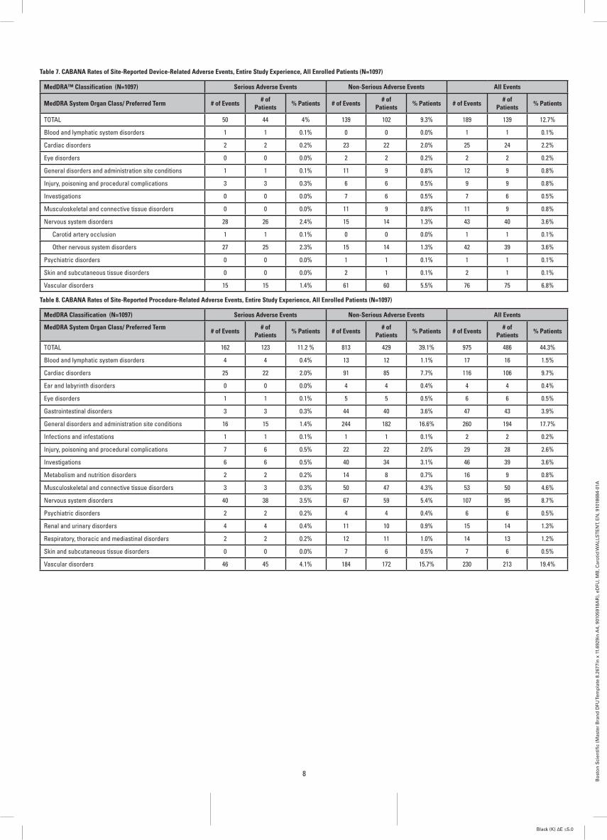
Table 7. CABANA Rates of Site-Reported Device-Related Adverse Events, Entire Study Experience, All Enrolled Patients (N=1097)
MedDRA™ Classification (N=1097) Serious Adverse Events Non-Serious Adverse Events All Events
MedDRA System Organ Class/ Preferred Term # of Events # of Patients % Patients # of Events # of
Patients % Patients # of Events # of Patients % Patients
TOTAL 50 44 4% 139 102 9.3% 189 139 12.7%
Blood and lymphatic system disorders 1 1 0.1% 0 0 0.0% 1 1 0.1%
Cardiac disorders 2 2 0.2% 23 22 2.0% 25 24 2.2%
Eye disorders 0 0 0.0% 2 2 0.2% 2 2 0.2%
General disorders and administration site conditions 1 1 0.1% 11 9 0.8% 12 9 0.8%
Injury, poisoning and procedural complications 3 3 0.3% 6 6 0.5% 9 9 0.8%
Investigations 0 0 0.0% 7 6 0.5% 7 6 0.5%
Musculoskeletal and connective tissue disorders 0 0 0.0% 11 9 0.8% 11 9 0.8%
Nervous system disorders 28 26 2.4% 15 14 1.3% 43 40 3.6%
Carotid artery occlusion 1 1 0.1% 0 0 0.0% 1 1 0.1%
Other nervous system disorders 27 25 2.3% 15 14 1.3% 42 39 3.6%
Psychiatric disorders 0 0 0.0% 1 1 0.1% 1 1 0.1%
Skin and subcutaneous tissue disorders 0 0 0.0% 2 1 0.1% 2 1 0.1%
Vascular disorders 15 15 1.4% 61 60 5.5% 76 75 6.8%
Table 8. CABANA Rates of Site-Reported Procedure-Related Adverse Events, Entire Study Experience, All Enrolled Patients (N=1097)
MedDRA Classification (N=1097) Serious Adverse Events Non-Serious Adverse Events All Events
MedDRA System Organ Class/ Preferred Term # of Events # of Patients % Patients # of Events # of
Patients % Patients # of Events # of Patients % Patients
TOTAL 162 123 11.2 % 813 429 39.1% 975 486 44.3%
Blood and lymphatic system disorders 4 4 0.4% 13 12 1.1% 17 16 1.5%
Cardiac disorders 25 22 2.0% 91 85 7.7% 116 106 9.7%
Ear and labyrinth disorders 0 0 0.0% 4 4 0.4% 4 4 0.4%
Eye disorders 1 1 0.1% 5 5 0.5% 6 6 0.5%
Gastrointestinal disorders 3 3 0.3% 44 40 3.6% 47 43 3.9%
General disorders and administration site conditions 16 15 1.4% 244 182 16.6% 260 194 17.7%
Infections and infestations 1 1 0.1% 1 1 0.1% 2 2 0.2%
Injury, poisoning and procedural complications 7 6 0.5% 22 22 2.0% 29 28 2.6%
Investigations 6 6 0.5% 40 34 3.1% 46 39 3.6%
Metabolism and nutrition disorders 2 2 0.2% 14 8 0.7% 16 9 0.8%
Musculoskeletal and connective tissue disorders 3 3 0.3% 50 47 4.3% 53 50 4.6%
Nervous system disorders 40 38 3.5% 67 59 5.4% 107 95 8.7%
Psychiatric disorders 2 2 0.2% 4 4 0.4% 6 6 0.5%
Renal and urinary disorders 4 4 0.4% 11 10 0.9% 15 14 1.3%
Respiratory, thoracic and mediastinal disorders 2 2 0.2% 12 11 1.0% 14 13 1.2%
Skin and subcutaneous tissue disorders 0 0 0.0% 7 6 0.5% 7 6 0.5%
Vascular disorders 46 45 4.1% 184 172 15.7% 230 213 19.4%

9
Bo
sto
n S
cien
tifi
c (M
aste
r B
ran
d D
FU Te
mp
late
8.2
677i
n x
11.
6929
in A
4, 9
0105
918A
R),
eD
FU, M
B, C
aro
tid
WA
LLS
TE
NT,
EN
, 910
1868
4-01
A
Black (K) ∆E ≤5.0
6.3 Potential Adverse EventsBased on the literature, and on clinical and commercial experience with carotid stents and embolic protection systems, potential adverse events include, but are not limited to the following:• Abrupt vessel closure• Additional interventional or surgical treatment (e.g.,
stenting or carotid endarterectomy)• Allergic reactions (including to antiplatelet agents, contrast
medium or stent materials)• Aneurysm• Angina/coronary ischemia• Arrhythmia• Arteriovenous fistula• Bacteremia or septicemia• Bleeding• Bradycardia• Cerebral vascular event such as edema• Cerebral ischemia/transient ischemic attack• Congestive heart failure (CHF)• Death• Detachment and/or implantation of a component• Emboli (air, tissue, plaque, thrombus, device or other)• Fever• Filter thrombosis/occlusion• Hematoma• Hemorrhage• Hyperperfusion syndrome• Hypotension/hypertension• Hypotonia• Infection • Ischemia/infarction of tissue or organ• Myocardial Infarction (MI)• Pain• Pseudoaneurysm• Renal failure/insufficiency• Restenosis of stented segment• Seizure• Severe unilateral headache• Stent embolization• Stent/filter entanglement or damage• Stent migration• Stent malposition• Stent thrombosis/occlusion• Stroke/cerebrovascular accident (CVA)• Vessel injury/dissection/perforation/rupture/trauma• Vessel occlusion or thrombosis• Vessel spasm or recoilAny device related adverse event involving the Carotid WALLSTENT™ Monorail™ Endoprosthesis (Carotid WALLSTENT Endoprosthesis) should be reported immediately to Boston Scientific, Customer Service, at (888) 272-1001.
7. CLINICAL STUDIES7.1 BEACHBEACH, (Boston Scientific EPI: A Carotid Stenting Trial for High-Risk Surgical Patients), was a prospective, single-arm, multi-center trial to evaluate the safety and efficacy of the Carotid WALLSTENT Endoprosthesis in conjunction with the FilterWire EX®/FilterWire EZ™ Embolic Protection System to treat high-surgical-risk, symptomatic (≥50% stenosis) and asymptomatic (≥80% stenosis) patients with disease in the carotid artery. A trial design utilizing a roll-in phase for initial clinical experience was employed in the study. In addition, a bilateral registry was included for patients presenting with bilateral carotid artery disease requiring treatment. A total of 747 patients were enrolled at 47 centers in the United States, including 189 roll-in patients, 480 pivotal patients and 78 bilateral registry patients. This trial is summarized in Table 9.
Table 9. Overview of BEACH Trial Study Design
Product Evaluated: Carotid WALLSTENT Endoprosthesis and FilterWire EX/FilterWire EZ System
Sample Size for Pivotal Patients: 480Number of Centers: 47Primary Endpoint: 1-Year Morbidity and Mortality: Non Q-wave MI through 24 hours Death, Stroke, Q-wave MI through 30 days Neurologic Death, Ipsilateral Stroke from 31-360 daysSecondary Endpoints: FilterWire EX/FilterWire EZ System Technical Success1
Carotid WALLSTENT Endoprosthesis Technical Success2
System Technical Success3
Angiographic Success4
Procedure Success5
30-Day Clinical Success6
Peri-Procedural Morbidity and Mortality7
Peri-Procedural Overall Morbidity8
1-Year Clinical Success9
Late Stroke, TIA and Death10
Study Hypothesis: Non-inferiority to historical controlPatient Follow-up: Neurological assessment by independent neurologist CK/CKMB to 24 hours ECG: discharge and 30 days Carotid ultrasound: discharge, 30 days, 6 months
and 1 year to 3 years AEs: discharge, 30 days, 6 months, 1 year to 3 years
1 FilterWire EX/FilterWire EZ System successfully delivered and deployed beyond the target lesion and successfully retrieved after completion of the stent placement. Calculated based on the number of FilterWire® System uses attempted.
2 Deployment of the Carotid WALLSTENT Endoprosthesis at the intended location and successful retrieval of the delivery catheter after stent placement. Calculated based on the number of stent implantations attempted.
3 Includes FilterWire System Technical Success combined with Carotid WALLSTENT Endoprosthesis Technical Success. Calculated based on the number of system placement attempts.
4 System Technical Success with a residual diameter stenosis ≤30% immediately after post-dilatation as determined by angiographic core lab. Calculated based on number of patients on whom a procedure is attempted.
5 Includes System Technical Success and Angiographic Success without death, stroke and MI (Q-wave and non Q-wave) immediately following the index procedure. Calculated based on number of patients attempted to be treated.
6 Procedure Success without any death, stroke or MI (Q-wave) up to and including 30 days post procedure. Calculated based on number of patients on whom a procedure is attempted.
7 Non Q-wave MI through 24 hours post procedure and death, stroke and Q-wave MI through 30 days post procedure.
8 Morbidity occurring up to and including 30 days after the index procedure, including complications associated with routine catheterization, e.g., infection, hematoma, etc.
9 Defined as a patent vessel by Duplex Ultrasound (as assessed by core laboratory to be <50% stenosis and confirmed by angiogram in patients that develop symptoms post procedurally) combined with freedom from stroke and death through 30 days, ipsilateral stroke and neurologic death 31-360 days and interim target vessel revascularization through 360 days. One-year clinical success was calculated based on the number of patients treated.
10 Defined as the incidence of any stroke (major or minor), TIA or death occurring after 30 days and up to and including 1 year post procedure. Major stroke: a new focal ischemic neurological deficit of abrupt onset, which is present after 7 days and increases the NIH Stroke Scale by ≥4. Minor stroke: a new focal ischemic neurological deficit of abrupt onset, lasting >24 hours and increases the NIH Stroke Scale by ≤3. TIA: a focal ischemic neurological deficit of abrupt onset and of presumed vascular etiology that resolves completely within 24 hours of onset.
The BEACH trial was designed to show non-inferiority between carotid stenting and a historical control, based on standard of care. The historical control was established based on a review of the current literature on carotid endarterectomy and was defined as a weighted Objective Performance Criterion (OPC). A criterion of 15% for patients who had comorbidity risk factors and a criterion of 11% for patients who had anatomic risk factors were selected. A spread of 4% for the “delta” definition of equivalency was selected.
Weighted OPC = (% Comorbid x 15%) + (% Anatomic x 11%)Two patients did not meet either the comorbid or anatomic high-risk criteria. Of the remaining 478 patients, 41.2% (197/478) were in the comorbid group and 58.8% (281/478) were in the anatomic group; therefore, the weighted OPC for BEACH was 12.6%. Note that 59 patients included in the comorbid group presented with both comorbid and anatomic risk factors.
12.6% = (41.2% x 15%) + (58.8% x 11%)Based on the weighted OPC of 12.6% and the pre-specified delta of 4%, the threshold for claiming non-inferiority to CEA is 16.6%, i.e., the one-sided upper 95% confidence limit of the primary endpoint must be <16.6% to conclude non-inferiority.
The protocol required regular patient follow-up by the treating physician and follow-up neurological assessments by an independent neurologist. Core laboratories provided independent assessments for angiographic, ultrasound, ECG and CT/MRI testing. Monitors reviewed all safety data to ensure appropriate reporting of adverse events. A Clinical Events Committee adjudicated suspected primary endpoint events. A Data Safety Monitoring Board reviewed adverse events to ensure patient safety.7.1.1 Eligibility Criteria SummaryThe study population consisted of male and female patients, at least 18 years of age, with discrete lesions in the common carotid artery (CCA), internal carotid artery (ICA) or carotid bifurcation. Patients had to be at high-risk for surgical intervention; both symptomatic (≥50% stenosis) and asymptomatic (≥80% stenosis) patients were eligible.The key inclusion criteria included the following:• Symptomatic: Carotid stenosis of ≥50% via angiography
with cerebral or retinal TIA or ischemic stroke symptoms determined to have occurred ipsilateral to the target lesion and to be reasonably attributable to the lesion within 180 days of the stenting procedure
• Asymptomatic: Carotid stenosis of ≥80% via angiography without cerebral or retinal TIA or ischemic stroke symptoms within 180 days of the stenting procedure
• Patient had to have an anatomic or comorbid high-risk condition as outlined below:
7.1.1.1 Anatomic High-Risk ConditionsONE (1) criterion qualifies1. Surgically inaccessible lesions at or above C2 or below
the clavicle2. Previous neck or head radiation therapy or surgery that
included the area of stenosis/repair or ipsilateral radical neck dissection for cancer
3. Spinal immobility of the neck due to cervical arthritis or other cervical disorders
4. Restenosis after a previous or unsuccessful attempt of CEA (≥50% symptomatic, ≥80% asymptomatic) at least 31 days prior to enrollment if arteriotomy was performed
5. Presence of laryngeal palsy or laryngectomy6. Presence of a tracheostoma7. Contralateral total occlusion with a qualifying lesion on
the ipsilateral side (Note: Applied to roll-in and pivotal groups only)
8. Bilateral carotid artery disease (Note: Patients with bilateral disease were placed in the Bilateral Registry provided that both ipsilateral and contralateral arteries required treatment at the time of enrollment.)
7.1.1.2 Comorbid High-Risk ConditionsCLASS I [ONE (1) criterion qualifies]1. Congestive heart failure (NYHA Class III/IV)2. Unstable angina (CCS Class III/IV)3. Requirement for staged and scheduled Coronary Artery
Bypass Graft (CABG) or valve replacement post carotid index procedure (Note: The staged procedure had to occur >30 days post index procedure.)
4. Chronic Obstructive Pulmonary Disease (COPD) manifested with a forced expiratory volume (FEV) ≤30%
5. Known severe left ventricular ejection fraction (LVEF) ≤30%CLASS II [TWO (2) criteria qualify]1. Age ≥75 years2. Recent MI (Q-wave and/or non Q-wave) >72 hours and ≤30
days, with any elevation in CK-MB greater than the local laboratory upper limit of normal values
3. Two or more major diseased coronary arteries with ≥70% stenosis at the time of index procedure in patients with a history of angina
4. Requirement for staged and scheduled peripheral vascular surgery or other major surgeries [e.g., abdominal aortic aneurysm (AAA)] post carotid index procedure
7.1.1.3 Specific Inclusion Criteria for the Carotid WALLSTENT Monorail Endoprosthesis (Carotid WALLSTENT Endoprosthesis) and FilterWire EZ System
1. Target lesion in the common carotid artery (CCA), internal carotid artery (ICA) or carotid bifurcation
2. Diameter of the target arterial segment to be stented ≥4.0 mm and ≤9.0 mm

10
Bo
sto
n S
cien
tifi
c (M
aste
r B
ran
d D
FU Te
mp
late
8.2
677i
n x
11.
6929
in A
4, 9
0105
918A
R),
eD
FU, M
B, C
aro
tid
WA
LLS
TE
NT,
EN
, 910
1868
4-01
A
Black (K) ∆E ≤5.0
3. Vessel diameter distal to the target lesion ≥3.5 mm and ≤5.5 mm as an optimal “landing zone” for placement of the FilterWire EZ™ System with visual angiographic recommendations
7.1.2 Description of Patients EvaluatedTable 10 summarizes patient follow-up at the endpoint evaluation time points of 30 days, 6 months, and 12 months. Patients were considered to have been evaluated if they had physician contact as evidenced by at least one of the following at the given time point: office visit, neurologic evaluation, AE log, stroke scales, event forms such as Repeat Carotid Angiography Form, SAE Notification Form, Subsequent Hospitalization Form, Vascular Event Form, Neurological Event Form, etc.
Table 10. BEACH Patient Follow-up
Pivotal (N=480)
Primary Analysis Sample (ITT1) 480
30-day Follow-up Evaluation Completed 466
6-month Follow-up Evaluation Completed 435
12-month Follow-up Evaluation Completed 418
12-month Follow-up Evaluation not Completed 62
Death 36
Lost to Follow-up 10
Missed Visit 16
Patients with Ultrasound Data Pre-Procedure 455
Patients with Ultrasound Data at 30 Days 446
Patients with Ultrasound Data at 6 Months 418
Patients with Ultrasound Data at 12 Months 3771ITT is Intent to Treat
Baseline patient demographics, lesion characteristics, and High-Risk Inclusion Criteria for the study are presented in Table 11. All reported angiographic data on the treated lesions are based on measurements obtained by the centralized angiographic core laboratory.
Table 11. Baseline Patient Demographics, Lesion Characteristics, and High-Risk Inclusion Criteria
Demographic and Medical History Value 95% CI
Age (years)
Mean ± SD (N) 70.9±9.3 (480) [70.0, 71.7]
Range (min, max) (41.0, 92.0)
Gender %
Male 65.2% (313/480) [60.8%, 69.5%]
History %
Diabetes mellitus 33.8% (162/480) [29.5%, 38.2%]
Hypertension 89.4% (429/480) [86.3%, 92.0%]
Hyperlipidemia 86.5% (415/480) [83.1%, 89.4%]
Current or history of smoking 74.6% (358/480) [70.4%, 78.4%]
Number of Symptomatic Patients 23.3% (112/480) [19.6%, 27.4%]
Baseline Lesion Characteristics
Calcification % 48.8% (234/480) [44.2%, 53.3%]
Lesion Length (mm)
Mean ± SD (N) 15.13±7.25 (480) [14.48, 15.78]
Range (min, max) (2.46, 57.60)
Minimal Lumen Diameter (mm)
Mean ± SD (N) 1.33±0.58 (480) [1.27, 1.38]
Range (min, max) (0.12, 3.51)
Percent Diameter Stenosis (%DS)
Mean ± SD (N) 71.61%±10.71% (480) [70.65%, 72.58%]
Range (min, max) (36.75%, 96.52%)
High-Risk Inclusion Criteria Value
Anatomic High-Risk Conditions (One Criterion Qualifies)
Surgically inaccessible lesions 9.2% (44/480)
Previous head/neck radiation therapy or radical neck surgery 10.8% (52/480)
Spinal immobility 7.3% (35/480)
Restenosis after previous, or unsuccessful attempt, of CEA 34.2% (164/480)
Presence of laryngeal palsy or laryngectomy 1.0% (5/480)
Presence of tracheostoma 2.1% (10/480)
Contralateral total occlusion 18.1% (87/480)
Comorbid High-Risk Conditions- Class I (One Criterion Qualifies)
Congestive heart failure (NYHA Class III/IV) 11.7% (56/480)
Unstable angina (CCS Class III/IV) 12.5% (60/480)
Requirement for CABG or valve replacement 6.5% (31/480)
COPD manifested with a forced expiratory volume (FEV) ≤30% 2.3% (11/480)
Known severe left ventricular ejection fraction (LVEF) ≤30% 12.1% (58/480)
Comorbid High-Risk Conditions - Class II (Two Criteria Qualify)
Age ≥75 years old 39.0% (187/480)
Recent MI (Q-wave and/or non Q-wave) >72 hours and ≤30 days 1.3% (6/480)
Two or more major diseased coronary arteries with ≥70% stenosis 21.7% (104/480)
Requirement for peripheral vascular or other major surgery 2.9% (14/480)
7.1.3 ResultsThe primary endpoint for the BEACH trial was 1-year morbidity and mortality defined as the cumulative incidence of any non Q-wave myocardial infarction within the 24 hours following carotid stenting, peri-procedural (≤30 days) death, stroke, Q-wave myocardial infarction, and late ipsilateral stroke or death due to neurologic events from 31 to 360 days. The 1-year morbidity and mortality rate was 8.9%. Rates for each contributor to the composite primary endpoint rate are presented along with the secondary endpoints in Table 12.The trial utilized the FilterWire EX® and the FilterWire EZ embolic protection devices. A total of 195 patients were enrolled using the FilterWire EX System and 285 patients were enrolled using the FilterWire EZ System. Poolability analysis was conducted to determine baseline homogeneity. No significant differences between the groups were found. In addition, a group difference on peri-procedural outcome analysis was performed. There was no evidence found against pooling the FilterWire EX System and FilterWire EZ System groups for purposes of estimating the treatment effect on 1-year morbidity and mortality.The primary objective of the BEACH trial was met. The observed 1-year morbidity and mortality rate of 8.9% with an upper confidence limit of 11.5% fell well below the predefined weighted OPC + delta of 16.6%, demonstrating that carotid stenting with the Carotid WALLSTENT™ Endoprosthesis and the FilterWire® Embolic Protection System is non-inferior to surgical treatment for carotid artery disease in patients who were at high risk for CEA.

11
Bo
sto
n S
cien
tifi
c (M
aste
r B
ran
d D
FU Te
mp
late
8.2
677i
n x
11.
6929
in A
4, 9
0105
918A
R),
eD
FU, M
B, C
aro
tid
WA
LLS
TE
NT,
EN
, 910
1868
4-01
A
Black (K) ∆E ≤5.0
Table 12. Clinical Results Through 360 Days Follow-up
Primary Endpoint Measures Pivotal (N=480) 95% CI1
1-Year Morbidity and Mortality 8.9% (40/448) [11.5%]
Non Q-wave MI (Through 24 hours) 0.9% (4/448) [0.2%, 2.3%]
Death, Stroke, Q-wave MI (Through 30 days) 5.4% (24/448) [3.5%, 7.9%]
Death 1.6% (7/448) [0.6%, 3.2%]
Neurologic 0.4% (2/448) [0.1%, 1.6%]
Cardiac 0.7% (3/448) [0.1%, 1.9%]
General 0.4% (2/448) [0.1%, 1.6%]
Stroke 4.5% (20/448) [2.8%, 6.8%]
Ipsilateral2 3.3% (15/448) [1.9%, 5.5%]
Major Ischemic 1.1% (5/448) [0.4%, 2.6%]
Minor Ischemic 2.0% (9/448) [0.9%, 3.8%]
Hemorrhagic (excludes Subarachnoid Hemorrhages) 0.2% (1/448) [0.0%, 1.2%]
Contralateral 1.1% (5/448) [0.4%, 2.6%]
Major Ischemic 0.0% (0/448) [0.0%, 0.8%]
Minor Ischemic 0.7% (3/448) [0.1%, 1.9%]
Hemorrhagic (excludes Subarachnoid Hemorrhages) 0.4% (2/448) [0.1%, 1.6%]
Subarachnoid Hemorrhagic 0.0% (0/448) [0.0%, 0.8%]
Q-wave MI 0.2% (1/448) [0.0%, 1.2%]
Neurologic Death, Ipsilateral Stroke (31 days through 360 days) 3.1% (14/448) [1.7%, 5.2%]
Neurologic Death 1.6% (7/448) [0.6%, 3.2%]
Ipsilateral Stroke 2.5% (11/448) [1.2%, 4.4%]
Major Ischemic 1.3% (6/448) [0.5%, 2.9%]
Minor Ischemic 0.4% (2/448) [0.1%, 1.6%]
Hemorrhagic (excludes Subarachnoid Hemorrhages) 0.7% (3/448) [0.1%, 1.9%]
Freedom from 1-Year Morbidity and Mortality – KM Estimate 91.6% [89.0%, 94.2%]
Secondary Endpoint Measures Pivotal (N=480) 95% CI
FilterWire EX® and FilterWire EZ™ System Technical Success3 97.1% (475/489) [95.2%,98.4%]
Carotid WALLSTENT™ Endoprosthesis Technical Success4 94.1% (475/505) [91.6%,96.0%]
System Technical Success5 98.3% (469/477) [96.7%,99.3%]
Angiographic Success6 90.8% (433/477) [87.8%,93.2%]
Procedure Success7 87.6% (418/477) [84.3%,90.5%]
30-Day Clinical Success8 85.3% (405/475) [81.8%,88.3%]
Peri-Procedural Morbidity and Mortality9 5.6% (27/478) [3.8%,8.1%]
Peri-Procedural Overall Morbidity10 68.5% (328/479) [64.1%,72.6%]
1-Year Clinical Success11 69.9% (297/425) [65.3%,74.2%]
Late Stroke, TIA and Death (31 days through 360 days)12 10.6% (49/462) [7.9%,13.8%]
Post-procedure In-lesion Minimal Lumen Diameter (mm):Mean + SD (N)Range (min, max)
4.2±0.8 (478)
(2.3, 7.9)
[4.1, 4.2]
Post-procedure In-lesion Percent Diameter Stenosis: Mean + SD (N)Range (min, max)
10.6%±14.4% (478)
(-73.3%,51.9%)[9.4%, 11.9%]
Target Vessel Revascularization (TVR) Rate (≤ 360 days)13 4.7% (20/425) [2.9%, 7.2%]
1-Year Restenosis Rate (> 50% Stenosis via Duplex U/S) 18.7% (72/385) [14.9%, 23.0%]
Carotid Duplex Ultrasound ICA/CCA Ratio:Pre-ProcedurePost-ProcedureAt 1 monthAt 6 monthsAt 12 months
5.3±3.1 (420)
1.4±0.5 (438)
1.4±0.5 (434)
1.9±1.2 (399)
1.9±1.1 (362)
[5.0, 5.6]
[1.4, 1.5]
[1.4, 1.5]
[1.8, 2.1]
[1.8, 2.0]
Numbers are % (count/sample size) or %.
1 1-sided 95% upper confidence limit is presented for 1-year morbidity and mortality.
2 Patient 42-014 was originally denoted to have suffered a minor ipsilateral stroke 27 days post-procedure. This event was sent back to the CEC for additional review after the CT/MRI core lab provided a review of films made available to them. Based upon the core lab report, the CEC adjudicated the event as a TIA.
3 FilterWire EX/ FilterWire EZ System successfully delivered and deployed beyond the target lesion and successfully retrieved after completion of the stent placement. Calculated based on the number of FilterWire® uses attempted.
4 Deployment of the Carotid WALLSTENT Monorail™ Endoprosthesis (Carotid WALLSTENT Endoprosthesis) at the intended location and successful retrieval of the delivery catheter after stent placement. Calculated based on the number of stent implantations attempted. Three patients did not have a Carotid WALLSTENT Endoprosthesis implantation attempted.
5 Includes FilterWire System Technical Success combined with Carotid WALLSTENT Endoprosthesis Technical Success. Calculated based on the number of system placement attempts.
6 System Technical Success with a residual diameter stenosis ≤30% immediately after post-dilatation as determined by angiographic core lab. Calculated based on number of patients on whom a procedure is attempted.
7 Includes System Technical Success and Angiographic Success without death, stroke and MI (Q-wave and non Q-wave) immediately following the index procedure. Calculated based on number of patients attempted to be treated.
8 Procedure Success without any death, stroke or MI (Q-wave) up to and including 30 days post procedure. Calculated based on number of patients on whom a procedure is attempted.
9 Non Q-wave MI through 24 hours post procedure and death, stroke and Q-wave MI through 30 days post procedure.
10 Morbidity occurring up to and including 30 days after the index procedure, including complications associated with routine catheterization, e.g., infection, hematoma, etc.
11 Defined as a patent vessel by Duplex Ultrasound (as assessed by core laboratory to be <50% stenosis and confirmed by angiogram in patients that develop symptoms post procedurally) combined with freedom from stroke and death through 30 days, ipsilateral stroke and neurologic death 31-360 days and interim target vessel revascularization through 360 days. One-year clinical success was calculated based on the number of patients treated.
12 Defined as the incidence of any stroke (major or minor), TIA or death occurring after 30 days and up to and including 1-year post procedure. Major stroke: a new focal ischemic neurological deficit of abrupt onset, which is present after 7 days and increases the NIH Stroke Scale by ≥4. Minor stroke: a new focal ischemic neurological deficit of abrupt onset, lasting >24 hours and increases the NIH Stroke Scale by ≤3. TIA: a focal ischemic neurological deficit of abrupt onset and of presumed vascular etiology that resolves completely within 24 hours of onset.
13 Defined as any surgical or percutaneous attempt to revascularize the target lesion after the initial treatment. The target lesion is defined as the stented segment including 0.5 cm at the proximal and distal margins of the stented segment.

12
Bo
sto
n S
cien
tifi
c (M
aste
r B
ran
d D
FU Te
mp
late
8.2
677i
n x
11.
6929
in A
4, 9
0105
918A
R),
eD
FU, M
B, C
aro
tid
WA
LLS
TE
NT,
EN
, 910
1868
4-01
A
Black (K) ∆E ≤5.0
The Kaplan-Meier curve through 360 days for all pivotal patients is presented in Figure 2. As can be seen, most major adverse events occur within 30 days with acceptable adverse event rates within 1 year.
Cum
ulat
ive
Even
t Fre
e fro
m M
orbi
dity
and
Mor
talit
y to
360
Day
s
100%
90%
80%
70%
0 30 60 90 120 150 180 210 240 270 300 330 360 390
Time after initial procedure (days)
Figure 2. All Pivotal Patients, Freedom from Morbidity and Mortality through 360 Days
Time After Initial Procedure 0 7 14 30 90 180 270 360
PIVOTAL# Entered 480 471 460 456 450 441 432 422
# Censored 0 2 0 0 6 5 8 17# At Risk 480 470 460 456 447 439 428 414
# Patients with Events 9 9 4 6 3 4 2 3% Event-Free 98.1% 96.2% 95.4% 94.2% 93.5% 92.7% 92.2% 91.6%
SE 0.6% 0.9% 1.0% 1.1% 1.1% 1.2% 1.3% 1.3%
Figures 3 and 4 present the Kaplan-Meier curves through 360 days for symptomatic and asymptomatic patients, respectively.
Cum
ulat
ive
Even
t Fre
e fro
m M
orbi
dity
and
Mor
talit
y to
360
Day
s
100%
90%
80%
70%
0 30 60 90 120 150 180 210 240 270 300 330 360 390
Time after initial procedure (days)
Figure 3. Symptomatic Patients, Freedom from Morbidity and Mortality through 360 Days
Time After Initial Procedure 0 7 14 30 90 180 270 360PIVOTAL
# Entered 112 107 104 104 103 100 100 96# Censored 0 0 0 0 1 0 3 5
# At Risk 112 107 104 104 103 100 99 94# Patients with Events 5 3 0 1 2 0 1 1
% Event-Free 95.5% 92.9% 92.9% 92.0% 90.2% 90.2% 89.3% 88.3%SE 2.0% 2.4% 2.4% 2.6% 2.8% 2.8% 3.0% 3.2%
Cum
ulat
ive
Even
t Fre
e fro
m M
orbi
dity
and
Mor
talit
y to
360
Day
s
100%
90%
80%
70%
0 30 60 90 120 150 180 210 240 270 300 330 360 390
Time after initial procedure (days)
Figure 4. Asymptomatic Patients, Freedom from Morbidity and Mortality through 360 Days
Time After Initial Procedure 0 7 14 30 90 180 270 360
PIVOTAL# Entered 368 364 356 352 347 341 332 326
# Censored 0 2 0 0 5 5 5 12# At Risk 368 363 356 352 345 339 330 320
# Patients with Events 4 6 4 5 1 4 1 2
% Event-Free 98.9% 97.3% 96.2% 94.8% 94.5% 93.4% 93.1% 92.6%SE 0.5% 0.9% 1.0% 1.2% 1.2% 1.3% 1.4% 1.4%

13
Bo
sto
n S
cien
tifi
c (M
aste
r B
ran
d D
FU Te
mp
late
8.2
677i
n x
11.
6929
in A
4, 9
0105
918A
R),
eD
FU, M
B, C
aro
tid
WA
LLS
TE
NT,
EN
, 910
1868
4-01
A
Black (K) ∆E ≤5.0
7.2 CABANA CABANA (A Carotid Stenting Boston Scientific Surveillance Program) was a nonrandomized, open-label study intended to: 1) compile early clinical outcomes data for the Carotid WALLSTENT™ and FilterWire EZ™ Embolic Protection System (FilterWire EZ System) in routine clinical practice; 2) evaluate clinical outcomes using a composite rate of death, stroke, and myocardial infarction (MI) rate ≤30 days, in total and by center experience tier; 3) assess the adequacy of the BSC Carotid Stenting Device Training Program. The trial is summarized in Table 13.
Table 13. Overview of CABANA Trial Study Design
Product Evaluated: Carotid WALLSTENT Endoprosthesis and FilterWire EZ Embolic Protection SystemSample Size: 1097Number of Centers: 99Registry Endpoint: Composite of major adverse events (MAEs) stroke, death, and myocardial infarction (MI) ≤30 days.Additional Data Analyses:MAEs reported in the following subgroups:
Death (≤30 days)Stroke (≤30 days)MI (≤30 days)Death, stroke and MI (<24 hours)Death, stroke and MI (>24 hours ≤30 days)Death, stroke and MI (≤ 30 days) by center experience tier1
Death, stroke and MI (≤ 30 days) by physician training categories2
Adverse Events:Device RelatedIndex Procedure-relatedNot related to index procedure or devices
System Technical Success3
Device MalfunctionTarget Lesion Revascularization4 Objectives:
To compile early clinical outcomes data for the Carotid WALLSTENT Endoprosthesis and FilterWire EZ System in routine clinical practice.To evaluate clinical outcomes using the death, stroke, and myocardial infarction (MI) rate ≤30 days, in total and by center experience tier.To assess the adequacy of the Boston Scientific Corporation (BSC) Device Training Program.
Patient Follow-up:Neurological assessment by independent neurologist : pre-discharge and at 30 days (±7 days) post-procedureNIH stroke scale: pre-discharge and at 30 days (±7 days) post-procedureMedication History: pre-discharge and at 30 days (±7 days) post-procedureAEs: pre-discharge and at 30 days (±7 days) post-procedure
Experience Tier CAS Procedures by Principal Investigator
Tier 1 (High) ≥ 75 procedures; ≥15 with the Carotid Wallstent Endoprosthesis and FilterWire System
Tier 2 (Medium) ≥ 40 procedures with any carotid stent and embolic protection system
Tier 3 (Low) ≥ 25, but < 40 procedures with any carotid stent and embolic protection system, ≥ 13 as primary operator
1 Center Experience Tier Designations
Investigator Category
CAS Credentials Required Proctoring
Required Device Module Training
Category 1 ≥ 5 CAS procedures with Carotid Wallstent Endoprosthesis and FilterWire EX/EZ System
None Optional
Category 2 < 5 CAS procedures with Carotid Wallstent Endoprosthesis and FilterWire EX/EZ System or ≥ 3 CAS procedures/month with any device as primary operator.
3 live cases Optional
Category 3 0 CAS procedures with Carotid Wallstent Endoprosthesis and FilterWire EX/EZ System or≤ 2 CAS procedures/month with any device as primary operator.
3 live cases Yes
CAS=Carotid Artery Stenting
2 Proctoring and Device Module Training Requirements for CABANA Physicians
3 System Technical Success: System technical success includes successful delivery and deployment of the FilterWire EZ System beyond the target lesion site, delivery and deployment of the Carotid WALLSTENT Endoprosthesis at the intended location, and successful retrieval of the delivery catheter and FilterWire EZ System after stent placement.
4 Target Lesion Revascularization: Any surgical or percutaneous attempt to revascularize the target lesion after the initial treatment
7.2.1 Eligibility Criteria SummaryThe study population consisted of male and female patients who provided consent and were willing and able to comply with all follow-up requirements. Patients requiring bilateral carotid artery stenting (as long as the contralateral procedure did not occur within the 30-day follow-up window from the initial procedure) and patients at high risk for adverse events from carotid endarterectomy due to anatomic or comorbid conditions who required carotid revascularization in the treatment of ipsilateral or bilateral carotid artery disease were eligible. The key inclusion criteria included the following:
CMS-Approved Comorbidity High Risk Criteria
• Age ≥80
• Recent (< 30 days) Myocardial Infarction (MI)
• Left Ventricle Ejection Fraction (LVEF) < 30%
• Contralateral carotid occlusion
• New York Heart Association (NYHA) Class III or IV congestive heart failure
• Unstable angina: Canadian Cardiovascular Society (CCS) Class III/IV
• Renal failure: end stage renal disease on dialysis
• Severe chronic lung disease
CMS-Approved Anatomical High Risk Criteria
• Common Carotid Artery (CCA) lesion(s) below clavicle
• Previous neck radiation
• High cervical Internal Carotid Artery (ICA) lesion(s) (C2 or above)
• Restenosis of prior carotid endarterectomy (CEA)
• Tracheostomy
• Laryngeal nerve palsy
High risk inclusion criteria from recent CAS trials such as ARCHER, CABERNET, SAPPHIRE, BEACH, and MAVERIC II.
7.2.1.1 Specific Inclusion Criteria for the Carotid WALLSTENT MonorailTM Endoprosthesis (Carotid WALLSTENT Endoprosthesis) and FilterWire EZ System
Patients with neurological symptoms, including history of cerebral or retinal TIA or ischemic stroke symptoms, who have ≥50% stenosis of the common, internal carotid artery and/or the bifurcation by ultrasound or angiogram
Patients without neurological symptoms, but with ≥80% stenosis of the common, internal carotid artery and/or the bifurcation by ultrasound or angiogram.
Reference vessel diameter (RVD) of target lesion ≥4.0 mm and ≤9.0 mm
Vessel diameter distal to the target lesion of ≥3.5 mm and ≤5.5 mm, providing an optimal “landing zone” for placement of the FilterWire EZ System.
7.2.2 Description of Patients EvaluatedThe CABANA study enrolled 1097 patients from December 17, 2008 to September 28, 2010. Table 14 provides the follow-up status for the referenced time period for the 30-day follow-up visit.
Table 14. CABANA Patient Follow-up through the 30-Day Postprocedure Visit
Number (N=1097)
30-Day Follow-up Completed 1040
Death <30 Days 13
Missed Visit 22
Lost to Follow-up 16
Withdrawn 6
Baseline patient demographics and clinical characteristics for the study are presented in Table 15 below.

14
Bo
sto
n S
cien
tifi
c (M
aste
r B
ran
d D
FU Te
mp
late
8.2
677i
n x
11.
6929
in A
4, 9
0105
918A
R),
eD
FU, M
B, C
aro
tid
WA
LLS
TE
NT,
EN
, 910
1868
4-01
A
Black (K) ∆E ≤5.0
Table 15. CABANA Baseline Demographics and Clinical Characteristics
Demographics and Clinical Characteristics N=1097 Patients* 95% CI
Age (years) 71.3*9.2 (1097)(40.0, 96.0)
[70.7, 71.8]
Male Gender 62.3% (683/1097) [59.3%, 65.1%]
History %
History of TIA 25.1% (275/1097) [22.5%, 27.7%]
History of Stroke 21.1% (231/1097) [18.7%, 23.6%]
Family History of Cerebrovascular Accidents 18.9% (207/1097) [16.6%, 21.3%]
History of Carotid Atherosclerosis 64.4% (707/1097) [61.5%, 67.3%]
History of Carotid Restenosis 21.7% (238/1097) [19.3%, 24.3%]
History of CEA 29.3% (321/1097) [26.6%, 32.1%]
History of Carotid PTA 6.0% (66/1097) [4.7%, 7.6%]
History of Carotid Stenting 10.3% (113/1097) [8.6%, 12.3%]
Current Carotid Bruit 54.2% (595/1097) [51.2%, 57.2%]
Smoking Status
Current 22.3% (245/1097) [19.9%, 24.9%]
Previous 51.6% (566/1097) [48.6%, 54.6%]
Never 23.7% (260/1097) [21.2%, 26.3%]
Unknown 2.4% (26/1097) [1.6%, 3.5%]
Current Diabetes Mellitus 39.5% (433/1097) [36.6%, 42.4%]
History of Hyperlipidemia 86.0% (943/1097) [83.8%, 88.0%]
History of Hypertension 90.5% (993/1097) [88.6%, 92.2%]
*Denominators may not reflect total number of patients due to missing or incomplete data.Numbers are presented as mean ± SD (n) (minimum, maximum) or % (count/sample size). Abbreviations: CI=confidence interval; PTA=percutaneous transluminal angioplasty; TIA=transient ischemic attack.
The CABANA study also evaluated the relationship between Principal Investigator (PI) experience and patient outcomes. Results are presented in Table 16. The results show that patients treated at Tier 1 study centers, had a lower rate of MAEs than patients treated at Tier 2 centers; the patients treated at Tier 3 centers had the lowest rate of MAE.The results from Tier 3 centers should be interpreted with caution since the centers in this study tier treated a substantially smaller proportion of the CABANA population (34 patients in total), relative to the Tier 1 and Tier 2 centers. Furthermore, it is generally difficult to draw conclusions about the relationship between the experience of the PI at a given center and the center outcomes, because the carotid stenting experience of the center PI may not accurately reflect the experience of the participating sub-investigators at the same center. Also, the high complexity and co-morbidities of the patients enrolled in the study make it difficult to predict treatment outcomes based on PI experience alone.Outcomes in CABANA demonstrate that physicians (PIs and sub-investigators) who received the greatest amount of training prior to the start of the CABANA trial (Category 3 physicians) had the lowest rate of patients with major adverse events compared to Category 1 and Category 2 physicians. Also, Category 1 physicians, who received the least amount of training but had the highest level of prior carotid stenting experience, and Category 2 physicians, who received moderate training, also produced relatively low rates of MAE. Based on these results, it can be concluded that training programs (like BSC’s) and prior experience are associated with a lower rate of MAEs in treated patients.
Table 16. Additional Data Analysis
CEC-Adjudicated Major Adverse Events, 30 Days, Stratified by Center Experience Tier, All Enrolled Patients (N=1097)
Center Experience Tier N = 1097 Patients [95% CI]
Tier 1: High 4.2% (23/546) [2.7%, 6.3%]
Tier 2: Medium 5.2% (23/445) [3.3%, 7.7%]
Tier 3: Low 2.9% (1/34) [0.1%, 15.3%]
CEC-Adjudicated Major Adverse Events, 30 Days, Stratified by Physician Training Category, All Enrolled Patients (N=1097)
Physician Training Category N = 1097 Patients [95% CI]
Category 1 5.8% (5/86) [1.9%, 13.0%]
Category 2 4.8% (35/724) [3.4%, 6.7%]
Category 3 3.3% (7/215) [1.3%, 6.6%]
System Technical Success N= 1097 Patients [95% CI]
Evaluable for System Technical Success* 97.7% (1072/1097) [96.7%, 98.5%]
System Technical Success 97.1% (1041/1072) [95.9%, 98.0%]
System Technical Failure 2.9% (31/1072) [2.0%, 4.1%]
FilterWire EZ™ System Technical Failure 2.2% (24/1072) [1.4%, 3.3%]
Carotid WALLSTENT™ Technical Failure 0.7% (8/1072) [0.3%, 1.5%]
*Evaluable patients for System Technical Success are patients who had Carotid WALLSTENT deployment attempted. Numbers are % (count/sample size).
A total of 1,150 Carotid WALLSTENTs were used or opened for use in CABANA procedures. Of these 1,150 devices, a total of 61 device malfunctions were reported. One such malfunction was a stent fracture that occurred as a result of a mechanical problem during preparation of the stent. This stent was not implanted, but instead, a second Carotid WALLSTENT was successfully implanted in the patient. The stent that fractured was not returned by the study site for analysis. Device malfunctions that occurred with the FilterWire EZ System are also listed in Table 17.
Table 17. Device Malfunctions
Carotid WALLSTENT Malfunctions N=1150 Devices
Prep/flush related 1.3% (15/1150)
Stent failed to cross lesion 0.8% (9/1150)
Stent deployment difficulties 0.3% (3/1150)
Stent kinked or deformed 0.2% (2/1150)
Sheath/shaft malfunction damage 0.4% (5/1150)
Guidewire related 0.5% (6/1150)
Suboptimal placement 0.5% (6/1150)
Distal migration of stent 0.2% (2/1150)
Other 1.1% (13/1150)
FilterWire EZ System Malfunctions N=1144 Devices
Delivery sheath failed 0.5% (6/1144)
Failed to cross lesion 2.6% (30/1144)
Guidewire bent/kinked 1.0% (12/1144)
Tip damaged/stretched 0.7% (8/1144)
Tip detached 0.0% (0/1144)
Unable to deploy 0.3% (4/1144)
Unable to retract FilterWire EZ System (sheath)
0.4% (5/1144)
Other 1.7% (19/1144)
Numbers are % (count/sample size)

15
Bo
sto
n S
cien
tifi
c (M
aste
r B
ran
d D
FU Te
mp
late
8.2
677i
n x
11.
6929
in A
4, 9
0105
918A
R),
eD
FU, M
B, C
aro
tid
WA
LLS
TE
NT,
EN
, 910
1868
4-01
A
Black (K) ∆E ≤5.0
Stent thrombosis (ST) was any imaging-confirmed thrombus in a target lesion, whether associated with a clinically significant ischemic event, or not.At the conclusion of the study, there were no CEC-adjudicated Target Lesion Revascularizations (TLRs). There were two CEC-adjudicated ST events. Figure 5 presents the Kaplan-Meier (KM) analysis for freedom from ST through the 30-day follow-up period. Figure 5. CABANA: Freedom from ST to 30 Days
90%
95%
100%
85%0 5 10 15 20 25 30Cu
mul
ativ
e Ev
ent F
ree
from
ST
to 3
0 Da
ys
Time after initial procedure (days)
ST Freedom 0 7 14 30 (1-month)
Entered 1097 1095 1050 1041
Censored 2 43 9 529
At Risk 1096 1073.5 1045.5 776.5
Events 0 2 0 0
Event Free 100% 99.8% 99.8% 99.8%
Standard Error 0% 0.1% 0.1% 0.1%
The 30-day MAE rate of the CABANA patients was compared with the 30-day results of the BEACH Pivotal cohort. Between-group differences, their two-sided 95% confidence intervals and P values are shown in Table 18.
Table 18. CEC-Adjudicated Major Adverse Events: Comparison to BEACH
ParameterRate of Patients with
Events (BEACH-Pivotal, N=480)
Rate of Patients with Events (CABANA,
N=1061)Difference 95% CI for Difference P value*
30-Day MAE 5.7% (27/473) 4.7% (47/991) 1.0% [-1.5%, 3.4%] 0.4303
Death 1.5% (7/473) 1.3% (13/991) 0.2% [-1.1%, 1.5%] 0.7955
Neurologic Death 0.4% (2/473) 0.5% (5/991) -0.1% [NA] 1.0000*
Cardiac Death 0.6% (3/473) 0.5% (5/991) 0.1% [NA] 0.7183*
Non-neurologic and Non-cardiac Death 0.4% (2/473) 0.3% (3/991) 0.1% [NA] 0.6606
Stroke 4.2% (20/473) 3.4% (34/991) 0.8% [-1.3%, 2.9%] 0.4490
Classification 1: Ipsilateral or Contralateral
Ipsilateral Stroke 3.2% (15/473) 3.0% (30/991) 0.1% [-1.8%, 2.0%] 0.8813
Contralateral Stroke 1.1% (5/473) 0.4% (4/991) 0.7% [NA] 0.1584*
Classification 2: Ischemic or Hemorrhagic
Ischemic Stroke 3.6% (17/473) 2.9% (29/991) 0.7% [-1.3%, 2.6%] 0.4934
Hemorrhagic Stroke 0.6% (3/473) 0.5% (5/991) 0.1% [NA] 0.7183*
MI 1.1% (5/473) 0.5% (5/991) 0.6% [NA] 0.3077*
Q-wave MI 0.2% (1/473) 0.0% (0/991) 0.2% [NA] 0.3231*
Non-Q-wave 0.8% (4/473) 0.5% (5/991) 0.3% [NA] 0.4816*
*Fisher’s Exact Test P values (indicated with *) are presented.Abbreviations: CI=confidence interval; MAE=major adverse event; MI=myocardial infarction
Results:The primary endpoint of the CABANA Registry was the composite rate of all major adverse events (MAE), including CEC-adjudicated death, stroke, and myocardial infarction (MI), through 30-day follow-up. Based on the final study results, the Carotid WALLSTENT™ demonstrates an acceptable safety profile, with a low 30-day rate of composite death, stroke, and MI, as well as low rates of the individual events.When the results of the CABANA study were compared to 30-day MAE rates in the BEACH Pivotal cohort (Table 18), which tested the Carotid WALLSTENT in a similar, though more rigorously defined, patient population, they were demonstrated to be similar. There were no statistical differences between overall 30-day MAE rates in BEACH (5.7%) and CABANA (4.7%; P=0.4303), nor were there statistical differences in the rates of any of the individual components of 30-day MAE (death, stroke and MI) between studies. The similarity between outcomes in the CABANA study and those reported in BEACH offers further evidence that treatment with the Carotid WALLSTENT, when used in conjunction with the FilterWire EZ™ System, is safe for use in patients at high risk for adverse events from CEA. Study Limitations:The CABANA Registry provides valuable data from a large, real-world experience; however, interpretation of the data is limited by the 30-day follow-up duration.

16
Bo
sto
n S
cien
tifi
c (M
aste
r B
ran
d D
FU Te
mp
late
8.2
677i
n x
11.
6929
in A
4, 9
0105
918A
R),
eD
FU, M
B, C
aro
tid
WA
LLS
TE
NT,
EN
, 910
1868
4-01
A
Black (K) ∆E ≤5.0
8. CLINICAL USE INFORMATIONOnly physicians who have received appropriate training and are familiar with the principles, clinical applications, complications, side effects and hazards commonly associated with carotid stent placement should use the device.
Warning: Do not use after the “Use By” date specified on the package. Assure that the device has been properly stored in a cool, dry, dark place prior to use.
Warning: The Carotid WALLSTENT™ Monorail™ Endoprosthesis (Carotid WALLSTENT Endoprosthesis) is supplied STERILE and intended for single use only. Do not use if the package is open or damaged. Do not reuse. Do not resterilize as this can compromise device performance and increase the risk of cross-contamination due to inappropriate reprocessing.
Refer to Section 1 for a description and diagram (Figure 1) of device components and a chart (Table 1) with available stent sizes and sizing information.8.1 Materials Recommended1. Guiding Catheter or Guiding Sheath
• Guiding Catheter Compatibility: 7F (min. internal diameter 1.85 mm [0.073 in]): use with
71-900 to 71-903 8F (min. internal diameter 2.18 mm [0.086 in]): use with
71-904 to 71-906• Guiding Sheath Compatibility: 5F (min. internal diameter 1.85 mm [0.073 in]): use with
71-900 to 71-903 6F (min. internal diameter 2.18 mm [0.086 in]): use with
71-904 to 71-9062. 5-ml sterile syringe for flushing3. Small basin containing heparinized sterile isotonic saline4. Guiding sheath or guiding catheter equipped with a
rotating hemostatic valve (RHV) Touhy-Borst Tip (The use of a guiding sheath or guiding catheter with a fixed hemostasis valve may cause the embolic protection device filter membrane to tear at the hemostasis valve upon removal.)
5. Boston Scientific embolic protection device with a 0.014 in (0.36 mm) guidewire
6. 0.014 in (0.36 mm) balloon dilatation catheter8.2 Periprocedural Care In the BEACH trial, it was recommended that patients receive aspirin and a total of at least 450mg of clopidogrel prior to the procedure. After the procedure, it was recommended that patients receive clopidogrel 75mg qd for 30 days and aspirin 325mg qd indefinitely, if possible. If clopidogrel was contra-indicated, ticlopidine was recommended as an alternative to clopidogrel.
Warning: The appropriate antiplatelet and anticoagulation therapy should be administered pre- and post-procedure as suggested in these instructions. Special consideration should be given to those patients with recently active gastritis or peptic ulcer disease.
8.3 Pre-procedurePlacement of the Carotid WALLSTENT Endoprosthesis in a stenotic or obstructed carotid artery should be done in an angiography procedure room. Angiography should be performed to map out the extent of the lesion(s) and the collateral flow. If thrombus is present, do not proceed with stent deployment. Access vessels must be sufficiently patent or sufficiently recanalized to proceed with further intervention. Patient preparation and sterile precautions should be the same as for any angioplasty procedure.8.4 Stent Size DeterminationSelect the proper size Carotid WALLSTENT Endoprosthesis (see Table 1) based on the largest diameter of the artery adjacent to the stenosis and the length of the segment to be stented. The unconstrained diameter of the Carotid WALLSTENT Endoprosthesis should be at least 1 mm to 2 mm larger than the diameter of the largest vessel to be stented. The Carotid WALLSTENT Endoprosthesis should overlap healthy tissue by at least 5 mm on each side of the lesion.
Note: For carotid bifurcation stenting, select the Carotid WALLSTENT Endoprosthesis size based on the diameter of the largest vessel (normally the CCA). The unconstrained diameter of the Carotid WALLSTENT Endoprosthesis should be at least 1 mm to 2 mm larger than the largest artery diameter. The Carotid WALLSTENT Endoprosthesis should overlap healthy tissue by at least 5 mm into the CCA and 5 mm into the ICA.
Warning: Appropriate sizing of the stent to the vessel is required to reduce the possibility of stent migration.
8.5 Inspection Prior to Use1. Carefully remove the Carotid WALLSTENT Endoprosthesis from
its packaging. Do not remove the packaging stylus from the inner lumen.
Caution: The delivery system has an internal hypotube. Take care to avoid unnecessary handling, which may kink or damage the delivery system. Do not use if the device is kinked.
2. Visually inspect the entire Carotid WALLSTENT Endoprosthesis for damage and check that the stent and the distal radiopaque marker (3a in Figure 1) are fully covered by the distal end of the outer sheath.
Caution: Special care must be taken not to handle or in any way disrupt the stent on the delivery system. This is most important during catheter removal from packaging, stylus removal, placement over the guidewire and advancement through a hemostatic valve and guiding catheter or guiding sheath hub.
Caution: Do not remove the stent from its delivery system as removal may damage the stent. The stent on the delivery system is intended to perform as a system. If removed, the stent cannot be put back on the delivery system.
8.6 Preparation8.6.1 Carotid WALLSTENT Endoprosthesis Preparation
Caution: Do not expose the delivery system to organic solvents (e.g., alcohol) as structural integrity and/or function of the device may be impaired.
1. Carefully remove the Carotid WALLSTENT Endoprosthesis from its protective hoop and place it uncoiled on the sterile field.
2. Keep the packaging stylus in the guidewire lumen and check that the stent and the distal radiopaque marker (3a in Figure 1) are fully covered by the distal end of the outer sheath.
3. Attach a 5-ml syringe filled with sterile heparinized saline to the T-connector (9 in Figure 1) and vigorously inject the saline into the annular space between the coaxial inner shaft and outer sheath until the fluid comes out of the guidewire hole (14 in Figure 1).
4. Clamp the device between the fingers covering the guidewire hole (14 in Figure 1) and continue flushing until the saline solution comes out of the catheter tip and the outer sheath at the marker. If necessary, refill the syringe.
Caution: Ensure the stent delivery system is fully flushed with heparinized saline prior to use. Do not use if saline is not observed exiting the distal end of the outer sheath.
5. Hold the distal tip of the delivery system and gently remove the packaging stylus. If the packaging stylus does not remove easily, do not use the device.
Caution: Do not hold the outer sheath where the stent is present during stylus removal.
6. Flush again after removal of packaging stylus and observe saline exiting distal tip.
8.6.2 Embolic Protection System Preparation and DeliveryThe Carotid WALLSTENT Endoprosthesis is indicated for use in conjunction with a Boston Scientific carotid embolic protection system. Please refer to the Directions for Use included with the embolic protection system for information on device preparation and placement.
Warning: If a filter-based embolic protection system is used, allow for and maintain adequate distance between the filter and the stent delivery system or deployed stent to avoid potential entanglement. If filter basket entanglement or basket detachment occurs, surgical conversion or collapsing the basket with a second stent should be considered.
8.6.3 Lesion Preparation
Warning: Maintain an Activated Clotting Time (ACT) of ≥275 seconds (≥200 seconds if using GP IIb/IIIa inhibitors) to prevent thrombus formation on the devices.
Caution: Venous access should be available during carotid stenting to manage bradycardia and/or hypotension by either pharmaceutical intervention or placement of a temporary pacemaker, if needed.
Warning: The use of a guiding sheath or guiding catheter with a fixed hemostasis valve may cause the embolic protection device filter membrane to tear at the hemostasis valve upon removal.
Caution: When catheters are in the body, they should be manipulated only under fluoroscopy. Radiographic equipment that provides high quality images is needed.
Warning: To minimize the possible introduction of air into the delivery system, it is important to maintain tight catheter connections and to thoroughly flush the delivery system.
Warning: Maintain continuous flush while removing and reinserting devices on the guidewire. Perform all exchanges slowly to prevent air embolism or trauma to the artery.
1. Define the largest artery diameter and the proximal and distal limits of the stenosis.
2. Use the selected guiding catheter or guiding sheath.3. If needed, pre-dilate the lesion with an appropriate size
balloon dilatation catheter.
Note: If no pre-dilatation is performed, there must be an adequate luminal opening to enable passage of the stent delivery system.
4. Maintain the embolic protection system wire position across the stenosis and withdraw the balloon dilatation catheter. Do not remove the guiding catheter or guiding sheath.
8.7 Delivery Procedure1. After the pre-dilatation catheter has been removed,
backload the Carotid WALLSTENT Endoprosthesis over the 0.014 in (0.36 mm) embolic protection system wire.
Caution: For best device performance, the guidewire exit notch should remain within the guiding catheter or sheath.
Caution: The delivery system is not designed for use with power injection. Use of power injection may adversely affect device performance.
Caution: If the shaft kinks during preparation of the Carotid WALLSTENT Endoprosthesis or its insertion over the guidewire, remove the device and use another one.
2. When advancing (or retracting when necessary) the Carotid WALLSTENT Endoprosthesis and during deployment, loosen the hemostatic valve of the introducer to allow easy movement.
3. Maintain the stent delivery system as straight as possible outside the body removing all slack. As the Carotid WALLSTENT Endoprosthesis deploys, it shortens from both ends towards the middle. Therefore, place the proximal and distal radiopaque markers of the inner shaft overlapping both edges of the stenosis.
4. Immobilize the stainless steel tube and confirm stent position angiographically.

17
Bo
sto
n S
cien
tifi
c (M
aste
r B
ran
d D
FU Te
mp
late
8.2
677i
n x
11.
6929
in A
4, 9
0105
918A
R),
eD
FU, M
B, C
aro
tid
WA
LLS
TE
NT,
EN
, 910
1868
4-01
A
Black (K) ∆E ≤5.0
8.8 Stent Deployment
Figure 6. Stent Deployment
Caution: Do not push the stainless steel tube!
1. Deploy the Carotid WALLSTENT™ Monorail™ Endoprosthesis (Carotid WALLSTENT Endoprosthesis) stepwise a few millimeters at a time (see Figure 5) by sliding the T-connector gently towards, but not past, the black limit marker (11 in Figure 1) until the Carotid WALLSTENT Endoprosthesis is approximately 50% deployed.
2. During deployment, the radiopaque marker on the outer sheath (4 in Figure 1) is retracted from the distal marker (3a in Figure 1), which allows fluoroscopic control of the Carotid WALLSTENT Endoprosthesis release.
3. Check position of the partially deployed Carotid WALLSTENT Endoprosthesis within the stenosis.
4. Contrast medium can be injected through the guiding catheter or guiding sheath, if desired.
5. If the Carotid WALLSTENT Endoprosthesis does not need to be repositioned, continue with final deployment (see Section 8.9 for Repositioning instructions).
6. Immobilize the stainless steel tube once again.7. When the Carotid WALLSTENT Endoprosthesis is in
its final position, gently slide the T-connector on the immobilized stainless steel tube towards the heart shaped hub (12 in Figure 1), until complete deployment of the Carotid WALLSTENT Endoprosthesis (see Figure 6).
Figure 7. Final Stent Deployment
8. After full Carotid WALLSTENT Endoprosthesis deployment, carefully remove the stent delivery system under fluoroscopic guidance, leaving the embolic protection system in place.
Caution: If the tip catches on the distal stent filaments upon removal of the stent delivery system, free the tip with gentle movements!
Figure 8. Removal of Stent Delivery System
Note: Balloon dilatation with an undersized balloon inside the Carotid WALLSTENT Endoprosthesis is recommended.
Caution: When more than one stent is required to cover the lesion or if there are multiple lesions, the distal lesion should be stented first, followed by stenting of the proximal lesion allowing a minimal overlap of at least 5 mm.
Caution: Care must be exercised when crossing a newly deployed stent with other interventional devices to avoid disrupting stent placement.
Warning: Overstretching of the artery may result in rupture and life threatening bleeding.
8.9 Stent Repositioning (Only when absolutely necessary)As previously noted, reconstrainment and repositioning of the Carotid WALLSTENT Endoprosthesis should be strictly avoided when the partially deployed Carotid WALLSTENT Endoprosthesis is already in contact with the plaque of the stenosis.
Figure 9. Stent Repositioning
Caution: Do not reconstrain the Carotid WALLSTENT Endoprosthesis more than twice!
1. Repositioning of a partially deployed Carotid WALLSTENT Endoprosthesis is possible if the stent has not been deployed past the limit marker (11 in Figure 1).
2. Immobilize the T-connector and carefully pull back the stainless steel tube (see Figure 8), reconstraining the Carotid WALLSTENT Endoprosthesis into the outer sheath.
3. Position the Carotid WALLSTENT Endoprosthesis appropriately across the lesion and commence deployment steps outlined earlier (Section 8.8).
Caution: When reconstraining, do not pull the inner shaft with excessive force to avoid damage to the tip.
8.10 Post Stent Placement1. Following stent placement, perform a final angiogram to
confirm optimal angiographic appearance of the deployed stent and vessel patency.
Warning: The stent may cause a thrombus, distal embolization, or may migrate from the site of implant down the arterial lumen. Appropriate sizing of the stent to the vessel is required to reduce the possibility of stent migration. In the event of thrombosis of the expanded stent, thrombolysis and PTA should be attempted.
2. Upon completion of the angiogram, the embolic protection system should be removed according to the Directions for Use supplied with the device.
3. Patients should be put on an appropriate regimen of anticoagulants and/or antiplatelets.
Warning: In the event of complications such as infection, pseudoaneurysm, or fistulization, surgical removal of the stent may be required.
Warning: The long-term performance of the Carotid WALLSTENT Endoprosthesis has not been established.
9. PATIENT INFORMATIONThe Carotid WALLSTENT Endoprosthesis is packaged with a Patient Implant Card for the patient that contains specific information about the Carotid WALLSTENT Endoprosthesis. All patients should keep this card in their possession at all times for procedure and stent identification.A Patient Guide, which includes information on carotid artery disease and the carotid stent implant procedure using embolic protection, can be obtained from Boston Scientific by visiting the online Web site at www.bostonscientific.com or by contacting Customer Service at (888) 272- 1001.
10. HOW SUPPLIEDSterile: This device is sterilized and non-pyrogenic.Do not use if package is opened or damaged.Do not use if labeling is incomplete or illegible.10.1 StorageStore in a cool, dry, dark place.
11. WARRANTYBoston Scientific Corporation (BSC) warrants that reasonable care has been used in the design and manufacture of this instrument. This warranty is in lieu of and excludes all other warranties not expressly set forth herein, whether express or implied by operation of law or otherwise, including, but not limited to, any implied warranties of merchantability or fitness for a particular purpose. Handling, storage, cleaning and sterilization of this instrument as well as other factors relating to the patient, diagnosis, treatment, surgical procedures and other matters beyond BSC’s control directly affect the instrument and the results obtained from its use. BSC’s obligation under this warranty is limited to the repair or replacement of this instrument and BSC shall not be liable for any incidental or consequential loss, damage or expense directly or indirectly arising from the use of this instrument. BSC neither assumes, nor authorizes any other person to assume for it, any other or additional liability or responsibility in connection with this instrument. BSC assumes no liability with respect to instruments reused, reprocessed or resterilized and makes no warranties, express or implied, including but not limited to merchantability or fitness for a particular purpose, with respect to such instruments.MedDRA is a trademark of the International Federation of Pharmaceutical Manufacturers and Associations
Elgiloy is a trademark of LaSalle Bank National Association
EU AuthorizedRepresentative
Boston Scientific LimitedBallybrit Business ParkGalwayIRELAND
EC REP
LegalManufacturer
Boston Scientific Corporation300 Boston Scientific WayMarlborough, MA 01752USAUSA Customer Service 888-272-1001
RecyclablePackage
© 2015 Boston Scientific Corporation or its affiliates. All rights reserved.
AustralianSponsor Address
Boston Scientific (Australia) Pty LtdPO Box 332BOTANYNSW 1455AustraliaFree Phone 1800 676 133Free Fax 1800 836 666
AUS
Do not use if packageis damaged.
BrazilLocal ContactBRA
Para informações de contato da Boston Scientific do Brasil Ltda, por favor, acesse o link www.bostonscientific.com/bra
ArgentinaLocal ContactARG
Para obtener información de contacto de Boston Scientific Argentina SA, por favor, acceda al link www.bostonscientific.com/arg
Magnetic ResonanceConditionalMR