Embed Size (px)
Citation preview

CAROTID ARTERY DISEASE

Epidemiology
3rd most common cause of death in the US
Most common cause of long term disability
500,000 CVAs annually
Contributes 200,000 deaths annually
Of those that survive, 2/3 have disability, 1/3 require hospitalization for it.
16 trillion$ a year in costs

Risk Factors Nontreatable
Age
Ethnicity
Gender
Family History
Genetics

Risk FactorsTreatable
Hypertension
TIA’s
Previous CVA’s
Asx Bruit or Stenosis
Cardiac Disease
Aortic Arch atheromatosis
Diabetes Mellitus
Cigarette Smoking
↑fibrinogen, ↑homocysteine ↑anticardiolipin
Oral contraceptives
Obesity

Anatomy
Brain 2% of body weight but 17% of CO and 20% of O2 supply….so neural tissue can become necrotic within minutes
Branches of aortic arch; inominate (Brachiocephalic), L common carotid and L subclavian.

Anatomy
Inominate branches to form R subclavian and R common carotid.
10% of population L common comes of inominate.

Anatomy
Brain supplied by 2 internals and 2 vertebrals. The internal supply 80-90% of total blood flow.
The common carotids bifurcate at angle of mandible into external and internal.
Branches if external are lingual, ascending pharyngeal, superior thyroid, occipital, posterior auricular. The terminal branches are int. maxillary and superficial temporal a.

Anatomy
Extensive collaterals between external and vertebrals in case of occlusion
Periorbital collaterals connect through ophthalmic artery to internal carotid in case of occlusion in neck.
Extensive side to side collaterals between L and R externals and L and R vertebrals.

Anatomy
Internals branch into anterior cerebral and middle cerebral arteriesThe L and R middle cerebrals connect at the circle of Willis via anterior and posterior communicating arteries.15% have no connections between ant and post cerebral circulations, 35% lack connection between the two hemispheres.

Anatomy
Vertebrals arise from first portion of subclavian artery and enter 6th cervical vertebra and ascend in foramen. Unite to form Basilar artery. The Basilar terminates as L and R posterior cerebral arteries posterior communicating arteries of the circle of Willis.

Anatomy
Branches of external carotid can anastamose with orbital arteries supply internal carotid artery in case of proximal occlusion
Collateral between external and ophthalmic are most important of these.

Anatomy
Vertebral gives off branches to muscles of neck…if proximal vertebral gets occluded, the external can supply the distal vertebral via these branches.If common occluded, blood can go from vertebral to external branches to internalFinally branches of the L and R external can anastamose freely across the face.

PathophysiologyComplication of atherosclerosis (most common)High shear stress (bifurcations)Intimal injuryCarotid bulb plaquesAneurysms, kinks, coiling….FMD (thickened,beaded), Takayashu (women, branches of aorta) arteritis, Temporal arteritis (elderly, blindness).Trauma

Atherosclerosis
Locations of turbulence, like bifurcations
The common carotid is most common spot in the cerebral circulation
Occur along the outer wall of bifurcation, and only proximal portion of external.

Atherosclerosis
At bifurcation you get separation of flow, disruption of laminar flow, flow stasis, prolonged residence time, shear stress
Grossly the plaque is thickest at the bifurcation, extending 2cm into distal internal carotid.

Atherosclerosis
The plaque occupies the media and intima, sparing the outer media and adventitia.
The plaque tapers from the media into the normal intima.
Mature plaques are characterized by a heterogeneous core and fibrous cap. Disruption of the cap leads to embolization and thrombosis. Also exposes the non-endothelized intima to platelets (ulcer).

Plaque Composition
Fibroblast proliferation
Lipid accumulation
Calcification
Ulceration
Sub-intimal hemorrhage
Thrombosis

Clinical Presentation
TIA: resolves within 24h. Can present as a transient hemispheric event or monocular blindness (amaurosis fugax). A hemispheric attack presents with contralateral combined sensory and motor deficit or purely motor or purely sensory deficit.

Clinical Presentation
When ischemia occurs in the posterior circulation, it causes vertebrobasilar insufficiency presenting as vertigo, drop attacks, binocular vision loss, dysarthria, dysphagia, incoordination.
A stroke lasts more than 24h. Most are a result of emboli to branches of middle cerebral artery

Evaluation
Physical Exam
Duplex (most accurate in >50% stenosis)
MRA
Angiography (gold standard, but risks)

Duplex
Excellent screen for neurologic sympt.peak sys. Velocity > 220cm/sec
end dias. Velocity > 80cm/sec
post stenotic turbulence
Less reliable in anatomic variants
Operator dependant

Carotid Angiography
Gold Standard Remains the most definitive tool for decision to operate Complications ~ 1-4%
PseudoaneurysmStrokeDissection

Natural History- Symptomatic Dz
Cumulative risk for stroke at 5 years after a TIA is 30-50%.1/3 patients die within 5y of TIA, usually of CAD.Risk for stroke following TIA 10-30% in first year, 6% risk subsequent years.After stroke, a 20-30% mortality, risk of recurrent is 5-40%, with 30% of these fatal.

Asymptomatic Disease
Only 10% of stroke patients have had a TIA prior.
Asymptomatic bruits are present in 5% of population>50
Bruits are not diagnostic of significant stenosis. (only 23% have >50% stenosis)
Asymptomatic Disease
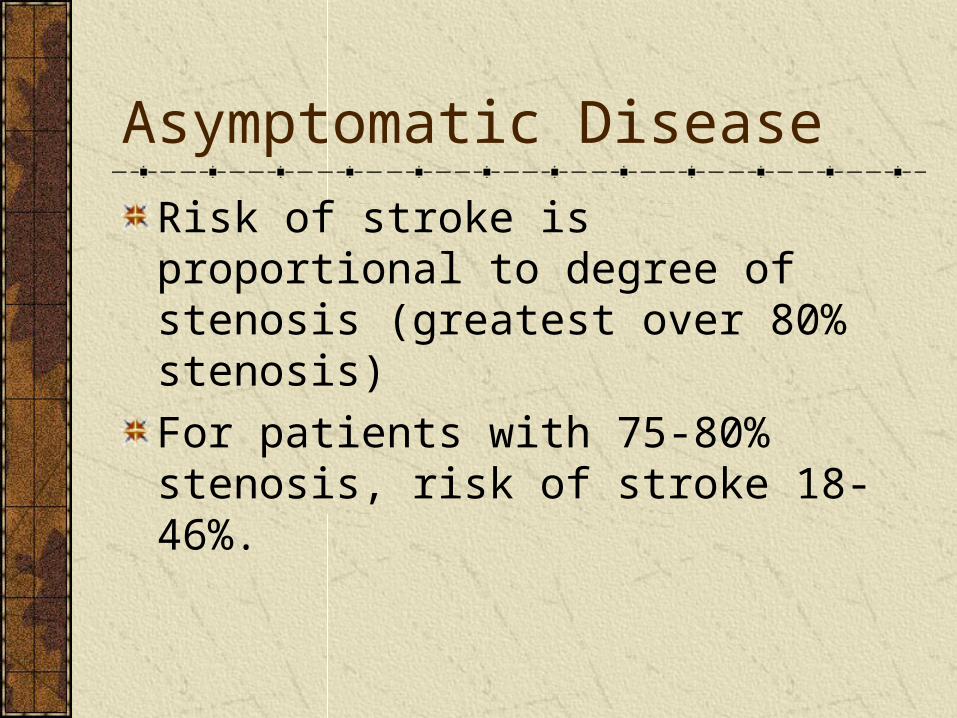
Risk of stroke is proportional to degree of stenosis (greatest over 80% stenosis)
For patients with 75-80% stenosis, risk of stroke 18-46%.
Asymptomatic Disease
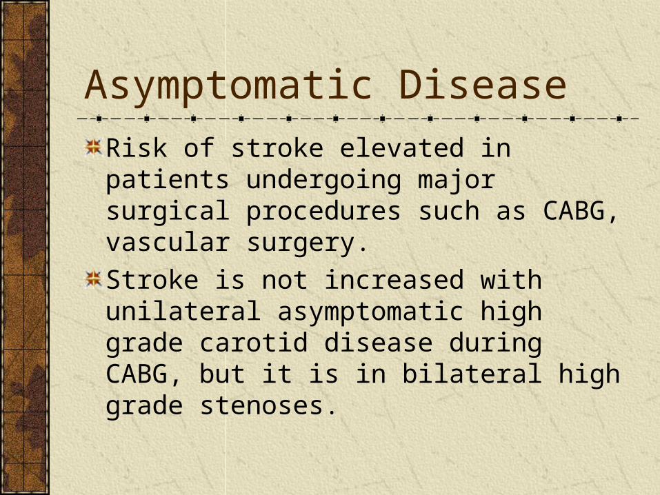
Risk of stroke elevated in patients undergoing major surgical procedures such as CABG, vascular surgery.
Stroke is not increased with unilateral asymptomatic high grade carotid disease during CABG, but it is in bilateral high grade stenoses.

Medical Treatment
Control risk factors
No drug therapy has been shown to reduce the risk of stroke in asymptomatic disease.

Medical TreatmentNo study has provided definitive evidence that systemic anticoagulation reduces the risk of stroke in patients who have had a stroke or TIA.ASA has been shown to decrease the morbidity and mortality from symptomatic diseaseIn patients with TIA or stroke, ASA demonstrated a 22% risk reduction in recurrent strokes, TIA, MI, or vascular death, compared with controls.Plavix and ASA offers no added benefit.

Symptomatic Disease
Degree of ICA stenosis is most important predictor of CVASeverity of stenosis is proportional to Risk of Stroke
Definite benefit of surgery in symptomatic pts with > 70% stenosis is established in three major studies (NASCET, ECST, VATCE)
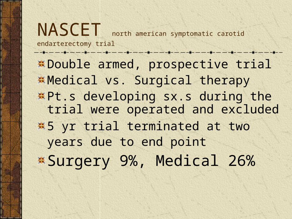
NASCET north american symptomatic carotid endarterectomy trial
Double armed, prospective trial Medical vs. Surgical therapyPt.s developing sx.s during the trial were operated and excluded
5 yr trial terminated at two years due to end point Surgery 9%, Medical 26%

NASCET (cont.)
Risk of major CVA was ↓ by 80% at 2yr follow-up.
CEA was beneficial in symptomatic pts with occlusion of contralateral carotid.

ECST european carotid surgery trial
Double armed prospective trial, 3y f/u
Medical vs. Surgical therapy
70-99 % stenosis
778 pts with carotid distribution CVA, TIA or retinal infarction
Surgery 12.3%, medical 22%
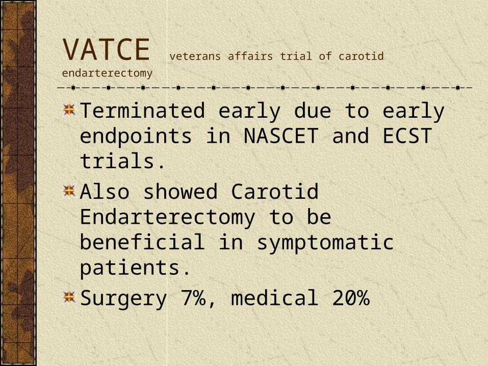
VATCE veterans affairs trial of carotid endarterectomy
Terminated early due to early endpoints in NASCET and ECST trials.
Also showed Carotid Endarterectomy to be beneficial in symptomatic patients.
Surgery 7%, medical 20%

Symptomatic Trials: Summary
0-29% CAS- medical therapy with anti-aggregate platelet therapy
30-69% CAS- medical therapy probably desirable in most patients*
50-69%- CAS- surgery provides modest benefit in hemispheric ischemia
≥ 70% CAS- surgical therapy indicated

Asymptomatic Disease
Prevalent in the elderly population
Asymptomatic CAS >70% rare
Asymptomatic bruit 1.5% risk of CVA per year X 5 yr.s
<75% ~ 1.3%/yr.
>75% ~ 10.5%/yr.

CASANOVA carotid artery surgery asymptomatic narrowing :
operation vs. aspirin
Asymptomatic pt.s with CAS 50-90%
Prospective double armed trial
Medical therapy (330 mg ASA QD + 75mg dypyridamole TID)
Surgical therapy- CEA

CASANOVA (cont)
No statistically significant difference in medical vs. surgically treated groups.
ACAS asymptomatic carotid atherosclerosis study
CEA, ASA and medical risk factor mgmt in patients < 80y/o with CAS>60%
Risk of CVA reduced over 5 yrs by 5.9%
Absolute yearly reduction of 1%
Benefit negated by many factors.

Asymptomatic Trials: Summary
Asymptomatic patients with CAS > 80% will benefit from surgery assuming the surgeon has complication rate <3%
Some investigators refrain from recommending surgery in any asymptomatic patient.
Endovascular TreatmentProblem of embolization from angioplastyUse of cerebral embolic protection devices4 prospective randomized trials comparing endo and surgery. 3 were in adequate risk, 1 in high risk only. CAVATAS, Wallstent, Sapphire (only one with protection device), the other was stopped 5/7 stroked after stenting!Long-term efficacy and durability is unknown.At present limited to high risk only