Embed Size (px)
Citation preview

CAROTENOIDS AFFECT THE STRUCTURE AND FUNCTIONS OF
THE CYANOBACTERIAL PHOTOSYNTHETIC COMPLEXES
Ph.D. Thesis
Sindhujaa Vajravel
Supervisors: Dr. Zoltán Gombos & Dr. Tünde N. Tóth
Biological Research Centre of the Hungarian Academy of Sciences
Institute of Plant Biology
Laboratory of Plant Lipid Function and Structure
University of Szeged
Doctoral School of Biology
Szeged, 2018

1
Contents
ABBREVIATIONS ....................................................................................................................... 3
1. INTRODUCTION..................................................................................................................... 5
1.1. General process of cyanobacterial photosynthesis .................................................................. 5
1.2. The light harvesting complex of cyanobacteria ....................................................................... 7
1.2.1. The structure of PBS ............................................................................................................. 7
1.2.2. PBS degradation process....................................................................................................... 8
1.3. Cyanobacterial photosystems: structure and function ........................................................... 11
1.3.1. Photosystem I ...................................................................................................................... 11
1.3.2. Photosystem II .................................................................................................................... 14
1.4. The structure, function, and biosynthesis of carotenoids ....................................................... 16
2. AIMS ........................................................................................................................................ 20
3. MATERIALS AND METHODS ........................................................................................... 21
3.1. Organisms and growth conditions ......................................................................................... 21
3.2. Construction of Synechocystis PCC 6803 mutants ................................................................ 22
3.3. Measurement of cell density and chlorophyll concentration ................................................. 22
3.4. 77 K fluorescence emission spectroscopy ............................................................................. 23
3.5. Circular-dichroism spectroscopy ........................................................................................... 23
3.6. Time-resolved fluorescence spectroscopy ............................................................................. 24
3.7. Photosynthetic oxygen polarography ..................................................................................... 24
3.8. Isolation of thylakoid membranes and cytosolic fraction ...................................................... 25
3.9. Sucrose density gradient separation of pigment-protein complexes ...................................... 25
3.9.1. Isolation of phycobilisome .................................................................................................. 25
3.9.2. Isolation of photosystem I ................................................................................................... 26
3.10. Protein analysis .................................................................................................................... 26
3.10.1. Determination of protein concentration and SDS-PAGE ................................................. 26
3.10.2. BN- and CN-PAGE........................................................................................................... 26
3.11. Chromatographic techniques ............................................................................................... 27
3.11.1. HPLC ................................................................................................................................ 27
3.11.2. FPLC ................................................................................................................................. 27
3.12. Statistical analysis ................................................................................................................ 28
4. RESULTS AND DISCUSSIONS ........................................................................................... 29
4.1. β-carotene influences the phycobilisome structure................................................................ 29

2
4.1.1. Fluorescence emission of cells with modified carotenoid composition or lack of
photosystems ................................................................................................................................. 29
4.1.2. Protein analysis of the cytosolic fraction from crtB cells ................................................... 32
4.1.3. High light-induced damage in carotenoid deficient mutants .............................................. 34
4.1.4. Effect of glucose on crtH cells ............................................................................................ 39
4.1.5. Additional myxoxanthophyll deficiency in crtH cells ......................................................... 40
4.1.6. Nitrogen shortage induced PBS degradation in crtH and crtB strains .............................. 41
4.2. Zeaxanthin and echinenone modify the structure of PSI trimer ............................................ 48
4.2.1. Pigment analysis of cells and PSI complexes ..................................................................... 48
4.2.2. Testing the PSI organization by in vivo spectroscopic methods ......................................... 51
4.2.3. Protein analysis of the thylakoid membrane ....................................................................... 55
4.2.4. Spectroscopic investigation of the PSI complexes .............................................................. 59
5. SUMMARY ............................................................................................................................. 64
6. SUMMARY IN HUNGARIAN .............................................................................................. 70
7. CONCLUSIONS ..................................................................................................................... 76
8. REFERENCES ........................................................................................................................ 78
9. ACKNOWLEDGEMENTS ................................................................................................... 87
10. LIST OF PUBLICATIONS ................................................................................................. 89

3
ABBREVIATIONS
1O2 - Singlet oxygen
APC - Allophycocyanin
BN/CN/SDS-PAGE - Blue and clear native/sodium dodecyl sulfate polyacrylamide gel
electrophoresis
CD - Circular dichroism
Chl - Chlorophyll
Cyt b6f - Cytochrome b6f
DAS - Decay associated spectrum
β-DM - n-Dodecyl β-D-maltoside
DMBQ - 2,6-Dimethylbenzoquinone
EDTA - Ethylenediaminetetraacetic acid
EET - Excitation energy transfer
FNR - Ferredoxin/NADP+ oxidoreductase
FPLC - Fast protein liquid chromatography
HPLC - High performance liquid chromatography
LAHG - Light-activated heterotrophic growth
LR30 - 30 kDa rod linker proteins
LR33 - 33 kDa rod linker proteins
LMC – Core membrane linker
LRC - Linker rod-core
LWC - Long wavelength chlorophyll
OD - Optical density
PAG - Photoautotrophic growth
PBS – Phycobilisome
PC - Phycocyanin

4
PMG - Photomixotrophic growth
PPFD - Photosynthetic photon flux density
PQ - Plastoquinone
PSI and PSII - Photosystem I and II
ROS - Reactive oxygen species
TE - Terminal emitter

5
1. INTRODUCTION
1.1. General process of cyanobacterial photosynthesis
Cyanobacteria are ecologically important prokaryotes that also serve as the popular model
organism for studying photosynthesis. The approximately 2.3 billion years ago, cyanobacteria
started performing an oxygenic photosynthesis by utilizing the visible range of light (300-700
nm) (Yagishita et al 1997). These prokaryotic photosynthetic organisms are the ancestors of
plastids in algae and plants due to the evolutionary event of endosymbiosis (Hohmann-
Marriott & Blankenship, 2011). Since cyanobacteria show a strong homology with the
photosynthetic machinery of eukaryotes, it can be used as a suitable model organism to study
different aspects and regulation of photosynthesis that is often difficult to approach in higher
plants and algae. The further advantages of cyanobacteria are its rapid growth under
laboratory culture conditions and its immense metabolic flexibility (Eberhard et al., 2008).
Due to its simplicity of single cell system, it is easy to manipulate genetically when compared
to the complex multicellular systems. These advantages are also making the cyanobacterium
as an important model organism to conduct basic and applied research in the field of
photosynthesis, abiotic stress responses and many other cellular processes.
Photosynthesis is a process to convert light energy into chemical energy providing the
basic energy source for life on earth and for the oxygenic atmosphere. Similarly to all
eukaryotic photosynthetic organisms, in cyanobacteria, the photosynthetic electron transport
occurs in the thylakoid membrane. The thylakoids accommodate the photosystem I (PS I),
photosystem II (PS II) as reaction centers (RCs), phycobilisome (PBS), cytochrome b6f (Cyt
b6f), and ATP synthase as the major photosynthetic protein complexes. The small electron
transport molecules, such as plastoquinone (PQ) pool, plastocyanin (PC), cytochrome c6, and

6
ferredoxin (Fd) (Fig. 1) are also involved in the electron transport mechanism (Eberhard et al
2008, Hohmann-Marriott & Blankenship 2011).
Figure 1. Schematic representation of the photosynthetic electron transport in cyanobacteria.
Abbreviations: PBS, Phycobilisome; PSI, Photosystem I; PSII, Photosystem II; PQ,
Plastoquinone; Cyt b6f, Cytochrome b6f; PC, Plastocyanin; Fd, Ferredoxin; ATPase, ATP
synthase.
Initial event of cyanobacterial photosynthesis is the absorption of photons by the light-
harvesting antennae, the PBS. The excitation energy is transferred from the PBS to the
chlorophyll a (Chl a) molecules of PSII reaction center where the charge separation occurs
and initiates electron transport. These electrons reduce PQ and transferred from PQ pool to
the Cyt b6f complex and to a mobile electron carrier, the PC (Fig. 1). PC reduces the photo-
oxidized PSI reaction center Chl a molecules; the oxidized form of PSI reaction center is
induced by the electron transfer from PSI to reduce NADPH. The above-described electron
transport mechanism plays a major role in the proton transfer across the thylakoid membrane,
and the created proton motive force is used by the ATPase to convert ADP to ATP. The
produced ATP and NADPH are subsequently used to produce carbohydrates in the Calvin-
Benson cycle (carbon-fixation reactions).

7
1.2. The light harvesting complex of cyanobacteria
1.2.1. The structure of PBS
In cyanobacteria, red algae and glaucophytes, the main light-harvesting antenna is the
PBS: a giant, multi pigment-protein complex, which is often the most abundant protein
complex of the cell. The PBS contains 200-500 phycobilin pigments covalently bound to
apoproteins, the so-called phycobiliproteins which are heterodimers composed of α and β
subunits. Based on the spectral properties, the phycobiliproteins are divided into three groups:
phycoerythrins, phycocyanins, and allophycocyanin. These phycobiliproteins are combined
to form a hierarchically ordered PBS megacomplex by the support of specific linker proteins
(MacColl 1998, Ughy & Ajlani 2004). Depending on the species and growth conditions,
several types of PBSs can be found. The most common form is the hemidiscoidal type of
PBSs which possess six to eight peripheral rods attached to the central allophycocyanin
(APC) core complex (Chang et al 2015, Watanabe & Ikeuchi 2013).
Among cyanobacteria, Synechocystis sp. PCC 6803 (hereafter Synechocystis) is one of the
most widely applied model organisms, and its PBS is dominantly present as a hemidiscoidal
structure that contains phycocyanobilins as the only chromophores. The incident light mostly
excites the pigments of the PBS rods then the excitation energy is transferred to the APC
core. This PBS has six radial rods, each consists of up to three hexameric phycocyanin (PC)
units attached together by LR10
, LR30
and LR33
rod linker proteins (Fig. 2).

8
Figure 2. A model of the hemidiscoidal structure of PBS (Arteni et al 2009). Side view (A)
and upward (B) view of PBS from the thylakoid membrane. Abbreviations: PC, Phycocyanin
(6 rods); APC, Allophycocyanin (3 cores); LR10
, LR30
, LR33
, LRC, and LC - linker proteins; T,
T8, B8 and M - four subunits of APC trimers.
The central core has three cylinders, each composed of four APC trimers. Some of the
APC core subunits are acting as terminal emitters (TEs) i.e. ApcD, ApcE, and ApcF which
ensure the excitation energy transfer to the photosystems (Ashby & Mullineaux 1999). As an
antenna for PSI and PSII (Chang et al 2015), the PBSs absorb in the wavelength range of
500-650 nm, which is less efficiently absorbed by the Chl a molecules (380-495 and 625-740
nm). Recently, the structure of a 16.8 MDa PBS from a red alga Griffithsia pacifica at 3.5 Å
resolution has been reported by using single-particle cryo-electron microscopy (Sarma et al
2016). This structure shows the important mechanisms behind the specific interactions
between linkers and chromophores. It also revealed the complicated assembly and the
complex mechanisms of energy transfer within the PBS and raises the question of similar
processing in the PBS of the less studied species.
1.2.2. PBS degradation process
Beside the light harvesting function, the PBSs also function as a nutrient reservoir.
Under macronutrient (e.g. nitrogen, phosphorus, and sulfur) limited conditions, the PBSs can
supply amino acid residues for the cell. During macronutrient starvation, in order to release
its important constituents, the PBSs go over a programmed proteolytic pathway (Schwarz &

9
Forchhammer 2005). Our present knowledge about PBS degradation was mostly obtained by
using Synechococcus elongatus sp. PCC 7942 (hereafter Synechococcus) and Synechocystis
strains (Schwarz & Forchhammer 2005). The studies in these species suggested that the
degradation pathway and its activation differ from species to species (Richaud et al 2001,
Schwarz & Forchhammer 2005), but the basic principles are similar.
Figure 3. The simplified model of the programmed PBS degradation process. NblA protein
involved in the sequential (A) rod degradation followed by (B) core degradation of PBSs.
The recent studies revealed that phosphorylational modifications of the PBS proteins
are prerequisite for the directed PBS degradation (Sendersky et al 2014). The degradation is
initiated by the cleavage of rod linker proteins and concomitant shortening of the rods; these
events are essential for the further degradation of PBSs. Several proteins encoded by nbl
(non-bleaching) genes are involved in the PBS degradation (Collier & Grossman 1994, Baier
et al 2014). The NblA protein is one of the key elements involved in PBS degradation and
functions as an adaptor for the PBS proteolytic enzyme (ClpC–ClpP) complex. The most
recent results suggest that NblA adaptor complex interacts with the N-terminus of β-
phycocyanin (Bernát et al 2009), presents them to the ClpC–ClpP complex (Karradt et al
2008) which eventually performs the cleavage of the PBS. In contrast to the earlier
hypothesis that NblA is required only for the rod degradation, recently it has been shown that
10
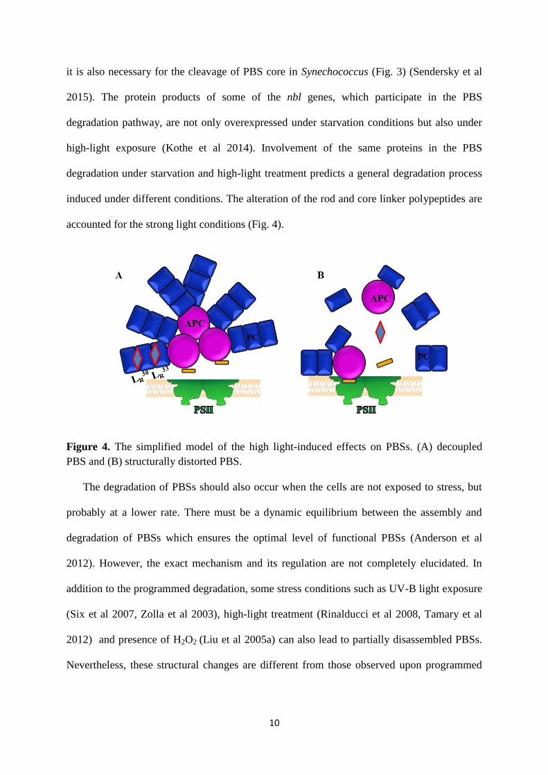
it is also necessary for the cleavage of PBS core in Synechococcus (Fig. 3) (Sendersky et al
2015). The protein products of some of the nbl genes, which participate in the PBS
degradation pathway, are not only overexpressed under starvation conditions but also under
high-light exposure (Kothe et al 2014). Involvement of the same proteins in the PBS
degradation under starvation and high-light treatment predicts a general degradation process
induced under different conditions. The alteration of the rod and core linker polypeptides are
accounted for the strong light conditions (Fig. 4).
Figure 4. The simplified model of the high light-induced effects on PBSs. (A) decoupled
PBS and (B) structurally distorted PBS.
The degradation of PBSs should also occur when the cells are not exposed to stress, but
probably at a lower rate. There must be a dynamic equilibrium between the assembly and
degradation of PBSs which ensures the optimal level of functional PBSs (Anderson et al
2012). However, the exact mechanism and its regulation are not completely elucidated. In
addition to the programmed degradation, some stress conditions such as UV-B light exposure
(Six et al 2007, Zolla et al 2003), high-light treatment (Rinalducci et al 2008, Tamary et al
2012) and presence of H2O2 (Liu et al 2005a) can also lead to partially disassembled PBSs.
Nevertheless, these structural changes are different from those observed upon programmed

11
degradation and are probably due to direct damage of proteins induced by extreme stress
conditions (Fig. 4).
1.3. Cyanobacterial photosystems: structure and function
1.3.1. Photosystem I
Photosystem I (PSI) is one of the major pigment-protein complexes of the oxygenic
photosynthetic organisms; it is required for Fd and NADP-reduction. In PSI, the excitation
energy trapping is faster than in PSII (Palsson et al 1998). The evolutionarily conserved PSI
structure possesses a high sequence homology from prokaryotic cyanobacteria to higher
plants. Hence, cyanobacterial PSI can be applied as a model system for investigating the
mechanism of energy transfer within the PSI core complex of plants. On the other hand,
while PSI complexes are present as monomers in plants, those are preferentially organized
into oligomers in cyanobacteria (Kruip et al 1994, Watanabe et al 2011). Most of the well-
studied cyanobacterial species possess trimeric PSI complexes and these trimers seem to have
a major physiological role at low light intensities (Fromme et al 2001) or elevated
temperatures (Klodawska et al 2015). Although the exact functions of trimers have not been
clarified yet, it seems that trimeric structure can improve the efficiency of the energy
conversion (Karapetyan et al 1999). Until now, a high resolution (2.5 Å) X-ray analysis of the
complete trimeric PSI was only obtained from a thermophilic cyanobacterium,
Thermosynechococcus elongatus (hereafter T. elongatus) (Jordan et al 2004). This complex is
a homotrimer of multiprotein subunits where each monomer contains 127 cofactors including
Chl a, β-carotene molecules and lipids (phosphatidyl-glycerol and monogalactosyl-
diglyceride). The monomers consist of 12 protein subunits; among those PsaA and PsaB are
the biggest and most of the cofactors are bound to these proteins (Fig. 5).

12
Figure 5. PSI trimeric complex of Thermosynechococcus elongatus at 2.5 Å resolutions as
seen from the stromal side of thylakoid membrane (Jordan et al 2001). Each monomer
representation (I, II and III) shows different structural components. The arrangement of
transmembrane α-helices is shown as cylinders and different protein subunits are labeled (I).
Lumenal loop regions are represented as ribbons (II). Complete set of cofactors (III):
quinones and Chls (blue), iron and sulfur atoms of the three Fe4S4 clusters (orange and yellow
spheres, respectively); Antenna system: Chl a (yellow), carotenoids (black) and lipids
(turquoise).
The oligomerization of PSI is ensured by a central domain containing the PsaL
protein (Chitnis & Chitnis 1993, Klodawska et al 2015). The association of PsaL into the
monomeric complex is stabilized by other subunits (i.e. PsaI and PsaM) and three β-carotene
molecules (Grotjohann & Fromme 2005, Toth et al 2015). This domain also contains the Ca2+
that is presumed to support the trimerization (Fromme et al 2001). Partial structure of PSI
monomer of Synechocystis has been also obtained and revealed some structural differences
between the two species (Mazor et al 2014), but further studies are needed to reveal the exact
structure of trimeric PSI from Synechocystis.

13
In cyanobacteria, around 90% of the total Chl a is bound to PSI as a consequence of
the higher Chl a content of PSI (96 Chl a/monomer) than PSII (35 Chl a/monomer) (Fromme
et al 2001, Gunerken et al 2015) and as high as 90 % of the total Chl a molecules are
associated with PSI complexes relative to PSII (Rakhimberdieva et al 2001, Tian et al 2011).
Around 5–10% of the PSI associated Chls absorb light energy at longer wavelengths; these
are the so-called red-shifted or long-wavelength Chls (LWCs). The LWCs were proposed to
originate from Chl-Chl interactions induced by the aggregation of Chls within the PSI
complex (Fromme et al 2001, Mazor et al 2014). Generally, the number of LWCs is higher in
the PSI trimers than in monomers in the studied cyanobacterial species, but their amount and
spectral properties vary among species (El-Mohsnawy et al 2010, Gobets & van Grondelle
2001). The difference in the number of LWCs in trimers and monomers were explained by
the presence of Chl a dimers at the trimerization domain of PSI which was also observed
from T. elongatus (Jordan et al 2001) and the interaction between these two Chls is probably
interrupted upon monomerization. The number of LWCs was mostly calculated based on
spectroscopic data (Gobets & van Grondelle 2001). The presence of LWCs in the
Synechocystis PSI monomer structure has also been also confirmed. Synechocystis contains
less LWCs than T. elongatus, but those LWCs are found in a similar orientation in the two
species (Mazor et al 2014). In the present study, we also consider the amount of LWCs as an
indicator for the organization of PSI complex.
In trimeric PSI complexes, a high amount of carotenoids, mostly β-carotenes were
observed (Ashikawa et al 1986, Bautista et al 2005, El-Mohsnawy et al 2010). In agreement
with these results, 22 β-carotenes per monomer were resolved in the crystal structure of PSI
from T. elongatus (Jordan et al 2001). These β-carotenes are important elements of the PSI
complex influencing its structure (Toth et al 2015) and function (Bautista et al 2005).
Moreover, the previous work in our laboratory demonstrated that even the xanthophyll
14
deficiency (i.e. the lack of oxygenated carotene derivatives) reduces the amount of PSI
oligomers in Synechocystis (Toth et al 2015).
1.3.2. Photosystem II
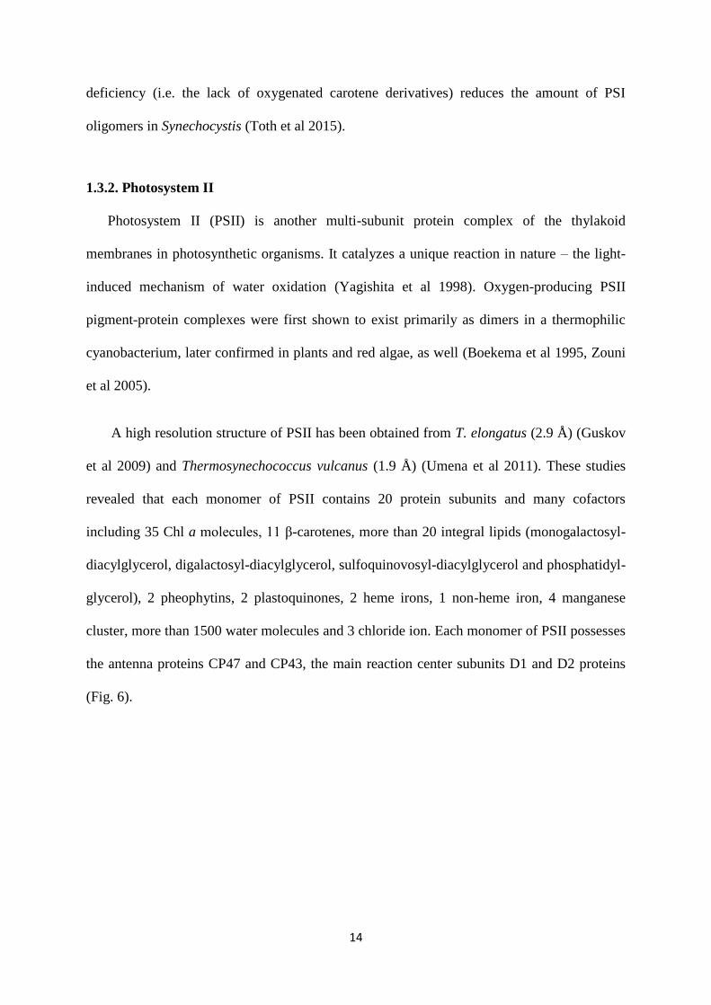
Photosystem II (PSII) is another multi-subunit protein complex of the thylakoid
membranes in photosynthetic organisms. It catalyzes a unique reaction in nature – the light-
induced mechanism of water oxidation (Yagishita et al 1998). Oxygen-producing PSII
pigment-protein complexes were first shown to exist primarily as dimers in a thermophilic
cyanobacterium, later confirmed in plants and red algae, as well (Boekema et al 1995, Zouni
et al 2005).
A high resolution structure of PSII has been obtained from T. elongatus (2.9 Å) (Guskov
et al 2009) and Thermosynechococcus vulcanus (1.9 Å) (Umena et al 2011). These studies
revealed that each monomer of PSII contains 20 protein subunits and many cofactors
including 35 Chl a molecules, 11 β-carotenes, more than 20 integral lipids (monogalactosyl-
diacylglycerol, digalactosyl-diacylglycerol, sulfoquinovosyl-diacylglycerol and phosphatidyl-
glycerol), 2 pheophytins, 2 plastoquinones, 2 heme irons, 1 non-heme iron, 4 manganese
cluster, more than 1500 water molecules and 3 chloride ion. Each monomer of PSII possesses
the antenna proteins CP47 and CP43, the main reaction center subunits D1 and D2 proteins
(Fig. 6).

15
Figure 6. PSII dimeric structure of Thermosynechococcus elongatus at 2.9 Å resolutions as
seen from the cytoplasmic side of thylakoid membrane (Guskov et al 2009). A black dash
line indicates the monomer-monomer interface. Different elements of structures have shown
in each monomers (I and II). (I) Cylindrical structures are exhibiting the helical parts, D1
subunits (yellow), D2 (orange), CP43 (magenta), CP47 (red). Cofactors are shown in stick
mode: Chl a (green), Carotenoids (orange), heme (blue), Pheophytin (yellow), PQ (red), the
Mn4Ca cluster (red and orange spheres). (II) Lipids and detergent molecules (yellow).
Chl a molecules are bound to the PSII reaction center and funnel the excitation energy
to the reaction center where charge separation occurs. PSII uses two types of PQ molecules
(QA and QB) as mobile electron carriers to perform electron transport towards the Cyt b6f
complex (Lamberg & Bren 2016, Barber et al 1997). PSII is the major source for the
production of harmful reactive oxygen species (ROS), especially singlet oxygen (Krieger-
Liszkay et al 2008) under high light stress. Singlet oxygen is highly reactive and unstable
form of oxygen which has a well-known damaging effect on proteins. The D1 protein is the
most sensitive to singlet oxygen but it also reacts with lipids and nucleic acids.
In cyanobacteria, the extra-membranous PBSs are attached to the PSII complex at its
cytoplasmic side, whereas in plants the membrane-embedded light-harvesting antenna II is
surrounded by PSII complexes (Lamberg & Bren 2016).

16
1.4. The structure, function, and biosynthesis of carotenoids
Carotenoids are the most widely spread pigments in nature. They are polyunsaturated
hydrocarbon substances which are classified into two subgroups, the oxygen containing
xanthophylls and the carotenes (oxygen free). Carotenoids are tetraterpenoids containing a
long conjugated chain of double bonds which determines their light absorption (400-500 nm)
properties (Sonoike et al 2001).
In photosynthetic organisms, together with tocopherol, carotenoids are the most important
elements of the non-enzymatic antioxidant system (Latifi et al 2009) and also capable to
quench triplet excited states of Chls (Frank & Cogdell 1996, Domonkos et al 2013).
Accordingly, in cyanobacteria the partial elimination of carotenoids decreases the stress
tolerance of the cells against high-light intensities (Schafer et al 2005, Zhu et al 2010); the
cells with complete carotenoid deficiency appeared to be extremely light sensitive and could
be maintained only in the dark under light-activated heterotrophic growth conditions (Sozer
et al 2010). Besides the protective functions, carotenoids are also important for the assembly
and stabilization of photosynthetic complexes in cyanobacteria (Sozer et al 2010, Toth et al
2015), as well as in plants (Croce et al 2002, Fiore et al 2012, Dall'Osto et al 2013,
Santabarbara et al 2013). The previous results in our laboratory suggested that complete
carotenoid deficiency also influence the PBS structure and evokes the presence of
unconnected PC rods and PBSs with shorter rods (Toth et al 2015). Although an indirect
effect of carotenoids on PBS structure is more likely, further studies are needed to investigate
the correlation between the carotenoid composition and the PBS organization.
Due to their physicochemical properties, the carotenoids are preferentially embedded into
lipid bilayers or integrated into pigment-protein complexes (Domonkos et al 2013). In
cyanobacteria, most of the β-carotene is bound to the PSs (Jordan et al 2001, Gunerken et al

17
2015) and to some less abundant proteins like high-light inducible proteins (HLIPs)
(Komenda & Sobotka 2016). Besides the protein location (Daddy et al 2015, Melnicki et al
2016), a substantial fraction of non-protein-bound xanthophylls are supposed to be present in
membranes (Dall'Osto et al 2013) where they can influence the molecular dynamics and
microviscosity of the membranes (Gruszecki & Strzalka 2005). Xanthophylls are often co-
purified with isolated PSI trimers but not considered as part of the complex ( Coufal et al
1989, Bautista et al 2005, Daddy et al 2015, Klodawska et al 2015) which predicts the
presence of xanthophylls within the thylakoid membrane at the vicinity of PSI. A wide range
of xanthophylls is present in various cyanobacterial species, depending on their natural
habitat and taxonomical position (Takaichi & Mochimaru 2007). In Synechocystis, the most
abundant xanthophylls are zeaxanthin, echinenone, and myxoxanthophyll. In spite of the high
amount of xanthophylls in the thylakoid membrane of Synechocystis, their exact localization
and structural roles in the photosynthetic complexes and the thylakoid organization have not
been completely unfolded yet. Several Synechocystis mutants have been generated by the
inactivation of various genes, which are involved in the carotenoid biosynthesis pathways
(Fig. 7).
In Synechocystis, the first step of carotenoid biosynthesis is the condensation of two
molecules of geranylgeranyl diphosphate into phytoene, catalyzed by the phytoene synthase
enzyme (CrtB). The carotenoid synthesis includes subsequent isomerization steps which can
be catalyzed by the carotene isomerase (CrtH) enzyme or occur via non-enzymatic photo-
isomerization. Previous results showed that the non-enzymatic isomerization process
obtained by photo-isomerization without CrtH enzyme activity results in largely decreased
carotenoid content (Masamoto et al 2004, Toth et al 2015).

18
Figure 7. The chemical structure of four carotenoid molecules (A) (Domonkos et al 2013)
and the major carotenoid biosynthesis pathways in Synechocystis (B). Abbreviations: CrtB,
phytoene synthase; CrtH, cis-trans carotene isomerase; CrtR, carotene β-hydroxylase; CrtO,
carotene β-ketolase; CruF, γ-carotene 1׳-hydroxylase.
Despite the low carotenoid level, crtH mutant cells are capable of growing under
photoautotrophic conditions (Toth et al 2015) and maintain wild-type like growth rate and
PSII activity (Masamoto et al 2004). In the following steps of the carotenoid biosynthesis, γ-
carotene is produced, which supplies the myxoxanthophyll and β-carotene synthesis in
parallel pathways.
Among accumulated carotenoids, β-carotene is also a key intermediate product for the
synthesis of hydroxylated zeaxanthin and ketolated echinenone derivatives (Domonkos et al
2013). Zeaxanthin is synthesized from β-carotene by the β-carotene hydroxylase (CrtR) by
sequentially adding hydroxyl groups at both ends of the molecule, whereas echinenone is
produced by β-carotene ketolase (CrtO), which introduces keto group at one end of β-
carotene (Masamoto et al 1998). The inactivation of both the crtR and crtO genes resulted in
the almost completely xanthophyll-deficient mutant, crtRO (Schafer et al 2005). Because the

19
CrtR enzyme is also involved in the myxoxanthophyll biosynthesis (Lagarde & Vermaas
1999), the crtRO and crtR mutants accumulate myxoxanthophyll intermediate (deoxymyxol-
fucoside) at a low level. In Synechococcus, the CruF (C-1′-hydroxylase) enzyme is essential
for the synthesis of myxoxanthophyll (Zhu et al 2010). The disruption of cruF gene resulted
in the generation of a myxoxanthophyll (myxol-2׳ fucoside) deficient mutant without relevant
alteration in the amount of other carotenoid species. Using these mutants, sequential
elimination of different carotenoid forms could be obtained, which allows studying their
specific roles.

20
2. AIMS
The present study was aimed at further investigating the influence of carotenoid
composition on the structure and organization of the major photosynthetic complexes. In the
proposed thesis, cyanobacterium Synechocystis sp. PCC 6803 was used as a photosynthetic
model organism. The main advantages of using this species are its entirely sequenced genome
and easy transformability. Carotenoids are the most abundant pigments in nature. They are
known to be important for photoprotection, regulation of the membrane properties, but less
information is available about their structural roles in the photosynthetic complexes. The
photosynthetic complexes should be organized and structurally interacted in the thylakoid
membrane to perform the efficient photosynthetic energy transfer.
The previous studies in our laboratory revealed that the absence or low level of
carotenoids lead to the co-existence of unconnected rod units and assembled PBSs with
shorter peripheral rods. It is rather surprising since the presence of carotenoids in the PBS
structure has not been reported. Therefore, our aim was;
1. To find out which specific carotenoid species involved and understand the mechanism
that eventually leads to the presence of unconnected PBS units, when the carotenoid
composition is altered.
A reduced level of PSI oligomers upon xanthophyll deficiency was also observed,
although xanthophylls are not considered to be part of any photosynthetic complexes in
cyanobacteria. But these predictions were not completely clarified. Hence, our aim was also;
2. To study, how xanthophylls influence the oligomerization of PSI in Synechocystis. More
specifically, which xanthophyll species contribute at what extent to the organization of
the PSI complex.

21
3. MATERIALS AND METHODS
3.1. Organisms and growth conditions
Cyanobacterial cells were cultivated in BG11 medium supplemented with 5 mM
HEPES–NaOH (pH 7.5). Synechocystis wild type (WT), crtH (Masamoto et al 2001), psaL
(Klodawska et al 2015), PSI-/PSII- (Ermakova-Gerdes et al 1995), cruF, crtH/cruF (present
study) crtRO, crtR and crtO (generous gift from Kazumori Masamoto) mutant strains were
grown under photoautotrophic growth (PAG) conditions. The crtB mutant (Toth et al 2015)
and its corresponding WT cells were cultivated under light-activated heterotrophic growth
(LAHG) conditions on a rotary shaker (100 rpm) at 30 °C.
Under PAG conditions, the cells were illuminated with continuous white light using
35 µmol photons m-2
s-1
PPFD (Photosynthetic Photon Flux Density). For photomixotrophic
growth (PMG) conditions, the media was supplemented with 10 mM glucose. Under LAHG
conditions, BG11 was supplemented with 10 mM glucose and daily light pulses of 20 µmol
photons m-2
s-1
PPFD were provided for 10 min. For oxygen polarography, the cultures were
grown under 40 µmol photons m-2
s-1
PPFD of continuous white light at 3 % CO2-enriched
atmosphere. High-light treatment was induced by 600 µmol photons m-2
s-1
for WT, and crtH
and 35 µmol photons m-2
s-1
PPFD for crtB cells, respectively. Mutant cells were maintained
in the presence of the appropriate antibiotics, 20 µg ml-1
of kanamycin for crtH and crtR,
spectinomycin for crtO, cruF, and crtB strains, respectively. For the double mutant crtRO
and crtH/cruF cells, 20 µg ml-1
kanamycin and spectinomycin were added. For the
experiments, the cells were cultivated without antibiotics and used in their logarithmic
growth phases.
22
3.2. Construction of Synechocystis PCC 6803 mutants
The cosmid clone cs0508 provided by S. Tabata, was cut with the restriction enzyme
HindIII. A 3.6 kb fragment containing the putative cruF gene (sll0814) was cloned between
the SmaI and EcoRV sites of pMPMA3 (Mayer 1995) A part of the putative cruF gene
coding region was removed by SmaI-EcoRI digestion and replaced with a Ω cassette (Sozer
et al 2010). This construct was used to transform into WT and crtH mutant cells of
Synechocystis. Transformants were selected under photomixotrophic growth conditions on
BG11 agar plates supplemented with glucose and gradually increasing amounts of
spectinomycin by several restreaking of single colonies. Complete segregation was confirmed
by PCR using the primers cruF forward (5′-TTAGCGGCACCTACTTTG-3′) and cruF
reverse (5′-TCTGCGTTGAGTTGAGTC-3′) and by pigment analysis using HPLC.
3.3. Measurement of cell density and chlorophyll concentration
Measurements of cell density and Chl concentration were performed by a Shimadzu
UV-1601 spectrophotometer. The cell density of the cultures was determined by measuring
optical density at 750 nm (OD750). For measuring absorption spectra of the cells, OD750 was
adjusted; non-transparent sides of cuvettes were positioned toward the detector to decrease
the effect of light scattering. Spectra were corrected to the light scattering measured at 750
nm. For calculation of phycobilin (630 nm) to Chl (683 nm) peak ratio, the amplitudes were
estimated by the use of reference wavelength at 750 and 550 nm, respectively. The Chl
concentration was calculated from absorbance at 665 nm, using 90% methanol extract
(Meeks & Castenholz 1971).

23
3.4. 77 K fluorescence emission spectroscopy
Low-temperature fluorescence emission spectra (615–800 nm) were measured using
spectrofluorometer (Fluorolog-3/Jobin–Yvon–Spex Instrument S.A., Inc.) equipped with a
liquid nitrogen cryostat. Prior to the measurements, cells containing 2 μg of Chls were
filtered onto the surface of a Whatman GF/C glass microfiber filter disc (25 mm diameter),
dark adapted for 5 min and frozen in liquid nitrogen. Due to the low Chl concentration in case
of crtB and PSI-/PSII- mutant strains, 1 ml of cell suspensions adjusted to 0.35 OD750 were
filtered. Fluorescence was excited at 590 and 436 nm from the same sample without changing
its orientation. The emission spectra excited at 590 and 436 nm were corrected for
photomultiplier sensitivity and normalized to the fluorescence intensity at 720 nm obtained
upon 436 nm excitation, otherwise as it is indicated in figure legends. For investigating the
contribution from LWCs of PSI complexes, 2 nm emission slit combined with 4 s integration
time was applied. For each spectrum, three biological samples were averaged.
3.5. Circular-dichroism spectroscopy
CD spectra were recorded with a JASCO J-815 dichrograph. Measurements were
carried out at room temperature using 3 nm band-pass and 1 nm step size. The Chl
concentration of the cell, thylakoid or PSI complex suspensions were adjusted to 10 μg ml−1
and measured in a cuvette with 1 cm optical path length (Klodawska et al 2015). For
obtaining CD spectra after CN-PAGE, the PSI-containing bands were excised and placed
between glasses and the uncovered part of the glass was masked. The recorded CD spectra
were baseline-corrected using the corresponding buffers or transparent gel slices and then
normalized to the 680 nm Chl absorption band. For each spectrum, three biological replicates
were measured. To determine the amplitude of the 515 nm CD band, 550 nm was used as a
reference wavelength.
24
3.6. Time-resolved fluorescence spectroscopy
Time-correlated single-photon counting was performed according to (Reguera et al
2007) using a FluoTime 200 spectrometer (PicoQuant, Germany) equipped with a
microchannel plate detector (Hamamatsu, Japan) and a PicoHarp 300 TCSPC system
(PicoQuant). Room- temperature fluorescence decays were recorded with picoseconds
resolution from cells in vivo at wavelengths between 620 and 720 nm with excitation pulses
at 580 nm, 0.1 pJ pulse energy and 20 MHz repetition rate, generated by a WhiteLase
supercontinuum laser (NKT Photonics, UK). The fluorescence decays were analyzed by
global lifetime fitting (Friebe et al 2017).
3.7. Photosynthetic oxygen polarography
Photosynthetic oxygen-evolving activity in intact cells of WT and crtH mutant strains
was measured with a Clark type oxygen electrode (Hansatech DW2). In order to provide
efficient histidine (His) uptake by the cells, those were grown under 3 % CO2-enriched
atmosphere at 40 µmol photons m-2
s-1
PPFD of white light intensity for oxygen evolution
experiments. Prior to the measurements, the Chl concentration of the cells was adjusted to 5
µg ml-1
by centrifugation at 6000 g for 5 min and kept it on the shaker for at least 1 h. In each
measurement, 2 ml of cells were used and three replicates were measured. The oxygen-
evolving activity of intact cells was measured in the presence or absence of 5 mM histidine
(His) using 2300 µmol photons m-2
s-1
PPFD of white light. Singlet oxygen production was
calculated as described earlier (Rehman et al 2013). PSII activity was measured in the
presence of PSII electron acceptor, 0.5 mM 2,6-dimethylbenzoquinone (DMBQ), and for the
photoinhibition experiments of PSII activity, 0.25 mM lincomycin were added prior to the
illumination.

25
3.8. Isolation of thylakoid membranes and cytosolic fraction
Thylakoids were isolated by breaking the freshly collected cells in medium A (25 mM
MES/NaOH buffer (pH 6.5), 10 mM CaCl2, 10 mM MgCl2) containing 25% glycerol, 0.5
mM phenyl-methanesulfonyl fluoride (PMSF) and 1 mM benzamidine by Bead Breaker at 5
times for 1 min using glass beads. The soluble fractions of WT LAHG and crtB LAHG cells
were pelleted out by 3000 g at 4 °C for 5 min. And for other cells (WT, crtRO, crtR, crtO,
cruF and psaL), the thylakoid membranes were collected at 35,000 g at 4 °C for 30 min.
The collected thylakoid membranes were solubilized with 1 or 2% n-Dodecyl β-D-
maltoside (β-DM) as it is indicated in the figure legends. Prior to the solubilization, the
thylakoid membranes were washed and resuspended in the medium containing 25 mM
MES/NaOH buffer (pH 6.5), 25 mM EDTA (ethylenediaminetetraacetic acid) and 25%
glycerol when it is indicated.
3.9. Sucrose density gradient separation of pigment-protein complexes
3.9.1. Isolation of phycobilisome
Phycobilisomes were isolated from Synechocystis of WT LAHG and crtB LAHG cells
according to (Toth et al 2015). The cells were pre-treated with 0.2 % lysozyme at 37 °C. The
cells were broken by an equal volume of glass beads (100 µm) in 0.75 M K-Na phosphate
buffer (pH 7.0) using Bead Breaker at 5 times for 1 min. The thylakoid membranes were
pelleted by centrifugation at 15,000 g after the treatment of Triton X-100 (5%, w/v) at room
temperature. After centrifugation, the supernatant was treated again with Triton X-100 (3%,
w/v) for 20 min before loading on to the discontinuous sucrose density gradient. The PBS-
containing layer was collected after centrifugation at 90,000 g for 20 h at 14 °C. The isolated
PBS samples can be stored at room temperature for further protein and spectroscopic
analysis.

26
3.9.2. Isolation of photosystem I
Freshly isolated thylakoid membranes were washed by medium A (see section 2.9.) and
solubilized with 2% β-DM. The solubilized samples were layered on a stepwise (6 steps, 0.2–
0.9 M) sucrose gradient containing 20 mM HEPES (pH 7) and 0.05% of β-DM.
Centrifugation was performed at 220,000 g for 14 h at 4 °C. PSI-containing fractions of the
gradient were withdrawn by syringe. The collected fractions of different complexes were
frozen in liquid N2 and stored at −80 °C for further analysis.
3.10. Protein analysis
3.10.1. Determination of protein concentration and SDS-PAGE
The protein content of isolated PSI and PBSs was determined by the Bradford method
(Bradford 1976). Protein composition of isolated PBSs and soluble fractions were studied
using SDS-PAGE with 6 M Urea of 15 % acrylamide. The isolated PBSs were precipitated
by adding an equal volume of 20 % trichloroacetic acid and incubating on ice for 5 min. The
samples were denatured at 85 °C for 5 min in Laemmli buffer before loading. PBS samples
and cytosolic fractions containing 7.5 µg and 15 µg of total proteins, respectively, were
loaded into the wells. The separated proteins were stained with Coomassie Blue.
3.10.2. BN- and CN-PAGE
Thylakoid membranes were isolated (see section 2.9.). The blue-native and clear-native
polyacrylamide gel electrophoresis (BN-PAGE and CN-PAGE) was performed at 4 °C in a
4–14 % polyacrylamide gel (Lagarde & Vermaas 1999, Komenda et al 2004). Samples were
loaded onto lanes at various Chl content as it is indicated in the figure legend (Fig. 25). The
Chl-containing protein bands were assigned based on their location in the gel (Kopecna et al
2012, Toth et al 2015).

27
3.11. Chromatographic techniques
3.11.1. HPLC
Pigments were extracted from cells with an acetone–methanol mixture at a ratio of 7:2
and for the isolated PSI trimer, 80% of acetone was used. The extracted pigment solutions
were passed through a 0.2 μm pore size PTFE syringe filter. Samples containing equivalent
amounts of Chls were separated using a 4.6 × 250 mm C18 column with 4 μm particle size
(Synergi Hydro-RP 80A, Phenomenex). The column was equilibrated with the solvent of
acetonitrile:water:triethylamine (9:1:0.01). Pigments were eluted with one-step linear
gradient from 0 to 100% ethylacetate in 25 min at a constant flow rate of 1 ml min−1
(Toth et
al 2015). The different pigments were identified according to their absorption spectrum and
retention time. The relative content of pigments was estimated based on peak areas in the
chromatograms recorded at 440 nm for xanthophylls (Mantoura & Llewellyn 1983), 664 nm
for Chl (Reguera et al 2006) and 454 nm for β-carotene (Hiyama et al 1969), respectively and
on their corresponding extinction coefficients. The values are the mean ± SD of at least three
independent experiments. The synechoxanthin peak was observed only in cells but its poor
resolution did not allow quantifying its amount.
3.11.2. FPLC
Purification of the PSI-containing fractions was achieved by using anion exchange
chromatography on a Mono Q HR 5/5 column. Elution of the PSI complexes from the
column was done with a linear gradient of MgSO4 (from 5 to 200 mM) in medium B (20 mM
MES/NaOH buffer (pH 6.5), 10 mM CaCl2, 10 mM MgCl2, 500 mM mannitol, 0.03 % β-DM
and 0.02 % sodium azide) at a flow rate of 0.4 ml min-1
(Fromme & Witt 1998).The eluted
fractions containing PSI trimers and monomers were pooled. The samples were concentrated
28
(Millipore, Amicon ultra, 2 ml, 100 K) and washed by medium A containing 0.03% β-DM.
The isolated complexes were frozen in liquid N2 and stored at −80 °C for further analysis.
3.12. Statistical analysis
Statistical analysis was performed using one-tailed Student’s t-test in OriginPro 9.0
(OriginLab, OriginPro Software, USA). Differences were considered statistically significant
with a P-value less than 0.05 (set at *P<0.05, **P<0.01). Each value represents the mean (±
SD) of at least three independent repetitions.

29
4. RESULTS AND DISCUSSIONS
4.1. β-carotene influences the phycobilisome structure
4.1.1. Fluorescence emission of cells with modified carotenoid composition or lack of
photosystems
First of all, we have investigated the correlation between carotenoids composition and
the structure of PBS antennae. In order to broaden the spectrum of light available for
photosynthesis, the various elements of PBS have distinct spectral properties. Based on the
spectral inhomogeneity, fluorescence spectroscopy can provide information about the energy
transfer between and within the photosynthetic complexes. For instance, recording the
fluorescence emission at 77 K (liquid nitrogen temperature) allows to separate the
fluorescence emission from different complexes. This way, the emission spectra obtained
upon PBS excitation refer to the energy transfer within the PBS and also from PBS to
photosystems. Upon excitation, the phycocyanin (PC) rods emit fluorescence at 640-650 nm,
the allophycocyanin (APC) core at around 660 nm, and the terminal emitter (TE) of APC has
maximal fluorescence at 680 nm.
Intensities of the fluorescence signal obtained upon PBS excitation (590 nm) and
normalized to the PSI peak of emission spectra recorded upon Chl excitation (436 nm)
contains important information about the efficiency of EET. As a first step, we identified the
specific carotenoid class accountable for the previously observed unconnected PC units (Toth
et al 2015). Table 1 shows the pigment composition of the various mutants used in this study.
77 K fluorescence emissions of carotenoid biosynthesis mutants (Table 1) such as crtRO
mutant which is deficient in xanthophylls (zeaxanthin and echinenone), cruF mutant, which
is deficient in myxoxanthophyll, crtB mutant does not contain any carotenoids and the less
carotenoids containing crtH mutant were monitored and compared (Fig. 8A).

30
Table 1. Distribution of the main carotenoid species in WT and partially carotenoid
depleted cells. Values are given as number of pigment molecules relative to 100 Chl
molecules. Each value represents the mean of at least three independent repetitions. *The
result is significant at P > 0.05 as compared to WT.
carotenoid
species
WT crtRO cruF crtH PSI-
/PSII-
crtH
PMG
crtH/
cruF
Myxo-
xanthophyll
6.9
±3.1
- - 7.0
±2.0
57.4
±1.4*
4.6
±2.9
-
Zeaxanthin 9.5
±1.2
- 16.7
±0.8*
3.3
±0.3*
120.3
±0.5*
3.1
±0.4*
4.7
±0.3*
Echinenone 6.4
±1.3
- 8.1
±0.8
1.9
±0.7*
18.6
±1.7*
1.6
±1.4*
3.1
±0.4*
β-carotene 15.6
±2.7
18.3
±2.3
17.8
±1.8
8.8
±0.5*
23.0
±1.9*
8.3
±0.8*
8.1
±0.3*
In the almost completely xanthophyll-deficient crtRO mutant (Table 1) the
fluorescence spectra were not significantly affected (Fig. 8A). Upon PBS excitation, the
emission spectra of WT and crtRO are dominated by three major bands; the PC related 640-
650 nm bands, the 685 and 695 nm ones predominantly originate from PSII, and at 720 nm,
the PSI complex has fluorescence emission. The crtRO strain contains significant amount of
desoxy-myxoxanthophyll, an intermediary product of myxoxanthophyll biosynthesis (Schafer
et al 2005). Since desoxy-myxoxanthophyll molecule might substitute for myxoxanthophyll
with certain functions, a completely myxoxanthophyll-deficient mutant (cruF) has been
generated (Table 1) and studied. Our experiments did not reveal any significant difference in
the fluorescence spectra of cruF cells relative to WT, whereas, in the completely carotenoid-
deficient mutant crtB, the PC as well as the TE-related 680 nm peaks was highly increased.
The increase of the PC peak indicates functionally unattached PC units which are unable to
transfer their energy towards the APC core of the PBSs. The similar effect was observed
earlier in time-resolved fluorescence experiments (Toth et al 2015). The high intensity
fluorescence emission from the TE indicates a disturbed EET from the PBSs to photosystems
due to the absence of PSII in the crtB cells (Sozer et al 2010, Toth et al 2015). The crtH cells

31
containing less carotenoid, including β-carotene, in comparison with WT (Table 1) and this
mutant also showed higher PC fluorescence amplitude similarly to crtB cells.
650 700 7500
1
2
3
4
5
650 700 7500
1
2
3
4
5
B
Wavelength (nm)
WT
PSI-/PSII-
Rela
tive F
luore
scence Inte
nsity
Wavelength (nm)
WT
crtRO
crtH
crtB
cruF
A
Figure 8. 77 K fluorescence emission spectra of Synechocystis WT, and crtRO, crtH, crtB,
cruF PSI-/PSII- mutant cells. Spectra were obtained upon excitation at 590 nm and
normalized to the amplitudes of the 720 nm band recorded upon 436 nm excitation of the
same sample for WT, crtRO, crtH, and cruF. The fluorescence emission spectrum of crtB
was normalized to the 640 nm amplitude of crtH cells upon 590 nm excitation and the
spectrum of PSI-/PSII- mutant was normalized to the 665 nm band amplitude of WT.
Based on these results, we confirm that none of the xanthophylls, including
myxoxanthophyll, can significantly influence the PBS structure. The previous hypothesis
from our laboratory (Toth et al 2015) confirmed here. Accordingly, β-carotene is the only
carotenoid that is required for assembled PBSs and the absence or decreased level of β-
carotene brings about the appearance of unconnected PC units. In the completely carotenoid-
deficient mutant, no active or assembled PSII complexes could be observed (Sozer et al 2010,
Toth et al 2015). One might argue that in the absence of functional PSII, the PBSs could be
partially degraded. In order to investigate the possible effect of photosystems on the
functional assembly of PBSs, we used the PSI and PSII-deficient strain, PSI-/PSII-. The 77 K
fluorescence emission spectrum of PSI-/PSII- mutant strain was dominated by the TE
fluorescence peak, but no increase in the PC originated band was observed (Fig. 8B). Similar
spectra as what is characteristic to detached PBSs were obtained from cells which are unable

32
to synthesize Chl and consequently to accumulate photosystems (Yu et al 1999). These
observations imply that the absence of photosystems in the cells cannot be held liable for the
disconnected PC units.
In cyanobacteria, the majority of β-carotene molecules are present in the two
photosystems (Domonkos et al 2013). Accordingly, in the PSI-/PSII- mutant, xanthophylls
showed up to 10-fold increase in their amount relative to Chl (Table 1) and the β-carotene
exhibited an almost parallel decrease with Chl. These results suggest that the small fraction of
non-photosystem-bound β-carotenes is necessary to avoid the presence of the unconnected
PC units. Although it was proposed that all β-carotenes should be bound to proteins in
cyanobacteria (Ashikawa et al 1986), this idea has not yet been proven (Domonkos et al
2013). It is noteworthy that some members of the high-light-inducible protein (HLIP) family
were recently confirmed to participate in the formation of carotenoid-containing complexes
(Knoppova et al 2014) (Daddy et al 2015) and known to perform mostly protective roles
during high light stress. Results showed that the complex containing HliA/B binds to
myxoxanthophyll and zeaxanthin (Daddy et al 2015), while the pigment-protein complex
involving HliC/D possesses β-carotene (Knoppova et al 2014). It was also demonstrated that
in the absence of all five HLIP proteins, the carotenoid content was changed as compared to
WT, especially the myxoxanthophyll and β-carotene level (Xu et al 2004). It is possible that
these proteins indirectly influence the PBS protection or photorecovery; but, up to now, there
is no indication of PBS related function of these proteins.
4.1.2. Protein analysis of the cytosolic fraction from crtB cells
In order to investigate the presence of rod linker proteins in the cells, we have
performed the protein analysis. In the PBS complex, the LR30
and LR33
rod linker proteins
connect the PC units to each other within the rods (Ughy & Ajlani 2004). The previous study

33
in our laboratory revealed assembled PBSs in crtB and crtH cells, but with substantially
shorter PC rods. The shorter rods were reflected by the reduced level of the LR30
and LR33
proteins as compared to the other linker proteins in both strains (Toth et al 2015). Cytosol of
the cells contains both the assembled PBSs and disconnected PC rods. In this study,
denaturing SDS-PAGE was carried out to compare the relative amount of rod linker proteins
in the cytosol and in the isolated PBSs of WT and completely carotenoid deficient crtB cells
(Fig. 9).
Figure 9. SDS-PAGE of the isolated PBS and cytosolic fractions of WT and crtB LAHG
cells. The identities of the corresponding polypeptides such as core-membrane linker (LMC),
ferredoxin/NADP+ oxidoreductase (FNR), 33 kDa rod linker proteins (LR
33), 30 kDa rod
linker proteins (LR30
) and linker rod-core (LRC) are indicated on the left side of the gel and
the molecular weight markers in kDa are indicated on the right side. Isolated PBS samples
containing 7.5 µg of protein and cytosolic fractions containing 15 µg of protein were loaded.
We observed a reduced level of rod linker proteins (LR30
and LR33
) in crtB strain, not
only in its isolated PBS sample, but also in its cytosolic fraction. These results suggest that a
higher rate of degradation or reduced synthesis of linker proteins can be accounted for the
lowered amount of linkers in the absence of carotenoids.

34
4.1.3. High light-induced damage in carotenoid deficient mutants
The completely carotenoid-deficient (crtB) strain suffers from severe effects such as
extreme light sensitivity, fragmented thylakoid membranes and absence of PSII (Sozer et al
2010, Toth et al 2015). For these reasons, crtH cells grown under photoautotrophic conditions
were preferentially used in the further experiments. In these cells, the photosynthetic
functions were almost completely restored (Masamoto et al 2004) (Table 2), but the β-
carotene dependent changes regarding the PBS structure were still noticeable. The crtH strain
exhibited WT level of CO2 dependent O2 evolution rates (Table 2) and somewhat higher PSII
activity than WT cells, similarly to the literature data (Masamoto et al 2004). These results
support the presence of functional photosynthetic electron transport chain in the crtH mutant.
Thus, using the crtH strain facilitates the understanding of the correlation between β-carotene
and PBS structure via decreasing the complexity of the model system.
Table 2. Photosynthetic electron transport activities [µmol O2 (mg Chl)–1
h–1
] measured in
terms of electron transport from water to CO2 in presence or absence of histidine (His) from
water to DMBQ by a Clark type oxygen electrode. * The result is significant at P > 0.1 or
** at P >0.05 as compared to WT.
strain from DMBQ
to CO2
from H2O to
CO2
H2O to CO2
in presence of
His
calculated
O2 uptake by
His
WT 201 ± 68 141 ± 18 120 ± 19 20 ± 3
crtH 282 ± 59* 139 ± 25 103 ± 23* 37 ± 5**
In this present study, we have investigated the possibility of the reactive oxygen
species (ROS) induced direct damage on the rod linker proteins. It is generally accepted that
carotenoids have paramount importance in scavenging ROS (Schafer et al 2005, Latifi et al
2009, Domonkos et al 2013). In case of reduced carotenoid content, the lowered capacity to
scavenge ROS can result in an increased level of these harmful agents. ROS induced damage
of PBSs was previously observed (Liu et al 2005b, Rinalducci et al 2008 ), as well as the

35
ROS production by the phycobiliproteins (He et al 1997) and PBSs (Rinalducci et al 2008);
therefore direct damage of the PBS or the rod linkers caused by ROS is also plausible in the
carotenoid deficient mutant or the cells possessing lower amount of carotenoids and
subsequently, largely reduced ROS scavenging activity. Alternatively, singlet oxygen (1O2)
may inhibit the synthesis of the linker proteins, similarly to that shown for the case of D1
protein subunit of the PSII reaction center complex (Nishiyama et al 2001, Latifi et al 2009).
Neither specific degradation of PBS rod linker proteins nor inhibition of their synthesis by
1O2 has been directly demonstrated.
We applied histidine trapping method (Rehman et al 2013) to measure the 1O2
producing capacity of WT and crtH cells (Table 2). In our experiments, the crtH mutant
showed an almost two-fold increase in the rate of 1O2 production as represented by the
calculated O2 uptake of histidine. It is generally known that upon high light stress, the ROS
production especially that of 1O2 increases (Rehman et al 2013).
PSII activity of cells with enhanced 1O2 production should be more light sensitive
(Rehman et al 2013); therefore, we followed the time-course of PSII activity under high light
(600 μmole photons m-2
s-1
PPFD) conditions by recording the DMBQ-dependent O2
evolution of the cells. The light treatment was performed in the presence of lincomycin, in
order to exclude the possible influence of altered PSII re-synthesis. In line with our 1O2
measurements, the crtH cells exhibited a more severe decrease in the PSII activity (Fig. 10).
36
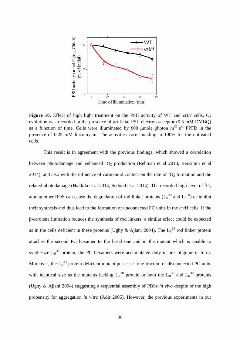
Figure 10. Effect of high light treatment on the PSII activity of WT and crtH cells. O2
evolution was recorded in the presence of artificial PSII electron acceptor (0.5 mM DMBQ)
as a function of time. Cells were illuminated by 600 μmole photon m-2
s-1
PPFD in the
presence of 0.25 mM lincomycin. The activities corresponding to 100% for the untreated
cells.
This result is in agreement with the previous findings, which showed a correlation
between photodamage and enhanced 1O2 production (Rehman et al 2013, Bersanini et al
2014), and also with the influence of carotenoid content on the rate of 1O2 formation and the
related photodamage (Hakkila et al 2014, Sedoud et al 2014). The recorded high level of 1O2
among other ROS can cause the degradation of rod linker proteins (LR33
and LR30
) or inhibit
their synthesis and thus lead to the formation of unconnected PC units in the crtH cells. If the
β-carotene limitation reduces the synthesis of rod linkers, a similar effect could be expected
as in the cells deficient in these proteins (Ughy & Ajlani 2004). The LR33
rod linker protein
attaches the second PC hexamer to the basal one and in the mutant which is unable to
synthesize LR33
protein, the PC hexamers were accumulated only in one oligomeric form.
Moreover, the LR33
protein deficient mutant possesses one fraction of disconnected PC units
with identical size as the mutants lacking LR30
protein or both the LR33
and LR30
proteins
(Ughy & Ajlani 2004) suggesting a sequential assembly of PBSs in vivo despite of the high
propensity for aggregation in vitro (Adir 2005). However, the previous experiments in our
37
laboratory revealed two fractions of PC units in different aggregation forms in crtH and crtB
cells (Toth et al 2015). We hypothesize that the presence of aggregated PC hexamers could
indicate that those were previously part of assembled PBSs. Thus, the appearance of the
detached PC units is more likely to occur as the results of PBS disassembly than of an
improper assembly.
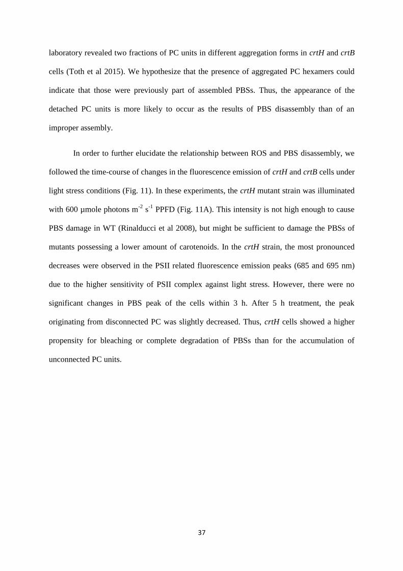
In order to further elucidate the relationship between ROS and PBS disassembly, we
followed the time-course of changes in the fluorescence emission of crtH and crtB cells under
light stress conditions (Fig. 11). In these experiments, the crtH mutant strain was illuminated
with 600 µmole photons m-2
s-1
PPFD (Fig. 11A). This intensity is not high enough to cause
PBS damage in WT (Rinalducci et al 2008), but might be sufficient to damage the PBSs of
mutants possessing a lower amount of carotenoids. In the crtH strain, the most pronounced
decreases were observed in the PSII related fluorescence emission peaks (685 and 695 nm)
due to the higher sensitivity of PSII complex against light stress. However, there were no
significant changes in PBS peak of the cells within 3 h. After 5 h treatment, the peak
originating from disconnected PC was slightly decreased. Thus, crtH cells showed a higher
propensity for bleaching or complete degradation of PBSs than for the accumulation of
unconnected PC units.
38
650 700 750
6
5
4
3
2
1
crtH 0h
crtH 2h
crtH 5hR
ela
tive
Flu
ore
sce
nce
In
ten
sity
Wavelength (nm)
0650 700 750
B
crtB 0h
crtB 12h
crtB 24h
Wavelegnth (nm)
A
Figure 11. Photoinhibition of crtH and crtB LAHG strains were followed by recording 77K
fluorescence emission spectra. The light intensity during the treatment was 600 and 35 µmole
photons m-2
s-1
for crtH (A) and crtB (B) cells, respectively. The spectra of crtH were
obtained upon excitation at 590 nm and normalized on 720 nm band recorded upon 436 nm
excitation. For crtB cells, the spectra were recorded upon 436 nm excitation and the
measurements were performed on cells possessing the same optical density at 750 nm.
The dark grown crtB mutant strain was also exposed to light stress (Fig. 11B), but by
using low light intensity (35 μmole photons m−2
s−1
PPFD) due to its extreme light sensitivity
(Sozer et al 2010). In the crtB mutant, the increased peak of unconnected PC units was
observed only after 24 h when Chl bleaching (Sozer et al 2010) and most probably cell death
was already occurring. It seems that light stress and consequently the increase of ROS is most
likely insufficient for accelerating the damage of PBS structure more than that of
photosystems in crtH and crtB mutants. Therefore, ROS are unlikely able to directly damage
the rod linkers which are already integrated into PBSs.

39
4.1.4. Effect of glucose on crtH cells
Oxidative stress to some extent is induced by the presence of glucose in the growth
media (Narainsamy et al 2013) and is accompanied by a concomitant increase of carotenoid
synthesis (Ryu et al 2004). Therefore, we also studied the fluorescence emission spectra of
WT and crtH cells grown under photomixotrophic (PMG) conditions in glucose-
supplemented media (Fig. 12A). Under PMG conditions, only slight differences were
detected by the fluorescence emission spectra of WT cells. However, a higher level of
disconnected PC units was observed in crtH PMG strain relative to the PSI fluorescence as
reflected by the increased contribution of 640 nm band to the emission spectra. The
absorption spectra of these cells were also recorded (Fig. 12B).
640 680 720 7600
1
2
3
4
5
550 600 650 700 7500.00
0.01
0.02
0.03
0.04
0.05A
Re
lative F
luo
resce
nce
In
ten
sity
Wavelength (nm)
WT
WT PMG
crtH
crtH PMG
B WT
WT PMG
crtH
crtH PMG
Ab
so
rption
Wavelength (nm)
Figure 12. 77 K fluorescence emission (A) and absorption (B) spectra of WT and crtH strain
grown under photoautotrophic (WT and crtH) and photomixotrophic (WT PMG and crtH
PMG) conditions. Emission spectra were obtained upon excitation at 590 nm and normalized
to the amplitudes of 720 nm band recorded upon 436 nm excitation from the same sample.
The absorption spectra were obtained from cultures with the same OD750 values and corrected
for baseline.
Prior to the measurement, the cultures were adjusted to a same OD750 value that
allows estimating the relative pigment content of the cells. Cultivation of the cells in the
presence of glucose resulted in significantly reduced amount of pigments in both the WT and
crtH cells as reflected by the substantially lower PBS and Chl peaks. The increased level of

40
unconnected PC units in crtH PMG cells could be attributed to a decrease in β-carotene
content when cells are cultivated in glucose-supplemented media. However, we could not
establish any significant difference between the pigment composition of the crtH cells grown
under autotrophic and PMG conditions (Table 1).
4.1.5. Additional myxoxanthophyll deficiency in crtH cells
Based on the pigment composition, the crtH cells possess similar myxoxanthophyll
content as WT; while the amount of all other carotenoids is much lower (Table 1).
Myxoxanthophyll is a very efficient scavenger of ROS (Domonkos et al 2013) and a high
level of this carotenoid may indicate an increased demand for the protection against ROS-
mediated damage. Hence, in this present study, we generated a double mutant in which both
the CrtH and CruF enzyme functions are missing (Table 1). If ROS influenced PBS distortion
(Liu et al 2005a, Rinalducci et al 2008) in crtH cells, somewhat higher level of the
disconnected PC units would be expected in the double mutant crtH/cruF cells; but in our 77
K fluorescence measurements, no significant increase of the emission peak originating from
PC was observed (Fig. 13).
650 700 750
0
1
2
3
4
Re
lative F
luo
resce
nce I
nte
nsity
Wavelength (nm)
crtH
crtH/cruF
Figure 13. 77 K fluorescence emission spectra of crtH and crtH/cruF strains obtained upon
590 nm excitation. The spectra normalized on 720 nm bands recorded upon 436 nm
excitation.

41
In fact, neither photoinhibition nor absence of myxoxanthophyll could enhance the level of
unconnected PC units in crtH strain.These results support the ideas that ROS are not directly
involved in the distortion of PBSs upon limited β-carotene availability and also that the
increased amount of unconnected PC units in the presence of glucose is probably induced by
a mechanism not related to ROS.
4.1.6. Nitrogen shortage induced PBS degradation in crtH and crtB strains
Our knowledge about enzymatic phycobilisome degradation has been primarily
obtained by performing macronutrient-starvation experiments on various cyanobacterial
strains (Schwarz & Forchhammer 2005). Here, we used nitrogen-deficiency to trigger an
enhanced PBS degradation in WT, crtH, and crtB cells. In nitrogen-depleted media, the
amount of the pigment-protein complexes especially that of PBSs were largely reduced
within a short time as represented by the color change of the cell cultures (Fig. 14) in
agreement with earlier published data in the literature. During the process of nitrogen
starvation-induced proteolysis, the cyanobacterial cells were changing their color from blue-
green to yellow-green (Fig. 14); this process is known as bleaching or chlorosis (Blankenship
& Chen 2013). In our experiments, the crtH mutant showed greenish color even at the later
stage of nitrogen starvation. The observed effect could be explained by the lower level of the
yellow color carotenoids in crtH cells relative to the bluish PBS or by the delayed
degradation of phycobiliproteins under β-carotene limited conditions as compared to the WT
(Fig. 14).
WT WT-N crtH crtH-N

42
Figure 14. Effect of nitrogen starvation on the Synechocystis WT and crtH mutant cells.
Cells were incubated in nitrogen-free media for 48 h.
The absorption spectra of WT cells showed a stronger decrease in the PBS peak
relative to the Chl peak after 24 h (from the ratio of 0.69 to 0.48) and this difference was
more pronounced after 48 h (Fig. 15) (to 0.17) treatment.
550 600 650 700 7500.00
0.01
0.02
0.03
0.04
0.05
Wavelength (nm)Wavelength (nm)
WT
WT -N 24h
WT -N 48h
Ab
so
rption
A B
550 600 650 700 7500.00
0.01
0.02
0.03
0.04
0.05
crtH
crtH -N 24h
crtH -N 48h
Figure 15. Effect of nitrogen starvation on the absorption spectra of WT and crtH strains.
Cells were cultivated in nitrogen-free media (-N) for 24 h or 48 h. The absorption spectra
were baseline corrected and normalized to the Chl peak.
In agreement with our absorption measurements, the 77 K fluorescence emission
spectra also implied smaller antenna size of the photosystems in WT cells, especially after 48
h (Fig. 16) of nitrogen starvation. The reduced PBS level led to decreased EET to
photosystems represented by lowered PSII fluorescence bands upon PBS excitation. Based on
absorption spectra, the rate of PBS degradation seemed to be similar in the crtH strain to that
of WT after 24 h nitrogen starvation (PBS to Chl ratio decreases from 0.80 to 0.55), but there
was a pronounced difference after 48 h (Fig. 15) (0.36 PBS to Chl ratio). The absorption
spectra of crtH cells after 48 h treatment implied almost twice as a high amount of
phycobilins than in the WT suggesting lower degradation rate of phycobiliproteins.
Interestingly, the 77 K fluorescence emission spectra of nitrogen deprived crtH cells upon
43
590 nm excitation revealed an increased intensity of the PC peak despite of the decrease in
phycobilin content relative to Chl (Fig. 16).
650 700 7500
1
2
3
4
Wavelength (nm)Wavelength (nm)
WT
WT -N 24h
WT -N 48h
Re
lative
Flu
ore
sce
nce
In
ten
sity
A B
650 700 7500
1
2
3
4
crtH
crtH-N 24h
crtH-N 48h
Figure 16. Effect of nitrogen starvation on 77 K fluorescence emission spectra of WT and
crtH strains. Cells were cultivated on nitrogen-free media (-N) for 24 h or 48 h. Fluorescence
emission spectra were obtained upon 590 nm excitation and normalized on 720 nm band
recorded upon 436 nm excitation.
The increase in the relative amount of the energetically disconnected PC subunits
especially those which are in smaller aggregation form are resulting in the shift from 650 nm
towards 640 nm. However, the fluorescence emission spectra obtained upon 590 nm
excitation were normalized; therefore, the increased PC peak did not necessarily indicate a
higher level of unconnected rods on the cell basis.
We performed time-resolved fluorescence measurements with picoseconds (ps) time
resolution on crtH mutant and WT cells incubated in nitrogen free media. This technique is
useful to confirm the presence of undisturbed EET processes or to demonstrate the distortion
of it. We have recorded fluorescence decays at various wavelengths upon 590 nm excitation
before and after 24 h nitrogen starvation. The data were fitted as the sum of exponential
decays. The wavelength distributions of the amplitudes corresponding to the individual decay
components are called decay-associated spectra (DAS). Upon 590 nm excitation, the

44
fluorescence decays exhibited at least five exponential decay components resolved by global
analysis (Fig. 17). In WT cells, the fastest-decaying component reflects the downhill EET
from PC to APC660 (Fig. 17A, blue curves). The second component (red curves) has a
characteristic peak with a positive amplitude at shorter wavelengths and negative amplitude
at longer wavelengths and therefore reflects the EET from APC660 to APC680 + Chls. The 170
ps component (yellow) reflects energy trapping by the RCs, which in turn shows the
equilibrated EET over PBS subunits and Chls. The long lifetime components (~ 534 ps,
purple, and ~ 1.42 ns, green) have very small amplitudes. These components probably reflect
the competition between secondary charge separation and charge recombination (Tian et al
2011).
Figure 17. The decay-associated spectra of Synechocystis WT (A), WT –N (nitrogen free)
(B), crtH (C) and crtH –N (nitrogen free) (D) upon 590 nm excitation before and after (-N)
24 h of nitrogen starvation. The corresponding lifetimes are given in the figures.
WT
A
WT -N
B
crtH -N
D
crtH
C
45
In our experiments even after 24 h of nitrogen shortage, the WT cells did not show
any major changes in the EET only a slight change in the corresponding life-times (Fig. 17B).
On the other hand, the obtained DAS for untreated crtH cells are strongly different from
those of WT cells. In the crtH cells, a decrease in the amplitude of the red component reflects
the decrease in energy transfer from PBS to the APC680 + Chls (Fig. 17C).
Most notably, the crtH cells were exhibited the dominant long living components, the
purple and green components with 521 ps and 1.42 ns lifetime and fluorescence maxima at
around 640-650 nm. These components originate from the functionally disconnected PC rods
of PBS. Similar DAS for crtH mutant was obtained from the previous study in our laboratory
(Toth et al 2015). In case of nitrogen shortage, the DAS of crtH cells displayed a component
peaking around 640-650 nm wavelengths with even higher amplitude (~ 1.06 ns) (Fig. 17D)
and no clear component for EET from was observed. These observations support the
enrichment of the cells in energetically disconnected PC rods of PBS under nitrogen
starvation.
Nitrogen starvation in carotenoids-deficient crtB cells was also monitored. The crtB
cells can only grow in light-activated heterotrophic growth (LAHG) conditions (Sozer et al
2010), thus the PBS degradation induced by nitrogen starvation can be investigated without
the possible influence of light. Prior to both the absorption and fluorescence emission
measurements, the cultures were adjusted to the same OD750 value in order to compare the
pigment content and corresponding fluorescence intensity on the cell basis. Based on the
absorption spectra, the crtB cells possessed low Chl content relative to the amount of
phycobilins (Fig. 18A).

46
550 600 650 700 7500.00
0.01
0.02
0.03
640 680 720 7600.0
2.0x10⁴
4.0x10⁴
6.0x10⁴
Absorp
tion
Wavelength (nm)
crtB
crtB -N 4.5h
crtB -N 9h
A B
Rela
tive F
luore
scence Inte
nsity
Wavelength (nm)
crtB
crtB -N 4.5h
crtB -N 9h
Figure 18. Effect of nitrogen starvation on the absorption and 77 K fluorescence emission
spectra of crtB strains. Cells were cultivated on nitrate free media (-N) for 4.5 h or 9 h. The
absorption spectra (A) were obtained from cells possesses the same OD750 and baseline
corrected. The fluorescence spectra (B) were recorded upon 436 nm excitation of cells with
identical OD750.
Both the Chl and phycobilin related peaks showed a decrease after 4.5 h of nitrogen
starvation which further reduced after 9 h treatment. In crtB cells similarly to WT, the
absorption of phycobilins decreases faster than that of Chl. After 4.5 h treatment, the
fluorescence emission spectra of the crtB cells (Fig. 18B) revealed a substantial decrease in
the TE fluorescence emission, while PSI fluorescence only slightly lowered. After 9 h of
nitrogen starvation, the TE peak and PSI peak further decreased, while the intensity of PC
band remained almost at the original level.
The results from nitrogen starvation experiments could be explained by a slower
degradation of unconnected PC rods, or an enhanced disassembly of PBSs upon β-carotene
shortage in both the crtB and crtH strains. However, no fluorescence from TE of crtH cells
appeared during the starvation, which supports the idea of the delayed degradation of PC rod
units. This way, a role of β-carotene in the recognition or degradation of detached
phycobiliproteins can be hypothesized. It is plausible that even under normal growth
conditions, the degradation of unconnected rods is delayed to a certain extent; this delayed

47
degradation of rods can result in the observed phenotype of the low β-carotene containing
cells. The delayed degradation of detached PC units would also explain their increased
presence in crtH cells grown under PMG conditions, where the cells have lower PBS and Chl
content than under autotrophic conditions.
On the other hand, the delayed degradation of PC units itself would not explain the
shorter rods of assembled PBSs. It is known that the degradation of PBSs is sequential
(Schwarz & Forchhammer 2005, Bernát et al 2009), thus the detachment of the last two rod
units (Toth et al 2015) cannot be part of an active mechanism targeting to reduce the antenna
size. Two mechanisms could explain such shortening of the rods. The detachment of the rods
should occur even under non-stressed conditions (Anderson & Toole 1998). In case of β-
carotene shortage, there might be a defect of the PBS repair system. More likely, under β-
carotene limitation PC detachment could occur due to damage of PBS linkers by a factor
different from ROS. Indeed, cyanobacterial cells with linker protein deficiency accumulate a
high level of disconnected phycobiliproteins (Anderson & Toole 1998, Ughy & Ajlani 2004),
while the mutations related to the phycobiliprotein structure lead to a decreased amount of
these proteins (Anderson & Toole 1998). It seems that the cyanobacterial cells are unable to
efficiently eliminate the disconnected PC oligomers although those have a capacity to
produce harmful ROS (He et al 1997, Anderson & Toole 1998). It is plausible that
carotenoids participate in a complex similar to those formed by HLIP proteins (Knoppova et
al 2014, Daddy et al 2015 ) and can protect the PBSs similarly to chaperons (Sato et al 2010).
Thus, an important function of β-carotene may be the prevention of PBS disassembly.

48
4.2. Zeaxanthin and echinenone modify the structure of PSI trimer
4.2.1. Pigment analysis of cells and PSI complexes
Firstly, we tested whether the xanthophyll deficiency influences the pigment content of
PSI or if we can detect xanthophylls in the PSI complex of WT cells. We analyzed the
pigment composition of whole cells and isolated PSI complexes by HPLC (Table 3).
Table 3. Relative distribution of the main carotenoid species from the whole cell extract, as
well as isolated PSI trimers of WT, crtRO, crtR, crtO, cruF and monomers of psaL. Values
are given as the number of pigment molecules relative to 100 Chl molecules. Each value
represents the mean (± SD) of at least three independent repetitions. Abbreviation of
carotenoids: Myx, myxoxanthophyll; Zea, zeaxanthin; Ech, echinenone; β-car, β-carotene.
*The result is significant at P < 0.05 or ** at P <0.01 as compared to WT using the t-test for
equal variance.
Whole cells Isolated PSI trimer/monomer
Myx Zea Ech β-car Zea Ech β-car
WT 15.1±4.9 21.1±2.2 9.8±0.4 26.0±0.9 1.3±0.1 8.0±0.9 17.7±0.2
crtRO - - - 34.3±0.1** - - 23.2±1.0**
crtR - - 10.0±1.3 21.6±1.1** - 9.2±1.2 15.9±0.2**
crtO 7.9±1.4* 18.2±0.7* - 38.6±2.9** 1.5±0.18* - 23.9±0.3**
cruF - 27.1±2.7* 12.1±2.6 30.3±1.2** 1.5±0.2 8.3±0.3 17.6±0.2
psaL 9.1±1.9* 26.4±1.2** 17.1±1.5** 43.2±4.3** 1.5±0.2* 7.2±0.3 14.9±0.8**
Besides the almost completely xanthophyll-deficient (crtRO) mutant, we also
investigated the PSI complex of crtR, crtO and cruF mutants which are deficient in various
xanthophyll species. Furthermore, we applied psaL mutant, which contains only PSI
monomers due to the absence of the PsaL protein (Chitnis & Chitnis 1993).
In order to minimize the amount of xanthophylls that co-purify, but unspecifically
bind to PSI, 2% β-DM was used for solubilization of the thylakoid membrane. Our data
confirmed the presence of xanthophyll molecules, zeaxanthin and echinenone in the purified
PSI complexes. In line with our findings, both zeaxanthin (Bautista et al 2005, Klodawska et

49
al 2015, Komenda & Sobotka 2016) and echinenone (Klodawska et al 2015, Daddy et al
2015) molecules were previously detected in Synechocystis PSI samples using lower (1% β-
DM) detergent concentration.
In spite of its large quantity in the cells, myxoxanthophyll was not observed in the
isolated PSI complexes so far (Table 3) (Bautista et al 2005, Klodawska et al 2015). No
compensation of the absence of echinenone by zeaxanthin was observed, but the echinenone
deficiency (in crtO and crtRO) coincided with an increase in the amount of β-carotene (Fig.
19) in the trimeric PSI complex.
The results are expressed relative to the Chl content, thus the increased amount of β-
carotene in crtO and crtRO can be explained by a higher β-carotene content of the PSI (Fig.
19B) or a decrease in the Chl content of the complexes. The loss of peripheral Chl molecules
during isolation of PSI complexes has been reported previously (Rogner et al 1990, El-
Mohsnawy et al 2010) and could provide a potential explanation for the apparent increase in
the amount of β-carotene in the mutant PSI.
Figure 19. The β-carotene content of the whole cell extracts of WT, crtRO, crtR, crtO, and
psaL (A) and the corresponding PSI complexes (B). The values are given as the number of β-
carotene molecules relative to 100 Chl molecules. Each value represents the mean ± SD of at
least three independent repetitions.
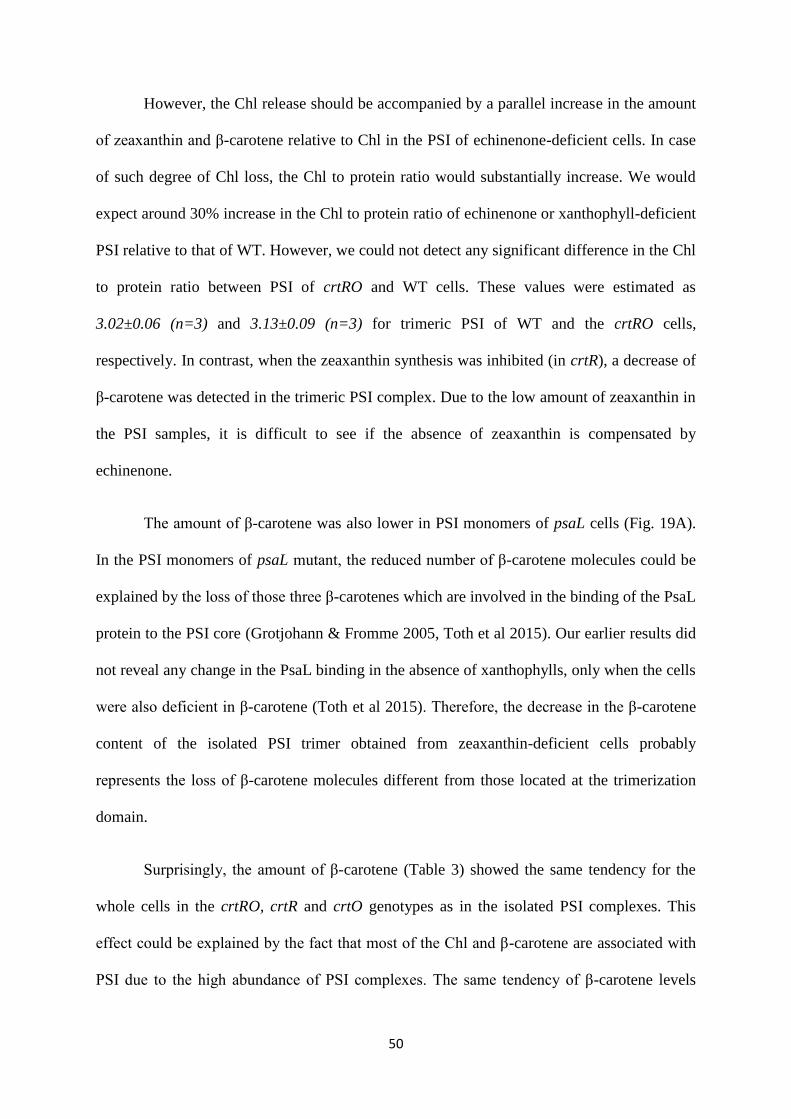
50
However, the Chl release should be accompanied by a parallel increase in the amount
of zeaxanthin and β-carotene relative to Chl in the PSI of echinenone-deficient cells. In case
of such degree of Chl loss, the Chl to protein ratio would substantially increase. We would
expect around 30% increase in the Chl to protein ratio of echinenone or xanthophyll-deficient
PSI relative to that of WT. However, we could not detect any significant difference in the Chl
to protein ratio between PSI of crtRO and WT cells. These values were estimated as
3.02±0.06 (n=3) and 3.13±0.09 (n=3) for trimeric PSI of WT and the crtRO cells,
respectively. In contrast, when the zeaxanthin synthesis was inhibited (in crtR), a decrease of
β-carotene was detected in the trimeric PSI complex. Due to the low amount of zeaxanthin in
the PSI samples, it is difficult to see if the absence of zeaxanthin is compensated by
echinenone.
The amount of β-carotene was also lower in PSI monomers of psaL cells (Fig. 19A).
In the PSI monomers of psaL mutant, the reduced number of β-carotene molecules could be
explained by the loss of those three β-carotenes which are involved in the binding of the PsaL
protein to the PSI core (Grotjohann & Fromme 2005, Toth et al 2015). Our earlier results did
not reveal any change in the PsaL binding in the absence of xanthophylls, only when the cells
were also deficient in β-carotene (Toth et al 2015). Therefore, the decrease in the β-carotene
content of the isolated PSI trimer obtained from zeaxanthin-deficient cells probably
represents the loss of β-carotene molecules different from those located at the trimerization
domain.
Surprisingly, the amount of β-carotene (Table 3) showed the same tendency for the
whole cells in the crtRO, crtR and crtO genotypes as in the isolated PSI complexes. This
effect could be explained by the fact that most of the Chl and β-carotene are associated with
PSI due to the high abundance of PSI complexes. The same tendency of β-carotene levels

51
was recorded in PSI complexes of the xanthophyll mutants (crtRO, crtR, crtO) at cellular
level supporting that those differences in β-carotene to Chl ratio of PSI probably exist in vivo
(Table 3) (Kusama et al 2015), as well. But for the psaL mutant where the decrease of the β-
carotene content of PSI was accompanied by an increase of the cellular β-carotene level. This
effect could be explained by the increased level of carotenoid-binding proteins (Daddy et al
2015).
4.2.2. Testing the PSI organization by in vivo spectroscopic methods
We addressed the question whether the absence of xanthophylls alters the
organization of PSI in vivo. The circular dichroism (CD) and fluorescence measurements
were proved to exhibit distinct spectral features for trimeric and monomeric PSI-containing
samples both in vitro (Shubin et al 1993, Gobets et al 2001, El-Mohsnawy et al 2010,
Karapetyan et al 2014 ) and in vivo (Klodawska et al 2015, Toth et al 2015) based on the
specific pigment-protein interactions, the CD spectroscopy can be applied to investigate the
role of xanthophylls in terms of structural rearrangement of PSI complex.
The circular dichroism (CD) spectra possess a characteristic fingerprint for PSI
complex at around 515 nm wavelength based on specific pigment-protein interactions within
the complex (Shubin et al 1993, Klodawska et al 2015, Zakar et al 2017). In this study, the
CD spectra were obtained from cell suspensions of xanthophyll deficient mutant crtRO, and
psaL relative to that of WT and revealed differences in the 515 nm peak (Fig. 20A). The PSI-
related signal was expressed by the CD amplitudes measured at 515 nm using a reference
wavelength of 550 nm. Values are also given in percentage (Fig. 20B), where 0%
corresponds to the level of 515 nm signal in the psaL (only PSI monomers) and 100% to that
in WT (predominantly PSI trimers).
52
Figure 20. Circular dichroism spectroscopy analysis of WT and various mutants of
Synechocystis cells. A: Overlaid CD spectra of psaL, crtRO, and WT cells. The spectra were
normalized to the Chl absorption measured at 680 nm. B: Comparison of the amplitudes of
the PSI-related 515 nm bands. Error bars represent standard deviation. * The result is
significant at P < 0.05 or ** at P <0.01 as compared to WT.
In our experiments, the PSI-related CD signal did not decrease in crtO and only to a
small extent in cruF cells, while approximately 30% and 40% reduction of the PSI signal was
observed in crtR and crtRO cells, respectively. Thus, mostly the absence of zeaxanthin could
be accounted for the decrease in the PSI-related CD signal observed in vivo upon complete
xanthophyll deficiency.
The CD spectra were normalized to the total Chl absorption of the cell suspensions;
consequently, any changes in the PSI to PSII ratio of cells could alter the intensity of the PSI-
related CD signal. The influence of xanthophyll content on PSI to PSII ratio was investigated
by the fluorescence emission of cells upon Chl (436 nm) excitation (Fig. 21) at low
temperature. This way both PSI and PSII are excited and any changes in the relative ratio of
fluorescence emitted by the photosystems suggest the alteration of their approximate ratios.
The spectra of the strains investigated in this study exhibited no or only a small (less than
5%) decrease in PSII peaks (685 and 695 nm) relative to the PSI band (720 nm).

53
The spectra were obtained from dark-adapted cells, thus the effect of light-induced
non-photochemical quenching on the intensity of the PSII peaks should not be considerable.
In line with these results, the time-resolved fluorescence measurements also did not reveal
any significant changes in the fluorescence lifetimes of the photosystems (Toth et al 2015).
Therefore, the difference observed in the CD spectra of the cells cannot be strongly
influenced by the change in the PSI to PSII ratio of the mutants.
Figure 21. 77K fluorescence emission spectra of Synechocystis WT and crtRO, crtR, crtO,
cruF and psaL mutant cells. The spectra were recorded upon 436 nm excitation of samples
with equivalent Chl content and normalized to their maxima.
The fluorescence technique was also useful to obtain further information about the
PSI trimer in vivo, based on the spectroscopic feature of the LWCs. Although LWCs are
responsible for only a small fraction of the total absorption, they have a strong effect on the
fluorescence properties of the PSI, especially when recorded at low (77K or less) temperature
(El-Mohsnawy et al 2010). Around five-six LWCs in the trimer and three-four in the
monomer of Synechocystis were proposed based on experimental results (Gobets et al 2001,
Mazor et al 2014). These LWCs absorb light around 700 nm and emit fluorescence above 710
nm depending on temperature (van der Lee et al 1993, Gobets et al 2001, Karapetyan et al

54
2014). Hence, here we recorded fluorescence emission at 77K temperature to obtain
information about the level of PSI trimer in vivo, based on the “red” Chl interaction.
According to our results, a decrease in the LWC signal was observed not only in psaL but
also in crtRO cells (Fig. 22A).
Figure 22. 77K fluorescence emission spectra recorded on WT and mutant Synechocystis
cells. A: The spectra were recorded upon 436 nm excitation of samples with equivalent Chl
content and normalized to their maxima. The inset shows the WT minus mutant difference
spectra for various cells. B: Difference in the fluorescence emission between the WT and
mutants at 735nm. Error bars represent standard error (n=3). * The result is significant at P <
0.05 or ** at P <0.01 as compared to WT.
We found around 60% decrease in the amplitude of the LWC signal obtained from the
crtRO mutant (Fig. 22B), in conditions when WT and psaL represent 0% and 100 %,
respectively. Our results showed around 45% reduction of the LWC signal in crtO cells, and
no significant difference (P >0.05) in crtR and cruF cells. Consequently, mostly the CrtO
mutation contributed to the PSI-assigned fluorescence change observed in the crtRO mutant.
The changes of the PSI-related CD signal do not essentially coincide with alteration
of the LWC fluorescence of the PSI (Fig. 20 and 22). Based on these results, we suppose that
both zeaxanthin and echinenone influence the structure of PSI, specifically, in a different
manner in vivo. In order to support our hypothesis biochemical investigation of the
xanthophyll-deficiency-induced effect on PSI were carried out.

55
4.2.3. Protein analysis of the thylakoid membrane
To investigate the importance of the specific xanthophylls in the organization of PSI
trimer, protein analysis was performed on the same set of mutants. We applied CN-PAGE
(Fig. 23) which allows comparing the intensity of Chl-containing bands. Identification of the
bands on the gel was performed based on the relative electrophoretic mobility (Kopecna et al
2012, Toth et al 2015).
Figure 23. CN-PAGE analysis of thylakoid membrane protein complexes of various
xanthophyll biosynthesis mutants. A: Thylakoid membranes of WT, crtRO, crtR, crtO, cruF
and psaL cells were solubilized with 1% β-DM (w/v). Samples containing 1, 0.5 (50%) or
0.25 (25%) µg of Chl were loaded, as it is indicated. The positions of PSI trimeric or PSI and
PSII monomeric complexes are indicated in the left side. B: Quantitative densitometry results
for the Chl-containing protein bands separated by the CN-PAGE. The results obtained from
the same sample loaded at various concentrations have been averaged after normalization.
Thylakoid sample of the psaL mutant with 50% and 25% of Chl concentration was
loaded on the gel to achieve a better assessment of the amount of monomers in the mutant
cells. It is noteworthy, that most of the Chl molecules (approximately 90%) bound to PSI;
thus, the intensity changes of the green bands are well correlating with the alterations in PSI

56
organization. In our experiments, the electrophoretic separation of native protein complexes
revealed a somewhat higher amount of monomers at the expense of PSI trimers in crtRO,
crtR, and crtO thylakoids, but these monomeric PSI bands were still much fainter than that of
the two-fold diluted psaL sample (Fig. 23A). Based on densitometry results, the crtRO, crtR,
and crtO mutants exhibited around 25% increase in the intensity of the monomeric band
relative to the WT independently of the applied Chl concentration (Fig. 23B).
The stability of PSI trimers was also tested using various treatments, like EDTA
washing, increased detergent concentration and high temperature (Fig. 24). We found that the
PSI trimer of xanthophyll-deficient crtRO mutant possessed a strongly increased propensity
for decomposing into monomers, as compared to WT upon EDTA treatment. Due to
technical limitations, the detailed structure of PSI trimer has only been obtained from T.
elongatus (Jordan et al 2001), while the only partial structure of PSI monomer is available
from Synechocystis PSI complex (Mazor et al 2014).
Figure 24. Protein analysis of Synechocystis thylakoid membrane of WT and the
xanthophyll-deficient mutant, crtRO by BN-PAGE. The membranes of WT and crtRO
mutant were solubilized with 1% β-DM (w/v) and treated with 25 mM EDTA, 5% β-DM or
60°C heat where indicated. Samples containing 2.5 µg of Chl were loaded on each lane. The
positions of PSI trimeric (PSI3), PSI and PSII monomeric (PSI1+PSII1) complexes are
indicated on the left side.
The high-resolution structure supposes a Ca2+
ion in the PSI trimeric complex which
is released during the transition of trimer to monomers (Jordan et al 2001). Based on the

57
structural homology of PSI between Synechocystis and T. elongatus, we assume that in our
experiments, the structural differences at the monomer-monomer interface in the absence of
xanthophylls could lead to the easier release of Ca2+
and eventually the disassembly of the
trimer into monomers. This hypothesis is supported by the substantial sensitivity of the
mutant trimer to the chelating agent, EDTA (Fig. 24). Thus, a weaker binding of divalent
cations (i.e. Ca2+
) in the absence of xanthophylls can be accounted for the reduced PSI trimer
stability. However, PSI trimer was not showing increased susceptibility to other factors (e.g.
detergent, high temperature). Interestingly, in crtRO samples, the PSI monomers exhibited
multiple bands with poor resolution upon solubilization with 1% β-DM, while separated into
one distinct band after solubilization with 5% β-DM (Fig. 24). This suggests a modified
interaction of the monomeric PSI with the surrounding lipid molecules
We performed sucrose density gradient centrifugation during purification of the PSI
complexes from solubilized thylakoids. Mostly, trimeric PSI was obtained from the WT and
cruF mutants, while only monomers were observed in psaL samples (Fig. 25). Surprisingly,
our results revealed an extreme instability of the trimeric PSI complexes in crtRO and crtR
mutants under these conditions and to some extent in crtO. In crtRO, crtR and crtO samples
the ratio of PSI trimers to monomers varied from isolation to isolation (Fig. 25 and 26); but in
crtRO mutant cells, we could not retain more than 50% of the trimers. These observations
correlate with the changes observed in CN and BN-PAGE (Fig. 23 and 24), but show a more
pronounced effect.

58
Figure 25. Sucrose density gradient fractionation of solubilized thylakoid membranes.
Thylakoid membranes were solubilized by 2% β-DM and samples with equivalent Chl
content were loaded onto each tube. The band designation is indicated next to the
corresponding bands.
These results also demonstrate a weakening of the driving force responsible for
maintaining the trimeric structure upon the absence of xanthophylls. It seems that the
instability of the PSI trimer of crtRO mutant is mostly due to the crtR and, at less extent, to
the crtO mutation. Accordingly, predominantly zeaxanthin and, in a certain degree,
echinenone could be accountable for the stabilization of the PSI trimeric complex.
Figure 26. Sucrose density gradient fractionation of solubilized thylakoid membranes.
Thylakoid membranes were solubilized by 2% β-DM and samples with equivalent Chl
content was loaded onto each tube. The band designation is indicated next to the
corresponding bands.
Such important role of xanthophylls in the trimeric structure of Synechocystis PSI
raises the question as to where those structurally important xanthophylls are located and how

59
they influence the protein structure. Although it was observed that some carotenoid-binding
proteins can also bind xanthophylls (Melnicki et al 2016), it is unlikely that those are co-
purified with PSI complexes. However, the influence of certain membrane-localized
carotenoid molecules on the structure and function of membrane proteins was previously
demonstrated (Chegeni et al 2016, Janik et al 2016). Such effect of non-protein-bound
xanthophylls on the PSI trimer structure could occur via their influence on the lipid
environment in the vicinity of the complex e.g. modifying the width or fluidity of the
membrane (Gruszecki & Strzalka 2005, Klodawska et al 2015) or via direct interaction of the
peripheral xanthophylls with specific parts of the transmembrane protein domains. In fact, the
impact of the membrane lipid compositions on the xanthophyll content and PSI
oligomerization was already established in Synechocystis cells (Domonkos et al 2009, Zakar
et al 2017).
4.2.4. Spectroscopic investigation of the PSI complexes
We have investigated the PSI trimeric complex in vivo by spectroscopic methods in
order to reveal the location of structurally important xanthophylls in the PSI complex and
understand how they influence it. Our idea of structurally modified PSI trimers was
confirmed in vitro by the CD spectra of the PSI complexes separated by native gel
electrophoresis (Fig. 27). The difference between PSI trimer of WT and monomer of psaL
spectra was similar to what was observed for whole cells. Moreover, trimers of both the
crtRO and crtR retained their spectral difference from WT, while crtO and cruF are still do
not differ significantly (Fig. 27A).

60
Figure 27. Circular dichroism spectroscopy analysis of PSI complex of WT and various
mutants of Synechocystis cells. The thylakoid membranes were solubilized by 1% β-DM. A:
Overlaid CD spectra of CN-PAGE separated PSI complex of psaL, crtRO, and WT. The
spectra were normalized to the Chl absorption measured at 680 nm. B: Comparison of the
amplitudes of the PSI-related 515 nm bands. Error bars represent standard deviation. ** The
result is significant at P <0.01 as compared to WT.
Similar results were obtained on sucrose density gradient fractions containing
dominantly PSI complexes (Fig. 28). Thus, specific action sites for the xanthophylls at the
PSI complex were predicted from the spectroscopic results.
Figure 28. Circular dichroism spectroscopy analysis of thylakoid membranes isolated from
WT, crtRO, and psaL cells. The spectra were normalized to the Chl absorption measured at
680 nm.

61
The pigment analysis revealed the same amount of xanthophylls per trimer as in the
monomer (see section 4.2.1) in spite of the high (2%) detergent concentration. We have also
tested by spectroscopic methods the PSI trimeric complexes isolated by a multistep
purification method (sucrose density gradient followed by anion exchange chromatography)
from thylakoid membrane solubilized with higher detergent concentration (2% β-DM). Under
these conditions, the CD spectrum of crtRO trimer appeared to be almost identical to the WT
one (Fig. 29A).
On the other hand, the similar difference between WT trimer and psaL monomer was
observed, as it was previously observed for these oligomers of PSI (Fig. 29A) (Shubin et al
1993).
Figure 29. Spectroscopic analysis of PSI complexes obtained by multistep isolation. The
thylakoid membranes were solubilized with 2% β-DM. A: Circular dichroism spectra of the
PSI complexes. The spectra were normalized to the Chl absorption at 680 nm. B: Absorption
spectra of the trimeric PSI isolated from WT and crtRO overlaid with the spectrum of
monomeric PSI from psaL. The inset shows the absorption region of the LWCs.
In order to monitor the presence of LWCs, we recorded the absorption spectra of the
trimeric PSI from WT and crtRO, as well as the PSI monomer of psaL cells (Fig. 29B).
Interestingly, the absorption spectrum of crtRO PSI trimer also did not show any difference
from WT trimer in the region of 690-720 nm, while PSI monomer of psaL has a lower

62
absorbance at this range (Fig. 29B). Thus, the PSI complexes seemed to undergo structural
modification during multistep isolation, which eventually can lead to the loss of the spectral
difference between WT and xanthophyll-deficient PSI trimers (Fig. 29). Based on our
observations, we predict the presence of the xanthophylls in the peripheral part of the protein
in the membrane plane where they can also interact with the surrounding lipid molecules.
We suppose that xanthophylls are performing mostly fine regulation of the PSI
structure. Very recently, our predictions regarding the presence of zeaxanthin and
echinenone molecules in the PSI trimeric complex of Synechocystis were confirmed by X-ray
crystallography (Malavath et al 2018). The high resolutions structure of the partial (subunit
depleted) PSI trimeric complex was obtained. Their measurements suggest at least 4
zeaxanthin and 7 echinenone and one hydroxyl-echinenone molecule in the PSI trimeric
complex. Their data also support the presence of the xanthophylls mostly at the periphery of
the monomeric complexes.
Furthermore, our experiments on the isolated trimeric PSI complex of T. elongatus
also suggest that around two molecules of zeaxanthin and echinenone are present per PSI
monomer (Table 4).
Table 4. Relative distribution of the main carotenoid species of PSI trimers isolated from WT
Synechocystis and T. elongatus. Values are given as the number of pigment molecules
relative to 100 Chl molecules. Each value represents the mean ± SD of at least three
independent repetitions.
Zea Ech β-car
Synechocystis PCC 6803 1.3±0.1 8.0±0.9 17.7±0.2
Thermosynechococcus elongatus 2.1±0.3 1.7±0.3 24.3±3.0
Based on the sequence homology, the structure of PSI complexes are very similar in
various cyanobacterial species; this predicts that the involvement of xanthophylls in the
stabilization of the complexes could be a more general phenomenon, not specific for

63
Synechocystis strain. Our previous measurements in the xanthophyll-deficient mutant of
Synechocystis revealed slightly lower stability of PSII dimer as well (Toth et al 2015). Thus,
the analogous effect of xanthophylls on both photosystems is plausible, but its confirmation
requires further studies on the PSII structure and its related functions.
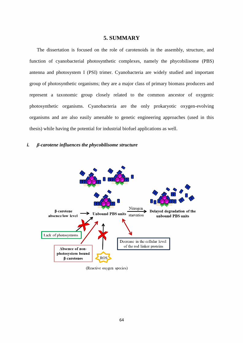
64
5. SUMMARY
The dissertation is focused on the role of carotenoids in the assembly, structure, and
function of cyanobacterial photosynthetic complexes, namely the phycobilisome (PBS)
antenna and photosystem I (PSI) trimer. Cyanobacteria are widely studied and important
group of photosynthetic organisms; they are a major class of primary biomass producers and
represent a taxonomic group closely related to the common ancestor of oxygenic
photosynthetic organisms. Cyanobacteria are the only prokaryotic oxygen-evolving
organisms and are also easily amenable to genetic engineering approaches (used in this
thesis) while having the potential for industrial biofuel applications as well.
i. β-carotene influences the phycobilisome structure
65
In the first part of the thesis, we have investigated the relationship between the
carotenoid composition and the structure of PBS antenna. It was previously found that the
complete carotenoid deficiency (crtB cells) and also the low level of carotenoids (crtH cells)
resulted in the presence of disconnected PC rods of PBS besides the assembled PBSs with
shorter rods. We have proved with a set of mutants, including a newly generated
myxoxanthophyll deficient mutant (cruF, in this thesis), that none of the xanthophylls could
significantly influence the structure of PBS. It is generally known that most of the β-carotene
is bound to the photosystems. Although the absence of photosystems was observed upon the
complete carotenoid deficiency, we showed that it does not affect the PBS structure. Thus, a
small fraction of β-carotene, which is not connected to the photosystems, is important for the
properly assembled PBS.
The presence of unconnected rod units could be explained by the absence or improper
function of the rod linker proteins which connect the PC rods to the rest of the PBS complex.
We found that the amounts of these linker proteins were reduced at the cellular level in the
complete carotenoid deficient mutant (crtB). Thus, a higher rate of degradation or inhibition
of linker protein synthesis can occur upon β-carotene shortage and results in the altered PBS
structure. The complete carotenoid deficient (crtB) mutant possesses an extreme light
sensitivity and does not show photosynthetic activity. On the other hand, the cells with lower
carotenoid content (crtH mutant) possess the same PBS related distortion as the complete
carotenoid deficient cells (crtB) without severe effects on the basic photosynthetic functions.
In order to decrease the complexity of our model system, the mutant with lower carotenoid
content was used in most of the further experiments for understanding the correlation
between β-carotene and PBS structure.

66
One of the main functions of carotenoids is the protection against ROS induced
damage. The effects of β-carotene shortage on the PBSs are in fact can be caused by an
increased production of ROS. Accordingly, the cells with lower carotenoid content (crtH)
exhibited an increased production of a harmful ROS, so-called 1O2. These cells were also
more susceptible to high light treatment but displayed a higher tendency for complete
degradation of PBSs rather than the increased accumulation of disconnected PC rods. In spite
of the fact that the mutant cells possess a lower amount of carotenoids, they contain a similar
level of myxoxanthophyll as wild-type cells. The remaining myxoxanthophyll could ensure
the ROS protection of the PBS linker proteins in this mutant. This hypothesis was
conclusively rejected by the use of the newly created double mutant (crtH/cruF, in this
thesis), which also lacks myxoxanthophyll. Based on our observations, we propose that ROS
induced direct damage is unlikely to cause a significant reduction in the level of the PBS rod
linker proteins upon limited β-carotene availability.
As a next approach, the enzymatic PBS degradation was induced by the nitrogen
starvation condition in the mutants (crtH and crtB) possessing disconnected PBS units.
During nitrogen-limited conditions, the wild-type cells degrade their phycobiliproteins
through a programmed pathway to release the composing nutrients and ensure the survival of
cells. Surprisingly, a slower degradation of the disconnected PBS units was observed in the
mutants under the nitrogen shortage. Consequently, our results suggest that the increased
amount of unconnected PC units upon limited β-carotene availability is most probably due to
an insufficient degradation rate of phycobiliproteins and/or an increased disassembly of PBS.
ii. Zeaxanthin and echinenone modify the structure of photosystem I trimer
In the second part of this thesis, the function of xanthophylls in the organization and
structure of PSI trimer was demonstrated. Our pigment analysis confirmed the presence of

67
xanthophylls, namely zeaxanthin and echinenone in the PSI trimeric complex. Both the
zeaxanthin and echinenone molecules can be part of the complex or be embedded in the
membrane in the vicinity of PSI. Despite the presence of a high amount of myxoxanthophyll
in the cells, we could not observe it in the isolated complex, thus only specific xanthophyll
molecules are present in PSI. In the xanthophyll-deficient mutant, a compensatory increase in
the level of β-carotene was observed, which suggest that xanthophylls might have important
functions in the complex.
We have monitored the organization of PSI trimer upon xanthophyll deficiency in
vivo and in vitro as well. The spectroscopic methods used in this study are based on specific
pigment-protein and pigment-pigment interactions within the PSI complex. One of our
methods, the circular dichroism, showed that only the absence of zeaxanthin resulted in a
noticeable change in the in vivo PSI trimer signal. On the other hand, the absence of
echinenone, much more than that of zeaxanthin, led to a reduced level of LWC fluorescence.
These LWCs are present at the PSI trimeric complex in a higher amount. These results
showed that both x have specific and not interchangeable roles in the structural organization
of PSI trimer in vivo.

68
The further biochemical investigations were performed to study the influence of
specific xanthophylls in the organization of PSI. The poor resolution of monomeric PSI bands
was observed in the xanthophyll deficient mutants, but this effect was eliminable by higher
detergent concentration. Thus, we presume a modified interaction of monomers with the
surrounding lipids upon xanthophyll-deficiency. A most conspicuous result was the
propensity of PSI trimers to disassemble into monomers in the absence of xanthophylls upon
treatment by a chelating agent, ethylenediaminetetraacetic acid (EDTA). We propose that the
structural differences in the monomer-monomer interface of the protein probably led to an
easier removal of the important Ca2+
upon xanthophyll deficiency.
The xanthophylls-related spectral differences were also detected in the isolated PSI
trimers, which prove that the monomerization is not the only change brought about the lack
of xanthophylls. It also predicts the specific sites for the xanthophylls at the PSI complex. A
more rigorous purification protocol was able to eliminate the spectral difference between
wild-type and xanthophyll deficient PSI trimers. This finding led to the proposition that the
xanthophylls are either closely associated or present in the peripheral part of the PSI complex
and can also interact with the surrounding lipid matrix. We propose that xanthophylls are
performing mostly fine regulation of the PSI structure. It is rather surprising because
xanthophylls were not considered to have a vital structural role in the photosynthetic
complexes of cyanobacteria. It is noteworthy that based on this work the presence of
xanthophylls in PSI presumes has been supported by the very recently published partial
structure of Synechocystis PSI trimer. This high-resolution crystal structure reveals that the
PSI complex contains both zeaxanthin and echinenone.
We also observed a low number of zeaxanthin and echinenone molecules in the PSI
complexes of T. elongatus. The stability of the PSII complexes is also influenced by the

69
absence of xanthophylls, based on the previous work in our laboratory. It seems that the
structural function of xanthophylls for photosystems could be a more general phenomenon,
not only specific for Synechocystis species or PSI.

70
6. ÖSSZEFOGLALÓ
A jelen tézis a karotinoidok szerkezeti jelentőségét vizsgálja biokémiai és biofizikai
módszerekkel a Synechocystis sp. PCC 6803 (Synechocystis) cianobaktérium fénybegyűjtő
fikobiliszóma (FB) antennájának és az első fotokémiai rendszerének (PS1) szerveződésében
és működésében. A cianobaktériumok széles körben tanulmányozott és nagy jelentőséggel
bíró fotoszintetikus szervezetek. Kiemelkedő jelentőségűek az elsődleges biomassza
termelésben és taxonómiailag közel állnak a mai oxigéntermelő fotoszintetikus szervezetek
őseihez. A prokarióták között egyedül a cianobaktériumok oxigénfejlesztők, genetikailag is
könnyen módosíthatóak és az ipar számára potenciálisan felhasználható biomassza vagy
bioüzemanyag termelők.
i. A β-karotin befolyásolja a fikobiliszóma antenna szerkezetét
A tézis első részében a karotinoidösszetétel és a FB antenna szerveződése közötti
összefüggést vizsgáltam. Korábban megfigyelték, hogy a teljes karotinoidhiány (crtB sejt) és

71
az alacsony karotinoid szint (crtH sejt) is szabadon álló, FB-hoz nem kapcsolt fikocianin
(FC) FB alegységek, valamint kevesebb FC egységet tartalmazó FBk megjelenéséhez vezet.
Tanulmányoztunk számos mutáns vonalat és előállítottunk egy teljes mixoxantofillhiányos
mutánst (cruF, jelen tézisben). Méréseinkkel megerősítettük, hogy egyik xantofill sem
befolyásolja jelentősen a FB szerkezetét, tehát egyedül a β-karotin hiánya felelős a nem-
kapcsolt FC egységek jelenlétéért. Az általánosságban ismert, hogy a legtöbb β-karotin
molekula a fotokémiai rendszerekhez kötve található a sejtben. Ugyanakkor, a fotokémiai
rendszerek hiánya, ami a karotinoidhiányos sejtekre is jellemző, nem befolyásolta a FB
szerkezetét az elvégzett kísérletekben. Valószínűleg egy kis mennyiségű, nem a fotokémiai
rendszerekhez kötött β-karotin szükséges a FB megfelelő szerveződéséhez.
A nem-kapcsolt FC egységek megjelenése magyarázható a FC alegységeket a FB
többi részéhez csatoló linker fehérjék hiányával vagy nem megfelelő működésével. A teljes
karotinoidhiányos mutánssal végzett kísérleteink azt mutatják, hogy a FC linkerek
mennyisége sejtszinten és a tisztított FB-ban egyaránt alacsonyabb volt. Következésképpen,
karotinoidok hiányában a módosult FB szerkezet valószínűleg a linker fehérjék nagyobb
mértékű lebontódásának vagy az azok szintézisében bekövetkező zavarnak a következménye.
A teljes karotinoidhiányos (crtB) mutáns extrém fényérzékeny és fotoszintetikus
aktivitással sem rendelkezik. Ugyanakkor, az alacsonyabb karotinoidtartalommal rendelkező
sejtek (crtH) az alapvető fotoszintetikus funkciók nagymértékű zavara nélkül tartalmazzák
ugyanazt a FB szerkezeti zavart, mint a teljes karotinoidhiányos mutáns (crtB). Ezért a
további kísérleteinkben túlnyomórészt az alacsonyabb karotinoidtartalommal rendelkező
mutáns sejteket használtuk a β-karotin és a FB szerkezet kapcsolatának feltárására, a
modellrendszerünk komplexitásának csökkentése érdekében.

72
A karotinoidok egyik legfontosabb szerepe a reaktív oxigén gyökök (ROS) elleni
védelem. Tehát csökkent β-karotin szint esetén felmerül a ROS által indukált direkt FB
károsodás lehetősége. Kísérleteinkben az alacsonyabb karotinoidtartalommal rendelkező
sejtek (crtH) megnövekedett szinglet oxigén (1O2) termelődést mutattak, amely egy igen
reaktív ROS. Ezek a sejtek továbbá érzékenyebbnek bizonyultak az erős fénnyel szemben is.
Ugyanakkkor a mutáns sejtben, magas fényintenzitások mellett, erősebb tendenciát
figyeltünk meg a teljes FB lebontódására, mint a szabadon álló FC alegységek további
felhalmozódására. Habár a mutáns sejtek alacsonyabb karotinoidtartalommal rendelkeznek,
mégis a vad típushoz hasonló mennyiségű mixoxantofill található bennük. A mutánsban
jelenlévő mixoxantofill képes lehet a PB linker fehérjéinek a ROS elleni védelmét
részlegesen ellátni. Ezt a hipotézist kizártuk az általunk az alacsony karotinoidtartalommal
rendelkező sejtekből létrehozott, mixoxantofill bioszintézisére sem képes kettős mutáns
(cruF/crtH, jelen tézis) vizsgálatával. Ezen eredmények alapján azt feltételezzük, hogy nem a
ROS-k okozta direkt károsodás a felelős a FC linker fehérjék mennyiségének csökkenéséért
korlátozott β-karotin hozzáférhetőség esetén.
Következő megközelítési módként a FB-k nitrogénhiány által indukált enzimatikus
lebontását használtuk a nem kapcsolt FC alegységeket tartalmazó sejtek (crtB és crtH)
esetében. Korlátozott nitrogén forrás esetében a vad típusú sejtek a saját fikobiliproteinjeik
programozott lebontásából nyernek vissza szükséges anyagokat a túlélésük érdekében.
Meglepő módon, a kísérleteinkben a nem-kapcsolt FC egységek alacsonyabb lebontódási
sebességét figyeltük meg a mutáns sejtekben. Ezért feltételezzük, hogy a FC egységek
lassabb lebontása vagy a FB-k fokozott szétesése okozza a nem-kötött FC egységek
feldúsulását β-karotin korlátozottság esetén.

73
ii. A zeaxantin és az echinenon módosítja az első fotokémiai rendszer trimerének
szerkezetét
Dolgozatom második felében a xantofilloknak a PS1 trimer szerveződésében játszott
szerepét vizsgáltuk meg. Az általunk végzett pigmentanalízis zeaxantin és echinenon
molekulák jelenlétét mutatta ki a tisztított PS1 komplex esetében. Tehát a zeaxantin és az
echinenon pigmentek a komplex részei, vagy a membránba ágyazva a PS1 komplex
közvetlen közelében helyezkednek el. A mixoxantofillt azonban a sejtben jelenlevő nagy
mennyisége ellenére sem tudtuk kimutatni a PS1 komplexben, vagyis csak bizonyos xantofill
molekulák vannak jelen a PS1-ben. A xantofillhiányos sejtek PS1 komplexében a β-karotin
molekulák számának növekedését figyeltünk meg, ami arra utal, hogy a xantofillok fontos
szerepet játszhatnak a PS1-ben.
Megvizsgáltuk a xantofillek szerepét a PS1 trimer szerveződésében spektroszkópiai
módszerekkel in vivo és in vitro is. Ezek a módszerek a PS1 komplexben levő specifikus
pigment-fehérje vagy pigment-pigment kölcsönhatásokon alapulnak. Az egyik módszerünk, a
cirkuláris dikroizmus, azt mutatta, hogy csak a zeaxantin hiánya okozott számottevő változást
az in vivo PS1 trimer jelében. A másik módszer azt az eredményt adta, hogy a zeaxantin nem,
de az echinenon hiánya csökkenti a hosszú-hullámhosszú klorofillok (LWC)

74
fluoreszcenciáját a PS1 komplexben. Ezek a LWC-k a PS1 trimerben nagyobb mennyiségben
vannak jelen mint a monomerben. A különböző xantofillek hiányának látszólag
ellentmondásos hatása a PS1 komplex szerkezetére arra utal, hogy a mind a zeaxantin, mind
az echinenon képes befolyásolni a PS1 komplexet in vivo körülmények között, méghozzá
specifikus módon.
További biokémiai vizsgálatokat végeztünk a különböző xantofillek PS1-ben betöltött
specifikus szerepének tisztázására. Natív gélelektroforézis segítségével rosszul elválasztódó
monomer PS1 sávot kaptunk a xantofillhiányos mintákban. Ezt a hatást azonban képesek
voltunk magasabb detergens koncentráció használatával megszüntetni. Ezen eredmények
alapján feltételezzük a PS1 fehérjéi és az őket körülvevő lipidek közti fehérje-lipid
kölcsönhatások zavarát a xantofilllhiányos mutánsban.
A legszembetűnőbb eredmény a teljes xantofillhiányos mutánsból izolált PS1
trimerek monomerekké szétesése a kelátképző etilén-diamin-tetraecetsav (EDTA) kezelés
hatására. Az eredményeink alapján azt tételezzük fel, hogy a xantofillok hiányában létrejövő
szerkezeti módosulás a monomer-monomer határon levő kiemelkedő jelentőségű Ca2+
ion
gyengébb kötődését eredményezi.
A xantofillfüggő spektrális különbségek az izolált PS1-ben is megfigyelhetőek voltak,
ami arra utal, hogy a monomerizáció nem az egyetlen változás, amit a xantofillek
előidéznek. Továbbá, feltételezi specifikus xantofillkötő helyek jelenlétét a PSI komplexben.
A vad típusú és xantofillhiányos PS1 trimer közötti szerkezeti különbség képes eltűnni a
soklépéses tisztítási eljárás során. Ennek eredményeképp arra következtettünk, hogy a
xantofillok a komplex membránhoz közel eső részén helyezkednek el a fehérjébe ágyazódva,
vagy annak közvetlen közelében, ahol képesek kölcsönhatni a komplexet körülvevő
lipidmolekulákkal. Feltételezhető tehát, hogy a xantofillek főleg a PS1 szerkezetének finom

75
szabályozásában játszanak szerepet. Ezen feltételezés meglepő annak függvényében, hogy a
xantofilleknek nem tulajdonítottak kiemelkedő szerepet a cianobakteriumok fotoszintetikus
komplexeiben.
Figyelemre méltó, hogy a jelen munka eredményeként a PS1-ben feltételezett
xantofillek jelenlétét a közelmúltban a Synechocystisből nyert részleges PS1 trimer
szerkezetben is kimutatták. A nagyfelbontású kristályszerkezet alapján tudjuk, hogy mind
zeaxantin mind echinenon molekula detektálható a PS1 trimer komplexben. Továbbá kis
mennyiségű zeaxantin és echinenon molekulát a Thermosynechococcus elongatus PS1
monomerében is megfigyeltünk. A csoportunkban elért korábbi eredmények azt mutatják,
hogy a xantofillok hiánya a PS2 szerkezetét is képes befolyásolni. Ez arra enged
következtetni, hogy a xantofillok szerkezetet befolyásoló szerepe egy általánosabb jelenség
és nem kizárólag a Synechocystis fajokra vagy a PS1-re jellemző.

76
7. CONCLUSIONS
We confirmed the role of carotenoids in the photosynthetic pigment-protein complexes:
i) PBS and ii) PSI trimeric complex of Synechocystis.
Role of carotenoids in the PBS structure
The non-photosystem bound β-carotene population is important for the assembly of PBS.
The carotenoid deficiency also resulted in a reduced cellular level of rod linker proteins
which explains the presence of unconnected rods of PBS.
In spite of the reduced antioxidant protection of the cell, ROS induced direct damage did
not seem to affect the phycobiliproteins or rod linkers of a cell possessing limited β-
carotene.
The enzymatic PBS degradation induced by nitrogen starvation demonstrated a slower
degradation of the unconnected PC units.
We propose that an insufficient degradation rate of phycobiliproteins and/or an increased
disassembly of PBS occur under the limited β-carotene availability.
Effect of xanthophylls on the PSI trimer
We confirmed the presence of two kinds of xanthophylls, zeaxanthin and echinenone in
the PSI trimeric complex. These xanthophylls should be part of the complex or be
embedded in the membrane in the vicinity of PSI.
Both of these PSI-associated xanthophylls appeared to be structurally important and
perform specific structural modifications in the PSI complex.
Although zeaxanthin is present at a much lower level in the isolated PSI complex, it is
structurally more important than echinenone for maintaining the trimeric PSI complex.

77
We propose that xanthophylls are important for the fine-tuning of the PSI trimer.

78
8. REFERENCES
Adir N. 2005. Elucidation of the molecular structures of components of the phycobilisome:
reconstructing a giant. Photosynth. Res. 85: 15-32
Anderson JM, Horton P, Kim EH, Chow WS. 2012. Towards elucidation of dynamic
structural changes of plant thylakoid architecture. Phil. Trans. R. Soc. B 367: 3515-24
Anderson LK, Toole CM. 1998. A model for early events in the assembly pathway of
cyanobacterial phycobilisomes. Mol. Microbiol. 30: 467-74
Arteni AA, Ajlani G, Boekema EJ. 2009. Structural organisation of phycobilisomes from
Synechocystis sp strain PCC6803 and their interaction with the membrane. Biochim.
Biophys. Acta 1787: 272-79
Ashby MK, Mullineaux CW. 1999. The role of ApcD and ApcF in energy transfer from
phycobilisomes to PSI and PSII in a cyanobacterium. Photosynth. Res. 61: 169-79
Ashikawa I, Miyata A, Koike H, Inoue Y, Koyama Y. 1986. Light-induced structural-
change of beta-carotene in thylakoid membranes. Biochemistry 25: 6154-60
Baier A, Winkler W, Korte T, Lockau W, Karradt A. 2014. Degradation of
phycobilisomes in Synechocystis sp. PCC6803: evidence for essential formation of an
NblA1/NblA2 heterodimer and its codegradation by A Clp protease complex. J. Biol.
Chem. 289: 11755-66
Barber J, Nield J, Morris EP, Zheleva D, Hankamer B. 1997. The structure, function and
dynamics of photosystem two. Physiol. Plant. 100: 817-27
Bautista JA, Rappaport F, Guergova-Kuras M, Cohen RO, Golbeck JH, Wang JY, Beal
D, Diner BA. 2005. Biochemical and biophysical characterization of photosystem I
from phytoene desaturase and ζ-carotene desaturase deletion mutants of Synechocystis
sp PCC 6803. J. Biol. Chem. 280: 20030-41
Bernát G, Waschewski N, Rögner M. 2009. Towards efficient hydrogen production: the
impact of antenna size and external factors on electron transport dynamics in
Synechocystis PCC 6803. Photosynth. Res. 99: 205-16
Bersanini L, Battchikova N, Jokel M, Rehman A, Vass I, Allahverdiyeva Y, Aro EM.
2014. Flavodiiron protein Flv2/Flv4-related photoprotective mechanism dissipates
excitation pressure of PSII in cooperation with phycobilisomes in cyanobacteria.
Plant Physiol. 164: 805-18
Blankenship RE, Chen M. 2013. Spectral expansion and antenna reduction can enhance
photosynthesis for energy production. Curr. Opin. Chem. Biol. 17: 457-61
Boekema EJ, Hankamer B, Bald D, Kruip J, Nield J, Boonstra AF, Barber J, Rogner
M. 1995. Supramolecular structure of the Photosystem II complex from green plants
and cyanobacteria. Proc. Natl. Acad. Sci. U.S.A. 92: 175-79

79
Bradford MM. 1976. A rapid and sensitive method for the quantitation of microgram
quantities of protein utilizing the principle of protein-dye binding. Anal. Biochem. 72:
248-54
Chang L, Liu X, Li Y, Liu CC, Yang F, Zhao J, Sui SF. 2015. Structural organization of
an intact phycobilisome and its association with photosystem II. Cell Res. 25: 726-37
Chegeni FA, Perin G, Gupta KBSS, Simionato D, Morosinotto T, Pandit A. 2016. Protein
and lipid dynamics in photosynthetic thylakoid membranes investigated by in-situ
solid-state NMR. Biochim. Biophys. Acta 1857: 1849-59
Chitnis VP, Chitnis PR. 1993. PsaL subunit is required for the formation of photosystem I
trimers in the cyanobacterium Synechocystis sp PCC 6803. FEBS Lett. 336: 330-34
Collier JL, Grossman AR. 1994. A small polypeptide triggers complete degradation of
light-harvesting phycobiliproteins in nutrient-deprived cyanobacteria. EMBO J. 13:
1039-47
Coufal J, Hladik J, Sofrova D. 1989. The carotenoid content of photosystem I pigment-
protein complexes of the cyanobacterium Synechococcus elongatus. Photosynthetica
23: 603-16
Croce R, Morosinotto T, Castelletti S, Breton J, Bassi R. 2002. The Lhca antenna
complexes of higher plants photosystem I. Biochim. Biophys. Acta 1556: 29-40
Daddy S, Zhan J, Jantaro S, He C, He Q, Wang Q. 2015. A novel high light-inducible
carotenoid-binding protein complex in the thylakoid membranes of Synechocystis
PCC 6803. Sci Rep-Uk 5: 1-8
Dall'Osto L, Piques M, Ronzani M, Molesini B, Alboresi A, Cazzaniga S, Bassi R. 2013.
The Arabidopsis nox mutant lacking carotene hydroxylase activity reveals a critical
role for xanthophylls in photosystem I biogenesis. Plant Cell 25: 591-608
Domonkos I, Kis M, Gombos Z, Ughy B. 2013. Carotenoids, versatile components of
oxygenic photosynthesis. Prog. Lipid Res. 52: 539-61
Domonkos I, Malec P, Laczko-Dobos H, Sozer O, Klodawska K, Wada H, Strzalka K,
Gombos Z. 2009. Phosphatidylglycerol depletion induces an increase in
myxoxanthophyll biosynthetic activity in Synechocystis PCC 6803 Cells. Plant Cell
Phys. 50: 374-82
Eberhard S, Finazzi G, Wollman FA. 2008. The dynamics of photosynthesis. Annu. Rev.
Genet. 42: 463-515
El-Mohsnawy E, Kopczak MJ, Schlodder E, Nowaczyk M, Meyer HE, Warscheid B,
Karapetyan NV, Rogner M. 2010. Structure and function of intact photosystem I
monomers from the cyanobacterium Thermosynechococcus elongatus. Biochemistry
49: 4740-51

80
Ermakova-Gerdes S, Shestakov S, Vermaas WFJ. 1995. Development of a photosystem I-
less strain of Synechocystis sp. PCC 6803 for analysis of mutations in the photosystem
II proteins D2 and CP43 In Photosynthesis: From Biology to Biosphere, ed. P Mathis,
pp. 483–86. Dordrecht: Kluwer
Fiore A, Dall'Osto L, Cazzaniga S, Diretto G, Giuliano G, Bassi R. 2012. A quadruple
mutant of Arabidopsis reveals a beta-carotene hydroxylation activity for
LUT1/CYP97C1 and a regulatory role of xanthophylls on determination of the
PSI/PSII ratio. BMC Plant Biol. 12: 1-15
Frank HA, Cogdell RJ. 1996. Carotenoids in photosynthesis. Photochem. Photobiol. 63:
257-64
Friebe VM, Millo D, Swainsbury DJK, Jones MR, Frese RN. 2017. Cytochrome c
Provides an Electron-Funneling Antenna for Efficient Photocurrent Generation in a
Reaction Center Biophotocathode. ACS Appl. Mater. Inter. 9: 23379-88
Fromme P, Jordan P, Krauss N. 2001. Structure of photosystem I. Biochim. Biophys. Acta
1507: 5-31
Fromme P, Witt HT. 1998. Improved isolation and crystallization of Photosystem I for
structural analysis. Biochim. Biophys. Acta 1365: 175-84
Gobets B, van Grondelle R. 2001. Energy transfer and trapping in photosystem I. Biochim.
Biophys. Acta 1507: 80-99
Gobets B, van Stokkum IH, Rogner M, Kruip J, Schlodder E, Karapetyan NV, Dekker
JP, van Grondelle R. 2001. Time-resolved fluorescence emission measurements of
photosystem I particles of various cyanobacteria: a unified compartmental model.
Biophys. J. 81: 407-24
Grotjohann I, Fromme P. 2005. Structure of cyanobacterial photosystem I. Photosynth.
Res. 85: 51-72
Gruszecki WI, Strzalka K. 2005. Carotenoids as modulators of lipid membrane physical
properties. Biochim. Biophys. Acta 1740: 108-15
Gunerken E, D'Hondt E, Eppink MHM, Garcia-Gonzalez L, Elst K, Wijffels RH. 2015.
Cell disruption for microalgae biorefineries. Biotechnol. Adv. 33: 243-60
Guskov A, Kern J, Gabdulkhakov A, Broser M, Zouni A, Saenger W. 2009.
Cyanobacterial photosystem II at 2.9-angstrom resolution and the role of quinones,
lipids, channels and chloride. Nat. Struct. Mol. Biol. 16: 334-42
Hakkila K, Antal T, Rehman AU, Kurkela J, Wada H, Vass I, Tyystjarvi E, Tyystjarvi
T. 2014. Oxidative stress and photoinhibition can be separated in the cyanobacterium
Synechocystis sp. PCC 6803. Biochim. Biophys. Acta 1837: 217-25
He JA, Hu YZ, Jiang LJ. 1997. Photodynamic action of phycobiliproteins: In situ
generation of reactive oxygen species. Biochim. Biophys. Acta 1320: 165-74
81
Hiyama T, Nishimura M, Chance B. 1969. Determination of carotenes by thin-layer
chromatography. Anal. Biochem. 29: 339-42
Hohmann-Marriott MF, Blankenship RE. 2011. Evolution of Photosynthesis. Annu. Rev.
Plant Biol. 62: 515-48
Janik E, Bednarska J, Zubik M, Sowinski K, Luchowski R, Grudzinski W, Matosiuk D,
Gruszecki WI. 2016. The xanthophyll cycle pigments, violaxanthin and zeaxanthin,
modulate molecular organization of the photosynthetic antenna complex LHCII. Arch.
Biochem. Biophys. 592: 1-9
Jordan P, Fromme P, Witt HT, Klukas O, Saenger W, Krauss N. 2001. Three-
dimensional structure of cyanobacterial photosystem I at 2.5 angstrom resolution.
Nature 411: 909-17
Karapetyan NV, Bolychevtseva YV, Yurina NP, Terekhova IV, Shubin VV, Brecht M.
2014. Long-wavelength chlorophylls in photosystem I of cyanobacteria: Origin,
localization, and functions. Biochemistry 79: 213-20
Karapetyan NV, Holzwarth AR, Rögner M. 1999. The photosystem I trimer of
cyanobacteria: molecular organization, excitation dynamics and physiological
significance. FEBS Lett. 460: 395-400
Karradt A, Sobanski J, Mattow J, Lockau W, Baier K. 2008. NblA, a key protein of
phycobilisome degradation, interacts with ClpC, a HSP100 chaperone partner of a
cyanobacterial Clp protease. J. Biol. Chem. 283: 32394-403
Klodawska K, Kovacs L, Varkonyi Z, Kis M, Sozer O, Laczko-Dobos H, Kobori O,
Domonkos I, Strzalka K, Gombos Z, Malec P. 2015. Elevated growth temperature
can enhance photosystem I trimer formation and affects xanthophyll biosynthesis in
cyanobacterium Synechocystis sp. PCC6803 Cells. PCP 56: 558-71
Knoppova J, Sobotka R, Tichy M, Yu J, Konik P, Halada P, Nixon PJ, Komenda J. 2014. Discovery of a chlorophyll binding protein complex involved in the early steps
of photosystem II assembly in Synechocystis. Plant Cell 26: 1200-12
Komenda J, Reisinger V, Muller BC, Dobakova M, Granvogl B, Eichacker LA. 2004.
Accumulation of the D2 protein is a key regulatory step for assembly of the
photosystem II reaction center complex in Synechocystis PCC 6803. J. Biol. Chem.
279: 48620-29
Komenda J, Sobotka R. 2016. Cyanobacterial high-light-inducible proteins - Protectors of
chlorophyll-protein synthesis and assembly. Biochim. Biophys. Acta 1857: 288-95
Kopecna J, Komenda J, Bucinska L, Sobotka R. 2012. Long-term acclimation of the
cyanobacterium Synechocystis sp PCC 6803 to high light is accompanied by an

82
enhanced production of chlorophyll that is preferentially channeled to trimeric
Photosystem I. Plant Physiol. 160: 2239-50
Kothe T, Poller S, Zhao FY, Fortgang P, Rogner M, Schuhmann W, Plumere N. 2014.
Engineered electron-transfer chain in Photosystem I based photocathodes outperforms
electron-transfer rates in natural photosynthesis. Chem.-Eur. J. 20: 11029-34
Krieger-Liszkay A, Fufezan C, Trebst A. 2008. Singlet oxygen production in photosystem
II and related protection mechanism. Photosynth Res 98: 551-64
Kruip J, Bald D, Boekema E, Rogner M. 1994. Evidence for the existence of trimeric and
monomeric photosystem-I complexes in thylakoid membranes from cyanobacteria.
Photosynth. Res. 40: 279-86
Kusama Y, Inoue S, Jimbo H, Takaichi S, Sonoike K, Hihara Y, Nishiyama Y. 2015.
Zeaxanthin and echinenone protect the repair of photosystem II from inhibition by
singlet oxygen in Synechocystis sp. PCC 6803. Plant Cell Phys. 56: 906-16
Lagarde D, Vermaas W. 1999. The zeaxanthin biosynthesis enzyme beta-carotene
hydroxylase is involved in myxoxanthophyll synthesis in Synechocystis sp. PCC
6803. FEBS Lett. 454: 247-51
Lamberg P, Bren KL. 2016. Extracellular electron transfer on sticky paper electrodes:
Carbon paste paper anode for microbial fuel cells. Acs Energy Lett 1: 895-98
Latifi A, Ruiz M, Zhang CC. 2009. Oxidative stress in cyanobacteria. FEMS Microbiol.
Rev. 33: 258-78
Liu LN, Chen XL, Zhang YZ, Zhou BC. 2005a. Characterization, structure and function of
linker polypeptides in phycobilisomes of cyanobacteria and red algae: An overview.
Biochim. Biophys. Acta 1708: 133-42
Liu XG, Zhao JJ, Wu QY. 2005b. Oxidative stress and metal ions effects on the cores of
phycobilisomes in Synechocystis sp PCC 6803. FEBS Lett. 579: 4571-76
MacColl R. 1998. Cyanobacterial Phycobilisomes. J. Struct. Biol. 124: 311-34
Malavath T, Caspy I, Netzer-El SY, Klaiman D, Nelson N. 2018. Structure and function of
wild-type and subunit-depleted photosystem I in Synechocystis. Biochim. Biophys.
Acta
Mantoura RFC, Llewellyn CA. 1983. The rapid-determination of algal chlorophyll and
carotenoid-pigments and their breakdown products in natural-waters by reverse-phase
high-performance liquid-chromatography. Anal. Chim. Acta 151: 297-314
Masamoto K, Hisatomi S, Sakurai I, Gombos Z, Wada H. 2004. Requirement of carotene
isomerization for the assembly of photosystem II in Synechocystis sp PCC 6803.
Plant Cell Phys. 45: 1325-29

83
Masamoto K, Misawa N, Kaneko T, Kikuno R, Toh H. 1998. Beta-carotene hydroxylase
gene from the cyanobacterium Synechocystis sp. PCC6803. Plant Cell Phys. 39: 560-
64
Masamoto K, Wada H, Kaneko T, Takaichi S. 2001. Identification of a gene required for
cis-to-trans carotene isomerization in carotenogenesis of the cyanobacterium
Synechocystis sp PCC 6803. Plant Cell Phys. 42: 1398-402
Mayer MP. 1995. A new set of useful cloning and expression vectors derived from
pBlueScript. Gene 163: 41-46
Mazor Y, Nataf D, Toporik H, Nelson N. 2014. Crystal structures of virus-like photosystem
I complexes from the mesophilic cyanobacterium Synechocystis PCC 6803. Elife 3: 1-
17
Meeks JC, Castenholz RW. 1971. Growth and photosynthesis in an extreme thermophile,
Synechococcus lividus (Cyanophyta). Arch. Microbiol. 78: 25-41
Melnicki MR, Leverenz RL, Sutter M, Lopez-Igual R, Wilson A, Pawlowski EG,
Perreau F, Kirilovsky D, Kerfeld CA. 2016. Structure, diversity, and evolution of a
new family of soluble carotenoid binding proteins in Cyanobacteria. Molecular Plant
9: 1379–94
Narainsamy K, Cassier-Chauvat C, Junot C, Chauvat F. 2013. High performance analysis
of the cyanobacterial metabolism via liquid chromatography coupled to a LTQ-
Orbitrap mass spectrometer: evidence that glucose reprograms the whole carbon
metabolism and triggers oxidative stress. Metabolomics 9: 21-32
Nishiyama Y, Yamamoto H, Allakhverdiev SI, Inaba M, Yokota A, Murata N. 2001.
Oxidative stress inhibits the repair of photodamage to the photosynthetic machinery.
EMBO J. 20: 5587-94
Palsson LO, Flemming C, Gobets B, van Grondelle R, Dekker JP, Schlodder E. 1998.
Energy transfer and charge separation in photosystem I: P700 oxidation upon
selective excitation of the long-wavelength antenna chlorophylls of Synechococcus
elongatus. Biophys. J. 74: 2611-22
Rakhimberdieva MG, Boichenko VA, Karapetyan NV, Stadnichuk IN. 2001. Interaction
of phycobilisomes with photosystem II dimers and photosystem I monomers and
trimers in the cyanobacterium Spirulina platensis. Biochemistry 40: 15780-88
Reguera G, Nevin KP, Nicoll JS, Covalla SF, Woodard TL, Lovley DR. 2006. Biofilm
and nanowire production leads to increased current in Geobacter sulfurreducens fuel
cells. Appl. Environ. Microbiol. 72: 7345-8
Reguera G, Pollina RB, Nicoll JS, Lovley DR. 2007. Possible nonconductive role of
Geobacter sulfurreducens pilus nanowires in biofilm formation. J. Bacteriol. 189:
2125-7

84
Rehman AU, Cser K, Sass L, Vass I. 2013. Characterization of singlet oxygen production
and its involvement in photodamage of photosystem II in the cyanobacterium
Synechocystis PCC 6803 by histidine-mediated chemical trapping. Biochim. Biophys.
Acta 1827: 689-98
Richaud C, Zabulon G, Joder A, Thomas JC. 2001. Nitrogen or sulfur starvation
differentially affects phycobilisome degradation and expression of the NblA gene in
Synechocystis strain PCC 6803. J. Bacteriol. 183: 2989-94
Rinalducci S, Pedersen JZ, Zolla L. 2008. Generation of reactive oxygen species upon
strong visible light irradiation of isolated phycobilisomes from Synechocystis PCC
6803. Biochim. Biophys. Acta 1777: 417-24
Rogner M, Muhlenhoff U, Boekema EJ, Witt HT. 1990. Monomeric, dimeric and trimeric
PSI reaction center complexes isolated from the thermophilic cyanobacterium
Synechococcus sp - size, shape and activity. Biochim. Biophys. Acta 1015: 415-24
Ryu JY, Song JY, Lee JM, Jeong SW, Chow WS, Choi SB, Pogson BJ, Park YI. 2004.
Glucose-induced expression of carotenoid biosynthesis genes in the dark is mediated
by cytosolic pH in the cyanobacterium Synechocystis sp. PCC 6803. J. Biol. Chem.
279: 25320-25
Santabarbara S, Casazza AP, Ali K, Economou CK, Wannathong T, Zito F, Redding
KE, Rappaport F, Purton S. 2013. The requirement for carotenoids in the assembly
and function of the photosynthetic complexes in Chlamydomonas reinhardtii. Plant
Physiol. 161: 535-46
Sarma MK, Kaushik S, Goswami P. 2016. Cyanobacteria: A metabolic power house for
harvesting solar energy to produce bio-electricity and biofuels. Biomass. Bioenergy
90: 187-201
Sato T, Minagawa S, Kojima E, Okamoto N, Nakamoto H. 2010. HtpG, the prokaryotic
homologue of Hsp90, stabilizes a phycobilisome protein in the cyanobacterium
Synechococcus elongatus PCC 7942. Mol. Microbiol. 76: 576-89
Schafer L, Vioque A, Sandmann G. 2005. Functional in situ evaluation of photo synthesis-
protecting carotenoids in mutants of the cyanobacterium Synechocystis PCC 6803. J.
Photochem. Photobiol. B: Biol. 78: 195-201
Schwarz R, Forchhammer K. 2005. Acclimation of unicellular cyanobacteria to
macronutrient deficiency: emergence of a complex network of cellular responses.
Microbiology 151: 2503-14
Sedoud A, Lopez-Igual R, Rehman AU, Wilson A, Perreau F, Boulay C, Vass I,
Krieger-Liszkay A, Kirilovsky D. 2014. The cyanobacterial photoactive orange
carotenoid protein is an excellent singlet oxygen quencher. Plant Cell 26: 1781-91

85
Sendersky E, Kozer N, Levi M, Garini Y, Shav-Tal Y, Schwarz R. 2014. The proteolysis
adaptor, NblA, initiates protein pigment degradation by interacting with the
cyanobacterial light-harvesting complexes. Plant J. 79: 118-26
Sendersky E, Kozer N, Levi M, Moizik M, Garini Y, Shav-Tal Y, Schwarz R. 2015. The
proteolysis adaptor, NblA, is essential for degradation of the core pigment of the
cyanobacterial light-harvesting complex. Plant J. 83: 845-52
Shubin VV, Tsuprun VL, Bezsmertnaya IN, Karapetyan NV. 1993. Trimeric forms of the
photosystem I reaction center complex pre-exist in the membranes of the
cyanobacterium Spirulina platensis. FEBS Lett. 334: 79-82
Six C, Joubin L, Partensky F, Holtzendorff J, Garczarek L. 2007. UV-induced
phycobilisome dismantling in the marine picocyanobacterium Synechococcus sp.
WH8102. Photosynth Res 92: 75-86
Sonoike K, Hihara Y, Ikeuchi M. 2001. Physiological significance of the regulation of
photosystem stoichiometry upon high light acclimation of Synechocystis sp. PCC
6803. Plant Cell Phys. 42: 379-84
Sozer O, Komenda J, Ughy B, Domonkos I, Laczko-Dobos H, Malec P, Gombos Z, Kis
M. 2010. Involvement of carotenoids in the synthesis and assembly of protein
subunits of photosynthetic reaction centers of Synechocystis sp. PCC 6803. Plant Cell
Phys. 51: 823-35
Takaichi S, Mochimaru M. 2007. Carotenoids and carotenogenesis in cyanobacteria: unique
ketocarotenoids and carotenoid glycosides. Cell. Mol. Life Sci. 64: 2607-19
Tamary E, Kiss V, Nevo R, Adam Z, Bernat G, Rexroth S, Roegner M, Reich Z. 2012.
Structural and functional alterations of cyanobacterial phycobilisomes induced by
high-light stress. Biochim. Biophys. Acta 1817: 319-27
Tian L, van Stokkum IH, Koehorst RB, Jongerius A, Kirilovsky D, van Amerongen H. 2011. Site, rate, and mechanism of photoprotective quenching in cyanobacteria. J.
Am. Chem. Soc. 133: 18304-11
Toth TN, Chukhutsina V, Domonkos I, Knoppova J, Komenda J, Kis M, Lenart Z,
Garab G, Kovacs L, Gombos Z, van Amerongen H. 2015. Carotenoids are
essential for the assembly of cyanobacterial photosynthetic complexes. Biochim.
Biophys. Acta 1847: 1153-65
Ughy B, Ajlani G. 2004. Phycobilisome rod mutants in Synechocystis sp strain PCC6803.
Microbiology 150: 4147-56
Umena Y, Kawakami K, Shen JR, Kamiya N. 2011. Crystal structure of oxygen-evolving
photosystem II at a resolution of 1.9 angstrom. Nature 473: 55-U65

86
van der Lee J, Bald D, Kwa SL, van Grondelle R, Rogner M, Dekker JP. 1993. Steady-
state polarized light spectroscopy of isolated Photosystem I complexes. Photosynth.
Res. 35: 311-21
Watanabe M, Ikeuchi M. 2013. Phycobilisome: architecture of a light-harvesting
supercomplex. Photosynth. Res. 116: 265-76
Watanabe M, Kubota H, Wada H, Narikawa R, Ikeuchi M. 2011. Novel supercomplex
organization of photosystem I in Anabaena and Cyanophora paradoxa. PCP 52: 162-
8
Xu H, Vavilin D, Funk C, Vermaas W. 2004. Multiple deletions of small cab-like proteins
in the cyanobacterium Synechocystis sp PCC 6803 - Consequences for pigment
biosynthesis and accumulation. J. Biol. Chem. 279: 27971-79
Yagishita T, Sawayama S, Tsukahara K, Ogi T. 1997. Behavior of glucose degradation in
Synechocystis sp. M-203 in bioelectrochemical fuel cells. Bioelectrochem. Bioenerget.
43: 177-80
Yagishita T, Sawayama S, Tsukahara KI, Ogi T. 1998. Performance of photosynthetic
electrochemical cells using immobilized Anabaena variabilis M-3 in
discharge/culture cycles. J. Ferment. Bioeng. 85: 546-49
Yu JJ, Wu QY, Mao HB, Zhao NM, Vermaas WFJ. 1999. Effects of chlorophyll
availability, on phycobilisomes in Synechocystis sp PCC 6803. IUBMB Life 48: 625-
30
Zakar T, Herman E, Vajravel S, Kovacs L, Knoppova J, Komenda J, Domonkos I, Kis
M, Gombos Z, Laczko-Dobos H. 2017. Lipid and carotenoid cooperation-driven
adaptation to light and temperature stress in Synechocystis sp. PCC6803. Biochim.
Biophys. Acta 1858: 337-50
Zhu Y, Graham JE, Ludwig M, Xiong W, Alvey RM, Shen G, Bryant DA. 2010. Roles
of xanthophyll carotenoids in protection against photoinhibition and oxidative stress
in the cyanobacterium Synechococcus sp. strain PCC 7002. Arch. Biochem. Biophys.
504: 86-99
Zolla L, Timperio AM, Walcher W, Huber CG. 2003. Proteomics of light-harvesting
proteins in different plant species. Analysis and comparison by liquid
chromatography-electrospray ionization mass spectrometry. Photosystem II. Plant
Physiol. 131: 198-214
Zouni A, Kern J, Frank J, Hellweg T, Behlke J, Saenger W, Irrgang KD. 2005. Size
determination of cyanobacterial and higher plant photosystem II by gel permeation
chromatography, light scattering, and ultracentrifugation. Biochemistry 44: 4572-81
87
9. ACKNOWLEDGEMENTS
I would like to express my sincere gratitude to my supervisors Zoltán Gombos, for
providing me the great opportunity and a big support to work in this promising research and
Tunde N. Toth whose continuous support and friendly guidance made my Ph.D. work
possible and for her great effort to explain things clearly, her patience and motivation that,
taken together, she has been a great mentor, a friend, and an inspiration.
My sincere thanks go to Mihály Kis and László Kovács for actively interested in my
work and for always available to advise me.
I would like to thank our collaborators Imre Vass, Przemysław Malec, Peter H.
Lambrev, Kinga Kłodawska, Ateeq Ur Rehman, Parveen Akhtar, and Sandeesha Kodru for
sparing their valuable time and showing me the way ahead. They have been great
contributors to the completion of my Ph.D. work.
I would like to express my gratitude to Ildikó Domonkos, Anna Kunné Sallai,
Hajnalka Laczkó-Dobos, Tomas Zakar, Éva Hermann and my other colleagues who have
been so helpful and co-operative at all times.
A very special gratitude goes to the Research Funds: The National Research,
Development and Innovation Office of Hungary NKFIH (K108411 to Z.G, T.N.T. and
PD108551 to H.L-D), The Hungarian Governmental Grant (GINOP-2.3.2-15-2016-00001),
The Hungarian Ministry for National Economy (GINOP-2.3.2-15-2016-00001), Hungarian
Scholarship Board (Balassi Institute), and European Biophysical Societies’ Association
(EBSA) Bursary for supporting our work.

88
My warmest thanks to my family for their moral support. Words cannot express how
grateful I am to my family. Thanks a lot for keep supporting me and especially all your
prayers for me. And, my beloved grandparents: To them, I dedicate this thesis.
Many thanks to my friends for all the unforgettable memories.
Thank you for all your encouragement.

89
10. LIST OF PUBLICATIONS (MTMT: 10058037)
Publications included in the thesis
1. Vajravel S, Kis M, Kłodawska K, Laczko-Dobos H, Malec P, Kovács L, Gombos Z, Toth
T.N (2017) Zeaxanthin and echinenone modify the structure of photosystem I trimer in
Synechocystis sp. PCC 6803. Biochim. Biophys. Acta 1858(5): 510-518. (IF: 4.93).
2. Vajravel S, Kovács L, Kis M, Rehman A. U, Vass I, Gombos Z, Toth T.N (2016) β-
Carotene influences the phycobilisome antenna of cyanobacterium Synechocystis sp. PCC
6803. Photosynthesis Res 130(1): 403-415. (IF: 3.86).
Other publications
1. Zakar T, Herman E, Vajravel S, Kovacs L, Knoppová J, Komenda J, Domonkos I, Kis M,
Gombos Z, Laczko-Dobos H (2017) Lipid and carotenoid cooperation-driven adaptation
to light and temperature stress in Synechocystis sp. PCC6803. Biochim. Biophys. Acta
1858(5): 337-350. (IF: 4.93).
2. Petrova N, Todinova S, Laczko-Dobos H, Zakar T, Vajravel S, Taneva S, Gombos Z,
Krumova S (2018) Structural integrity of Synechocystis sp. PCC 6803 phycobilisomes
evaluated by means of differential scanning calorimetry. Photosynthesis Res (in-press).
(IF: 3.86).
3. Zakar T, Kovacs L, Vajravel S, Herman E, Kis M, Laczko-Dobos H, Gombos Z (2018)
Determination of PS I oligomerisation in various cyanobacterial strains and mutants by
non-invasive methods. Photosynthetica (in-press). (IF: 1.4).