Embed Size (px)
Citation preview

Randomized Comparison of Strategies for Type BAortic Dissection
The INvestigation of STEnt Grafts in Aortic Dissection (INSTEAD) Trial
Christoph A. Nienaber, MD, PhD; Hervé Rousseau, MD, PhD; Holger Eggebrecht, MD;Stephan Kische, MD; Rossella Fattori, MD, PhD; Tim C. Rehders, MD; Gunther Kundt, PhD;
Dierk Scheinert, MD, PhD; Martin Czerny, MD, PhD; Tilo Kleinfeldt, MD; Burkhart Zipfel, MD;Louis Labrousse, MD, PhD; Hüseyin Ince, MD, PhD;
for the INSTEAD Trial
Background—Thoracic endovascular aortic repair (TEVAR) represents a novel concept for type B aortic dissection.Although life-saving in acute emergencies, outcomes and survival of TEVAR in stable dissection are unknown.
Methods and Results—One hundred forty patients in stable clinical condition at least 2 weeks after index dissection wererandomly subjected to elective stent-graft placement in addition to optimal medical therapy (n�72) or to optimal medicaltherapy alone (n�68) with surveillance (arterial pressure according to World Health Organization guidelines �120/80 mm Hg). The primary end point was all-cause death at 2 years, whereas aorta-related death, progression (with need forconversion or additional endovascular or open surgery), and aortic remodeling were secondary end points. There was nodifference in all-cause deaths, with a 2-year cumulative survival rate of 95.6�2.5% with optimal medical therapy versus88.9�3.7% with TEVAR (P�0.15); the trial, however, turned out to be underpowered. Moreover, the aorta-related death ratewas not different (P�0.44), and the risk for the combined end point of aorta-related death (rupture) and progression (includingconversion or additional endovascular or open surgery) was similar (P�0.65). Three neurological adverse events occurred inthe TEVAR group (1 paraplegia, 1 stroke, and 1 transient paraparesis), versus 1 case of paraparesis with medical treatment.Finally, aortic remodeling (with true-lumen recovery and thoracic false-lumen thrombosis) occurred in 91.3% of patients withTEVAR versus 19.4% of those who received medical treatment (P�0.001), which suggests ongoing aortic remodeling.
Conclusions—In the first randomized study on elective stent-graft placement in survivors of uncomplicated type B aorticdissection, TEVAR failed to improve 2-year survival and adverse event rates despite favorable aortic remodeling.
Clinical Trial Registration—URL: http://www.clinicaltrials.gov. Unique identifier: NCT00525356.(Circulation. 2009;120:2519-2528.)
Key Words: aneurysm � aorta � aortic dissection � stents � remodeling � prognosis
In 1999, thoracic endovascular aortic repair (TEVAR) wasintroduced as an alternative treatment option for patients
with type B aortic dissection. TEVAR is considered life-saving in patients with acute type B aortic dissection com-plicated by contained rupture or organ malperfusion syn-drome,1–3 whereas its role in improving outcomes ofuncomplicated type B aortic dissection is yet unknown.Traditionally, stable patients are managed with medicaltreatment (annual survival rate �80%); however, long-termoutcomes remain sobering because of aneurysmal expansionof the false lumen and late complications.4–6 Consistently,
persistent false-lumen perfusion has been identified as a riskfactor for adverse outcomes, whereas complete thrombosishas been associated with improved outcome.7–10 It was thusour hypothesis that nonsurgical reconstruction of the dissec-tion with a membrane-coated stent might improve outcomeprognosis in these patients.11
Editorial see p 2513Clinical Perspective on p 2528
Although traditional management had focused on opensurgery or medical interventions, the feasibility and efficacy
Received June 11, 2009; accepted September 24, 2009.From the University of Rostock, Divisions of Cardiology and Biomedical Statistics (C.A.N., S.K., T.C.R., G.K., T.K., H.I.), Rostock, Germany; University
of Duisburg-Essen (H.E.), Essen, Germany; University Heart Center Leipzig (D.S.), Leipzig, Germany; German Heart Institute Berlin (B.Z.), Berlin, Germany;the Centre Hospitalière Universitaire, Hôpital de Rangueil (H.R.), Toulouse, France; the Universitá St. Orsola Malpighi (R.F.), Bologna, Italy; Department ofCardiothoracic Surgery (M.C.), University of Vienna, Vienna, Austria; and Bordeaux Heart University Hospital (L.L.), Bordeaux, France.
All members of the Investigation of Stent Grafts in Aortic Dissection (INSTEAD) Study Group are listed in the Appendix in the online-only Data Supplement.The online-only Data Supplement is available with this article at http://circ.ahajournals.org/cgi/content/full/CIRCULATIONAHA.109.886408/DC1.Correspondence to Christoph A. Nienaber, MD, FESC, FACC, Department of Internal Medicine, Division of Cardiology, University of Rostock,
Ernst-Heydemann-Straße 6, 18055 Rostock, Germany. E-mail [email protected]© 2009 American Heart Association, Inc.
Circulation is available at http://circ.ahajournals.org DOI: 10.1161/CIRCULATIONAHA.109.886408
2519
Cardiovascular Surgery
by guest on April 19, 2018
http://circ.ahajournals.org/D
ownloaded from
by guest on A
pril 19, 2018http://circ.ahajournals.org/
Dow
nloaded from
by guest on April 19, 2018
http://circ.ahajournals.org/D
ownloaded from
of endovascular repair of aortic dissection are evident.1,2,12–14
Although endovascular strategies are therapeutic options forcomplicated aortic dissection as rescue maneuvers,5,6,15–18
there is ongoing controversy about clinically stable type Baortic dissection, with current consensus in support of sur-veillance and tight control of hypertension.19,20 Conversely,with a death rate up to 30% at 2 years11 and a survival rate�50% in the long term,21 attention has shifted to TEVAR asa viable alternative. The INvestigation of STEnt grafts inAortic Dissection (INSTEAD) trial was designed to clarifythe impact of endovascular stent grafts as an adjunct tomedical treatment and surveillance in patients with type Bdissection considered uncomplicated at the time of trialinclusion.
MethodsStudy DesignMethodological aspects of the INSTEAD trial have been describedpreviously11; the rationale of INSTEAD was to compare conserva-tive with endovascular interventional treatment for improved out-comes.4,9,22,23 Sponsorship and external monitoring of theinvestigator-initiated INSTEAD trial were provided through anunrestricted research grant by Medtronic Bakken Research Institute,Maastricht, Belgium, and accompanied by research specialists unin-volved in the planning and execution of the trial. Supplementalsupport from the Institutional Research Unit at Rostock Universityincluded minor funding and in-kind (mostly statistical) support.
The study protocol was approved by the human rights andethics committee at the coordinating center and by the localinstitutional review board at each participating center. An inde-pendent data and safety monitoring board oversaw conduct,safety, and efficacy of the trial in scheduled adjudication meet-ings and decided to continue the trial on the basis of an interimanalysis after it enrolled half the required number of patients; datamanagement and statistical analyses were performed by thecoordinating center with oversight by members of the INSTEAD
executive committee (see Appendix in the online-only Data Supple-ment). No company providing financial support or products had any rolein the design, analysis, or interpretation of the study.
Study PopulationIn brief, consecutive patients at 7 centers in Germany, Italy, andFrance who had uncomplicated type B aortic dissection between 2and 52 weeks after onset were considered candidates for randomassignment to TEVAR plus optimal medical therapy or to medicaltreatment alone between November 2003 and the end of 2005.Patients were considered unsuitable for randomization in the pres-ence of traditional indications for endovascular or open surgery(diameter �6 cm), with recurrence of acute complications, and whenanatomic conditions for TEVAR were not met, such as aortic kinking�75° or complete false-lumen thrombosis. After an interim period of�14 days to identify early complications and exclude spontaneousfalse-lumen thrombosis, all INSTEAD patients were considereduncomplicated chronic dissection cases. After 597 patients wereevaluated and 140 were enrolled, randomization was performedcentrally at a 1:1 ratio by means of a computer-generated permuted-block sequence with variable block size, with stratification accordingto study center (Figure 1); written informed consent was obtained.
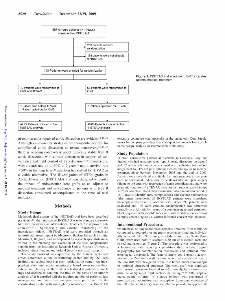
Interventional ProceduresOn the basis of diagnostic measurements obtained from multislicecomputed tomography or magnetic resonance imaging, individu-ally selected TALENT stent grafts (Medtronic, Inc, Santa Rosa,Calif) were used both to scaffold �20 cm of dissected aorta andto seal major entries (Figure 2). The procedure was performed ina laboratory with imaging capabilities that included digitalangiography for catheterization maneuvers and optional trans-esophageal ultrasound. The femoral artery could usually accom-modate the 24F stent-graft system, which was advanced over a260-cm stiff wire navigated in the true lumen under fluoroscopicor optional ultrasound guidance. The stent graft was deployedwith systolic pressure lowered to �50 mm Hg by sodium nitro-prusside or by rapid right ventricular pacing.24,25 After deploy-ment, gentle inflation of a latex balloon was performed ifproximal wall apposition was incomplete. Intentional coverage ofthe left subclavian artery was accepted to provide an appropriate
Figure 1. INSTEAD trial enrollment. OMT indicatesoptimal medical treatment.
2520 Circulation December 22/29, 2009
by guest on April 19, 2018
http://circ.ahajournals.org/D
ownloaded from
landing zone and avoid endoleak; prophylactic surgical revascu-larization of the left subclavian artery was left to the discretion ofthe investigator. Magnetic resonance angiography was used toidentify potential supra-aortic variants (eg, presence of a lusorianartery, incomplete circle of Willis, or dominant left vertebralartery) in case of intentional occlusion of the left subclavianartery.17,26
Clinical Outcome and End PointsClinical outcome was adjudicated by an independent committeewith expert members; events were classified in approximation tothe reporting standards of the Ad Hoc Committee for Standard-ized Reporting Practices in Vascular Surgery/International Soci-ety for Cardiovascular Surgery.27 Three classes of complications(systemic, local nonvascular, and local vascular) and 3 grades ofseverity (mild, moderate, and severe) were used; mild complica-tions were not considered for the present analysis.
An outcomes adjudication committee that consisted of a cardiacsurgeon, 2 vascular surgeons, and 2 cardiac interventionalists as-sessed each complication independently in blinded fashion; potentialdisagreements were to be resolved by consensus. The primary endpoint was all-cause death at 2 years; secondary end points wereaorta-related death, a composite end point of progressive aorticpathology (including crossover/conversion or additional endovascu-lar or open surgery for rupture, expansion, or malperfusion), andmorphological evidence of aortic remodeling. With half the requirednumber of patients enrolled at the interim analysis, the committeedecided to continue the trial, although the incidence of death and(moderate and severe) complications was monitored continuously tosafeguard against divergent outcomes.28
Assessment of Aortic RemodelingWith serial tomographic imaging at 3 months and at 1 and 2 years bycomputed tomography or magnetic resonance, all patients underwentevaluation for false-lumen thrombosis and recording of true- andfalse-lumen diameter at defined transversal levels: Levels A and Breflect nondissected aorta, whereas levels C and D reflect dissectedproximal and distal descending thoracic aortic segments (Figure 2).Furthermore, individual maximum diameter was documented.
Statistical AnalysisConsidering the primary end point as a binary outcome rather thanusing a time-to-event calculation, we projected that 20% of patientsin the medical group would have a primary end point event within 2years, with an expected reduction from 20% to 3% to 5% in thestent-graft group. On the assumption of equal allocation in bothgroups, a sample size of 140 patients was required for 80% power todetect a difference with a 2-sided �-error of 0.05. Sample size wasdetermined with the study planning software nQuery Advisor 7.0(Statcon, Witzenhausen, Germany).
Patients were classified according to randomized allocation forall analyses; data were processed with the SPSS/PC softwarepackage version 15.0 (SPSS, Munich, Germany). Means (�SD)and medians and ranges were used to describe continuousvariables; absolute numbers and percentage frequencies wereused for categorical factors. For continuous variables, differencesbetween groups were evaluated by use of a 2-sample t test ornonparametric Mann–Whitney U test depending on the distribu-tion of variables. Categorical variables were compared by theFisher exact test or �2 test. Longitudinal data within groups werecompared by standard general linear model repeated-measuresANOVA. Time-to-event curves were calculated by the Kaplan–Meier method and compared by log-rank test on an intention-to-treat basis. Cox proportional hazards regression models were usedto estimate hazard ratios and 95% confidence intervals. All testswere 2 tailed, and P�0.05 was considered statistically significant.
ResultsPatient Characteristics and Treatment AssignmentBetween November 2003 and November 2005, of 597screened patients, 140 who met the inclusion criteria wererandomly assigned to elective TEVAR in addition to optimalmedical therapy or to optimal medical treatment alone (Fig-ure 1). Two patients failed to undergo stent-graft placementafter randomization because of declined consent in 1 andsudden death in another; 2 patients eventually declinedmedical treatment and opted for early stent-graft placement
Figure 2. Endovascular stentgraft in type B dissection. Car-toon demonstrating the typicalfeatures of type B dissectionwith flow in both the true andthe expanded false lumenresulting from a major proximalentry tear (left); planes A to Dwere followed up longitudinallyin every patient. A stent graftwas placed to scaffold the dis-sected aorta and to seal theentry to the false lumen, result-ing in reconstruction of thetrue lumen with subsequentfalse-lumen thrombosis (right).Levels were defined as (A) atthe sinotubular junction, (B) atthe center of the arch betweentruncus brachiocephalicus andleft common carotid artery, (C)at the level of the maximumaortic diameter, and (D) at thehiatus.
Nienaber et al Endovascular Treatment of Aortic Dissection 2521
by guest on April 19, 2018
http://circ.ahajournals.org/D
ownloaded from
although randomized differently. Overall, 140 patients werefollowed up in both groups, with 72 patients in the endovas-cular treatment arm and 68 in the medical treatment arm on anintention-to-treat basis; all patients underwent completeprotocol-guided follow-up.
Baseline and demographic characteristics, comorbidityprofiles and risk factors, distribution of American Society ofAnesthesiologists classification, and dissection morphologywere evenly distributed. Moreover, the time interval betweenonset of dissection and randomization was identical betweengroups, with a median of 45 and 39 days, respectively, whichreflects the early phase of chronic disease (Table 1). Themedian interval between randomization and stent-graft place-ment was 12 days (range 1 to 29 days); procedural details andhospital stay are listed in Table 2.
TEVAR was completed successfully in 70 patients withno intraprocedural conversion to open surgery; there wereno complications related to general anesthesia or ventila-tion. One stent graft was inserted in 58 patients (82.9%), 2
grafts in 8 (11.4%), and 3 grafts in 4 (5.7%). Intentionalocclusion of the left subclavian artery without prior revas-cularization was documented in 17 cases (24.3%) with noneurological sequelae or need for revascularization. In 3cases, calcification at the level of the femoral arteriesrequired retroperitoneal access to the common iliac artery,with patch repair in 1 case. Although the majority ofpatients (74.3%) spent �24 hours under intensive care,median hospitalization in the TEVAR group was 8 days,which was required for imaging logistics and antihyper-tensive medication adjustment. Periprocedural outcomes(30 days) included 3 vascular injuries that required ancil-lary procedures and 3 cases of neurological complications,with 1 paraplegia, 1 transient paraparesis in the presence ofextensive coverage (3 stent grafts) with left subclavianartery occlusion (without prior revascularization), and 1stroke (Table 3); normalized arterial pressure according toWorld Health Organization criteria (�120/80 mm Hg) wasdocumented in all patients 1 month after randomizationand at follow-up visits in both groups.
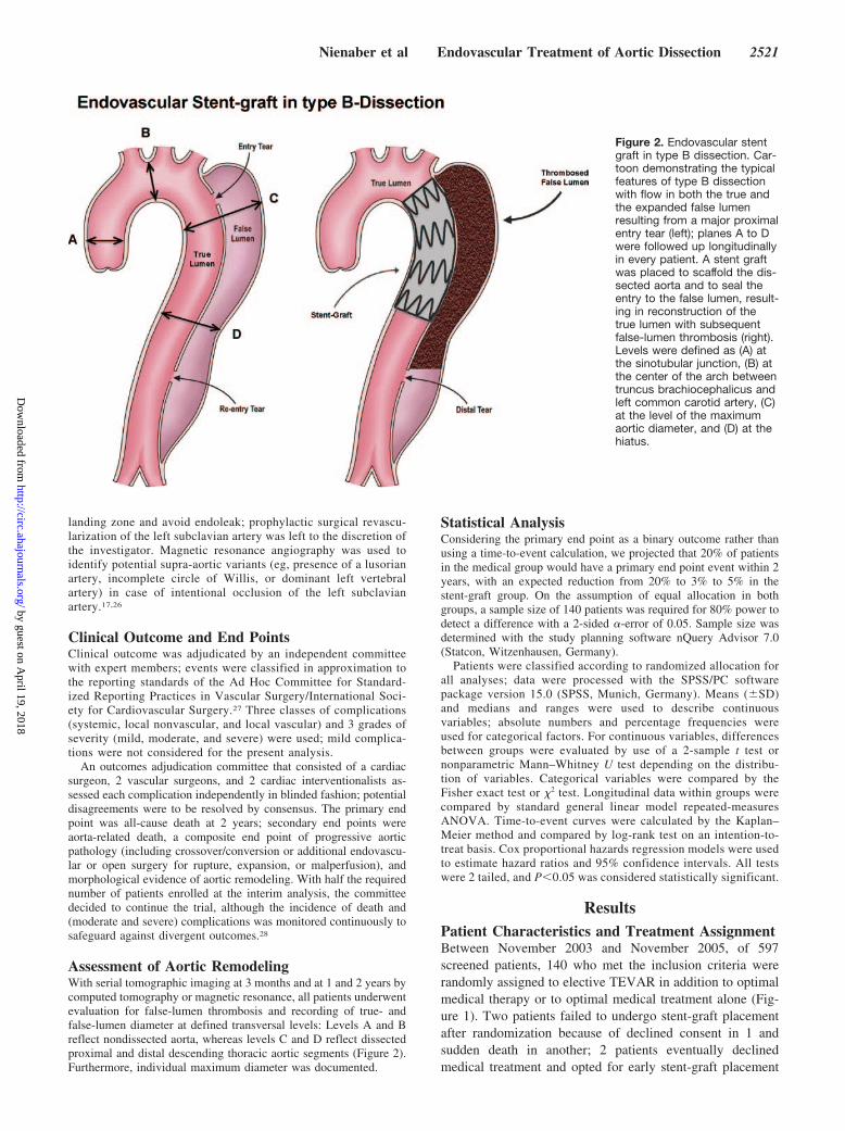
Primary OutcomeFigure 3A shows cumulative all-cause survival rate (esti-mated with the use of Kaplan-Meier curves) in both groups.Comparison between curves revealed no significant differ-ence (log-rank test P�0.15). Survival probability at 2 yearswas 88.9�3.7% with TEVAR and 95.6�2.5% with medical
Table 1. Baseline Characteristics of Patients
Characteristics OMT (n�68)OMT�TEVAR
(n�72)
Age, y, mean�SD 60.1�11.7 60.3�10.7
Male sex, n (%) 56 (82.3) 62 (86.1)
Atherosclerosis/hypertension, n (%) 56 (82.3) 61 (84.7)
Marfan syndrome, n (%) 0 (0) 2 (2.8)
Hypertension only, n (%) 11 (16.2) 7 (9.7)
Unknown, n (%) 2 (2.9) 2 (2.8)
Diabetes mellitus, n (%) 6 (8.8) 5 (6.9)
Active smoking, n (%) 17 (25.0) 14 (19.4)
Pulmonary disease, n (%) 9 (13.2) 7 (9.7)
Body mass index, kg/m2,mean�SD
27.7�5.5 26.7�4.4
New York Heart Associationclassification, n (%)
I 51 (75.0) 55 (76.4)
II 13 (19.1) 16 (22.2)
III 4 (5.9) 1 (1.4)
American Society of Anesthesiologyclass, n (%)
I (healthy status) 20 (29.4) 23 (31.9)
II (mild systemic disease) 41 (60.3) 34 (47.2)
III (severe systemic disease) 7 (10.3) 15 (20.8)
Maximum diameter of dissectedaorta, mm, mean�SD
43.5�9.3 44.2�9.5
Dissection morphology, n (%)
Confined to descendingthoracic aorta
5 (7.4) 8 (11.1)
Thoracoabdominal extension 63 (92.6) 64 (88.9)
False lumen, n (%)
Perfused 45 (66.2) 46 (63.9)
Perfused with partial thrombosis 23 (33.8) 26 (36.1)
Days from dissection to randomization,median (range)
45 (20–252) 39 (18–252)
Table 2. Procedural Characteristics in TEVAR Group
Days from randomization to stent-graft, median (range) 12 (1–29)
General anesthesia, n (%) 68 (97.1)
Duration of procedure, min, median (range) 108 (20–200)
Intraprocedural death, n (%) 0 (� � �)
Procedural success, n (%) 67 (95.7)
Stent grafts per patient, median (range) 1.34 (1–3)
Femoral access, n (%) 66 (94.3)
Occlusion of left subclavian artery, n (%) 17 (24.3)
Carotid-subclavian bypass, n (%) 2 (2.9)
Access-vessel patch repair, n (%) 1 (1.4)
Hospital stay, d, median (range) 8 (5–29)
Intensive care unit stay, h, median (range) 23 (12–128)
Table 3. Periprocedural Outcomes After TEVAR (30 Days)
Deaths, n (%) 2 (2.8)
Periprocedural events, n (%)
Retrograde type A dissection 1 (1.5)
Rupture of iliac access vessel 1 (1.5)
Conversion to open surgery 0 (� � �)
Ancillary procedures/injuries 3 (4.5)
Stenting of iliac artery 1 (1.5)
Aortic stent-graft extension 1 (1.5)
Aortic bare-stent extension 1 (1.5)
Periprocedural neurological events, n (%)
Paraplegia/paraparesis 2 (2.9)
Major stroke 1 (1.5)
2522 Circulation December 22/29, 2009
by guest on April 19, 2018
http://circ.ahajournals.org/D
ownloaded from

treatment. Unadjusted Cox regression analysis for all-causesurvival revealed a hazard ratio of 0.34 with a 95% confi-dence interval from 0.068 to 1.670 (P�0.183); with 11fatalities, the 2-year death rates did not achieve the assump-tion of 28 events to achieve the desired statistical power.
Secondary End Points and Adverse EventsFigure 3B depicts the estimated cumulative freedom fromaorta-related death (log-rank test P�0.44). At 2 years, thesurvival probabilities were 94.4�2.7% with TEVAR and97.0�2.0% with medical treatment alone. Analysis ofindividual fatalities revealed that 4 patients had beenincluded despite protocol violation; with acute malperfu-sion in 1 case after dissection-related renal dysfunction ondialysis, 2 cases with acute leg ischemia, and 1 case withongoing pain and extra-aortic blood collection since onset
of dissection, none of these 4 patients should have enteredthe study. A detailed list of case fatalities is summarized inTable 4.
Figure 3C illustrates the Kaplan-Meier analysis of acombined end point of aorta-related death, crossover/conversion for expansion, and ancillary procedures, withno differences between groups (log-rank test P�0.65). At2 years, cumulative freedom from the combined end pointwas 72.5�5.5% with optimal medical treatment and77.2�5.0 with additional stent grafting.
Table 5 summarizes all events including overall andaorta-related deaths within 2 years of randomization. Aorticexpansion �60 mm occurred more frequently with medicaltreatment and was followed by crossover to TEVAR in 16.2%and by conversion to open surgery in 4.4% of patients; 1patient crossed over because of additional late malperfusion
Figure 3. A, Kaplan–Meier estimates of 2-year overall cumulative survival rate in both groups; P�0.15 by log-rank test. B, Kaplan–Meier estimates of 2-year aorta-related survival rate in both groups; P�0.44 by log-rank test. C, Kaplan–Meier estimates of 2-yearcumulative freedom from combined end point of progression and adverse events. The combined end point consisted of related death,conversion, and ancillary interventions (including a second stent-graft procedure, access revision, and peripheral interventions). Endo-vascular interventions (conversion to TEVAR in the control group or additional TEVAR in the stent-graft group) are an integral part ofthe combined end point of progressive aortic pathology. There was no difference between groups (log-rank test P�0.65). Pat. at riskindicates patients at risk; OMT, optimal medical therapy.
Nienaber et al Endovascular Treatment of Aortic Dissection 2523
by guest on April 19, 2018
http://circ.ahajournals.org/D
ownloaded from

syndrome. There were 2 cases of ischemic spinal injury afterstent grafting and 1 with medical therapy (P�0.90); the lattercase developed true-lumen collapse with malperfusion tovarious pairs of intercostal arteries 11 months after dissectionfollowed by conversion to late stent-graft placement. In thestent-graft group, all aorta-related deaths had occurred within2 months; an additional stent graft for false-lumen flow anddiameter expansion was implanted in 6 cases, whereas 3patients were converted to open surgery for expansion,retrograde type A dissection, or malperfusion. Interestingly,
all crossover cases from medical treatment to TEVAR haduneventful outcomes, no deaths, and documented aorticremodeling.
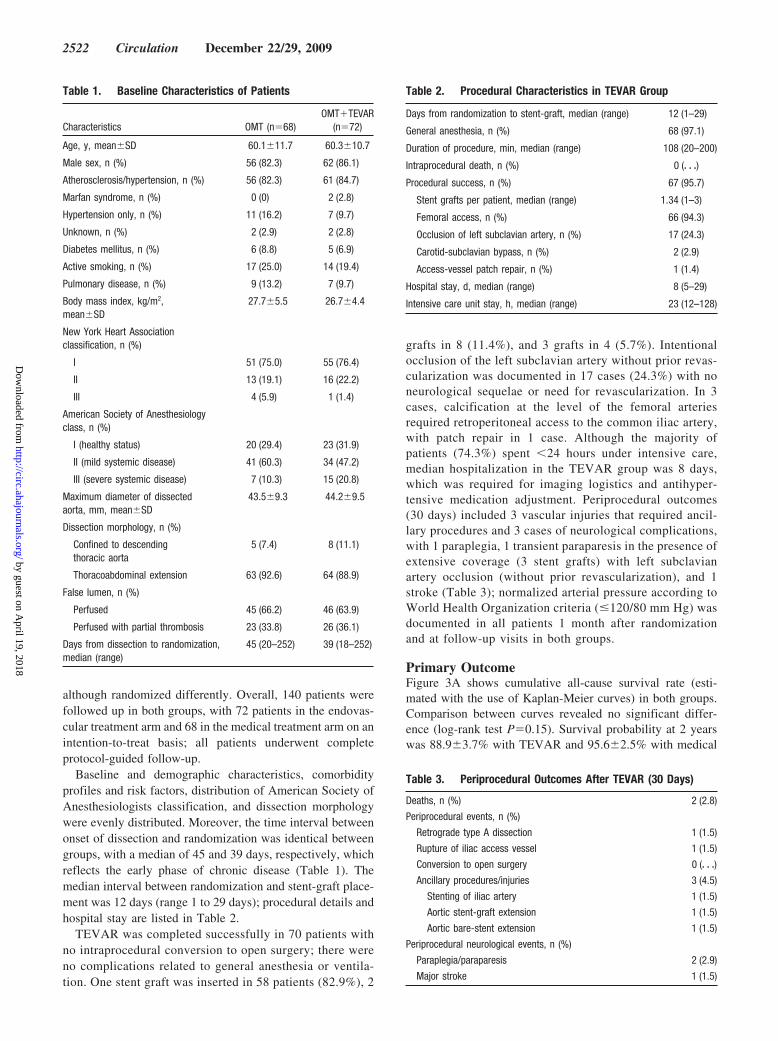
Clinical Follow-Up and Aortic RemodelingTable 6 summarizes morphological evolution over time inboth groups and evidence of aortic remodeling. Althoughbaseline dimensional variables were similar in nondis-sected (A and B) and dissected (C and D) segments of theaorta, placement of a stent graft was followed by expan-sion of the thoracic true lumen from 17.4�10.7 to25.7�6.7 mm at 3 months, with further expansion to27.0�7.3 mm at 2 years (P�0.001) at level D; similarchanges were documented at level C. Simultaneously,maximal false-lumen diameter shrank from 26.9�10.9 to17.2�13.7 mm at 3 months after stent grafting (P�0.001)and to 13.8�14.9 mm at 2 years (P�0.001) at level D,with similar changes at level C. Moreover, the process offalse-lumen thrombosis in the thoracic aorta was enhancedafter stent-graft placement, with 91.3% complete false-lumen thrombosis and morphological evidence of aorticremodeling (P�0.001), as demonstrated in Figure 4. Con-versely, medical treatment alone failed to demonstratesignificant true-lumen recovery or false-lumen shrinkageand revealed false-lumen thrombus formation only in aminority of patients.
DiscussionThe INSTEAD trial, as the first randomized comparisonbetween elective endovascular surgery and best medical
Table 4. Case Fatalities After Randomization
Interval, d
PatientAge,
y Sex GroupDissection to
RandomizationRandomization to
Stent Graft Thoracic False-Lumen Status Related Death Detailed Information
1 65 M OMT 244 N/A Minimal partial thrombosis Yes Delayed rupture of enlargingfalse lumen
2 73 M TEVAR 71 1 Complete thoracic thrombosis Yes* Postprocedural rupture ofaccess vessel
3 53 M TEVAR 30 29 Complete thoracic thrombosis Yes* Abdominal redissection withintestinal malperfusion
4 66 F TEVAR 15 1 Complete thoracic thrombosis Yes* Postprocedural type Adissection with tamponade
5 68 M OMT 73 N/A Minimal partial thrombosis Yes Rupture of thoracic aorta
6 56 M TEVAR 53 40 Entry closed, partialthrombosis
Yes* Rupture of thoracic aorta
7 61 M TEVAR 293 22 Type I endoleak, partialthrombosis
No Fatal hemorrhagic stroke insevere hypertension
8 74 M TEVAR 112 12 Complete thoracic thrombosis No Sudden cardiac death fromventricular fibrillation
9 63 M OMT 15 N/A Complete thoracic thrombosis No Metastasized renal cancer
10 70 M TEVAR 17 Died 2 days afterrandomization but
before TEVAR
No false-lumenthrombosis
No Pulmonary embolism
11 77 M OMT 90 Died 10 days afterrandomization;
opted out for stentgraft and diedbefore TEVAR
No false-lumen thrombosis No Myocardial infarction
M indicates male; F, female; OMT, optimal medical treatment; and N/A, not available.*Patients with violation of inclusion protocol.
Table 5. Events Within 2 Years of Randomization
OMT OMT�TEVAR P
Overall deaths, n (%) 3 (4.4) 8 (11.1) 0.20
Aorta-related deaths, n (%) 2 (2.9) 4 (5.6) 0.68
Secondary interventions, n (%) 15 (22.1) 13 (18.1) 0.74
Crossover 11 (16.2) N/A N/A
Conversion to surgery 3 (4.4) 3 (4.2) 1.00
Stent-graft extension N/A 6 (8.3) N/A
Aortic bare-stent extension N/A 1 (1.4) N/A
PTA/access-vessel repair 1 (1.5) 3 (4.2) 0.62
Adverse events, n (%)
Persistent paraplegia/paraparesis
1 (1.4) 2 (2.8) 0.90
Major stroke 0 (� � �) 2 (2.8) 0.53
OMT indicates optimal medical treatment; N/A, not applicable; and PTA,percutaneous transluminal angioplasty.
2524 Circulation December 22/29, 2009
by guest on April 19, 2018
http://circ.ahajournals.org/D
ownloaded from

treatment, justifies medical management for uncomplicatedtype B aortic dissection and corroborates excellent survivalrate with tight blood pressure control and close surveil-lance.20,29,30 Moreover, for patients who fail to respond tomedical management and with progressive expansion or latemalperfusion, deferred endovascular therapy is feasible andsafe.
Although the concept of endovascular stent grafting hasbeen embraced to replace open surgery for managingcomplications of type B dissection (even without anyrandomized data),18,31,32 the revelations of INSTEAD donot challenge the perception of an endovascular alternativeto open surgery. Instead, the potential of endografting toremodel a dissected aorta33 and to successfully deal withlate expansion and distal malperfusion has been con-firmed.32 Although there were 3 late conversions to opensurgery in both groups, there was no case of induced distal
malperfusion after placement of a stent graft. Spinal injuryoccurred in 2 cases after stent grafting and in 1 casespontaneously. Nevertheless, endovascular therapy in clin-ically stable, low-risk patients failed to improve 2-yearsurvival rate and was associated with spinal injury in 2.8%,as expected from previous observations.14,34 –36 Thus, theperception of prophylactic scaffolding as a better alterna-tive to tailored medical management was diminished givenongoing complications such as distal expansion and laterupture regardless of therapy. In light of documentedsuccessful medical management with monitored pharma-cotherapy, TEVAR appeared appropriate in cases ofemerging complications. Interestingly, all crossover pa-tients survived elective TEVAR for expansion and had anuneventful follow-up with remodeling despite rather lateintervention.37
Thus, INSTEAD supports the notion of a complication-specific approach instead of endovascular surgery for all typeB dissections; patients who survive type B dissection andwho are subjected to best medical management with surveil-lance show an excellent 2-year survival rate and acceleratedprogression in only a few cases.28,30 Moreover, with surveil-lance, progression was identified by follow-up imaging andwas used to qualify patients for timely crossover to TEVARor to ancillary procedures in the primary endovascular group.Finally, anatomic remodeling of the dissected aorta was notonly feasible in the initial phase of dissections but also incrossover patients after false-lumen expansion.
INSTEAD was initiated with an assumption of a latedeath rate of up to 30% in type B dissections,4,19,30 which,however, was not confirmed with current modern medicalmanagement and surveillance. Although the concept ofprophylactic scaffolding to initiate remodeling is intrigu-ing and intuitively promising, a follow-up period longerthan 2 years in larger cohorts is probably warranted toreveal differences. INSTEAD was designed to exert a levelof power that it eventually failed to reach because theprojected absolute difference in mortality rate of 15% froman estimated 20% first-year mortality rate was not seen.Nevertheless, the observed mortality rate in both themedical and endovascular groups was considerably lowerthan expected.
Thus, INSTEAD calls for a reappraisal of standardizedcare with blood pressure control and surveillance forpatients with distal dissection regardless of treatment.Tailored medical management (in uncomplicated type Bdissection) avoids procedure-related adverse events, butpatients should be followed up for late complications. In es-sence, given the outcome of modern medical management,INSTEAD was underpowered, a characteristic, however, that isgermane to controlled randomized trials based on historicalmortality data. Finally, corroborating previous findings, IN-STEAD confirmed that stent grafts enhance false-lumen throm-bosis and aortic remodeling in 90% of cases.10,33
Study LimitationsINSTEAD focused on uncomplicated dissections likely todevelop late complications; thus, potential benefits ofTEVAR may emerge in some patients beyond the 2-year
Table 6. Morphological Characteristics Over Time(Remodeling)
CharacteristicsOMT
(n�68)OMT�TEVAR
(n�72) P
Baseline type B dissection
Maximum aortic diameter 43.6�9.2* 44.1�9.6 0.65
True-lumen diameter at level C 20.3�9.3 19.4�8.0* 0.55
False-lumen diameter at level C 27.7�11.6 29.3�12.4* 0.65
True-lumen diameter at level D 17.3�8.7 17.4�10.7* 0.91
False-lumen diameter at level D 24.0�10.4 26.9�10.9* 0.13
3-Month follow-up
Maximum aortic diameter 46.2�11.1 44.7�8.3 0.75
True-lumen diameter at level C 21.9�8.8 30.6�6.0 �0.001
False-lumen diameter at level C 29.4�15.0 14.0�14.2† �0.001
True-lumen diameter at level D 17.1�8.8 25.7�6.7 �0.001
False-lumen diameter at level D 27.4�12.9 17.2�13.7† �0.001
1-Year follow-up
Maximum aortic diameter 45.5�7.9 44.7�11.9 0.37
True-lumen diameter at level C 23.9�9.9 31.8�5.9 �0.001
False-lumen diameter at level C 24.7�15.5 13.1�18.9 �0.001
True-lumen diameter at level D 19.3�9.0 27.1�7.0 �0.001
False-lumen diameter at level D 24.8�11.5 14.6�14.7 �0.001
2-Year follow-up
Maximum aortic diameter 48.3�13.1 43.8�12.5 0.31
True-lumen diameter at level C 22.7�10.9 32.3�6.4 �0.001
False-lumen diameter at level C 26.8�9.4 12.5�16.7 �0.001
True-lumen diameter at level D 18.3�7.8 27.0�7.3 �0.001
False-lumen diameter at level D 26.9�10.3 13.8�14.9 �0.001
False-lumen thrombosis at 2 y, n (%)‡
Complete 13 (19.4) 63 (91.3) �0.001
Incomplete 6 (9.1) 6 (8.7) 0.79
Values are mean�SD.*P�0.001 vs 3 months, 1 year, and 2 years.†P�0.001 vs 1 and 2 years (repeated-measures analysis).‡At the level of descending thoracic aorta.
Nienaber et al Endovascular Treatment of Aortic Dissection 2525
by guest on April 19, 2018
http://circ.ahajournals.org/D
ownloaded from

window of INSTEAD, whereas all patients were exposedto the risk of TEVAR. Given that high-risk patients withearly complications did not qualify for INSTEAD (butwere readily treated with TEVAR), stent grafting inINSTEAD was of a prophylactic nature. Chronic dissec-tion ranging from 2 to 52 weeks of onset may includepatients with a heterogeneous risk; nevertheless, the trialturned out to be underpowered on the basis of previousoutcome assumptions. Both advancing TEVAR technologyand growing operator skills are likely to lead to anavoidance of procedure-related adverse events, thus low-ering the threshold to use TEVAR in asymptomatic pa-tients at risk despite best medical management.38 Given thecurrent lack of reliable prognostic tools, new risk condi-tions such as partial false-lumen thrombosis39 or criticalfalse-lumen diameter40 may become important for identi-fication of patients for prophylactic TEVAR.
OutlookThe current picture of clinical care is transient, and ourcurrent views of best management will soon be outdated;both may be supplanted by growing insight into diseaseprogression in patients with “asymptomatic” or “uncom-plicated” dissection. New interventional platforms andimproved devices will emerge to address current stent-graft inadequacies.41 Future trials should focus on definedsubgroups to test the prophylactic use of refined anddedicated endografts.
AcknowledgmentsWe thank our referring physicians for patients to be considered ascandidates for the INSTEAD trial, as well as all nursing staff inparticipating centers for their continuous support and diligent patientcare. We are indebted to Gitta Knoop for her support in preparingthis manuscript.
Sources of FundingThis study was supported by an unrestricted research grant fromMedtronic Bakken Research Institute, Maastricht, Belgium; supple-mental support was received from the Institutional Research Unit atRostock University (minor funding for statistical support).
DisclosuresDr Nienaber reports receiving lecture and consulting fees orhonoraria from Boston Scientific, Inc, Cook, Inc, and Medtronic,Inc, and serving as an expert witness in the John Ritter case; DrRousseau, lecture fees and honoraria from Gore, Inc, and Medtronic,Inc; Dr Eggebrecht, lecture fees and honoraria from Bolton, Inc, andMedtronic, Inc; Dr Kische, honoraria from Medtronic Inc; Dr Fattori,lecture fees and honoraria from Medtronic, Inc; Dr Labrousse,lecture fees and honoraria from Gore, Inc; and Dr Ince, honorariafrom Medtronic, Inc. The remaining authors report no conflicts.
References1. Dake MD, Kato N, Mitchell RS, Semba CP, Razavi MK, Shimono T,
Hirano T, Takeda K, Yada I, Miller DC. Endovascular stent-graftplacement for the treatment of acute aortic dissection. N Engl J Med.1999;340:1546–1552.
2. Nienaber CA, Fattori R, Lund G, Dieckmann C, Wolf W, von Kodo-litsch Y, Nicolas V, Pierangeli A. Nonsurgical reconstruction of
Figure 4. Gadolinium-enhanced sagittal magnetic resonance images of type B dissection before and after endovascular repair. Theimage at baseline shows a patient at the time of randomization to stent-graft placement; the center image demonstrates the aorta 3months after TEVAR; and the 12-month scan shows remodeling with complete resolution of false lumen.
2526 Circulation December 22/29, 2009
by guest on April 19, 2018
http://circ.ahajournals.org/D
ownloaded from
thoracic aortic dissection by stent-graft placement. N Engl J Med.1999;340:1539 –1545.
3. Mukherjee D, Eagle KA. Aortic dissection: an update. Curr ProblCardiol. 2005;30:287–325.
4. Tsai TT, Fattori R, Trimarchi S, Isselbacher E, Myrmel T, EvangelistaA, Hutchison S, Sechtem U, Cooper JV, Smith DE, Pape L, FroehlichJ, Raghupathy A, Januzzi JL, Eagle KA, Nienaber CA; InternationalRegistry of Acute Aortic Dissection. Long-term survival in patientspresenting with type B acute aortic dissection: insights from theInternational Registry of Acute Aortic Dissection. Circulation. 2006;114:2226 –2231.
5. Svensson LG, Kouchoukos NT, Miller DC, Bavaria JE, Coselli JS, CuriMA, Eggebrecht H, Elefteriades JA, Erbel R, Gleason TG, Lytle BW,Mitchell RS, Nienaber CA, Roselli EE, Safi HJ, Shemin RJ, Sicard GA,Sundt TM III, Szeto WY, Wheatley GH III; Society of Thoracic SurgeonsEndovascular Surgery Task Force. Expert consensus document on thetreatment of descending thoracic aortic disease using endovascular stent-grafts. Ann Thorac Surg. 2008;85(suppl):S1–S41.
6. Erbel R, Alfonso F, Boileau C, Dirsch O, Eber B, Haverich A, RakowskiH, Struyven J, Radegran K, Sechtem U, Taylor J, Zollikofer C, KleinWW, Mulder B, Providencia LA; Task Force on Aortic Dissection,European Society of Cardiology. Diagnosis and management of aorticdissections. Eur Heart J. 2001;22:1642–1681.
7. Erbel R, Oelert H, Meyer J, Puth M, Mohr-Katoly S, Hausmann D,Daniel W, Maffei S, Caruso A, Covino FE. Effect of medical andsurgical therapy on aortic dissection evaluated by transesophagealechocardiography: implications for prognosis and therapy: theEuropean Cooperative Study Group on Echocardiography. Circu-lation. 1993;87:1604 –1615.
8. Kato N, Shimono T, Hirano T, Suzuki T, Ishida M, Sakuma H, Yada I,Takeda K. Midterm results of stent-graft repair of acute and chronic aorticdissection with descending tear: the complication-specific approach.J Thorac Cardiovasc Surg. 2002;124:306–312.
9. Akutsu K, Nejima J, Kiuchi K, Sasaki K, Ochi M, Tanaka K, TakanoT. Effects of the patent false lumen on the long-term outcome of typeB acute aortic dissection. Eur J Cardiothorac Surg. 2004;26:359 –366.
10. Schoder M, Czerny M, Cejna M, Rand T, Stadler A, Sodeck GH, GottardiR, Loewe C, Lammer J. Endovascular repair of acute type B aorticdissection: long-term follow-up of true and false lumen diameter changes.Ann Thorac Surg. 2007;83:1059–1066.
11. Nienaber CA, Zannetti S, Barbieri B, Kische S, Schareck W, Rehders TC.Investigation of STEnt grafts in patients with type B aortic Dissection:design of the INSTEAD trial: a prospective, multicenter European ran-domized trial. Am Heart J. 2005;149:592–599.
12. Wheatley GH III, Gurbuz AT, Rodriguez-Lopez JA, Ramaiah VG, OlsenD, Williams J, Diethrich EB. Midterm outcome in 158 consecutive GoreTAG thoracic endoprostheses: single center experience. Ann ThoracSurg. 2006;81:1570–1577.
13. Kaya A, Heijmen RH, Overtoom TT, Vos JA, Morshuis WJ, SchepensMA. Thoracic stent grafting for acute aortic pathology. Ann Thorac Surg.2006;82:560–565.
14. Eggebrecht H, Nienaber CA, Neuhäuser M, Baumgart D, Kische S,Schmermund A, Herold U, Rehders TC, Jakob HG, Erbel R. Endo-vascular stent-graft placement in aortic dissection: a meta-analysis. EurHeart J. 2006;27:489–498.
15. Nienaber CA, Ince H, Weber F, Rehders T, Petzsch M, Meinertz T,Koschyk DH. Emergency stent-graft placement in thoracic aortic dis-section and evolving rupture. J Card Surg. 2003;18:464–470.
16. Eggebrecht H, Lönn L, Herold U, Breuckmann F, Leyh R, Jakob HG,Erbel R. Endovascular stent-graft placement for complications of acutetype B aortic dissection. Curr Opin Cardiol. 2005;20:477–483.
17. Rehders TC, Petzsch M, Ince H, Kische S, Korber T, Koschyk DH,Chatterjee T, Weber F, Nienaber CA. Intentional occlusion of the leftsubclavian artery during stent-graft implantation in the thoracic aorta: riskand relevance. J Endovasc Ther. 2004;11:659–666.
18. Trimarchi S, Nienaber CA, Rampoldi V, Myrmel T, Suzuki T, BossoneE, Tolva V, Deeb MG, Upchurch GR Jr, Cooper JV, Fang J, IsselbacherEM, Sundt TM III, Eagle KA; IRAD Investigators. Role and results ofsurgery in acute type B aortic dissection: insights from the InternationalRegistry of Acute Aortic Dissection (IRAD). Circulation. 2006;114(supplI):I-357–I-364.
19. Nienaber CA, Eagle KA. Aortic dissection: new frontiers in diagnosisand management: part II: therapeutic management and follow-up.Circulation. 2003;108:772–778.
20. Tefera G, Acher CW, Hoch JR, Mell, Turnipseed WD. Effectiveness ofintensive medical treatment in type B aortic dissection: a single-centerexperience. J Vasc Surg. 2007;45:1114–1118.
21. Suzuki T, Mehta RH, Ince H, Nagai R, Sakomura Y, Weber F, SumiyoshiT, Bossone E, Trimarchi S, Cooper JV, Smith DE, Isselbacher EM, EagleKA, Nienaber CA; International Registry of Aortic Dissection. Clinicalprofiles and outcomes of acute type B aortic dissection in the current era:lessons from the International Registry of Aortic Dissection (IRAD).Circulation. 2003;108(suppl):II-312–II-317.
22. Takahashi J, Wakamatsu Y, Okude J, Kanaoka T, Sanefuji Y, Gohda T,Sasaki S, Matsui Y. Maximum aortic diameter as a simple predictor ofacute type B aortic dissection. Ann Thorac Cardiovasc Surg. 2008;14:303–310.
23. Onitsuka S, Akashi H, Tayama K, Okazaki T, Ishihara K, Hiromatsu S,Aoyagi S. Long-term outcome and prognostic predictors of medicallytreated acute type B aortic dissections. Ann Thorac Surg. 2004;78:1268–1273.
24. von Knobelsdorff G, Höppner RM, Tonner PH, Paris A, Nienaber CA,Scholz J, Schulte am Esch J. Induced arterial hypotension for interven-tional thoracic aortic stent-graft placement: impact on intracranial hae-modynamics and cognitive function. Eur J Anaesthesiol. 2003;20:134–140.
25. Nienaber CA, Kische S, Rehders TC, Schneider H, Chatterjee T, BüngerCM, Höppner R, Ince H. Rapid pacing for better placing: a comparison oftechniques for precise deployment of endograft in the thoracic aorta. JEndovasc Ther. 2007;14:506–512.
26. Fattori R. Towards a better management of descending aortic dissection:the importance of close ambulatory follow-up. Eur J Vasc EndovascSurg. 2006;32:356–537.
27. Chaikof EL, Blankensteijn JD, Harris PL, White GH, Zarins CK,Bernhard VM, Matsumura JS, May J, Veith FJ, Fillinger MF, RutherfordRB, Kent KC; Ad Hoc Committee for Standardized Reporting Practicesin Vascular Surgery of the Society for Vascular Surgery/American Asso-ciation for Vascular Surgery. Reporting standards for endovascular aorticaneurysm repair. J Vasc Surg. 2002;35:1048–1060.
28. Bolland K, Whitehead J. Formal approaches to safety monitoring ofclinical trials in life-threatening conditions. Stat Med. 2000;19:2899–2917.
29. Estrera AL, Miller CC, Goodrick J, Porat EE, Achouh PE, Dhareshwar J,Maeda R, Azizzadeh A, Safi HJ. Update in outcomes of acute type Baortic dissection. Ann Thorac Surg. 2007;83:842–845.
30. Winnerkvist A, Lockowandt U, Rasmussen E, Radegran K. A prospectivestudy of medically treated acute type B aortic dissection. Eur J VascEndovasc Surg. 2006;32:349–355.
31. Fattori R, Tsai TT, Myrmel T, Evangelista A, Cooper JV, Trimarchi S, LiJ, Lovato L, Kische S, Eagle KA, Isselbacher EM, Nienaber CA. Com-plicated acute type B dissection: is surgery still the best option? A reportfrom the International Registry of acute Aortic Dissection (IRAD). J AmColl Cardiol Intv. 2008;1:395–402.
32. Duebener LF, Lorenzen P, Richardt G, Misfeld M, Nötzold A, HartmannF, Sievers HH, Geist V. Emergency endovascular stent-grafting for life-threatening acute type B aortic dissection. Ann Thorac Surg. 2004;78:1261–1266.
33. Rodriguez JA, Olsen DM, Lucas L, Wheatley G, Ramaiah V, DiethrichEB. Aortic remodeling after endografting of thoracoabdominal aorticdissection. J Vasc Surg. 2008;47:1188–1194.
34. MacKenzie KS, LeGuillan MP, Steinmetz OK, Montreuil B. Man-agement trends and early mortality rates for acute type B dissection:a 10-year single-institution experience. Ann Vasc Surg. 2004;18:158 –166.
35. Buth J, Harris PL, Hobo R, van Eps R, Cuypers P, Duijm L, Tielbeek X.Neurological complications associated with endovascular repair ofthoracic aortic pathology: incidence and risk factors: a study from theEuropean Collaborators of Stent/Graft Techniques for Aortic AneurysmRepair (EUROSTAR) registry. J Vasc Surg. 2007;46:1103–1111.
36. Fattori R, Nienaber CA, Rousseau H, Beregi JP, Heijmen R, Gra-benwöger M, Piquet P, Lovato L, Dabbech C, Kische S, Gaxotte V,Schepens M, Ehrlich M, Bartoli JM; Talent Thoracic RetrospectiveRegistry. Results of endovascular repair of the thoracic aorta with theTalent Thoracic stent graft: the Talent Thoracic Retrospective Registry.J Thorac Cardiovasc Surg. 2006;132:332–339.
37. Akin I, Kische S, Ince H, Nienaber CA. Indication, timing and results ofendovascular treatment of type B dissection. Eur J Vasc Endovasc Surg.2009;37:289–296.
Nienaber et al Endovascular Treatment of Aortic Dissection 2527
by guest on April 19, 2018
http://circ.ahajournals.org/D
ownloaded from

38. Hagan PG, Nienaber CA, Isselbacher EM, Bruckman D, Karavite DJ,Russman PL, Evangelista A, Fattori R, Suzuki T, Oh JK, Moore AG,Malouf JF, Pape LA, Gaca C, Sechtem U, Lenferink S, Deutsch HJ,Diedrichs H, Marcos y Robles J, Llovet A, Gilon D, Das SK, ArmstrongWF, Deeb GM, Eagle KA. The International Registry of Aortic Dis-section (IRAD): new insights into an old disease. JAMA. 2000;283:897–903.
39. Tsai TT, Evangelista A, Nienaber CA, Myrmel T, Meinhardt G, CooperJV, Smith DE, Suzuki T, Fattori R, Llovet A, Froehlich J, Hutchison S,Distante A, Sundt T, Beckman J, Januzzi JL Jr, Isselbacher EM, Eagle
KA; International Registry of Acute Aortic Dissection. Partial thrombosisof the false lumen in patients with acute type B aortic dissection. N EnglJ Med. 2007;357:349–359.
40. Song JM, Kim SD, Kim JH, Kim MJ, Kang DH, Seo JB, Lim TH, LeeJW, Song MG, Song JK. Long-term predictors of descending aortaaneurysmal change in patients with aortic dissection. J Am Coll Cardiol.2007;50:799–804.
41. Nienaber CA, Kische S, Ince H. Thoracic aortic stent-graft devices:problems, failure modes and applicability. Semin Vasc Surg. 2007;20:81–89.
CLINICAL PERSPECTIVEINSTEAD, the first randomized comparison between elective endovascular stent grafting and best medical treatment,justifies medical management for uncomplicated type B aortic dissection and corroborates excellent survival rate with tightblood pressure control and close surveillance. For patients with complications such as progressive expansion or latemalperfusion who fail to respond to medical management, deferred endovascular therapy is feasible and safe. The resultsof INSTEAD do not challenge the endovascular treatment alternative to open surgery and confirm the potential ofendovascular therapy to successfully deal with late expansion and distal malperfusion. Nevertheless, primary endovasculartherapy in stable type B dissection failed to improve the 2-year survival rate and was associated with spinal injury in 2.9%of cases. Although low death and complication rates in both groups suggest a need for a reappraisal of standardized medicalmanagement with monitored blood pressure control, TEVAR is an appropriate crossover strategy in cases of emergingcomplications. Interestingly, all crossover patients survived elective TEVAR with uneventful follow-up and remodelingdespite rather late intervention. INSTEAD supports the notion of a complication-specific approach instead of TEVAR forall type B dissections; patients who survive type B dissection and are given best medical management with surveillanceshow an excellent 2-year survival rate, with progression to crossover/conversion in only 21%. Surveillance can be used toidentify patients with evidence of progression who qualify for safe crossover or conversion. Finally, INSTEAD confirmedthat stent-graft scaffolding enhances false-lumen thrombosis and aortic remodeling in type B dissection not only in theearly phase of dissections but also in the chronic phase after false-lumen expansion, a notion that may translate toprognostic benefits that could potentially be seen at longer (5-year) follow-up.
2528 Circulation December 22/29, 2009
by guest on April 19, 2018
http://circ.ahajournals.org/D
ownloaded from
for the INSTEAD TrialZipfel, Louis Labrousse and Hüseyin Ince
Tim C. Rehders, Günther Kundt, Dierk Scheinert, Martin Czerny, Tilo Kleinfeldt, Burkhart Christoph A. Nienaber, Hervé Rousseau, Holger Eggebrecht, Stephan Kische, Rossella Fattori,
STEnt Grafts in Aortic Dissection (INSTEAD) TrialRandomized Comparison of Strategies for Type B Aortic Dissection: The INvestigation of
Print ISSN: 0009-7322. Online ISSN: 1524-4539 Copyright © 2009 American Heart Association, Inc. All rights reserved.
is published by the American Heart Association, 7272 Greenville Avenue, Dallas, TX 75231Circulation doi: 10.1161/CIRCULATIONAHA.109.886408
2009;120:2519-2528; originally published online December 7, 2009;Circulation.
http://circ.ahajournals.org/content/120/25/2519World Wide Web at:
The online version of this article, along with updated information and services, is located on the
http://circ.ahajournals.org/content/suppl/2010/01/19/CIRCULATIONAHA.109.886408.DC1Data Supplement (unedited) at:
http://circ.ahajournals.org//subscriptions/
is online at: Circulation Information about subscribing to Subscriptions:
http://www.lww.com/reprints Information about reprints can be found online at: Reprints:
document. Permissions and Rights Question and Answer this process is available in the
click Request Permissions in the middle column of the Web page under Services. Further information aboutOffice. Once the online version of the published article for which permission is being requested is located,
can be obtained via RightsLink, a service of the Copyright Clearance Center, not the EditorialCirculationin Requests for permissions to reproduce figures, tables, or portions of articles originally publishedPermissions:
by guest on April 19, 2018
http://circ.ahajournals.org/D
ownloaded from
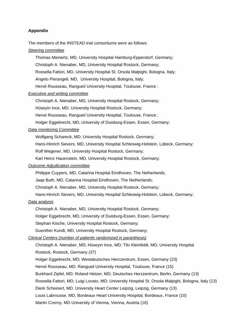
Appendix
The members of the INSTEAD trial consortiums were as follows:
Steering committee
Thomas Meinertz, MD, University Hospital Hamburg-Eppendorf, Germany;
Christoph A. Nienaber, MD, University Hospital Rostock, Germany;
Rossella Fattori, MD, University Hospital St. Orsola Malpighi, Bologna, Italy;
Angelo Pierangeli, MD, University Hospital, Bologna, Italy;
Hervé Rousseau, Rangueil University Hospital, Toulouse, France ;
Executive and writing committee
Christoph A. Nienaber, MD, University Hospital Rostock, Germany;
Hüseyin Ince, MD, University Hospital Rostock, Germany;
Hervé Rousseau, Rangueil University Hospital, Toulouse, France ;
Holger Eggebrecht, MD, University of Duisburg-Essen, Essen, Germany;
Data monitoring Committee
Wolfgang Schareck, MD, University Hospital Rostock, Germany;
Hans-Hinrich Sievers, MD, University Hospital Schleswig-Holstein, Lübeck, Germany;
Rolf Wegener, MD, University Hospital Rostock, Germany;
Karl Heinz Hauenstein, MD, University Hospital Rostock, Germany;
Outcome Adjudication committee
Philippe Cuypers, MD, Catarina Hospital Eindhoven, The Netherlands;
Jaap Buth, MD, Catarina Hospital Eindhoven, The Netherlands;
Christoph A. Nienaber, MD, University Hospital Rostock, Germany;
Hans-Hinrich Sievers, MD, University Hospital Schleswig-Holstein, Lübeck, Germany;
Data analysis
Christoph A. Nienaber, MD, University Hospital Rostock, Germany;
Holger Eggebrecht, MD, University of Duisburg-Essen, Essen, Germany;
Stephan Kische, University Hospital Rostock, Germany;
Guenther Kundt, MD, University Hospital Rostock, Germany;
Clinical Centers (number of patients randomized in paranthesis)
Christoph A. Nienaber, MD; Hüseyin Ince, MD; Tilo Kleinfeldt, MD; University Hospital
Rostock, Rostock, Germany (37)
Holger Eggebrecht, MD; Westdeutsches Herzzentrum, Essen, Germany (23)
Hervé Rousseau, MD. Rangueil University Hospital, Toulouse, France (15)
Burkhard Zipfel, MD; Roland Hetzer; MD; Deutsches Herzzentrum, Berlin, Germany (13)
Rossella Fattori, MD; Luigi Lovato, MD; University Hospital St. Orsola Malpighi, Bologna, Italy (13)
Dierk Scheinert, MD. University Heart Center Leipzig, Leipzig, Germany (13)
Louis Labrousse, MD, Bordeaux Heart University Hospital, Bordeaux, France (10)
Martin Czerny, MD University of Vienna, Vienna, Austria (16)