Embed Size (px)
Citation preview

2017.11.21.
1
Cardiovascular Physiology V.
46. The regulation of local blood flow.
47. Factors determining cardiac output, the Guyton diagram.
Ferenc Domoki, November 20 2017.
Control of circulation
Systemic control Local control
Major goal: to maintain
constant pressure gradient
(∆P) chiefly by regulating
mean arterial blood pressure
(MABP)
Major goal: to maintain
adequate blood flow to meet
locally the metabolic and
functional needs of the tissue.
Hemostasis, and immune
functions also affect local
blood flow.

2017.11.21.
2
Common local blood flow regulation responses
1. Autoregulation (constant blood flow despite changing pressure gradient)
2. Active hyperemia (increased blood flow to meet metabolic/functional demands)
3. Reactive hyperemia (increased blood flow following an interruption of flow – ischemia)
4. Decreased local blood flow when vessels are injured – hemostasis
5. Increased local blood flow in inflamed tissues –inflammatory hyperemia
LOCAL CONTROL OF BLOOD FLOW
1. Autoregulation
60 180 mmHg
Flo
w
Kidney
14020 mmHg
Flo
w
Skeletalmuscle
Flow is relatively constant
in a pressure range.
(Note that flow stops at
pressures above 0 mmHg:
Critical closing pressure).
2. Active hyperemia
Increased tissue
metabolism is associated
with enhanced blood
flow: the tissue is capable
of adjusting its own
perfusion.
3. Reactive hyperemia
Hypoxia causes vasodilation.
Flo
w
Time
Occlusion
Hyperemia
Occlusion is followed by
increased flow.

2017.11.21.
3
Control of local blood flow
• Local blood flow will depend on the
regional vascular resistance largely
determined and exclusively regulated by
the diameter of precapillary resistance
vessels.
• All local regulation will thus converge on
the contraction/relaxation of the vascular
smooth muscle in these vessels.
LOCALLY important components of arteriolar smooth muscle tone
RESTING TONE = BASAL TONE
Myogenic tone
Local vascular (endothelial) factors
Local tissue humoral factors
Systemic hormones
Sympathetic vasoconstrictor tone
+ NEUROGENIC TONE
+ PHASIC (not tonic) VASODILATORY INNERVATION CAN
INCREASE BLOOD FLOW LOCALLY
Intrinsic vascular factors!

2017.11.21.
4
Myogenic tone of arteriolar smooth muscle
• Arteriolar smooth muscles maintain spontaneous contraction – myogenic tone
• Bayliss effect: some arteriolar smooth muscles are sensitive to mechanical stretching: they respond with contraction to stretch:blood pressure↑- wall stretch↑ - vasoconstriction – arteriolar resistance ↑
• This simple response tends to stabilize flow when MABP changes
• Bayliss effect is the direct opposite of stress-relaxation observed in venous smooth muscle.
Robert W.Furchgott
Louis J.
Ignarro
Ferid
Murad
THE NOBEL PRIZE IN PHYSIOLOGY OR MEDICINE, 1998
„for their discoveries concerning
nitric oxide as a signalling
molecule in the cardiovascular
system”
Endothelial factors regulating arteriolar smooth muscle tone

2017.11.21.
5
Endothelial factors regulating arteriolar smooth muscle tone
Dilators
• Nitric oxide
• Prostacyclin
• EDHF (endothelium-derived hyperpolarizing factor)
Constrictor:
• endothelin
Depending on the species, developmental stage, and organ, the presence and the contribution of these factors to the local regulation of arteriolar tone varies greatly
Acetylcholine Acetylcholine
Vascular smooth
muscle (endothelial
cells are removed)
Contraction
Endothelial
lining is intact
Relaxation
The discovery of NO as „ENDOTHELIUM-DERIVED
RELAXING FACTOR (EDRF)

2017.11.21.
6
GqαPLC
IP3
Ca2+Calmodulin
Arg Citrulline + NONOS
GTP cGMP
Guanylyl-cyclase
PKG
Ca2+ decrease
RELAXATION
Inactivation
Phospho-diesterase
NOReceptor Viagra
Endothelial cell Smooth muscle
BIOSYNTHESIS AND ACTION OF NITRIC OXIDE
NITRIC OXIDE SYNTHASE ISOFORMS
NOS:
NOS-1 (nNOS): neural
NOS-2 (iNOS): phagocytes (cytokine-induced NOS, NO is
bactericide here)
NOS-3 (eNOS): endothelial cells
eNOS is stimulated by a number of mediators:
acetylcholine
histamine (H1)
bradykinin
VIP (vasoactive intestinal peptide)
SP (substance P)
NA (NO decreases NA-induced vasoconstriction)
shear stress

2017.11.21.
7
Prostacyclin
PGI2
Prostaglandins
PGD2 PGE2 PGF2α
Thromboxane A2
TXA2
vasodilation vasoconstriction
EICOSANOIDS
MembranePhospholipids
Arachidonic acid
PLA2
PGH2
Cyclooxygenase
COX-1, COX-2
Cortisol
Aspirin
Lipoxins
Leukotrienes
CytochromeP450 products
Inflammation,
Allergic reaction
EDHF?
?
EICOSANOIDS IN CIRCULATION
Eicosanoids are general inter- and intracellular signal molecules
that can be produced virtually by every cell.
Circulation:
1. Endothelial PGI2: continuous vasodilator tone in arteries.
It inhibits aggregation of platelets. (Receptor → cAMP)
2. PGE2: putative mediator of metabolically induced
vasodilation. (Receptor → cAMP)
3. TXA2: vasoconstrictor released from platelets.
(Receptor → IP3/Ca)
ASPIRIN can prevent coagulation/thrombosis:
COX COX
TXA2 PGI2 PGI2
cc. 4 hoursASPIRIN
COX COX
TXA2 PGI2 PGI2
2017.11.21.
8
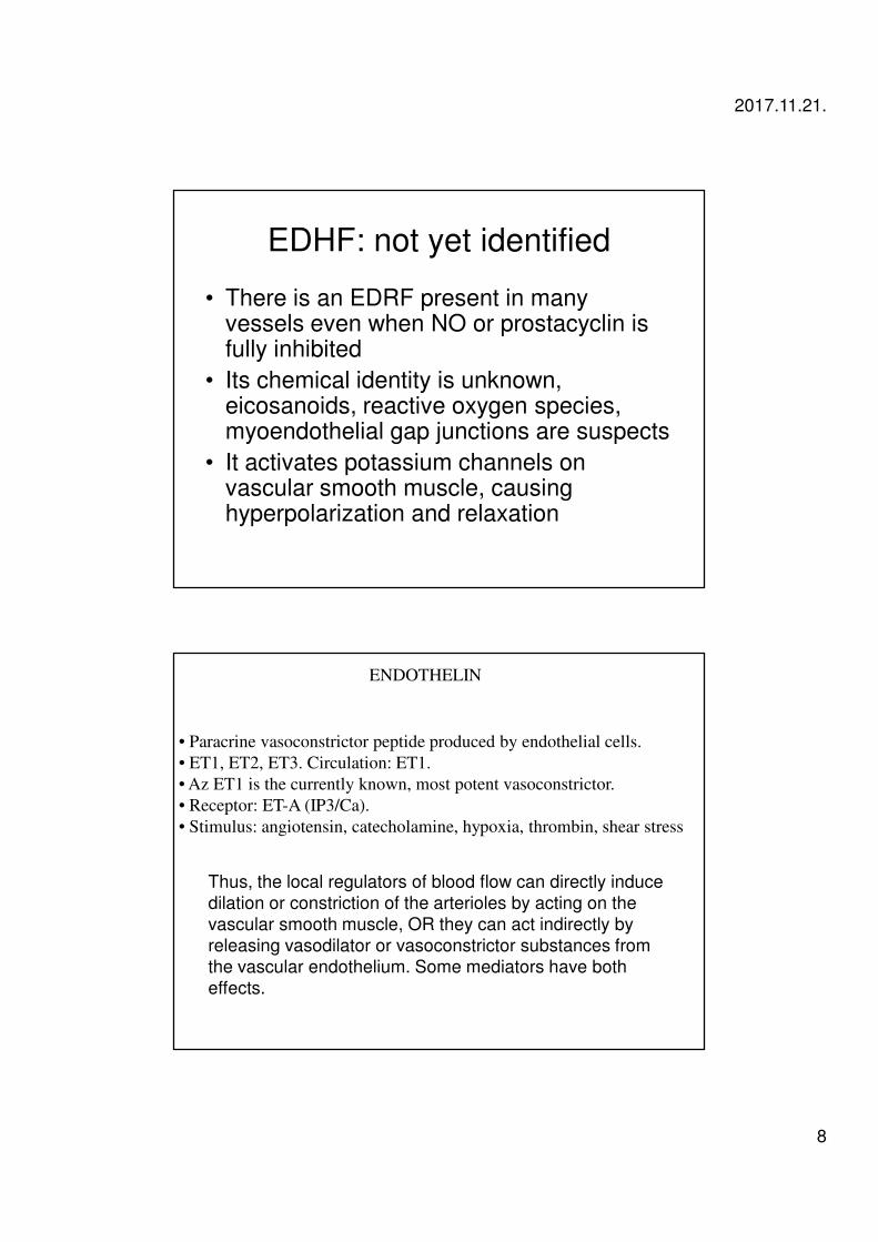
EDHF: not yet identified
• There is an EDRF present in many vessels even when NO or prostacyclin is fully inhibited
• Its chemical identity is unknown, eicosanoids, reactive oxygen species, myoendothelial gap junctions are suspects
• It activates potassium channels on vascular smooth muscle, causing hyperpolarization and relaxation
ENDOTHELIN
• Paracrine vasoconstrictor peptide produced by endothelial cells.
• ET1, ET2, ET3. Circulation: ET1.
• Az ET1 is the currently known, most potent vasoconstrictor.
• Receptor: ET-A (IP3/Ca).
• Stimulus: angiotensin, catecholamine, hypoxia, thrombin, shear stress
Thus, the local regulators of blood flow can directly induce dilation or constriction of the arterioles by acting on the
vascular smooth muscle, OR they can act indirectly by releasing vasodilator or vasoconstrictor substances from the vascular endothelium. Some mediators have both
effects.

2017.11.21.
9
LOCALLY important components of arteriolar smooth muscle tone
RESTING TONE = BASAL TONE
Myogenic tone
Local vascular (endothelial) factors
Local tissue humoral factors
+ PHASIC (not tonic) VASODILATORY INNERVATION CAN
INCREASE BLOOD FLOW LOCALLY
Intrinsic vascular factors!
Local vasodilator tissue metabolites
• Released from active cells or from cells of
energy distress
• Hypoxia, carbon dioxide, lactic acid,
acidosis, potassium ions
• NO
• PGE2
• adenosine

2017.11.21.
10
InosineS-adenozyl-
homocisteine
Coffein
A
Na+
A
ATP
A A
cAMP↑
A2
Adenosine
Vasodilation
Smooth muscle cell
Tissue cell
LOCALLY important components of arteriolar smooth muscle tone
RESTING TONE = BASAL TONE
Myogenic tone
Local vascular (endothelial) factors
Local tissue humoral factors
+ PHASIC (not tonic) VASODILATORY INNERVATION CAN
INCREASE BLOOD FLOW LOCALLY
Intrinsic vascular factors!

2017.11.21.
11
LOCALLY important VASODILATORY autonomic innervation
• Parasympathethic innervation (Ach, VIP,
NO) of salivary glands, external genitalia,
pial vessels
• Sympathetic innervation of skeletal muscle
arterioles (Ach, maybe not in humans)
• Enteral nervous system innervation of
arterioles in GI tract glands (Ach, VIP, NO)
Long-term changes in local blood flow: Angiogenesis
• Chronic elevation of metabolic activity, or hypoxia triggers angiogenesis
• HYPOXIA triggers humoral factors (vascular endothelial growth factor, fibroblast growth factor, angiopoietins)
• Steps: next slide
• Capillary density is determined by MAXIMUM flow need, not average flow need (muscle)
• Opposite phenomenon: arteriolar/ capillary rarefaction

2017.11.21.
12
The cellular steps involved in angiogenesis.
Clapp C et al. Physiol Rev 2009;89:1177-1215
©2009 by American Physiological Society
Autoregulation
• Present in every organ (except pulmonary circulation), however, most pronounced in the cerebral, coronary and renal circulation.
• Based on the parallel increase or decrease of LOCAL vascular resistance with changes in arterial blood pressure
• Mechanisms of acute autoregulation: 1. myogenic (Bayliss effect)2. metabolic (accumulation/washout of vasodilatory metabolites)3. functional (in the kidney autoregulation of glomerular filtration produces the flow autoregulation)
• Long-term autoregulation (weeks to months): new vessel growth (angiogenesis) – vessel degeneration –arteriolar and capillary rarefaction

2017.11.21.
13
Active/Reactive hyperemia
Long-term activation of the tissue leads to excess capillarization: angiogenesis!
Hemostasis-induced vasoconstriction
• Loss of functional endothelium: decreased release of NO, prostacyclin and EDHF
• Vasodilator mediators acting via endothelium will be ineffective or their effect will reverse (for instance Ach)
• Thrombin induces release of constrictor endothelin
• Platelets release vasoconstrictors: serotonin, catecholamines, thromboxane

2017.11.21.
14
Inflammation-induced vasodilation
1. Histamine
2. Bradykinin and kallidine (Lys-bradykinin)
3. PGE2
4. Neurogenic inflammation (Substance P,
Neurokinin A, Calcitonin-gene related
peptide)
INFLAMMATION:
Vasodilation
Increased permeability
Itching/pain
Tissue damage
Immune reaction
Histamine
H1-R(IP3/Ca)
H2-R(cAMP)
HISTAMINE
HISTAMINE IS THE
MOST IMPORTANT
INFLAMMATORY
MEDIATOR.

2017.11.21.
15
KININS
Tissue
kallikrein
Plasmakallikrein
Inactivepeptides
Kininase I
Kininase II =
ACE
XII XIIa Clotting
Prekallikrein
HMW kininogen
LMW kininogen
Bradykinin
Lysil-Bradykinin
Bradykinin and Lysil-bradykinin are both effective.

2017.11.21.
16
PAIN
To spinal cord
Trauma
Histamin
Local
Inflamma-
tion
Chemosensitive
C-fibres
CNS:
PAIN
Axon collaterals:
FLARE
Vasodilation +Local edemaCGRP/SP
LOCAL ERYTHEMA
EDEMA
Chemosensitive
Pain receptors
(C-fibres) release
neuropeptides:
•Calcitonin gene-
related peptide
(CGRP)
•Substance P (SP)
NEUROGENIC INFLAMMATIONTRIPLE RESPONSE: local erythema + local edema + flare
Mast cell:Histamin
TRAUMA
vasodilationvasodilation
FLARE FLARE
AXON-REFLEX
IP3/C
a2
+ ↑
NO
IP3/Ca2+ ↑
cAMP↓
cAMP↑
NO
ENDOTHELIAL CELLMUSCLE
Contraction
Relaxation
Contraction
Endothelin-1
ET-A
TXA2
TP
Serotonin
5HT2a
5HT2a
Bradykinin B1
B1
Neurokinin A NK2
NK2
Acetylcholine M1
M1
α2 Noradrenaline α1
α1
Histamine H1
H1
H2 CGRP ?? VIP ??
AdenosineA2
A1
PGE2
PGI2IPEP1-4
LOCALLY ACTING VASOACTIVE SUBSTANCES
NK1
Substance P NK1
cGMP↑

2017.11.21.
17
CARDIAC OUTPUT „regulation”
Factors determining cardiac output:
1. The heart
2. Blood volume
3. Venous compliance
4. Total peripheral resistance
Cardiac output does NOT have a homeostatic regulation as
arterial blood pressure, rather the complex interactions of the
above factors will determine the cardiac output.
Guyton model: the graphic representation of factors determining Cardiac Output
• Cardiac function curve: effect of atrial
pressure on cardiac output (Frank-
Starling)
• Systemic vascular function curve: effect of
atrial pressure on venous return (modified
by blood volume, TPR, venous
compliance)

2017.11.21.
18
Effect of atrial pressure on cardiac output (Frank-Starling mechanism)
Systemic vascular function curve: effect of atrial pressure on venous return
2017.11.21.
19
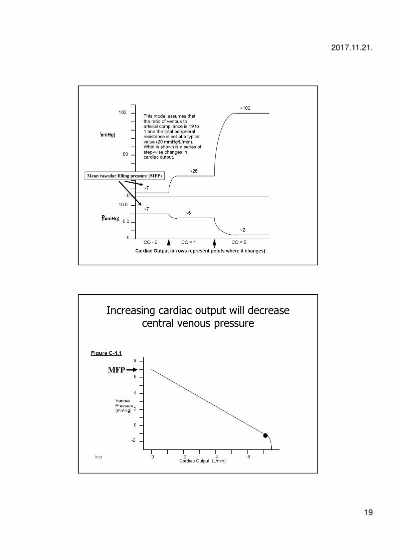
Mean vascular filling pressure (MFP)
Increasing cardiac output will decrease central venous pressure
MFP

2017.11.21.
20
Exchanging the two axes will yield the vascular function curve
MFP
Merging the two curves will show the „steady state” equilibrium cardiac output of
the system

2017.11.21.
21
Effect of sympathetic tone on the cardiac function curve
Sympathetic stimulation
Sympathetic inhibition
Effect of venous compliance/blood volume on the vascular function curve
Decreased blood volume
Increased compliance
(venodilation)
Increased blood volume
Decreased compliance
(venoconstriction)

2017.11.21.
22
Effect of total peripheral resistance on the vascular function curve
MFP
Increased TPR
Decreased TPR
Guyton diagram at work: cardiac output change during exercise
Sympathetic stimulation of the heart
Sympathetic venoconstriction
Decreased total peripheral resistance