Embed Size (px)
Citation preview

JOURNAL OF VIROLOGY, Dec. 2011, p. 13124–13132 Vol. 85, No. 240022-538X/11/$12.00 doi:10.1128/JVI.05725-11Copyright © 2011, American Society for Microbiology. All Rights Reserved.
Cardiotrophin-1 Promotes a High Survival Rate in Rabbits withLethal Fulminant Hepatitis of Viral Origin�†
Maria Jesus Tunon,1 Beatriz San Miguel,1 Irene Crespo,1 Jose Ignacio Riezu-Boj,3 Esther Larrea,3Marcelino Alvarez,2 Iranzu Gonzalez,3 Matilde Bustos,3
Javier Gonzalez-Gallego,1 and Jesus Prieto3,4*Institute of Biomedicine and CIBERehd1 and Department of Animal Health,2 University of Leon, Leon,
Spain, and Center for Applied Medical Research, CIMA, University of Navarra,3 and CIBERehd,Clinic of the University of Navarra,4 Pamplona, Spain
Received 20 July 2011/Accepted 28 September 2011
Rabbit hemorrhagic disease virus (RHDV) causes lethal fulminant hepatitis closely resembling acute liverfailure (ALF) in humans. In this study, we investigated whether cardiotrophin-1 (CT-1), a cytokine withhepatoprotective properties, could attenuate liver damage and prolong survival in virus-induced ALF. Twenty-four rabbits were infected with 2 � 104 hemagglutination units of RHDV. Twelve received five doses of CT-1(100 �g/kg) starting at 12 h postinfection (hpi) (the first three doses every 6 h and then two additional dosesat 48 and 72 hpi), while the rest received saline. The animals were analyzed for survival, serum biochemistry,and viral load. Another cohort (n � 22) was infected and treated similarly, but animals were sacrificed at 30and 36 hpi to analyze liver histology, viral load, and the expression of factors implicated in liver damage andrepair. All infected rabbits that received saline died by 60 hpi, while 67% of the CT-1-treated animals surviveduntil the end of the study. Treated animals showed improved liver function and histology, while the viral loadswere similar. In the livers of CT-1-treated rabbits we observed reduction of oxidative stress, diminishedPARP1/2 and JNK activation, and decreased inflammatory reaction, as reflected by reduced expression oftumor necrosis factor alpha, interleukin-1�, Toll-like receptor 4, VCAM-1, and MMP-9. In addition, CT-1-treated rabbits exhibited marked upregulation of TIMP-1 and increased expression of cytoprotective andproregenerative growth factors, including platelet-derived growth factor B, epidermal growth factor, platelet-derived growth factor receptor �, and c-Met. In conclusion, in a lethal form of acute viral hepatitis, CT-1increases animal survival by attenuating inflammation and activating cytoprotective mechanisms, thus rep-resenting a promising therapy for ALF of viral origin.
Acute liver failure (ALF) arises as a result of extensivehepatocellular damage that exceeds the liver’s capacity to re-generate. Depending on the interval between the onset ofjaundice and that of encephalopathy, ALF can be categorizedinto hyperacute (less than 1 week), acute (between 1 and 4weeks) and subacute (more than 4 weeks). Etiologic factorsinclude viral infections, drugs, biological toxins, metabolic dis-orders, and ischemia, but it is not unusual for this syndrome toarise without any known causative agent (22).
ALF is one of the most challenging human conditions re-quiring critical care. Therapy is merely supportive and orientedto the correction of complications. Survival is poor and livertransplantation is the only definitive treatment for patientswith severe ALF. However, the indication for liver transplan-tation relies on prognostic assessment, and this lacks accuracy.In transplanted patients mortality is about 10%, but ca. 30% ofpatients with ALF die without having access to transplantation(24). Thus, novel medical treatments for this condition are
urgently needed. With the exception of N-acetylcysteine, whichcan prevent glutathione depletion in paracetamol overdose(11), pathogenic therapies able to attenuate liver cell necrosisand stimulate regeneration are lacking.
Viral infections due to hepatitis B virus, hepatitis A virus,and hepatitis E virus are common causes of ALF, mainly inparticular geographical areas of the world (22). The mecha-nisms responsible for liver injury in acute severe viral hepatitisleading to ALF are complex and include the virus cytopathiceffect, the damage induced by a vigorous inflammatory re-sponse, and the cytotoxicity of immune effectors. Most animalmodels of ALF, including the administration of hepatotoxins,the injection of substances that activate immune cells (e.g.,concanavalin A [ConA]), and the infusion of molecules thatdirectly promote apoptosis of hepatocytes (e.g., Fas ligand), donot reproduce the complexity of cell-damaging mechanismsactivated in patients with severe acute viral hepatitis. An im-portant difficulty for testing innovative therapeutic approachesfor ALF of viral origin is the lack of appropriate animal modelsthat develop massive hepatocellular necrosis as result of viralinfection.
Recently, it has been shown that the rabbit hemorrhagicdisease virus (RHDV), a member of the Caliciviridae family,causes a life-threatening form of viral hepatitis that recapitu-lates many of the features of human ALF, including a tendencyto bleeding, encephalopathy, and intracranial hypertension (7,31). More than 90% of infected adult rabbits die as result of
* Corresponding author. Mailing address: Division of Hepatologyand Gene Therapy, Center for Applied Medical Research, and Uni-versity Clinic, University of Navarra, Avda. Pio XII 55, 31008 Pam-plona, Navarra, Spain. Phone: 34-948194700. Fax: 34-948194717.E-mail: [email protected].
† Supplemental material for this article may be found at http://jvi.asm.org/.
� Published ahead of print on 5 October 2011.
13124
on Novem
ber 20, 2018 by guesthttp://jvi.asm
.org/D
ownloaded from

massive liver damage within 3 days. Since RHDV infectionconstitutes a highly reproducible model of virus-induced ALF,it offers a valuable scenario to test the therapeutic potential ofhepatoprotective therapies in this condition.
Cardiotrophin-1 (CT-1) is a member of the interleukin-6(IL-6) family of cytokines, which is endowed with potent cyto-protective properties. CT-1 activates different cell survivalpathways, including STAT-3, AKT, and ERK1/2, and inducesthe expression of antiapoptotic factors (4, 15). It has beenreported that CT-1 defends the liver against ConA challengeand ischemia/reperfusion damage (4, 15). However, it is notknown whether this cytokine displays therapeutic effects inacute severe viral hepatitis. Therefore, the aim of the presentstudy was to investigate whether CT-1 is able to attenuate liverdamage and improve survival in a relevant model of virus-induced ALF.
MATERIALS AND METHODS
Animals. Nine-week-old New Zealand White rabbits were kept in the animalfacility of the University of Leon with 12-h light cycle at 21 to 22°C and 50%relative humidity. They were given water and standard dry rabbit food ad libitum.Animals received care according to the Guide for the Care and Use of LaboratoryAnimals (National Institutes of Health, 1985). The study protocols were reviewedand approved by the University of Leon Animal Care Committee.
Experimental Procedure. For survival studies, 24 rabbits (cohort 1) wereinjected intramuscularly with 2 � 104 hemagglutination units of a RHDV isolate(32). At 12, 18, 24, 48, and 72 h postinfection (hpi), 12 animals received anintravenous injection of rat CT-1 (100 �g/kg [body weight] dissolved into 3 ml ofsaline; Dro Biosystems, San Sebastian, Spain), and the other 12 animals receivedthe same volume of vehicle (saline). Animals from both groups were left to diespontaneously, and all of the surviving rabbits from the CT-1-treated group weresacrificed at 7 days postinfection.
To investigate the molecular changes taking place in liver tissue in the infecteduntreated and treated groups, we used another cohort of animals (cohort 2, n �22) which were infected and treated with saline or CT-1 as described above.Surviving animals from this cohort were sacrificed at 30 hpi (four from the salinegroup and six treated with CT-1) or 36 hpi (five from the saline group and six thatreceived CT-1). A group of noninfected rabbits (n � 6) of the same age andgender were used as healthy controls and were sacrificed at the time of the study.In addition, a group of six noninfected normal rabbits received three injectionsof CT-1 (100 �g/kg [body weight]) every 6 h and were sacrificed 6 h after the lastinjection in order to investigate the effects of CT-1 in normal rabbit liver.
Cell culture. SIRC rabbit corneal cells (ATCC CL-60) were grown in Dul-becco minimum essential medium (Invitrogen, Carlsbad, CA) supplementedwith 10% fetal bovine serum, penicillin (100 U/ml), and streptomycin (100�g/ml). SIRC cells were treated with 25 ng of rat CT-1/ml and were collected atdifferent time points.
Biochemical and virological analysis. Blood samples from cohort 1 werecollected from the marginal ear vein of surviving animals in heparin tubes at 12,18, 24, 36, and 48 hpi, and at 7 days postinfection for determination of aspartateaminotransferase (AST), alanine aminotransferase (ALT), bilirubin, and glu-cose. The RHDV viral load was measured in plasma and in liver extract byquantitative real-time PCR (for primers, see Table S1 in the supplementalmaterial). The copy number was determined by extrapolation from the cyclethreshold of each sample on a standard curve of known concentration. Thestandard was generated by insertion of the RHDV amplicon in a pCR2.1-TOPOvector (TOPO TA cloning kit; Invitrogen).
Histological analysis. Liver samples from surviving animals at 7 days postin-fection (cohort 1), and animals sacrificed at 30 hpi (cohort 2) were stained withhematoxylin and eosin for histological examination. Immunohistochemical anal-ysis of antigen Ki-67 (MM1 clone; Novocastra, Newcastle, United Kingdom) asa marker of cellular proliferation was carried out in liver specimens. Liverhistology was evaluated by an experienced veterinary pathologist and semiquan-titative assessment of different histological parameters was performed.
Western blot analysis. Western blot analyses in liver tissue homogenates wereperformed as described previously (7) using the following antibodies: anti-poly-(ADP-ribose)polymerase-1 (PARP1/2), anti-HGF, anti-PDGFR�, and anti-Lamin-B (Santa Cruz Biotechnology, Santa Cruz, CA), horseradish peroxidase-
conjugated antibody (Dako, Glostrup, Denmark), anti-�-actin (Sigma), and anti-JNK and anti-phospho-JNKThr183/Tyr185 (Cell Signaling, Danvers, MA). Thedensity of the specific bands was quantified with an imaging densitometer (ScionImage, Frederick, MD).
RNA extraction and reverse transcription-PCR. Total RNA extraction fromliver tissue and cDNA synthesis were performed as described previously (4).cDNA was amplified using TaqMan Universal PCR MasterMix (primers andprobes in Table S2 in the supplemental material) or QuantiTect SYBR greenPCR master mix (see primers in Table S1 in the supplemental material) (AppliedBiosystems, Foster City, CA). Relative changes in gene expression levels weredetermined by using the 2��CT method as described previously (19). The resultswere normalized according to GAPDH (glyceraldehyde-3-phosphate dehydro-genase).
Oxidative stress parameters. Oxidized and reduced glutathione analysis wasperformed fluorimetrically by the method of Hissin and Hilf (14). Lipid peroxi-dation products were quantified in 250 mg of tissue extract by determiningthiobarbituric acid reactive substances (TBARS) (34).
Statistical analysis. Results are expressed as mean values � the standarderrors of the mean. Statistical analyses were performed using nonparametric(Kruskal-Wallis and Mann-Whitney U) tests. All P values were two tailed andconsidered significant if �0.05. Kaplan-Meier plots and log-rank tests were usedto analyze survival. Values were analyzed with the statistical package Statistica7.0 (Statsoft, Inc., Tulsa, OK).
RESULTS
Treatment with CT-1 reduces liver damage and improvessurvival in RHDV-induced acute liver failure. Twelve hoursafter intramuscular administration of 2 � 104 hemagglutina-tion units of RHDV, the infected rabbits started to show pros-tration, side recumbency, respiratory agitation, and tachycar-dia. In infected animals given saline, these symptoms werefollowed by neurological disturbances (convulsions, ataxia, andposterior paralysis), which rapidly progressed to coma anddeath. Eight rabbits from this group died before 48 hpi, and noanimal survived at 60 hpi. In sharp contrast, 67% (n � 8) ofanimals that received CT-1 survived until they were sacrificedat the end of the study period (day 7 postinfection) (Fig. 1A).
Serum transaminases (which reflect the extent of hepatocellu-lar death) experienced a striking elevation after 24 hpi, but thevalues were significantly less increased in RHDV-infected ani-mals treated with CT-1 than in those receiving saline (Fig. 1B andC). Similarly, serum bilirubin levels (whose elevation results fromthe liver failure to transport bilirubin to bile) increased markedlyat 36 and 48 hpi in the two groups, but the rise was significantlylower in CT-1-treated animals (Fig. 1D). In CT-1-treated animalsthat survived, the biochemical parameters approached normalvalues at day 7 postinfection (Fig. 1B to D).
In ALF, plasma hypoglycemia develops because of impairedgluconeogenesis and reduced insulin clearance (23, 28). In un-treated infected rabbits, plasma glucose dropped profoundly asresult of liver failure, while it was maintained at significantlyhigher values in animals which received CT-1 therapy (Fig. 1E).
Effect of CT-1 therapy on liver histopathology. At 30 hpi thelivers of untreated RHDV-infected rabbits exhibited a severeacute inflammatory reaction with extensive areas of hepatocel-lular necrosis, intense hyperemia, edema and hemorrhage, andheterophil infiltration (rabbit heterophil leukocytes are equiv-alent to human neutrophils). Portal tracts showed moderatecell infiltration. Non-necrotic hepatocytes manifested cytoplas-mic swelling and vacuolation occurring mainly in the cen-trolobular area. In infected rabbits given CT-1 therapy, thelivers showed lobular inflammation with heterophil infiltration,but necrotic areas were markedly reduced compared to those
VOL. 85, 2011 CT-1 IN FULMINANT VIRAL HEPATITIS 13125
on Novem
ber 20, 2018 by guesthttp://jvi.asm
.org/D
ownloaded from

of infected controls, and cell swelling and vacuolation werealmost absent (Fig. 2). In normal noninfected rabbits receivinga similar dose of CT-1, the liver histology (and routine bio-chemical data) was comparable to that of normal rabbits givensaline (Fig. 2A and B and data not shown). At day 7 postin-fection, the livers of the surviving CT-1-treated animalsshowed inflammatory infiltrate in portal tracts and moderateportal and periportal fibrosis, together with the presence ofsome inflammatory foci in the lobule composed of mononu-clear cells and heterophils (Fig. 2E). In these animals, numer-ous Ki-67-positive nuclei were present in hepatocytes, a finding
consistent with the activation of a vigorous postnecrotic regen-erative process (Fig. 2F).
CT-1 has no influence on RHDV replication. To determinewhether improved survival in CT-1 treated rabbits was relatedto inhibition of viral replication, RHDV viral load was quan-tified in plasma and liver tissue from CT-1-treated and un-treated animals. We found that RHDV-RNA plasma levelsduring the course of the infection were similar in all RHDV-infected rabbits irrespectively of the treatment they received(saline or CT-1) (Fig. 3A). Moreover, the viral load in livertissue at 30 and 36 hpi was comparable in the two groups (Fig.
FIG. 1. Survival and liver function in RHDV-infected animals treated with saline or CT-1. (A) Percentage of surviving animals after RHDVinfection that were treated with CT-1 or saline. Alanine aminotransferase (ALT) (B), aspartate aminotransferase (AST) (C), bilirubin (D), andglucose (E) plasma levels in animals after RHDV infection untreated or CT-1 treated (cohort 1) were determined. Shaded areas represent therange of normal values (means � standard deviations). #, P � 0.05; ##, P � 0.01 (CT-1-treated RHDV-infected rabbits versus untreatedRHDV-infected animals).
13126 TUNON ET AL. J. VIROL.
on Novem
ber 20, 2018 by guesthttp://jvi.asm
.org/D
ownloaded from

3B). Plasma RHDV-RNA levels reached values of 108 ge-nomes/ml in both treated and untreated rabbits at 48 hpi, whenmortality was very high in the untreated group. Virus titersdeclined subsequently in the surviving CT-1-treated animals toreach low levels (103 viral genomes/ml) by day 7 when theanimals were sacrificed. Thus, the survival benefit afforded byCT-1 appears to be due to factors other than interference inRHDV replication.
CT-1 attenuates oxidative stress and the inflammatory re-action in the liver in rabbits with RHDV-induced acute liverfailure. As noted above, RHDV-induced ALF is characterizedby a severe acute inflammatory reaction in the liver. Highlevels of proinflammatory mediators ignite the production ofreactive oxygen species (ROS) leading to persistent JNK phos-phorylation, PARP activation, and necrapoptosis (17). Weevaluated the extent of oxidative stress by determining theoxidized/reduced glutathione (GSSG/GSH) ratio and the lev-
els of TBARS (as a reflection of lipid peroxidation) in livertissue from untreated and CT-1-treated infected animals sac-rificed at 30 and 36 hpi. At both time points, GSSG/GSH ratioand TBARS values were significantly increased in RHDV-infected rabbits given saline compared to healthy livers. Incontrast, in RHDV-infected animals treated with CT-1, boththe TBARS values and the GSSG/GSH ratio were significantlylower than in animals given saline and were similar to those ofhealthy controls (Fig. 4A and B). Interestingly, JNK phosphor-ylation was prominent at 30 hpi in livers from RHDV-infectedrabbits but was markedly attenuated in four of six animalstreated with CT-1 (Fig. 4C). In parallel with these findings,PARP1/2 cleavage was prominent at both 30 and 36 hpi in thegroup of infected rabbits given saline but inconspicuous in fourof six animals treated with CT-1 (Fig. 4C and data not shown).Thus, RHDV infection causes strong oxidative stress, JNKactivation, PARP1/2 cleavage, and massive necrapoptosis. The
FIG. 2. Liver histology from normal rabbits treated or not treated with CT-1 and from RHDV-infected animals treated with saline or CT-1.Hematoxylin and eosin staining of liver samples from a healthy rabbit (A), a normal rabbit treated with CT-1 (see Materials and Methods) (B),an RHDV-infected rabbit treated with saline at 30 hpi (C), an RHDV-infected rabbit treated with CT-1 at 30 hpi (D), and an RHDV-infectedrabbit treated with CT-1 that survived at day 7 postinfection (E) and Ki-67 staining of RHDV-infected rabbit treated with CT-1 that survived atday 7 postinfection (F) was performed. Cytoplasmic swelling and vacuolation (csv) and extensive areas of hepatocellular necrosis (hn) are seen inpanel C, while these changes are markedly attenuated in panel D. Residual foci of inflammation and increased numbers of Ki67-positive nuclei(in brown) are seen in panels E and F. Images show representative microphotographs from six animals in panel A, six animals in panel B, fouranimals in panel C, six animals in panel D, and eight animals in panels E and F.
VOL. 85, 2011 CT-1 IN FULMINANT VIRAL HEPATITIS 13127
on Novem
ber 20, 2018 by guesthttp://jvi.asm
.org/D
ownloaded from

surviving benefit afforded by CT-1 therapy is associated withmarked attenuation of this chain of events.
Proinflammatory cytokines have been shown to play a keyrole in acute liver injury of various etiologies (2, 35). We foundthat the mRNA levels of IL-1�, IL-6, and tumor necrosis factoralpha (TNF-) were strongly elevated in RHDV-infected liverscompared to healthy controls and that CT-1 therapy resulted ina significant reduction in the expression of all of these mole-cules (Fig. 5A, B, and C). Importantly, liver expression ofendogenous CT-1 dropped markedly in all infected animals
(possibly as a result of the profound hepatocellular damage)without differences between those that were treated with salineor CT-1 (data not shown).
Toll-like receptor 4 (TLR4) is also an important trigger ofthe inflammatory reaction. TLR4 is activated by both patho-gen-associated molecular patterns and endogenous ligands de-rived from extracellular matrix (ECM) degradation and necro/apoptotic cells (30). Marked TLR4 upregulation has beendescribed in different viral infections (13, 39). Activation ofTLR4 may ignite cascades of proinflammatory cytokines, thus
FIG. 3. RHDV viral load in RHDV-infected rabbits treated with saline or CT-1. RHDV viral load in plasma from animals of cohort 1 (A) andin liver tissue from animals of cohort 2 (B). Rabbits were RHDV infected and treated with CT-1 (RHDV�CT-1) or with saline (RHDV).
FIG. 4. Oxidative stress, JNK, and PARP1/2 in the livers of normal rabbits and RHDV-infected animals treated with saline or CT-1. TBARS(A) and GSSG/GSH ratio (B) are shown. (C) Western blot analysis of JNK activation and PARP1/2 cleavage in liver tissue from noninfected(control), RHDV-infected animals treated with CT-1 (RHDV�CT-1) or with saline (RHDV). #, P � 0.05; ##, P � 0.01 (CT-1-treatedRHDV-infected rabbits versus RHDV-infected animals that received saline). *, P � 0.05; **, P � 0.01 (RHDV-infected animals versusnoninfected controls).
13128 TUNON ET AL. J. VIROL.
on Novem
ber 20, 2018 by guesthttp://jvi.asm
.org/D
ownloaded from

aggravating hepatocellular damage in severe forms of acuteliver disease (10). Accordingly, gene deletion of TLR4 hasbeen shown to palliate liver injury in experimental models ofacute liver damage (38). We found that CT-1 therapy signifi-
cantly dampened the elevation of TLR4 expression that oc-curred in livers from RHDV-infected rabbits (Fig. 5D). Asimilar effect was observed with respect VCAM-1 expression.This membrane molecule is involved in transendothelial mi-
FIG. 5. Expression of inflammatory markers in livers from normal rabbits and RHDV-infected animals treated with saline or CT-1. mRNAexpression of IL-1� (A), IL-6 (B), TNF- (C), TLR4 (D), VCAM-1 (E), COX-2 (F), MMP-9 (G), and TIMP-1 (H) in liver tissue from uninfected rabbits(control) and RHDV-infected animals treated with saline (RHDV) or with CT-1 (RHDV�CT-1). #, P � 0.05; ##, P � 0.01 (CT-1-treatedRHDV-infected rabbits versus RHDV-infected rabbits given saline). *, P � 0.05; **, P � 0.01 (RHDV-infected animals versus noninfected controls).
VOL. 85, 2011 CT-1 IN FULMINANT VIRAL HEPATITIS 13129
on Novem
ber 20, 2018 by guesthttp://jvi.asm
.org/D
ownloaded from

gration of leukocytes and ROS production (21). We found thatit is highly induced in RHDV infection and that CT-1 therapystrongly inhibited its expression (Fig. 5E). COX-2 is also a keydriver of inflammation whose expression is enhanced by pro-inflammatory cytokines (18). COX-2 was markedly upregu-lated in the livers of rabbits with RHDV infection, and thiseffect was significantly reduced by CT-1 (Fig. 5F).
Activation of matrix metalloproteinases (MMPs), particu-larly MMP-9, is a critical event in the development of experi-mental ALF. By degrading ECM, MMPs facilitate leukocyteinflux, collapse of sinusoids, and parenchymal hemorrhage.MMP inhibitors or genetic deletion of MMP-9 defend miceagainst ALF elicited by LPS/D-(�)-galactosamine or TNF-/D-(�)-galactosamine (33, 37). TIMP-1 is an endogenous inhib-itor of MMPs, but it also exerts potent cell survival and growth-promoting activities independently of its MMPs blockingeffects (9, 12). In the livers of RHDV-infected animals, weobserved a vigorous overexpression of MMP-9 withoutchanges in TIMP-1 mRNA values. Strikingly, in the liver ofRHDV-infected rabbits which received CT-1, MMP-9 upregu-lation was considerably attenuated, whereas TIMP-1 washighly hyperexpressed (Fig. 5G and H). TIMP-1 appears to bea target gene of CT-1. In fact, we observed that incubation ofrabbit corneal cells with this cytokine elicited a striking up-regulation of TIMP-1 gene expression (Fig. 6A). Confirmingthese data, we found that normal noninfected rabbits treatedwith CT-1 (three injections of 100 �g/kg separated by 6 h) andsacrificed 6 h after the last dose showed a robust elevation ofTIMP-1 mRNA in liver tissue (Fig. 6B).
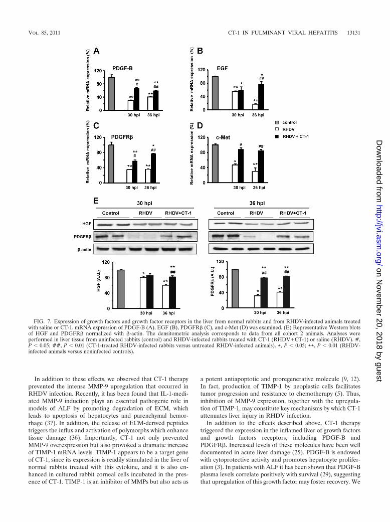
CT-1 stimulates cytoprotective and proregenerative factorsin RHDV-infected livers. Tissue defense against noxious insultsis orchestrated by a diversity of growth factors and cytokines,including HGF (8), EGFR ligands (16), PDGF (29), and CT-1(4), among others. In the livers of RHDV-infected rabbitsgiven saline, we found a marked decrease of HGF at 36 hpi andof its receptor c-Met at both 30 and 36 hpi. The infectedrabbits treated with CT-1 exhibited higher hepatic expressionof HGF at 36 hpi and of c-Met at 30 and 36 hpi than theinfected animals which received saline (Fig. 7D and E). Also,the PDGF-B gene expression and protein levels of PDGFR�in liver tissue dropped intensely at 30 and 36 hpi in control
RHDV-infected rabbits while those treated with CT-1 showedsignificantly higher values of both PDGF-B and its receptor atboth time points (Fig. 7A, C, and E). Furthermore, CT-1therapy prevented the profound decline of hepatic EGFmRNA levels taking place in RHDV infection, with values at36 hpi that were significantly higher in rabbits treated withCT-1 than in RHDV-infected rabbits given saline (Fig. 7B).
DISCUSSION
RHDV infection is a unique model that closely reproducesthe histopathological, biochemical, and clinical manifestationsof human fulminant viral hepatitis (31). Therefore, we selectedRHDV-induced ALF to determine whether CT-1 might rep-resent a potential therapy for this condition and to define theresponsible mechanisms.
Previously, we reported that CT-1 is a potent hepatoprotec-tive cytokine with the ability to attenuate ConA hepatitis, Fas-induced hepatocellular damage, and ischemia/reperfusion in-jury (4, 15, 20). However, its role in fulminant viral hepatitishas never been tested. In the present study, RHDV-infectedanimals received five doses of CT-1, the first one at 12 hpi. Thistime point was selected because of the rapid evolution ofRHDV infection. Later in the course of the disease, rabbits leftuntreated were so ill and prostrated that it was technicallydifficult to administer intravenous injections.
Notably, while 100% of rabbits with RHDV died within 60 h,CT-1 treatment allowed 67% survival at day 7 postinfection,which was the end of the study period (unpublished observa-tions from our group indicate that, after this time point, theanimals that were not sacrificed survived normally). This re-markable therapeutic efficacy could not be ascribed to anyantiviral effect of the cytokine since the plasma viral load wassimilar in treated and untreated animals up to 48 hpi, a timepoint where mortality was much higher in untreated than inCT-1-treated rabbits. Also, RHDV-RNA abundance in livertissue was comparable at 30 and 36 hpi in the two groups ofanimals. Our data indicate that the beneficial effect of CT-1appears to be due to the mitigation of inflammation, the re-duction of oxidative stress, and the activation of endogenouscytoprotective and growth-promoting mechanisms. Indeed,CT-1-treated animals exhibited a significant reduction in theexpression of key drivers of inflammation, such as TNF-,IL-1�, COX-2, and TLR4, which are known to be relevantmediators of tissue damage in the inflamed liver (2, 10, 18). Onthe other hand, in the treated rabbits the alleviation of theinflammatory reaction within the liver was accompanied by asignificant decrease in oxidative stress as reflected by de-creased levels of lipid peroxidation products and reducedGSSG/GSH ratio. The decrement of oxidative stress might bea consequence of the anti-inflammatory properties of CT-1,but it could also be due to the reported ability of this cytokineto stimulate antioxidant genes (15). Persisting production ofROS in the inflamed tissues causes prolonged JNK phosphor-ylation, leading to PARP1/2 activation and necrapoptosis (17).This chain of events appears to be inhibited by CT-1 adminis-tration. Supporting this view, we found that JNK activation andPARP1/2 cleavage were readily detectable in liver tissue at 30hpi in untreated RHDV-infected rabbits but not in four out ofsix CT-1-treated infected animals.
FIG. 6. Induction of TIMP-1 mRNA expression after treatmentwith CT-1. (A) Kinetics of TIMP-1 mRNA expression in SIRC rabbitcorneal cells treated with 25 ng of CT-1/ml. (B) Expression of TIMP-1mRNA in liver tissue from normal rabbits that received treatment withCT-1 or were left untreated (control). *, P � 0.05; **, P � 0.01(CT-1-treated versus untreated rabbits).
13130 TUNON ET AL. J. VIROL.
on Novem
ber 20, 2018 by guesthttp://jvi.asm
.org/D
ownloaded from
In addition to these effects, we observed that CT-1 therapyprevented the intense MMP-9 upregulation that occurred inRHDV infection. Recently, it has been found that IL-1-medi-ated MMP-9 induction plays an essential pathogenic role inmodels of ALF by promoting degradation of ECM, whichleads to apoptosis of hepatocytes and parenchymal hemor-rhage (37). In addition, the release of ECM-derived peptidestriggers the influx and activation of polymorphs which enhancetissue damage (36). Importantly, CT-1 not only preventedMMP-9 overexpression but also provoked a dramatic increaseof TIMP-1 mRNA levels. TIMP-1 appears to be a target geneof CT-1, since its expression is readily stimulated in the liver ofnormal rabbits treated with this cytokine, and it is also en-hanced in cultured rabbit corneal cells incubated in the pres-ence of CT-1. TIMP-1 is an inhibitor of MMPs but also acts as
a potent antiapoptotic and proregenerative molecule (9, 12).In fact, production of TIMP-1 by neoplastic cells facilitatestumor progression and resistance to chemotherapy (5). Thus,inhibition of MMP-9 expression, together with the upregula-tion of TIMP-1, may constitute key mechanisms by which CT-1attenuates liver injury in RHDV infection.
In addition to the effects described above, CT-1 therapytriggered the expression in the inflamed liver of growth factorsand growth factors receptors, including PDGF-B andPDGFR�. Increased levels of these molecules have been welldocumented in acute liver damage (25). PDGF-B is endowedwith cytoprotective activity and promotes hepatocyte prolifer-ation (3). In patients with ALF it has been shown that PDGF-Bplasma levels correlate positively with survival (29), suggestingthat upregulation of this growth factor may foster recovery. We
FIG. 7. Expression of growth factors and growth factor receptors in the liver from normal rabbits and from RHDV-infected animals treatedwith saline or CT-1. mRNA expression of PDGF-B (A), EGF (B), PDGFR� (C), and c-Met (D) was examined. (E) Representative Western blotsof HGF and PDGFR� normalized with �-actin. The densitometric analysis corresponds to data from all cohort 2 animals. Analyses wereperformed in liver tissue from uninfected rabbits (control) and RHDV-infected rabbits treated with CT-1 (RHDV�CT-1) or saline (RHDV). #,P � 0.05; ##, P � 0.01 (CT-1-treated RHDV-infected rabbits versus untreated RHDV-infected animals). *, P � 0.05; **, P � 0.01 (RHDV-infected animals versus noninfected controls).
VOL. 85, 2011 CT-1 IN FULMINANT VIRAL HEPATITIS 13131
on Novem
ber 20, 2018 by guesthttp://jvi.asm
.org/D
ownloaded from

also found that CT-1 induced the expression of EGF andc-Met. The latter is the receptor for HGF, a potent hepato-protective factor (8) whose protein levels are increased in theliver of CT-1-treated rabbits. EGF, on the other hand, exertsantiapoptotic effects and acts as a potent hepatomitogen (16).Thus, it is likely that upregulation of all of these moleculescontributes to diminish hepatocellular damage and to stimu-late regeneration following severe acute liver injury.
It has been shown that, in addition to hepatocytes, RHDVcan also infect endothelial cells and intravascular macrophagesby promoting the activation and apoptosis of these cells, whichmay play a pathogenic role in RHDV-induced ALF (1, 27). Ithas been reported that CT-1 reduces macrophage activation(6) and exerts a protective role on endothelial cells (26). Theseeffects on nonparenchymal cells might also contribute to theattenuation of liver damage observed in the infected animalstreated with CT-1.
To summarize, we have shown that, in rabbits with lethalviral hepatitis, CT-1 therapy was able to diminish liver necrosisand to improve survival dramatically. This beneficial effect wasassociated with attenuation of inflammation, reduction of pro-oxidant injury, downregulation of MMP-9, upregulation ofTIMP-1, and overexpression of several growth factors and re-ceptors. Since CT-1 does not affect viral replication, it seemsthat modulation of innate immunity and inflammation maysignificantly reduce hepatocellular damage in acute severe viralhepatitis. In conclusion, our data point to CT-1 as a moleculeof potential therapeutic value for patients with ALF of viralorigin.
ACKNOWLEDGMENTS
This study was supported in part by grants from CIBERehd, by theInstituto de la Salud Carlos III, by the Fondo de InvestigacionesSanitarias (PI/021121), by the Ministerio de Ciencia e Innovacion(BFU2011-30136 and SAF2011-30045), by grants from Fundacíon Pe-dro Barrié de la Maza and Condesa de Fenosa, and by UTE ProjectCIMA.
The technical help of Sandra Jusue, Beatriz Carte, and EdurneElizalde is acknowledged.
REFERENCES
1. Alonso, C., et al. 1998. Programmed cell death in the pathogenesis of rabbithemorrhagic disease. Arch. Virol. 143:321–332.
2. Antoniades, C. G., P. A. Berry, J. A. Wendon, and D. Vergani. 2008. Theimportance of immune dysfunction in determining outcome in acute liverfailure. J. Hepatol. 49:845–861.
3. Borkham-Kamphorst, E., et al. 2008. Platelet-derived growth factor isoformexpression in carbon tetrachloride-induced chronic liver injury. Lab. Invest.88:1090–1100.
4. Bustos, M., et al. 2003. Protection against liver damage by cardiotrophin-1:a hepatocyte survival factor up-regulated in the regenerating liver in rats.Gastroenterology 125:192–201.
5. Crocker, M., et al. 2011. Serum angiogenic profile of patients with glioblas-toma identifies distinct tumor subtypes and shows that TIMP-1 is a prog-nostic factor. Neurol. Oncol. 13:99–108.
6. Fernandez-Ruiz, V., et al. 2011. Treatment of murine fulminant hepatitiswith genetically engineered endothelial progenitor cells. J. Hepatol. 55:828–837.
7. Garcia-Lastra, R., et al. 2010. Signaling pathways involved in liver injury andregeneration in rabbit hemorrhagic disease, an animal model of virally in-duced fulminant hepatic failure. Vet. Res. 41:2.
8. Giebeler, A., et al. 2009. c-Met confers protection against chronic liver tissuedamage and fibrosis progression after bile duct ligation in mice. Gastroen-terology 137:297–308.
9. Guedez, L., et al. 1998. In vitro suppression of programmed cell death of Bcells by tissue inhibitor of metalloproteinases-1. J. Clin. Invest. 102:2002–2010.
10. Guo, J., and S. L. Friedman. 2010. Toll-like receptor 4 signaling in liverinjury and hepatic fibrogenesis. Fibrogenesis Tissue Repair 3:21.
11. Harrison, P. M., R. Keays, G. P. Bray, G. J. Alexander, and R. Williams.1990. Improved outcome of paracetamol-induced fulminant hepatic failureby late administration of acetylcysteine. Lancet 335:1572–1573.
12. Hayakawa, T., K. Yamashita, K. Tanzawa, E. Uchijima, and K. Iwata. 1992.Growth-promoting activity of tissue inhibitor of metalloproteinases-1(TIMP-1) for a wide range of cells. A possible new growth factor in serum.FEBS Lett. 298:29–32.
13. He, Q., C. S. Graham, E. Durante Mangoni, and M. J. Koziel. 2006. Differ-ential expression of Toll-like receptor mRNA in treatment non-respondersand sustained virologic responders at baseline in patients with chronic hep-atitis C. Liver Int. 26:1100–1110.
14. Hissin, P. J., and R. Hilf. 1976. A fluorometric method for determination ofoxidized and reduced glutathione in tissues. Anal. Biochem. 74:214–226.
15. Iniguez, M., et al. 2006. Cardiotrophin-1 defends the liver against ischemia-reperfusion injury and mediates the protective effect of ischemic precondi-tioning. J. Exp. Med. 203:2809–2815.
16. Jia, C. 2011. Advances in the regulation of liver regeneration. Expert Rev.Gastroenterol. Hepatol. 5:105–121.
17. Kim, Y. S., M. J. Morgan, S. Choksi, and Z. G. Liu. 2007. TNF-inducedactivation of the Nox1 NADPH oxidase and its role in the induction ofnecrotic cell death. Mol. Cell 26:675–687.
18. Kwon, O. K., et al. 2011. Ethanol extract of Elaeocarpus petiolatus inhibitslipopolysaccharide-induced inflammation in macrophage cells. Inflammationdoi:10.1007/s10753-011-9343-3.
19. Livak, K. J., and T. D. Schmittgen. 2001. Analysis of relative gene expressiondata using real-time quantitative PCR and the 2��CT method. Methods25:402–408.
20. Marques, J. M., et al. 2007. Cardiotrophin-1 is an essential factor in thenatural defense of the liver against apoptosis. Hepatology 45:639–648.
21. Muller, W. A. 2011. Mechanisms of leukocyte transendothelial migration.Annu. Rev. Pathol. 6:323–344.
22. Nguyen, N. T., and J. M. Vierling. 2011. Acute liver failure. Curr. Opin.Organ Transplant. 16:289–296.
23. O’Grady, J. G., G. J. Alexander, K. M. Hayllar, and R. Williams. 1989. Earlyindicators of prognosis in fulminant hepatic failure. Gastroenterology 97:439–445.
24. Ostapowicz, G., et al. 2002. The results of a prospective study of acute liverfailure at 17 tertiary care centers in the United States. Ann. Intern. Med.137:947–954.
25. Pinzani, M., et al. 1996. Expression of platelet-derived growth factor and itsreceptors in normal human liver and during active hepatic fibrogenesis.Am. J. Pathol. 148:785–800.
26. Pulido, E. J., et al. 1999. Cardiotrophin-1 attenuates endotoxin-inducedacute lung injury. J. Surg. Res. 84:240–246.
27. Ramiro-Ibanez, F., J. M. Martin-Alonso, P. Garcia Palencia, F. Parra, andC. Alonso. 1999. Macrophage tropism of rabbit hemorrhagic disease virus isassociated with vascular pathology. Virus Res. 60:21–28.
28. Sielaff, T. D., et al. 1995. An anesthetized model of lethal canine galac-tosamine fulminant hepatic failure. Hepatology 21:796–804.
29. Takayama, H., et al. 2011. Serum levels of platelet-derived growth factor-BBand vascular endothelial growth factor as prognostic factors for patients withfulminant hepatic failure. J. Gastroenterol. Hepatol. 26:116–121.
30. Tsan, M. F., and B. Gao. 2004. Endogenous ligands of Toll-like receptors.J. Leukoc. Biol. 76:514–519.
31. Tunon, M. J., M. Alvarez, J. M. Culebras, and J. Gonzalez-Gallego. 2009. Anoverview of animal models for investigating the pathogenesis and therapeu-tic strategies in acute hepatic failure. World J. Gastroenterol. 15:3086–3098.
32. Tunon, M. J., et al. 2003. Rabbit hemorrhagic viral disease: characterizationof a new animal model of fulminant liver failure. J. Lab. Clin. Med. 141:272–278.
33. Wielockx, B., et al. 2001. Inhibition of matrix metalloproteinases blockslethal hepatitis and apoptosis induced by tumor necrosis factor and allowssafe antitumor therapy. Nat. Med. 7:1202–1208.
34. Willis, E. D. 1985. Evaluation of lipid peroxidation in lipids and biologicalmembranes, p. 407–420. In K. Snell and B. Mullock (ed.), Biochemicaltoxicology: a practical approach. IRL Press, Oxford, England.
35. Wu, Z., M. Han, T. Chen, W. Yan, and Q. Ning. 2010. Acute liver failure:mechanisms of immune-mediated liver injury. Liver Int. 30:782–794.
36. Xu, X., et al. 2011. A self-propagating matrix metalloprotease-9 (MMP-9)dependent cycle of chronic neutrophilic inflammation. PLoS One 6:e15781.
37. Yan, C., L. Zhou, and Y. P. Han. 2008. Contribution of hepatic stellate cellsand matrix metalloproteinase 9 in acute liver failure. Liver Int. 28:959–971.
38. Zhai, Y., et al. 2004. Cutting edge: TLR4 activation mediates liver ischemia/reperfusion inflammatory response via IFN regulatory factor 3-dependentMyD88-independent pathway. J. Immunol. 173:7115–7119.
39. Zhang, Y., et al. 2010. Overexpression of Toll-like receptor 2/4 on monocytesmodulates the activities of CD4�CD25� regulatory T cells in chronic hep-atitis B virus infection. Virology 397:34–42.
13132 TUNON ET AL. J. VIROL.
on Novem
ber 20, 2018 by guesthttp://jvi.asm
.org/D
ownloaded from