Embed Size (px)
Citation preview

Protein kinases are intriguing molecular targets for therapeutic intervention. Through reversible phospho-rylation, kinases regulate a wide array of signalling path-ways that control metabolism, cell cycle progression, cell proliferation and cell death, differentiation and survival. There are over 500 kinases in the human kinome, and over 150 of these have been shown or are proposed to be involved in the onset and/or progression of various human diseases including inflammatory diseases, car-diovascular diseases, metabolic diseases, neurodegen-erative diseases and cancer1.
These kinases are therefore putative targets for drug development. Indeed, development of kinase inhibitors — predominantly targeting kinases that are dysregulated in various cancers — is a rapidly growing area of drug discov-ery, partly as a result of some remarkable early successes (see below), as well as the relative ease with which these agents can be designed. This ease in design is attri-butable to the fact that the structure of the key region of the kinase targeted by most inhibitors (the ATP binding pocket) has been well-studied and is highly conserved across the kinome2. Thus the vast majority of approved kinase inhibi-tors and drugs in development target this ATP binding pocket, preventing ATP from binding to the kinase. If ATP cannot bind, the kinase cannot phosphorylate its downstream targets and signalling is inhibited.
However, as discussed below, the conservation of the ATP binding pocket among kinases means that
inhibitors can also inhibit unintended kinases, and if any of these kinases serve important functions in the heart, ‘off-target’ cardiotoxicity can result. In addition, as we discuss some of the problems inherent to kinase inhibi-tors, it will become apparent that many of the pathways that regulate cancer cell survival also regulate essential processes in cardiomyocytes, including survival. So, although inhibiting those kinases in cancer is beneficial3, inhibiting them in the cardiomyocyte may not be.
On the market in the United States, there are cur-rently nine small molecule ATP-competitive inhibitors that target a range of kinases and three inhibitors of mTOR (mammalian target of rapamycin) that work through a novel inhibitory protein–protein interaction (TABLE 1). Numerous additional agents are also in devel-opment4. Although cases of cardiotoxicity associated with kinase inhibitors (herein defined as drug-related deterioration in cardiac function and/or development of congestive heart failure (CHF)) have been reported for several of these molecules, in general the major-ity of the approved agents appear to be well-tolerated from a cardiac safety perspective5. That said, albeit with the few exceptions that we note below, the true risk of cardiotoxicity is not known because thorough clinical assessments of left ventricular function have not been done. Furthermore, patients in early phase clinical trials typically lack significant cardiovascular co-morbidities. This makes it difficult to predict rates
*Center for Translational Medicine, Thomas Jefferson University, 1025 Walnut Street, 316 College Building Philadelphia, Pennsylvania 19107, USA.‡Early and Investigative Safety, Nonclinical Safety, Hoffmann-La Roche Inc., 340 N Kingsland Road Nutley, New Jersey, 07110, USA.Correspondence to T.F. e-mail: [email protected]:10.1038/nrd3252
Cardiotoxicity of kinase inhibitors: the prediction and translation of preclinical models to clinical outcomesThomas Force* and Kyle L. Kolaja‡
Abstract | Targeted therapeutics, particularly those that inhibit the activity of protein kinases that are mutated and/or overexpressed in cancer, have revolutionized the treatment of some cancers and improved survival rates in many others. Although these agents dominate drug development in cancer, significant toxicities, including cardiotoxicity, have emerged. In this Review, we examine the underlying mechanisms that result in on-target or off-target cardiotoxicities of small molecule kinase inhibitors. We also discuss how well the various preclinical safety models and strategies might predict clinical cardiotoxicity. It is hoped that a thorough understanding of the mechanisms underlying cardiotoxicity will lead to the development of safe, effective drugs and consequently, fewer costly surprises as agents progress through clinical trials.
R E V I E W S
NATURe RevIewS | Drug Discovery vOlUme 10 | FebRUARy 2011 | 111
© 2011 Macmillan Publishers Limited. All rights reserved
Regular type radionuclide ventriculographyThe use of radionuclides to study left ventricular function.
multi-targetedintentionally designed kinase inhibitor of more than one kinase.
Cardiac stressEither increased demand (for example, hypertension) or reduced supply (for example, ischaemia) of oxygen to the heart.
of cardiotoxicity when agents become approved and are subsequently used in a broader population.
To date, the only approved kinase inhibitor that is clearly associated with clinical cardiotoxicity is sunitinib (Sutent; Pfizer). In a retrospective review of a series of patients with gastrointestinal stromal tumours who underwent regular type radionuclide ventriculography, 18% of patients developed either CHF or a decline in left ven-tricular ejection fraction of ≥15 points — this was a clin-ically significant decline6. The only other large study of cardiotoxicity with a small molecule kinase inhibitor was with lapatinib (Tykerb; GlaxoSmithKline) (which inhib-its the epidermal growth factor receptor and HeR2 (also known as eRbb2). Although very low rates of cardio-toxicity were reported in the lapatinib trial, the majority of patients had been previously treated with trastuzu-mab (Herceptin; Roche/Genentech) (a monoclonal antibody targeting HeR2 that is associated with cardiac dysfunction) without obvious cardiotoxicity, and thus were presumably less likely to develop left ventricular dysfunction from an anti-HeR2 small molecule inhibi-tor7. Other than these two comprehensive cardiovascular safety studies on sunitinib and lapatinib, only data from series of case reports and limited cardiac safety informa-tion from Phase III registration trials are available.
Although clinically important cardiotoxicity appears to be limited to only a few currently marketed agents, a major concern is that a large number of drugs in devel-opment — many of which are multi-targeted — either intentionally or unintentionally inhibit pathways that maintain cardiomyocyte homeostasis, especially under
cardiac stress. Thus, it is imperative to develop strategies that accurately identify problematic agents early in the drug development process.
In this Review, we will discuss the growing connec-tion between preclinical models of kinase inhibitor-induced cardiotoxicity and clinical safety. As protein kinases regulate numerous aspects of cell signalling, developing inhibitors to prevent a tumour from having a selective growth advantage can lead to overlapping deleterious effects on cardiac biology and physiology. we discuss the challenges of making safe and selective kinase inhibitors by examining the cardiac effects of sunitinib. we also explore the vast field of genetically modified mouse models and discuss their merits in predicting more accurately which kinase inhibitors may have the potential to cause cardiotoxicity. Additional in vitro models used to predict cardiotoxicity are reviewed, with emphasis on human stem cell-derived cardiomyocytes, as they have the potential to mimic car-diac biology and cardiac energy homeostasis, and be a means of introducing phenotypic and genotypic diver-sity of the human race into preclinical safety assessment. lastly, we conclude with future perspectives in clinical studies, including biomarkers and imaging.
Small molecule kinase inhibitorsAs noted above, the vast majority of small molecule kinase inhibitors are designed to competitively target the ATP binding domain of the kinase of interest. Given the structural conservation of the ATP binding pocket across the 500+ kinases of the human kinome2, limited
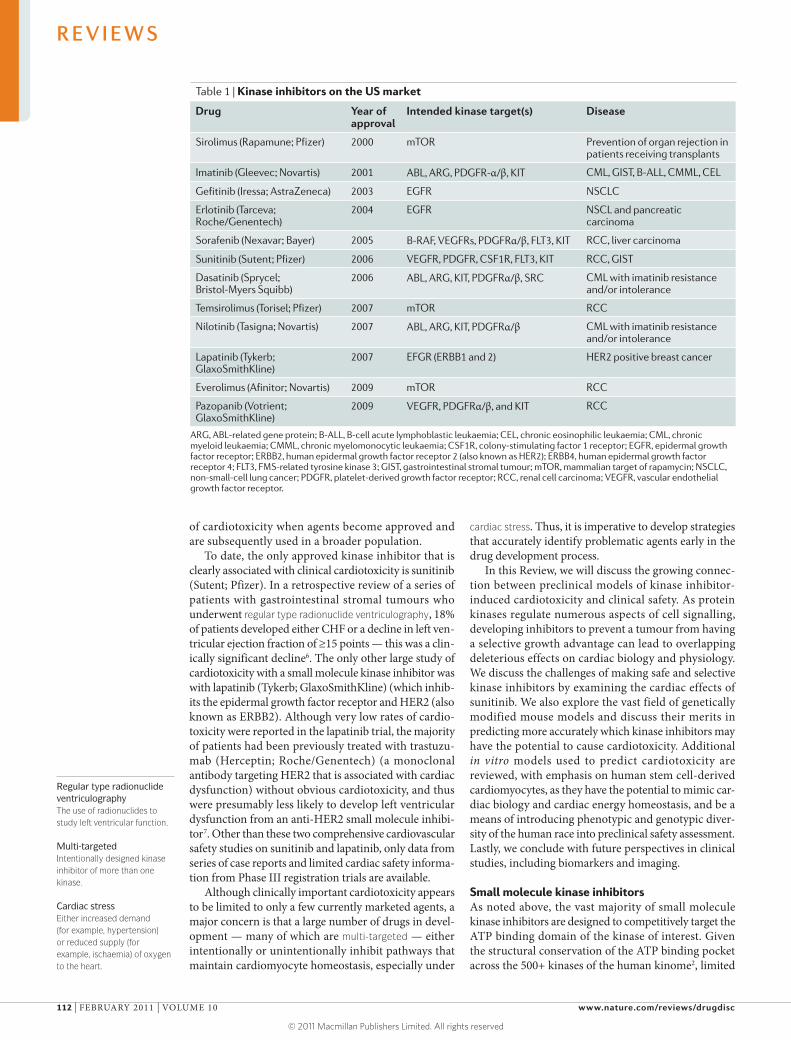
Table 1 | Kinase inhibitors on the US market
Drug year of approval
intended kinase target(s) Disease
Sirolimus (Rapamune; Pfizer) 2000 mTOR Prevention of organ rejection in patients receiving transplants
Imatinib (Gleevec; Novartis) 2001 ABL, ARG, PDGFR-α/β, KIT CML, GIST, B-ALL, CMML, CEL
Gefitinib (Iressa; AstraZeneca) 2003 EGFR NSCLC
Erlotinib (Tarceva; Roche/Genentech)
2004 EGFR NSCL and pancreatic carcinoma
Sorafenib (Nexavar; Bayer) 2005 B-RAF, VEGFRs, PDGFRα/β, FLT3, KIT RCC, liver carcinoma
Sunitinib (Sutent; Pfizer) 2006 VEGFR, PDGFR, CSF1R, FLT3, KIT RCC, GIST
Dasatinib (Sprycel; Bristol-Myers Squibb)
2006 ABL, ARG, KIT, PDGFRα/β, SRC CML with imatinib resistance and/or intolerance
Temsirolimus (Torisel; Pfizer) 2007 mTOR RCC
Nilotinib (Tasigna; Novartis) 2007 ABL, ARG, KIT, PDGFRα/β CML with imatinib resistance and/or intolerance
Lapatinib (Tykerb; GlaxoSmithKline)
2007 EFGR (ERBB1 and 2) HER2 positive breast cancer
Everolimus (Afinitor; Novartis) 2009 mTOR RCC
Pazopanib (Votrient; GlaxoSmithKline)
2009 VEGFR, PDGFRα/β, and KIT RCC
ARG, ABL-related gene protein; B-ALL, B-cell acute lymphoblastic leukaemia; CEL, chronic eosinophilic leukaemia; CML, chronic myeloid leukaemia; CMML, chronic myelomonocytic leukaemia; CSF1R, colony-stimulating factor 1 receptor; EGFR, epidermal growth factor receptor; ERBB2, human epidermal growth factor receptor 2 (also known as HER2); ERBB4, human epidermal growth factor receptor 4; FLT3, FMS-related tyrosine kinase 3; GIST, gastrointestinal stromal tumour; mTOR, mammalian target of rapamycin; NSCLC, non-small-cell lung cancer; PDGFR, platelet-derived growth factor receptor; RCC, renal cell carcinoma; VEGFR, vascular endothelial growth factor receptor.
R E V I E W S
112 | FebRUARy 2011 | vOlUme 10 www.nature.com/reviews/drugdisc
© 2011 Macmillan Publishers Limited. All rights reserved

Class effectA toxicity or pharmacological outcome that occurs with all molecules that inhibit a particular target (for example, all β-adrenergic receptor antagonists lead to heart rate slowing).
Side populationA form of stem cell that is characterized by a specific pattern using fluorescent activated sorting.
selectivity is an inherent property of small molecule inhibitors and a challenge in the design of these agents. Strategies to improve selectivity go beyond selectively targeting the ATP binding pocket and include consider-ing regions adjacent to the pocket (this is the mecha-nism used by Type II inhibitors) or targeting allosteric regions remote from the pocket (this is strategy adopted by Type III inhibitors)8–10. Targeting allosteric regions would be much more selective, but because they exploit novel structural aspects of individual kinases, they are more difficult to design. Although Type III inhibitors constitute a small minority of the kinase inhibitors in development, additional allosteric inhibitors may be identified as screening strategies migrate towards cell-based screening assays11.
The cardiotoxicological properties of small molecule kinase inhibitors will typically be due to (intended or unintended) kinase inhibition, although off-target effects on non-kinases could have a role. In some cases, on-target (intended) inhibition of a kinase which is appealing from a tumour progression and/or angiogenesis perspective could also be physiologically crucial in the heart and/or the vasculature (BOX 1). Off-target kinase inhibitor-mediated cardiotoxicity is a consequence of the inher-ent challenge of selectivity of most ATP-competitive kinase inhibitors. This leads to unintended inhibition of multiple kinases beyond the intended target kinase (or kinases). Although these off-target kinases may or may not have a role in tumour progression, they may be essential to the function of the heart. exacerbating this form of toxicity is the current trend of intentional ‘multi-targeting’ (inhibiting multiple kinases with a single drug)12. members of this class of agents typi-cally target factors driving both tumour progression and angiogenesis, and targets linked to angiogenesis include vascular endothelial growth factor recep-tor 2 (veGFR2), placenta growth factor (PlGF), and platelet-derived growth factor receptor-β (PDGFRβ).
It has become increasingly apparent that agents target-ing veGFRs, particularly veGFR2, induce significant hypertension13. Indeed, this appears to be a class effect of these agents, be they small molecules or monoclonal antibodies (mAbs), and can require at least temporary cessation of treatment. This additional stress of hyper-tension can be particularly problematic in patients with an already compromised cardiac reserve or advanced coronary artery disease.
An additional issue that is likely to exacerbate kinase inhibitor toxicity is pathway targeting; that is, the tar-geting of multiple kinases within a specific pathway to achieve maximal suppression of activity of that path-way. For example, inhibition of multiple components of the phosphoinositide 3-kinase (PI3K)–AKT pathway appears to be a viable strategy in a number of cancers14. However, this pathway also maintains cardiomyocyte homeostasis and protects cardiomyocytes from injury and death15. Thus, the potential cardiovascular toxic-ity of compounds that inhibit the PI3K–AKT pathway should be carefully examined.
A third mechanism by which small molecule kinase inhibitors could mediate toxicity would be through the inhibition of non-kinase targets. Any enzyme that requires ATP to perform its various functions could be inhibited by small molecule kinase inhibitors. Thus, even if one knew the complete selectivity profile of a drug against all kinases, it would be impossible (at least at this point in time) to have a full selectivity pro-file against the non-kinase targets. even the relatively selective kinase inhibitor imatinib (Gleevec; Novartis) is known to inhibit at least two non-kinase targets, the quinone oxidoreductase NQO2 and the AbC fam-ily member bCRP (also known as AbCG2)16. NQO2 is believed to have a protective role in the setting of oxidative stress. Although bCRP is not expressed in adult cardiomyocytes, it is expressed in a subset of stem/ progenitor cells, the so-called side population cells (in fact,
Box 1 | Kinase cardiac biology and the potential mechanistic links to cardiotoxicity
Cardiovascular energy homeostasis is predominantly regulated by protein kinases, including some that are the target of marketed inhibitors or those kinase inhibitors in late phase clinical trials 88. Beating cardiomyocytes consume vast amounts of ATP in vivo. The ATP is utilized to generate contractile activity of the sarcomeres (allowing the heart to contract) and to maintain the necessary gradients of calcium, sodium and potassium, which are driven by the various ion pumps and channels89. The most prominent cardiac ion pump is the sarco(endo)plasmic reticulum calcium ATPase (SERCA2A)89. To initiate systole, calcium must be released from the sarcoplasmic reticulum into the intracellular environment, and with every diastole, that calcium must be taken back up into the sarcoplasmic reticulum by SERCA2A, otherwise the heart would be unable to relax. It has been estimated that if all ATP production halted in the heart, ATP stores would be depleted within a matter of seconds90,91. Any perturbation in the delicate balance of ongoing energy generation and utilization (for example, myocardial ischaemia (reducing supply) or hypertension (increasing demand)), can lead to profound abnormalities in cardiac function.
Kinase signalling pathways are intimately linked with calcium homeostasis, and any dysregulation of calcium handling can lead to profound alterations, culminating in dysrhythmia, altered cardiac conduction and cardiac pathological changes such as cardiac hypertrophy92. This dysregulation of calcium can be detrimental, even when it occurs in microdomains within the cytosol and nucleus, and does not require global calcium dysregulation to lead to adverse left ventricular remodelling (that is, dilation and fibrosis). The changes in the ventricle are in part driven by calcium-mediated activation of calcineurin and subsequently, the transcription factors NFAT and GATA4 (REf. 93). Calcium/calmodulin-dependent protein kinase II is a key player in the pathology of hypertrophy and heart failure, as it regulates both calcium handling (via its effects on the sarcoplasmic reticulum and ryanodine receptor) and hypertrophy (via its effects on signalling pathways)94–96. Cardiomyocyte survival is also regulated by protein kinases, the most notable being components of the phosphoinositide 3-kinase–AKT pathway15. Taken together, there are multiple, critical kinase-regulated pathways that, if inhibited, could exacerbate cardiotoxicity.
R E V I E W S
NATURe RevIewS | Drug Discovery vOlUme 10 | FebRUARy 2011 | 113
© 2011 Macmillan Publishers Limited. All rights reserved

Hoechst dyeA family of fluorescent stains used for labelling DNA and detecting and/or separating cell populations of interest.
Pressure loadA pressure load on the heart can be induced by anything that raises blood pressure. Experimentally, this typically means constricting the transverse aorta or infusing a drug (for example, angiotensin ii).
mitochondrial permeability transition poreA regulatory opening within the mitochondria induced by certain types of cellular stress (for example, oxidant stress or ischemia). This results in loss of mitochondrial membrane integrity and swelling, ultimately inducing cell death.
Stem/progenitor cell compartmentThe small population of undifferentiated cells within a tissue that has the potential to regenerate and replenish the dying cells of an organ.
bCRP-mediated extrusion of Hoechst dye is what pro-duces the side population phenotype of these cells when they are analysed using fluorescence-activated cell sort-ing)17. The toxicological consequences of inhibiting non-kinase targets, if any, are unclear, but this issue is of concern as it significantly increases the potential for side effects and can complicate the toxicological screen-ing strategy. From a cardiotoxicity-focused perspective, alternatives to the traditional medicinal chemistry-based strategies to design Type I (ATP-competitive) inhibitors should be explored.
Sunitinib and cardiotoxicityTo illustrate the complexities inherent in identifying mechanisms of kinase inhibitor toxicity, we will use the example of sunitinib, which appears to have both on- and off-target effects contributing to cardiotoxicity. Clinical issues with this multi-targeted agent are discussed above. It has been presumed that a major contributor to the deterioration in left ventricular function is sunitinib-induced hypertension. In support of this idea, sunitinib-treated mice did not develop cardiomyocyte apoptosis until an increase in blood pressure was induced18.
A number of studies in various mouse models have also shown that angiogenesis (which is mediated in the heart by both veGFR2 and PDGFRβ, targets of sunitinib) is key to maintaining cardiac homeostasis in the setting of a pressure load or ischaemia19–22. Although this indicates that sunitinib-induced hypertension has an important role in cardiotoxicity, several patients who developed cardiotoxicity on sunitinib did not have hypertension, suggesting that additional mechanisms may be responsible 6. Although no head-to-head cardio-toxicity study has been conducted with veGFR antago-nists, the cardiotoxicity of sunitinib does appear to be greater than the other agents23. even though this could be due to the greater potency of sunitinib in inhibiting veGFR2, this difference suggests the possibility of addi-tional off-target effects.
Identifying the mechanisms of sunitinib toxicity is more difficult than for other more selective kinase inhibi-tors, as sunitinib inhibits at least 50 kinase targets at ther-apeutically relevant concentrations in in vitro assays24. Hints to the mechanisms were found in transmission electron micrographs of endomyocardial biopsies from a patient who presented with profound sunitinib-associated heart failure. The biopsies showed marked mitochondrial swelling indicative of the opening of the mitochondrial permeability transition pore, a response that typically leads to a necrotic form of cell death. This swelling appeared to be so widespread that it seemed likely that energy (ATP) homeostasis may have been compromised.
Studies in neonatal rat ventricular myocytes (NRvms) also confirmed that energy compromise does have a key role in sunitinib-mediated toxicity, but this decrease in available energy did not lead to activation of AmP-activated protein kinase, the central regula-tor of the response of the cell to energy perturbations. Surprisingly, the lack of a response to the energy com-promise appeared to be due to direct inhibition of 5′-AmP activated protein kinase (AmPK) by sunitinib18.
Gene transfer of an activated mutant of AmPK partially rescued the toxicity in NRvms, suggesting that inhibi-tion of this off-target kinase contributes to the toxicity. Although this suggests that redesigning sunitinib to pre-vent it from inhibiting AmPK could solve the problem of off-target toxicity, pivotal work on the protective role of AmPK in maintaining viability of hypoxic cancer cells suggests that this ‘off-target’ effect may be important in the anticancer efficacy of sunitinib25. Although the role of AmPK in cancer cell survival remains unclear26, AmPK is central to maintaining energy balance in the cardiomyocyte, and thus a compound that alters AmPK activity should be closely scrutinized for cardiotoxicity.
Given what we know about the role of veGFRs and PDGFRβ in the heart’s response to stressors, inhibition of these kinases most probably contributes to the on-target toxicity associated with sunitinib and the other multi-tar-geted agents. This toxicity is mediated via adverse effects on the vasculature that, at least in animal models, lead to loss of capillaries, impaired blood flow and regional ischaemia, especially in the setting of cardiac stress19–22. These processes are undoubtedly mechanistic contribu-tors to the hypertension that is characteristic of kinase inhibitors. Remarkably, it appears that hypertension is a ‘biomarker’ of the anticancer efficacy of the multi- targeted agents as several studies have shown that cancer patients who develop hypertension have better outcomes than those who do not27–30. That said, it is important to underline that hypertension is an off-target consequence and probably plays no part in the killing of tumour cells; it should therefore be aggressively treated.
This vignette on sunitinib illustrates how it can be difficult to identify the specific mechanisms of car-diotoxicity and manage the toxicity without hamper-ing treatment of the malignancy. However, it also underscores the crucial importance of knowing the kinase selectivity profile of an inhibitor to help develop informed hypothesis-driven research31,32. The kinase inhibition profile can be mathematically modelled to associate particular kinases with cardiotoxicity; this is an approach that has been used to correlate kinases with other types of toxicities33. One of the limitations of this approach is our limited understanding of the role that the vast majority of kinases expressed in the human kinome have in cardiac biology.
Cardiac biology and stem/progenitor cellsA potential mechanism of kinase inhibitor-mediated toxicity could be to ablate or suppress the proliferation of the stem/progenitor cell compartment of the heart. Given the limited ability of adult cardiomyocytes to re-enter the cell cycle and proliferate34, and the acute nature of car-diac injury, intrinsic mechanisms to repair the heart are not robust. Although these cardiac stem/progeni-tor cell populations are relatively quiescent, stress can induce these cells to generate significant numbers of new cardiomyocytes35 –37. The physiological significance of the replication of small subsets of progenitor cells is still under debate, but it is possible that these cell popula-tions are collateral targets of some of the small molecule kinase inhibitor anticancer agents.
R E V I E W S
114 | FebRUARy 2011 | vOlUme 10 www.nature.com/reviews/drugdisc
© 2011 Macmillan Publishers Limited. All rights reserved

Mitogens
• Cell cycle progression• Cell proliferation
• CPCs • Cardiomyocytes
Cardiomyocytes
Cardiomyocytes
• CPCs • Cardiomyocytes
Cardiomyocytes
Growth factorreceptors
ERKsCDKsAuroraPLK
PIM1
GSK3Cell typesAKT
CaMKII PKA
PKCαPI3Kγ
• Pathologicalhypertrophicstimuli (e.g. Angiotension IIand hypertension)
• Cell growth• Hypertrophy
PI3Kγ
NFATs
CAMKII
S6K
Calcineurin↑ [Ca2+]
JNK
GSK3
Physiologicalgrowth stimuli Autophagy
Energy stress(↓ATP)
Growth factor receptors (IGF1R, insulinreceptor, PDGFRs) Cell death
PI3K
PDK1
Growthfactorreceptors
LKB1CAMKKβ
PI3K
RAS
AKT etc.
RAF 1/B RAF MEK1/2 ERK1/2
Pro-apoptotic factors (e.g. Forkhead, BAD)
AMPK
AKT mTOR
• Oxidative stress • Cytokines
Calcium
• Contractile dysfunction• Adverse remodelling
JAK–STAT
PTEN
a
b
c
d
e
f
Nature Reviews | Drug Discovery
Figure 1 | crucial signalling pathways in the heart and consequences of their activation or inhibition. Various stimuli (green boxes) activate downstream signalling pathways in the heart, leading to physiological or pathological responses (blue boxes). The cell types proposed, or known to be affected, are indicated. a | Mitogens activate growth factor receptors that act through various kinases to promote cell cycle progression and cell proliferation. In addition to the core cell cycle regulators (cyclin-dependent kinases (CDKs) and serine/threonine protein kinases (Auroras and PLKs)), extracellular signal-regulated kinases (ERKs) and AKT appear to be key to promoting proliferation of cardiac progenitor cells (CPCs) and, possibly, cardiomyocytes. AKT acts through both activation of a serine/threonine protein kinase (Pim1) and inhibition of glycogen synthase kinase-3 (GSK3). b | Pathological hypertrophy is driven by increases in Ca2+ concentration that are triggered by various stimuli (angiotensin II, hypertension and so on). This activates calcineurin, a phosphatase. Calcineurin dephosphorylates members of the NFAT family of transcription factors, allowing them to enter the nucleus and activate gene expression programs promoting hypertrophy. Counteracting this are Jun N-terminal kinases (JNKs) and GSK3 family members that phosphorylate NFATs, leading to their exclusion from the nucleus. Other key factors promoting hypertrophy are calcium/calmodu-lin-dependent protein kinase 2 (CaMKII) and phosphoinositide 3-kinase γ (PI3Kγ). c | Physiological growth is driven by various growth factor receptors that activate the PI3K–AKT pathway, leading to inhibition of GSK3. Multiple inputs lead to the activation of mammalian target of rapamycin (mTOR) and downstream targets (for example, ribosomal protein S6 kinase (S6K)) promoting physiological and pathological hypertrophic growth. d | Another key regulator of mTOR is 5′-AMP activated protein kinase (AMPK), the master sensor of energy status in the cardiomyocyte. When ATP is depleted, AMP levels rise and this, together with two other kinases (serine/threonine protein kinase 11 (also known as
LKB1) and calcium/calmodulin-dependent protein kinase kinase (CAMKKβ), activates AMPK. Along with GSK3, AMPK inhibits mTOR-mediated protein synthesis, thereby conserving energy stores. Autophagy is a complicated response to perturbations in energy status that aims to preserve energy when needed. When the energy status is good, mTOR is active and autophagy (resulting in recycling of damaged organelles including mitochondria) is repressed. During energy stress, mTOR is inhibited and autophagy is activated, restoring homeostasis. e | Cardiomyocyte death is regulated by various mechanisms, but focusing on the role of kinases in the heart, the PI3K–AKT pathway (which is downstream of growth factor receptors), is central to prevention of apoptosis, by acting on a number of targets. These include the pro-apoptotic factors BCL2 antagonist of cell death (BAD) and members of the Forkhead family of transcription factors, which are inhibited by AKT. The RAS–RAF–MAPK/ERK kinase (MEK)–extracellular signal-regulated kinase (ERK) pathway is also anti-apoptotic in the heart. f | Cytokines and oxidative stress activate members of the Janus kinase (JAK) family and subsequently, their immediate targets, the signal transducer and activator of transcription (STAT) family of transcription factors. In the heart, this pathway protects against cell death via various mechanisms. Contractile function can also be directly regulated by various kinases such as PKA (which is downstream of β-adrenergic receptors in the ‘fight or flight’ response to stress) and CAMKII. Both kinases enhance contractile function but long-term activation leads to adverse remodelling in the heart. Protein kinase Cα (PKCα) and PI3Kγ (the latter acting via a scaffolding function, not its kinase activity) exert negative inotropic effects on cardiomyocytes. In the setting of heart failure, PKCα inhibition maintains contractile function, whereas inhibition of PI3Kγ appears to be harmful, worsening remodelling. IGF1R, insulin-like growth factor 1 receptor; PDGFR, platelet-derived growth factor. PKA; cAMP-dependent protein kinase.
R E V I E W S
NATURe RevIewS | Drug Discovery vOlUme 10 | FebRUARy 2011 | 115
© 2011 Macmillan Publishers Limited. All rights reserved

AnthracyclineA class of drugs (of which doxorubicin is a member) used extensively in treatment of various cancers.
Pressure overloadA consequence of thoracic aortic constriction, resulting in increased blood pressure and subsequent cardiac hypertrophy and LV dysfunction.
Thoracic aortic constriction A surgical procedure in which the aorta is banded, creating an acute and usually severe increase in blood pressure.
HERGThe human ether-a-go-go gene HERG (also known as KCNH2) codes for a specific potassium ion channel. Mutations in the gene cause one form of hereditary long QT syndrome.
In fact, adverse effects of cancer therapeutics on stem and progenitor cells may be exacerbated by kinase inhibitor-mediated effects on energy metabolism in the adult cardiomyocyte, as noted above for sunitinib. For example, treatment of juvenile mice with the anthracy-cline doxorubicin impairs cardiac progenitor cell func-tion and vascularization, leading to cardiotoxicity as the mice age38. This is similar to the effect observed in children treated with doxorubicin who developed heart failure later in life39. experimentally, infusion of cardiac stem cells can rescue rats from the cardiotoxicity of dox-orubicin, leading the authors of the study to hypothesize that cardiac stem cells are a key target of doxorubicin-induced cardiotoxicity36.
In other studies, inhibition in mice of KIT, the recep-tor for stem cell factor, resulted in enhanced cardiomyo-cyte proliferation and better-preserved left ventricular function when the mice were subjected to pressure over-load25,40. Although beneficial in the short term, chronic inhibition of KIT in this model raised concerns about long-term maintenance of the progenitor pool in the heart. Although the biological significance of these findings in humans is not known, concerns regarding the effects of kinase inhibitors on stem and progenitor cells or on immature cardiomyocytes that are capable of entering the cell cycle should continue to be an active area of research.
Identification of kinases of concernIn this section we will take a ‘virtual cardiotoxicity’ approach by briefly examining what is known from the literature concerning the role of kinase inhibitor targets in the heart. The approach towards understanding and predicting cardiotoxicity resulting from kinase inhibi-tion involves a combination of knowing what cell type (or types) of the heart a kinase is expressed in, and the detailed biology of that kinase in the heart. Drug targets that are potentially problematic (or as we refer to them, ‘kinases of concern’) can often be surmised based on gene targeting studies in mice. Thus, we speculate on possible kinases involved in cardiotoxicity, albeit in some cases with little data beyond genetically modified mice to sup-port our conclusions.
fiG. 1 and TABLE 2 contain several examples of kinases of concern that were identified by studying gene-targeted and/or transgenic mice. A classic example is the HeR2 conditional knockout mouse, which developed a dilated cardiomyopathy with ageing that was worsened by thoracic aortic constriction (TAC), mimicking severe acute hypertension41. more often, gene deletion may lead to no marked abnormalities unless an additional stressor is added. This pattern also suggests that stress-related recruitment of other normally quiescent pathways may be critical in the mechanism of cardiotoxicity. Other examples are the sorafenib (Nexavar; bayer) targets RAF1 and bRAF, and the PI3K pathway. In both RAF1 and p110αPI3K dominant negative transgenic mice, there were no obvious phenotypes until the mice were subjected to TAC, at which point they developed left ven-tricular dysfunction42,43. Additional examples in which TAC revealed significant cardiotoxicity are seen in mice
lacking the angiogenesis regulators veGFRs or PDGFRβ or in mice treated with mAbs that sequester veGF19,20,22,44. These mouse models offer insight into the role of stress in promoting cardiotoxicity with sunitinib (and possi-bly with other veGF and veGFR antagonists such as sorafenib, pazopanib (votrient; GlaxoSmithKline) and bevacizumab (Avastin; Roche/Genentech)).
However, caution must be taken in predicting kinase inhibitor toxicity based solely on findings in genetically modified mice. many studies involving gene targeting in mice used germline knockouts, meaning that the mouse phenotype could be confounded by unique develop-mental roles of a given gene in the embryo and/or the placenta. For example, p38α knockout mice die in mid-gestation due to malformed myocardium and vascula-ture; however, this was attributed to defects in placental development and not cardiac development per se45. This issue can be addressed by using conditional knockouts in which the gene of interest is deleted very late in gestation or, preferably, in adulthood and selectively within the tissue of interest. A second key confounder is that inhibi-tion of a kinase with a kinase inhibitor is never complete in magnitude or duration; by contrast, kinase inhibition as a result of gene deletion is almost always complete, both in magnitude and duration. Furthermore, protein–protein interactions will be disrupted in the knockout, but will generally not be disrupted with drug treat-ment. This could cause phenotypes in the knockouts that would not be seen with kinase inhibitor treatment. Finally, the stress of severe TAC is an extreme state that patients rarely — if ever — experience. Thus, the pheno-type may be significantly exaggerated in the knockouts exposed to TAC compared to a patient receiving a drug. As discussed extensively by molkentin and Robbins46, genetically modified mice are powerful tools that can be used to shed light on the importance of specific genes of interest, but they do not eliminate the need to thor-oughly understand the molecular underpinnings of any phenotype.
Although the focus of this article is on the cardiotoxic-ity mediated by kinase inhibitors, there are several exam-ples of cardiovascular diseases in which kinase inhibitors are predicted to produce beneficial effects (BOX 2).
Can preclinical models predict cardiotoxicity? Cardiotoxicity is routinely assessed throughout the drug development process, starting during the early stages of drug discovery. Depending on the target and/or the toxic-ity profile of the compounds being developed, assays that can predict cardiotoxicity can be introduced during lead identification and lead optimization stages of drug devel-opment. Initial models often evaluate overt cellular toxicity in basic cell lines and cardiac electrophysiological effects are initially examined using cell lines that overexpress HERG. The assessment of potential cardiotoxicity of kinase inhibitors typically involves specialized in vivo models in which left ventricular function, electrocardiogram read-ings and biomarkers of cardiac injury can be quantified.
Attempts to predict clinical cardiotoxicity of kinase inhibitors have had limited success to date. There is a plethora of empirical models available, which include
R E V I E W S
116 | FebRUARy 2011 | vOlUme 10 www.nature.com/reviews/drugdisc
© 2011 Macmillan Publishers Limited. All rights reserved

Table 2 | Kinases of importance in the heart and vasculature: findings in genetically modified or KI-treated mice
Kinase Mouse model(s) role of kinase in heart/vasculature other notes refs
RAF1/BRAF KO; DNTG Anti-apoptotic; preserves LV function under stress. KO: LV dysfunction and HF in the absence of additional stress; DNTG: reduced hypertrophy but LV dysfunction due to cell death
KO: effects mediated by ASK1; RAF inhibits ASK1 via a non-kinase-dependent mechanism. RAF mutations account for some cases of Noonan syndrome (HCM phenocopy)
42,113
PI3K (p110α) CATG; DNTG Physiological heart growth; cardiomyocyte survival
Greater LV dysfunction after TAC in DNTG and improved LV function in CATG mouse
43,114,115
PI3K (p110γ) KO Regulates contractility and pathological hypertrophy
Mice protected from isoproterenol-induced injury
116–118
PDK1 KO Cardiomyocyte survival and β-adrenergic responsiveness
Cardiac-specific KO displayed HF and DCM 119
AKT1, 2 or 3 CATG; DNTG; KO Regulators of cardiomyocyte survival, growth and metabolism
AKT1 promotes physiological hypertrophy and suppresses pathological hypertrophy; AKT2 promotes survival and insulin sensitivity
120,121
mTOR Kinase inhibitor (KI) administered
mTORC1 regulates protein synthesis, inhibition leads to energy preservation under stress; mTORC2 regulates AKT activation
Rapamycin is well-tolerated and blocks cardiac hypertrophy. It will be used in combination regimens in cancer (long-term administration inhibits mTORC2 and AKT)
122,123
AMPK Transgenic; KO Sensor of energy stress; inhibits mTORC1, preserving energy stores. KO of AMPKα2 increased hypertrophy and LV dysfunction after TAC
Activated by tumour suppressor LKB1 or CAMKKβ. Activated mutant leads to glycogen storage hypertrophic cardiomyopathy
124,125
GSK3α/β Transgenic; knock-in; KO
Together with AMPK, inhibits mTORC1; deletion of GSKβ protective in post-MI remodelling; deletion of GSK3α leads to HF in setting of stress
GSK3α important in β-adrenergic responsiveness. Deletion leads to HF with stress
126–128, 181
CDKs Conditional KO; KO CDK2 inhibition reduces ischaemia–reperfusion injury, mediated via effects on retinoblastoma protein
Deletion of CDK2 and CDK4 during development leads to HF, but conditional deletion of CDK2 in adult mice lacking CDK4 does not lead to abnormalities
129,130
Aurora kinases
Kinase inhibitor administered
M phase regulators KIs expected to disrupt CPC proliferation, karyokinesis and any cytokinesis of cardiomyocytes resulting in cell death; associated with cardiotoxicity
131,132
PLKs Kinase inhibitor administered
PLK1 involved in activation of CDC2, chromosome segregation, centrosome maturation, bipolar spindle formation and cytokinesis
As above 132,133
PDGFRs KO β isoform is crucial in angiogenesis and heart’s response to PO
Conditional KO: HF with pressure overload 21
VEGFRs VEGF trap; (monoclonal antibody); KO; Kinase inhibitor administered
Crucial in angiogenesis and the heart’s response to PO; antihypertensive effects
Heart-specific KO of VEGFR1 or VEGFR2 showed impaired response to ischaemia
19, 20,22, 44,53
EGFR (ERBB1)
Kinase inhibitor administered
Helps to maintain LV function in setting of chronic catecholamine stimulation; mediates pro-survival signalling
HF uncommon in clinical use 134
ERBB2 ERBB4
KO; conditional KO; transgenic
Cardiomyocyte survival and homeostasis; maintenance of LV function
KOs of HBEGF (ERBB ligand), neuregulin, ERBB2 and ERBB4 each develop DCM and HF. Neuregulin 1 KOs are more sensitive to doxorubicin-induced cardiotoxicity
135, 136–138
KIT W/Wv mouse, kinase inhibitor administered
Promotes CSC and immature cardiomyocyte differentiation; promotes homing to sites of MI, promoting repair.
Imatinib reduced restenosis after arterial injury; W/Wv mouse protected versus HF induced by PO
40, 104,106,
139
ABL/ARG Kinase inhibitor administered
Maintains ER homeostasis. LV dysfunction is seen in rodents treated with imatinib
Minimal HF with ABL inhibitors noted in clinical use
56
JAK2 Transgenic; KO JAK2 and STAT3 protective in many pathological settings
KO: displayed decreased ischaemia–reper-fusion injury, PO hypertrophy and peripartum cardiomyopathy
140–142
FAK KO Antihypertrophic and antifibrotic in heart Eccentric hypertrophy and dilated cardiomyopathy with PO or angiotensin II
143
R E V I E W S
NATURe RevIewS | Drug Discovery vOlUme 10 | FebRUARy 2011 | 117
© 2011 Macmillan Publishers Limited. All rights reserved

smooth and cardiac muscle cell lines, primary cardio-myocytes (neonatal and adult, and rodent and human), isolated three-dimensional organoids, isolated pro-genitor cardiomyocytes and, more recently, human embryonic stem cell (eSC) and inducible pluripotent stem (iPS) cell-derived cardiomyocytes47,48. Additional models include isolated heart preparations from vari-ous species and mammalian models used for short-term in vivo toxicology studies.
In vitro models of cardiotoxicityCell lines. Cell culture, especially with cell lines, is a frequently relied upon approach for predicting clinical side effects, including the potential risk of cardiotoxic-ity. A number of rodent and human cell lines, includ-ing myocardial or skeletal muscle, have been developed to detect cardiotoxicity (TABLE 3). Importantly, most of
the cardiomyocyte models listed in the table continue to contract in vitro, thereby maintaining the primary physiological and functional role of the cardiomyocyte, including the key in vivo demand of constant ATP pro-duction. This is a crucial feature for detecting any car-diotoxicity of small molecule kinase inhibitors in vitro, as energy stress is likely to contribute to the toxicity.
A point worthy of consideration with in vitro models is the use of media containing high concentrations of glucose. many cell lines are cultured in a manner that is suitable for immortalized, tumour-derived cell lines, which are metabolically adapted to grow quickly and generate energy with little reliance on mitochondrial fatty acid oxidation. Cells in this environment generate ATP through glycolysis and are less reliant on oxidative phosphorylation and mitochondria in general49. In car-diomyocytes, mitochondria are a crucial toxicological
Table 2 | (cont) Kinases of importance in the heart and vasculature: findings in genetically modified or KI-treated mice
Kinase Mouse model(s) role of kinase in heart/vasculature other notes refs
DMPK Transgenic Myotonic dystrophy type 1 is caused by excess repeats of the 3′ UTR region of DMPK
Overexpression in mouse heart leads to hypertrophic cardiomyopathy with dysrthymia
144
LTK Transgenic Activation of LTK results in cardiac hypertrophy and cardiomyocyte degeneration
Overexpression led to severe cardiac concentric hypertrophy
145
ROCK Transgenic; KO Pro-fibrotic and pro-apoptotic in the setting of PO
KO is protected from fibrosis, whereas transgenic shows accelerated hypertrophic cardiac decompensation
146,147
LKB1 KO Activates AMPK which is pro-angiogenic in heart
Cardiac-specific deletion leads to LV dysfunction, atrial fibrillation and death
148
LDB3, ZASP and/or Cypher
Conditional KO Mutations in ZASP are associated with skeletal muscle myopathies
Deletion in cardiomyocytes leads to a DCM 149
ERK1/2 KO; transgenic Generally promotes survival and may modulate physiological (but not pathological) hypertrophy
Activation of this pathway protects aginst cardiac ischaemic injury; inactivation promotes LV dysfunction after PO
150–153
PKCα KO; kinase inhibitor administered
Adverse effects on heart in setting of PO PKCα KO (as opposed to PKCβ or PKCγ) protects against TAC-induced CHF. Ruboxistaurin protects against TAC-induced CHF
154
cGMP- dependent PK
KO One of the four nodal kinases in HF; activated by PDE5 inhibitors; inhibits apoptosis, hypertrophy and β-adrenergic responses
Clinical trial with sildenafil in HF showed improved LV function
155,156
PIM Kinase KO Pro-survival; activated by AKT; regulated at level of gene expression
Promotes cardiac resident stem cell proliferation and survival
157
CAMKII KO Nodal kinase in HF; pro-hypertrophic; promotes decompensation in setting of PO
Mechanism of cardiotoxicity involves regulation of CAMKII gene expression and Ca2+ handling
94,158,159
GRK2 and/or GRK5
KO Downregulates β-adrenergic signalling through recruitment of β-arrestin
Peptide inhibitor protects against HF 160,161
ASK1 KO Promotes pathological hypertrophy and remodelling; pro-apoptotic
KO protected from PO and MI-induced remodelling
162,163
PTEN phosphatase
KO Anti-hypertrophic, impairs cardiomyocyte survival with stress and antagonizes PI3K signalling
PO in PTEN KO mice reduced pathological hypertrophy, interstitial fibrosis and apoptosis with preservation of LV function
163
AMPK, 5′ AMP-activated protein kinase; ARG, abl-related gene; ASK1, apoptosis signal-regulating kinase 1; CAMKII, Ca2+/calmodulin-dependent protein kinase II; CATG,constitutively active transgenic; CDK, cyclin dependent kinase; CHF, congestive heart failure; CPC, cardiac progenitor cells; CSC; DCM, dilated cardiomyopathy; DMPK, dystrophia myotonica protein kinase; DNTG, dominant negative transgenic; EGFR, epidermal growth factor receptor (also known as ERBB1); ERBB2, human epidermal growth factor receptor 2; ERBB4, human epidermal growth factor receptor 4; ERK2, extracellular signal-regulated kinase 2 (also known as MAP kinase 1); FAK, focal adhesion kinase; GSK3, glycogen synthase kinase 3; HCM, hypertrophic cardiomyopathy; HF, heart failure; JAK2, Janus kinase 2; LDB3, LIM domain binding 3; LKB1, serine threonine kinase 11 (also known as STK11); LTK, leukocyte receptor tyrosine kinase; LV, left ventricular; MI, myocardial infarction; mTOR C1, mammalian target of rapamycin complex 1; PDE5, phosphodiesterase 5; PDGFR, platelet-derived growth factor receptor; PDK1, phosphoi-nositide-dependent protein kinase 1; PI3K, phosphoinositide 3-kinase; PKC, protein kinase C; PLK, polo-like kinase; PO, pressure overload; PTEN, phosphatase and tensin homologue; ROCK, RHO-associated kinase (RHO-kinase); TAC, trans-aortic constriction; VEGFR, vascular endothelial growth factor receptor.
R E V I E W S
118 | FebRUARy 2011 | vOlUme 10 www.nature.com/reviews/drugdisc
© 2011 Macmillan Publishers Limited. All rights reserved
PhenocopiesA phenotype or trait that is similar among different individuals.
Noonan, Costello and Cranio-facio-cutaneous syndromesA group of syndromes that affect a number of organ systems and cause severe cardiac hypertrophy.
Ischaemia–reperfusion injuryA complex phenomenon that occurs when the blood supply from an organ is restored after a period of ischaemia.
target, and thus the use of galactose-containing media is optimal because it forces the generation of ATP through oxidative phosphorylation.
One of the more frequently used models to study cardiac biology has been the rat embryonic ventricle-derived H9C2 cell line. These cells have been studied as an in vitro model of cardiac muscle for biochemical and pathophysiological processes, including ischaemia– reperfusion injury, oxidative stress and tissue differen-tiation50,51. In addition, these cells have the versatility to allow the study of action potentials and cardiac electro-physiology, as the voltage channels that are expressed are similar to embryonic or neonatal cells, including l-type Ca2+ channels and ATP-sensitive K+ channels52. Studies using H9C2 cells grown in high or low glucose-
containing media showed that sorafenib was significantly more cytotoxic in galactose-containing media versus glucose-containing media, whereas imatinib, dasatinib (Sprycel; bristol-myers Squibb) and sunitinib showed no difference across the two media types53.Primary cells. NRvm isolation was optimized in the early 1980s54,55. This model has been extensively used to study hypertrophy, stress, cardiac failure, cardiac ion channels, contractility and basic cardiac biology, including the role of kinases in a large number of proc-esses (including hypoxia/reoxygenation (which mimics ischaemia), hypertrophy and apoptosis) as well as toxic-ity. An assessment of seven marketed kinase inhibitors (lapatinib, erlotinib (Tarceva; Roche/Genentech), gefit-inib (Iressa; AstraZeneca), imatinib, sorafenib, sunitinib
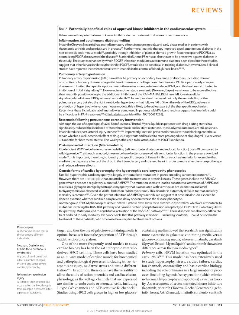
Box 2 | Potentially beneficial roles of approved kinase inhibitors in the cardiovascular system
Below we outline potential uses of kinase inhibitors in the treatment of diseases other than cancer.
inflammation and autoimmune diabetes mellitusImatinib (Gleevec; Novartis) has anti-inflammatory effects in mouse models, and early phase studies in patients with rheumatoid arthritis and psoriasis are in process97. Furthermore, imatinib therapy improved type I autoimmune diabetes in the non-obese diabetic mouse model98, probably through inhibition of platelet-derived growth factor receptors (PDGFRs), as neutralizing PDGF also reversed the disease98. Sunitinib (Sutent; Pfizer) was also shown to be protective against diabetes in this study. The exact mechanism by which PDGFR inhibition modulates autoimmune diabetes is not clear, but these studies suggest that other kinase inhibitors that inhibit PDGFR would also be beneficial in treating diabetes. However, small clinical studies have reported inconsistent results with imatinib in the control of blood glucose levels 99,100.
Pulmonary artery hypertensionPulmonary artery hypertension (PAH) can either be primary or secondary to a range of disorders, including chronic obstructive pulmonary disease, congenital heart disease and collagen vascular diseases. PAH is a particularly complex disease with limited therapeutic options. Imatinib reverses monocrotaline-induced PAH, and this has been attributed to inhibition of PDGFR signalling101. However, in another study, sorafenib (Nexavar; Bayer) was shown to be more effective than imatinib, possibly owing to the additional inhibition of the RAF–MAPK/ERK kinase (MEK)–extracellular signal-regulated kinase (ERK) pathway by sorafenib102. Indeed, sorafenib reduced not only the remodelling of the pulmonary artery but also the right ventricular hypertrophy that follows PAH. Given the role of the ERK pathway in promotion of hypertrophy in various mouse models, this is likely to be at least part of the therapeutic mechanism. Recently a Phase II clinical trial of imatinib was completed in patients with PAH, and results suggest that imatinib could be efficacious in PAH treatment103 (Clinicaltrials.gov identifier: NCT00477269).
restenosis following percutaneous coronary interventionAlthough the use of clopidogrel (Plavix; Sanofi Aventis / Bristol-Myers Squibb) in patients with drug eluting stents has significantly reduced the incidence of stent thrombosis and in-stent restenosis, these adverse outcomes are still observed. Imatinib reduces post-arterial injury stenosis104,105. Importantly, imatinib prevented stenosis without blocking endothelial repair, which is a well-described effect of drug-eluting stents and has led to more prolonged use of clopidogrel (1 year versus 3–6 months for bare metal stents). This was hypothesized to be attributable to PDGFR inhibition.
Post-myocardial infarction (Mi) remodellingKit–deficient W/Wv mice have worse remodelling (left ventricular dilatation and reduced function) post-MI compared to wild-type mice106, although as noted, these mice have better-preserved left ventricular function in the pressure overload model40. It is important, therefore, to identify the specific targets of kinase inhibitors (such as imatinib, for example) that mediate the disparate effects of the drug in the injured artery and stressed heart in order to more effectively target therapy and reduce adverse effects.
genetic forms of cardiac hypertrophy: the hypertrophic cardiomyopathy phenocopiesFamilial hypertrophic cardiomyopathy is largely attributable to mutations in genes encoding sarcomere proteins107. However, there are phenocopies that are attributable to mutations in protein kinases. These genes include the prkag2 gene which encodes a regulatory subunit of AMPK108. The mutation seems to lead to constitutive activation of AMPK and results in a glycogen storage hypertrophic myopathy that is associated with ventricular pre-excitation and atrial tachyarrythmias (as observed in Wolfe–Parkinson–White syndrome). This disorder is extremely difficult to treat and early mortality is common109. Given the potent inhibition of AMPK by sunitinib, we suggest that preclinical studies should be done to examine whether sunitinib can prevent, delay or even reverse the disease phenotype.Another group of HCM phenocopies is the Noonan, Costello and Cranio-facio-cutaneous syndromes, which are attributable to mutations involving the RAS–RAF pathway and tyrosine protein phosphatase non-receptor type 11 (PTPN11), which regulates this pathway. Mutations lead to constitutive activation of the RAS/RAF pathway110–112. These disorders are also very difficult to treat and lead to early mortality. It is conceivable that RAF pathway inhibitors — including sorafenib — could be used in the treatment of these patients, who otherwise have very limited treatment options.
R E V I E W S
NATURe RevIewS | Drug Discovery vOlUme 10 | FebRUARy 2011 | 119
© 2011 Macmillan Publishers Limited. All rights reserved

Table 3 | Advantages and disadvantages of the most commonly used models of cardiomyocytes
In vitro model Derivation or source Key feature Advantage Disadvantage refs
H9C2 Embryonic BDIX rat heart
Replicating cell line used to assess cardiac biology and electrophysiology
Expresses cardiac and skeletal muscle ion channels, and similar metabolic enzymes
Morphologically distinct from cardiomyocytes; embryonic phenotype can differentiate into myotube-like structures
50,164,165
HL1 Derived from atrial cell isolated from transgenic mice expressing SV40 large T antigen controlled by an atrial natriuretic factor promoter
Spontaneously contract and proliferate, maintain an adult cardiac phenotype. Maintain organized sarcomeric structures suitable for contraction yet are able to proliferate
Energy metabolism is different from cardiomyocytes, including lack of complex I activity in mitochondria and glycolytic phenotype
Limited toxicity screening conducted; mouse repolarization cascade is different from human cascade, so more relevant CV safety pharmacology screening systems are also used
166–169
Neonatal rat ventricular myocytes
Neonatal rat Readily available, can maintain contractility
Well characterized, long utility in culture during which the cells maintain the ability to beat
Many variables, including strain used, day postpartum used, isolation procedures, timing of post-isolation studies, media constituents, whether studies done in buffer or media and concentration and duration of drug exposure studied
54,55
Adult rat cardiomyocytes
Rat Can be maintained in culture for several days and maintain their contractile state
Cell yield can be increased using reversible transfection resulting in transient dedifferentiation
Limited toxicological data available
170,171
Neonatal mouse ventricular myocytes
Mouse Increasing use due to the ability to generate transgenic, KO and knock-in models from which cells can be isolated
Lack of response to certain stress stimuli, a lack of secretion of atrial natreutic factor, autonomous hypertrophy in serum-free growth factor deprived media
Cardiac repolarization of cardiomyocytes uses the Ito and ISUS instead of IKr (predominates in human cardiomyocyte repolarization); use in toxicological studies limited
172–174
Adult mouse ventricular myocytes
Mouse As above Studies are limited to contractile function, calcium handling and electrophysiology
Survival time in culture is very short so experimental study design has to be adjusted accordingly
182
Human cardiomyocytes
Human Electrophysiological properties can be maintained, albeit briefly
Most experiments of short duration and focus on life span, cells maintain their rod-like shape and striated, sarcomeric structure. Cells can dedifferentiate and proliferate if cultured for long time periods
Long-term duration culture and study of human primary cardiomyocytes has been complicated by dedifferentiation, which is attributed to media containing high concentrations of serum
175–179
AC16 line Human cells derived from fusion with SV40 transfected, mitochondrial devoid fibroblasts
Cells proliferate readily until placed in growth factor-deficient media, which leads to differentiation and acquisition of adult cardiac markers
Expandable source of maturing cells
Limited data have been published on toxicity screening
180
Stem cell-derived human cardiomyocytes
Derived through directed differentiation of pluripotent cells into cardiomyocytes
Have electrophysi-ological properties similar to embryonic cardiomyocytes, but are a maturing population of cells
Most methods yield cardiomyocytes with sarcomeric structures and channel activity similar to immature cardiomyocytes; require longer duration culturing to mature
Although early data are encouraging, more validation data are needed
47,48
IKr, inward delayed rectifier potassium current; ISUS
, sustained current during depolarization; Ito, transient outward k+ current; mTORC1, (derivative of) mammalian target of rapamycin complex 1; SV40, simian virus 40.
R E V I E W S
120 | FebRUARy 2011 | vOlUme 10 www.nature.com/reviews/drugdisc
© 2011 Macmillan Publishers Limited. All rights reserved

Endoplasmic reticulum stressA conserved response to excessive misfolded proteins resulting in an effort to repair and correct the situation; failing to do so leads to programmed cell death.
AblAn oncogene associated with chronic myelogenous leukaemia.
Embyroid bodiesAggregates of differentiating and undifferentiated cells formed from embryonic stem cells.
IKrThe inward delayed rectifier potassium current that is regulated in humans by HERG and is responsible for repolarization of cardiac action potential.
and dasatinib) in NRvms suggested that sunitinib and dasatinib are more cytotoxic compared to the others at equivalent concentrations. In addition, the rank order of cytotoxicity appeared to inversely correlate with selectiv-ity, with less selective molecules being more prone to cause cytotoxicity in vitro and cardiac injury in vivo31.
In another study, imatinib treatment of NRvms at low micromolar concentrations led to endoplasmic reticulum stress, energy rundown, collapse of mito-chondrial membrane potential and activation of apop-tosis and cell death56. The cytotoxicity of imatinib was largely inhibited when NRvms were transduced with imatinib-resistant ABL, suggesting that the on-target inhibition of Abl by imatinib contributes to the cyto-toxicity57. Sunitinib increased lactate dehydrogenase release in NRvms at submicromolar concentrations57, and pretreatment with the AmPK-activating molecule metformin did not attenuate sunitinib-mediated cyto-toxicity. In another study, sunitinib inhibited AmPK activity at low nanomolar concentrations and caused cell death; this was attenuated by adenoviral transduc-tion with constitutively active AmPK6,18. Given the cru-cial role that ATP and energy homeostasis have in the contraction of cardiomyocytes, the role of AmPK as a general modulator of cardiotoxicity should be explored further (fiG. 1).
Embryonic stem cells. New insights into how stem cells mature into cardiomyocytes have enabled us to generate stem cell-derived cardiomyocytes58. Human eSCs will grow into aggregates in suspension and differentiate into embyroid bodies that contain spontaneously beat-ing cardiomyocytes59 with electrophysiological prop-erties that are similar to embryonic cardiomyocytes60. Newer methods are based on directing embryological differentiation towards cardiomyogenic precursors47. In addition, depending on the derivation and purification methods, the resulting cells are a heterogeneous popula-tion of atrial, ventricular and/or nodal-like cells. most methods yield cardiomyocytes with immature sarcom-eric structures and channel activity similar to immature or embryonic cardiomyocytes47. The most prominent difference between eSC-derived cardiomyocytes and adult cardiomyocytes is the lack of a prominent iKr in eSC-derived cardiomyocytes60,61.
As noted above, no suitable models for adult human cardiac biology exist, but it is now feasible to use stem cell-derived cardiomyocytes for safety, pharmacology and toxicology assessment62. Initial publications have focused on the electrophysiological properties of stem cell-derived cardiomyocytes, noting that known cardio-active molecules like terfenadine, cisapride, verapamil, e4031, sotalol and quinidine show detectable, predicted changes using patch clamping and multi-electrode arrays63,48. Preliminary data suggest that the gene expres-sion pattern of stem cell-derived cardiomyocytes is similar to the gene expression pattern in the intact adult heart (K.l.K., unpublished observations). Although data are still forthcoming, stem cell-derived cardiomyocytes appear to be a considerable breakthrough in studying cardiac biology, pharmacology and toxicology.
In vivo models of cardiotoxicityZebrafish. Zebrafish (Danio rerio) have been extensively employed in embryology and vertebrate genetics, estab-lishing a well-conserved linkage to mammalian genet-ics. They have been used to attempt to predict drugs that will either cause reproductive or teratogenic effects or alter cardiac conduction and prolong the QT interval64. In addition, zebrafish are beginning to be explored as a means to predict overt cellular cardiotoxicity, although we are not aware of any published reports using this model.
The molecular aspects of the development of the cardiovascular system are well-conserved throughout vertebrate evolution, including in zebrafish (although the zebrafish heart has only two chambers). Of note, the heart maintains the ability to regenerate throughout adulthood, through dedifferentiation of adult cardio-myocytes and subsequent polo-like kinase-dependent proliferation65. Nonetheless, zebrafish cardiomyocytes express voltage-gated sodium channels, l-type and T-type calcium channels and potassium channels, in a similar way to other vertebrate hearts66,67. In addition to relevant cardiac biology and ion channels, zebrafish are small, amenable to screening in 384 well plates, and are transparent for up to several days of age, allowing for vis-ual inspection of the contractile function of the heart68. It can be difficult to parse out the cardiotoxic effects of drugs on the heart that are a result of a disturbance in development (and therefore not likely to be applicable to adult cancer patients taking that drug). However, the use of the casper zebrafish, which remain transparent throughout adulthood, circumvents this problem69. Another problem arises from differences between fish and human kinases at the ATP binding pocket, which could, in some cases, invalidate the use of the zebrafish as a model. Thus, the value of zebrafish in predicting kinase inhibitor toxicity remains to be defined.
Rodent models. Although the cell-based models described above were developed to improve the speed at which one could identify and understand the deleteri-ous effects of a molecule on the heart, some of the earli-est studies were conducted in rodents. Rabbit models were initially considered the standard model in which to profile cardiotoxicity — for example, the cardiotox-icity of doxorubicin was first confirmed in rabbits70,71. Studies with anthracyclines in rats and mice, including the spontaneously hypertensive rat model, confirmed that smaller rodent models could also be employed to study cardiac injury72.
Rodents offer an ideal model for examining car-diotoxicity, as extensive pharmacokinetic and pharm-codynamic data are available from studies examining the anticancer efficacy of kinase inhibitors in rodents. This, plus the extensive data from patients treated with kinase inhibitors, allows for matching of plasma levels in rodents with plasma levels in patients. However, there are many caveats to the use of rodents. First, appropri-ate end points to measure cardiotoxicity are not clearly defined. For example, left ventricular ejection fraction (lveF) has been examined in rodents treated with imatinib and sunitinib. For imatinib, deteriorations in
R E V I E W S
NATURe RevIewS | Drug Discovery vOlUme 10 | FebRUARy 2011 | 121
© 2011 Macmillan Publishers Limited. All rights reserved

Tissue Doppler imaging/strain rateA form of cardiac ultrasound used to assess functional parameters of the direction and speed of blood flow.
lveF were seen in two studies56,73, although the mag-nitude was small. with sunitinib, no abnormalities in lveF were seen, despite the fairly extensive mito-chondrial abnormalities observed using transmission electron microscopy. In fact, not until a pressure load was added was it possible to see increased cardiomyo-cyte apoptosis in the sunitinib-treated mouse6,18. This response was similar to the response observed in many of the genetically modified mice discussed above. This clearly suggests that there is a ‘reserve’ functional capac-ity present in the mouse heart, and when this is taken together with the lack of cardiovascular co-morbidities, it seems that there is no perfect predictive model when it comes to detecting the cardiotoxicity of kinase inhibi-tors in vivo. we believe that the addition of a pressure load will increase sensitivity, and this approach should be considered in future studies, even though TAC is a technically demanding procedure. However, more sen-sitive measures of left ventricular function (see below) should also be tried.
Future preclinical directions. A current trend in safety assessment is to develop human cell-based models early in drug development. This has the potential to improve the predictive accuracy of preclinical models by intro-ducing models that truly resemble the genotypic and phenotypic diversity present in the human population and, ultimately, the propensity to develop disease. This is an important transition, as early safety assessment has historically relied heavily upon animal models74. A plau-sible strategy incorporating this approach would be to create stem cells from adult cells (that is, iPS cells)75,76. by using iPS cells to derive cardiomyocytes, for exam-ple, one could prospectively explore the differences in electrophysiological and toxicological phenotypes of the various normal and disease states of interest48.
The most meaningful and direct approach, how-ever, would require prospective planning in clinical trial design, which would include skin punch biopsies to create iPS-derived cardiomyocytes from patients with positive or negative cardiovascular outcomes. In this direct post-hoc clinical trial, the ability to glean valuable cardiovascular, functional and toxicological information across an entire clinical trial could lead to the develop-ment of improved biomarkers. The eventual goal is to use these biomarkers to determine which patients should be included or excluded in future clinical trials.
Studies in patientsbefore exploring strategies to predict or detect patients at risk of cardiotoxicity, there are a few caveats worth mentioning. First, there are no guidelines for oncologists (or cardiologists) that address how to approach patients who will be treated with cancer therapeutics. Strategies for follow-up do not exist, and approaches involving treatment of patients who develop left ventricular dys-function generally rely on guidelines written for patients with more traditional forms of heart failure. There is, however, a significant amount of interest in this issue, and position papers are beginning to appear77. Second, although recovery of left ventricular function has been
seen in patients treated with trastuzumab and sunitinib, it is crucial to follow patients who have developed car-diotoxicity for longer periods of time. This lesson was learned from the studies on adult survivors of childhood cancer, in whom early recovery of left ventricular func-tion was followed by significant and persistent declines in function39. Although it appears that the cardiotoxicity of kinase inhibitors may be intrinsically different to the cardiotoxicity resulting from anthracyclines or radiation, that conclusion can only be drawn after longer-term follow-up of patients.
The third caveat is that postmarketing surveillance and vigilance are crucial. Patients typically enrolled in trials that are conducted before regulatory approval have limited co-morbidities, and they are often excluded if they have, for example, coronary artery disease (CAD) or any left ven-tricular dysfunction. Although it makes sense to control variables during drug development, after approval many patients with significant co-morbidities will be treated. Furthermore, experience with sunitinib, albeit limited in patient number, shows that the only predictor of patients who would go on to develop CHF was a history of CAD6.
Imaging studies. lveF is a very insensitive marker of cardiotoxicity because of the multiple compensatory mechanisms that can be recruited to maintain contrac-tile function. Thus, lveF can be maintained even when the intrinsic contractile function is depressed. Therefore, determinations of lveF may only detect those patients whose cardiovascular compensatory mechanisms are already compromised by co-morbidities (for example, CAD or uncontrolled hypertension). Furthermore, lveF determinations do not detect heart failure with normal systolic function (previously termed diastolic dysfunction), which is a major cause of admissions to hospital for heart failure. There is some evidence that tissue Doppler imaging/strain rate may be more sensitive than lveF, but the true utility of that approach remains to be determined78. below we examine other potential markers and biomarkers that are being explored.
Biomarkers. A few cardiovascular safety biomarker studies have been conducted, with troponin I emerging as a potential candidate. elevations in troponin I levels are the major biomarker for cardiomyocyte necrosis in acute coronary syndromes. New technology now allows us to detect serum levels of troponin I in the picomo-lar range, but the significance of small, transient drug-related changes in troponin I is not clear79,80. That said, elevations appear to be predictive of cardiotoxicity with anthracyclines81. In one study involving patients receiv-ing high-dose chemotherapy, an early elevation in tro-ponin I predicted future left ventricular dysfunction, and treatment of troponin I-positive patients with an angiotensin-converting enzyme inhibitor led to better preserved left ventricular function82.
Although this is encouraging, it is not clear that using troponin I as a biomarker of cardiotoxicity to follow patients receiving cancer therapies that are less cardio-toxic (including kinase inhibitors) will be as predictive. For example, in the study of sunitinib in gastrointestinal
R E V I E W S
122 | FebRUARy 2011 | vOlUme 10 www.nature.com/reviews/drugdisc
© 2011 Macmillan Publishers Limited. All rights reserved
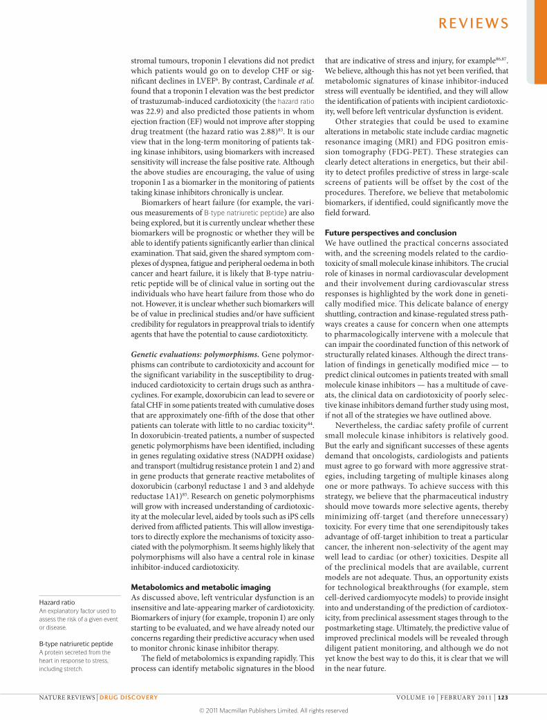
stromal tumours, troponin I elevations did not predict which patients would go on to develop CHF or sig-nificant declines in lveF6. by contrast, Cardinale et al. found that a troponin I elevation was the best predictor of trastuzumab-induced cardiotoxicity (the hazard ratio was 22.9) and also predicted those patients in whom ejection fraction (eF) would not improve after stopping drug treatment (the hazard ratio was 2.88)83. It is our view that in the long-term monitoring of patients tak-ing kinase inhibitors, using biomarkers with increased sensitivity will increase the false positive rate. Although the above studies are encouraging, the value of using troponin I as a biomarker in the monitoring of patients taking kinase inhibitors chronically is unclear.
biomarkers of heart failure (for example, the vari-ous measurements of B-type natriuretic peptide) are also being explored, but it is currently unclear whether these biomarkers will be prognostic or whether they will be able to identify patients significantly earlier than clinical examination. That said, given the shared symptom com-plexes of dyspnea, fatigue and peripheral oedema in both cancer and heart failure, it is likely that b-type natriu-retic peptide will be of clinical value in sorting out the individuals who have heart failure from those who do not. However, it is unclear whether such biomarkers will be of value in preclinical studies and/or have sufficient credibility for regulators in preapproval trials to identify agents that have the potential to cause cardiotoxiticty.
Genetic evaluations: polymorphisms. Gene polymor-phisms can contribute to cardiotoxicity and account for the significant variability in the susceptibility to drug-induced cardiotoxicity to certain drugs such as anthra-cyclines. For example, doxorubicin can lead to severe or fatal CHF in some patients treated with cumulative doses that are approximately one-fifth of the dose that other patients can tolerate with little to no cardiac toxicity84. In doxorubicin-treated patients, a number of suspected genetic polymorphisms have been identified, including in genes regulating oxidative stress (NADPH oxidase) and transport (multidrug resistance protein 1 and 2) and in gene products that generate reactive metabolites of doxorubicin (carbonyl reductase 1 and 3 and aldehyde reductase 1A1)85. Research on genetic polymorphisms will grow with increased understanding of cardiotoxic-ity at the molecular level, aided by tools such as iPS cells derived from afflicted patients. This will allow investiga-tors to directly explore the mechanisms of toxicity asso-ciated with the polymorphism. It seems highly likely that polymorphisms will also have a central role in kinase inhibitor-induced cardiotoxicity.
Metabolomics and metabolic imagingAs discussed above, left ventricular dysfunction is an insensitive and late-appearing marker of cardiotoxicity. biomarkers of injury (for example, troponin I) are only starting to be evaluated, and we have already noted our concerns regarding their predictive accuracy when used to monitor chronic kinase inhibitor therapy.
The field of metabolomics is expanding rapidly. This process can identify metabolic signatures in the blood
that are indicative of stress and injury, for example86,87. we believe, although this has not yet been verified, that metabolomic signatures of kinase inhibitor-induced stress will eventually be identified, and they will allow the identification of patients with incipient cardiotoxic-ity, well before left ventricular dysfunction is evident.
Other strategies that could be used to examine alterations in metabolic state include cardiac magnetic resonance imaging (mRI) and FDG positron emis-sion tomography (FDG-PeT). These strategies can clearly detect alterations in energetics, but their abil-ity to detect profiles predictive of stress in large-scale screens of patients will be offset by the cost of the procedures. Therefore, we believe that metabolomic biomarkers, if identified, could significantly move the field forward.
Future perspectives and conclusionwe have outlined the practical concerns associated with, and the screening models related to the cardio-toxicity of small molecule kinase inhibitors. The crucial role of kinases in normal cardiovascular development and their involvement during cardiovascular stress responses is highlighted by the work done in geneti-cally modified mice. This delicate balance of energy shuttling, contraction and kinase-regulated stress path-ways creates a cause for concern when one attempts to pharmacologically intervene with a molecule that can impair the coordinated function of this network of structurally related kinases. Although the direct trans-lation of findings in genetically modified mice — to predict clinical outcomes in patients treated with small molecule kinase inhibitors — has a multitude of cave-ats, the clinical data on cardiotoxicity of poorly selec-tive kinase inhibitors demand further study using most, if not all of the strategies we have outlined above.
Nevertheless, the cardiac safety profile of current small molecule kinase inhibitors is relatively good. but the early and significant successes of these agents demand that oncologists, cardiologists and patients must agree to go forward with more aggressive strat-egies, including targeting of multiple kinases along one or more pathways. To achieve success with this strategy, we believe that the pharmaceutical industry should move towards more selective agents, thereby minimizing off-target (and therefore unnecessary) toxicity. For every time that one serendipitously takes advantage of off-target inhibition to treat a particular cancer, the inherent non-selectivity of the agent may well lead to cardiac (or other) toxicities. Despite all of the preclinical models that are available, current models are not adequate. Thus, an opportunity exists for technological breakthroughs (for example, stem cell-derived cardiomyocyte models) to provide insight into and understanding of the prediction of cardiotox-icity, from preclinical assessment stages through to the postmarketing stage. Ultimately, the predictive value of improved preclinical models will be revealed through diligent patient monitoring, and although we do not yet know the best way to do this, it is clear that we will in the near future.
Hazard ratioAn explanatory factor used to assess the risk of a given event or disease.
b-type natriuretic peptideA protein secreted from the heart in response to stress, including stretch.
R E V I E W S
NATURe RevIewS | Drug Discovery vOlUme 10 | FebRUARy 2011 | 123
© 2011 Macmillan Publishers Limited. All rights reserved

1. Cohen, P. The role of protein phosphorylation in human health and disease. The Sir Hans Krebs medal lecture. Eur. J. Biochem. 268, 5001–5010 (2001).
2. Verkhivker, G. m. Exploring sequence-structure relationships in the tyrosine kinome space: functional classification of the binding specificity mechanisms for cancer therapeutics. Bioinformatics 23, 1919–1926 (2007).
3. Giamas, G. et al. Kinases as targets in the treatment of solid tumors. Cell. Signal. 22, 984–1002 (2010).
4. Zsila, F., Fitos, I., bencze, G., Keri, G. & Orfi, l. Determination of human serum α1-acid glycoprotein and albumin binding of various marketed and preclinical kinase inhibitors. Curr. Med. Chem. 16, 1964–1977 (2009).
5. Cheng, H. & Force, T. molecular mechanisms of cardiovascular toxicity of targeted cancer therapeutics. Circ. Res. 106, 21–34 (2010).
6. Chu, T. F. et al. Cardiotoxicity associated with tyrosine kinase inhibitor sunitinib. Lancet 370, 2011–2019 (2007).
7. Perez, E. A. et al. Cardiac safety of lapatinib: pooled analysis of 3,689 patients enrolled in clinical trials. Mayo Clin. Proc. 83, 679–686 (2008).
8. Ohren, J. F. et al. Structures of human mAP kinase kinase 1 (mEK1) and mEK2 describe novel noncompetitive kinase inhibition. Nature Struct. Mol. Biol. 11, 1192–1197 (2004).
9. Okram, b. et al. A general strategy for creating “inactive-conformation” Abl inhibitors. Chem. Biol. 13, 779–786 (2006).
10. Sebolt-leopold, J. S. et al. blockade of the mAP kinase pathway suppresses growth of colon tumors in vivo. Nature Med. 5, 810–816 (1999).
11. Zhang, J., Yang, P. l. & Gray, N. S. Targeting cancer with small molecule kinase inhibitors. Nature Rev. Cancer 9, 28–39 (2009).
12. morphy, R. Selectively nonselective kinase inhibition: striking the right balance. J. Med. Chem. 53, 1413–1437 (2010).
13. bhargava, P. VEGF kinase inhibitors: how do they cause hypertension? Am. J. Physiol. Regul. Integr Comp. Physiol. 297, R1–R5 (2009).
14. liu, P., Cheng, H., Roberts, T. m. & Zhao, J. J. Targeting the phosphoinositide 3-kinase pathway in cancer. Nature Rev. Drug. Discov. 8, 627–644 (2009).
15. matsui, T. et al. Akt activation preserves cardiac function and prevents injury after transient cardiac ischemia in vivo. Circulation 104, 330–335 (2001).
16. bantscheff, m. et al. Quantitative chemical proteomics reveals mechanisms of action of clinical Abl kinase inhibitors. Nature Biotech. 25, 1035–1044 (2007).A definitive application of open-ended proteomic technology used to gain insight into off-target effects of kinase inhibitors. This approach underscores the inherent challenges in being able to identify mechanisms of toxicity.
17. meissner, K. et al. The ATP-binding cassette transporter AbCG2 (bCRP), a marker for side population stem cells, is expressed in human heart. J. Histochem. Cytochem. 54, 215–221 (2006).
18. Kerkela, R. et al. Sunitinib-induced cardiotoxicity is mediated by off-target inhibition of AmP-activated protein kinase. Clin. Transl. Sci. 2, 15–25 (2009).
19. Thirunavukkarasu, m. et al. VEGFR1 (Flt-1+/–) gene knockout leads to the disruption of VEGF-mediated signaling through the nitric oxide/heme oxygenase pathway in ischemic preconditioned myocardium. Free. Radic. Biol. Med. 42, 1487–1495 (2007).
20. Thirunavukkarasu, m. et al. Heterozygous disruption of Flk-1 receptor leads to myocardial ischaemia reperfusion injury in mice: application of affymetrix gene chip analysis. J. Cell. Mol. Med. 12, 1284–1302 (2008).
21. Chintalgattu, V. et al. Cardiomyocyte PDGFR-β signaling is an essential component of the mouse cardiac response to load-induced stress. J. Clin. Invest. 120, 472–484 (2010).
22. Izumiya, Y. et al. Vascular endothelial growth factor blockade promotes the transition from compensatory cardiac hypertrophy to failure in response to pressure overload. Hypertension 47, 887–893 (2006).
23. Orphanos, G. S., Ioannidis, G. N. & Ardavanis, A. G. Cardiotoxicity induced by tyrosine kinase inhibitors. Acta Oncol. 48, 964–970 (2009).
24. Karaman, m. W. et al. A quantitative analysis of kinase inhibitor selectivity. Nature Biotech. 26, 127–132 (2008).
The technology described in this study opened the door for broad scale assessment of competitive inhibition of kinase inhibitors, thereby immediately allowing one to understand the challenges associated with making selective kinase inhibitors.
25. liu, l. et al. Hypoxia-induced energy stress regulates mRNA translation and cell growth. Mol. Cell 21, 521–531 (2006).
26. Shell, S. A. et al. Activation of AmPK is necessary for killing cancer cells and sparing cardiac cells. Cell Cycle 7, 1769–1775 (2008).
27. Rixe, O., billemont, b. & Izzedine, H. Hypertension as a predictive factor of sunitinib activity. Ann. Oncol. 18, 1117 (2007).
28. bono, P. et al. Hypertension and clinical benefit of bevacizumab in the treatment of advanced renal cell carcinoma. Ann. Oncol. 20, 393–394 (2009).
29. Rini, b. I. et al. Antitumor activity and biomarker analysis of sunitinib in patients with bevacizumab-refractory metastatic renal cell carcinoma. J. Clin. Oncol. 26, 3743–3748 (2008).
30. Goodwin, R. et al. Treatment-emergent hypertension and outcomes in patients with advanced non-small-cell lung cancer receiving chemotherapy with or without the vascular endothelial growth factor receptor inhibitor cediranib: NCIC Clinical Trials Group Study bR24. Ann. Oncol. 21, 2220–2226 (2010).
31. Hasinoff, b. b. The cardiotoxicity and myocyte damage caused by small molecule anticancer tyrosine kinase inhibitors is correlated with lack of target specificity. Toxicol. Appl. Pharmacol. 244, 190–195 (2010).
32. Fabian, m. A. et al. A small molecule-kinase interaction map for clinical kinase inhibitors. Nature Biotech. 23, 329–336 (2005).
33. Olaharski, A. J. et al. Identification of a kinase profile that predicts chromosome damage induced by small molecule kinase inhibitors. PLoS Comput. Biol. 5, e1000446 (2009).
34. bergmann, O. et al. Evidence for cardiomyocyte renewal in humans. Science 324, 98–102 (2009).A revolutionary application of airborne radiation; the assessment of radioisotopes in human tissues to estimate proliferation rates. This work definitively shows that the myocardium exhibits a baseline proliferation and has ushered out the notion that the adult heart is forever postmitotic.
35. Padin-Iruegas, m. E. et al. Cardiac progenitor cells and biotinylated insulin-like growth factor-1 nanofibers improve endogenous and exogenous myocardial regeneration after infarction. Circulation 120, 876–887 (2009).
36. De Angelis, A. et al. Anthracycline cardiomyopathy is mediated by depletion of the cardiac stem cell pool and is rescued by restoration of progenitor cell function. Circulation 121, 276–292 (2010).A thorough demonstration of the use of exogenous stem cells to attenuate cardiotoxicity induced by doxorubicin. These data provide rationale for the hypothesis that the stem cell compartment in the heart is a target of toxicity. This hypothesis was put forth to explain the increased incidence of heart failure in doxorubicin-treated children.
37. Kajstura, J. et al. Cardiac stem cells and myocardial disease. J. Mol. Cell. Cardiol. 45, 505–513 (2008).
38. Huang, C. et al. Juvenile exposure to anthracyclines impairs cardiac progenitor cell function and vascularization resulting in greater susceptibility to stress-induced myocardial injury in adult mice. Circulation 121, 675–683 (2010).
39. lipshultz, S. E. Exposure to anthracyclines during childhood causes cardiac injury. Semin. Oncol. 33, S8–S14 (2006).
40. li, m. et al. c-kit is required for cardiomyocyte terminal differentiation. Circ. Res. 102, 677–685 (2008).
41. Crone, S. A. et al. Erbb2 is essential in the prevention of dilated cardiomyopathy. Nature Med. 8, 459–465 (2002).
42. Harris, I. S. et al. Raf-1 kinase is required for cardiac hypertrophy and cardiomyocyte survival in response to pressure overload. Circulation 110, 718–723 (2004).
43. lin, R. C. et al. PI3K(p110α) protects against myocardial infarction-induced heart failure: identification of PI3K-regulated miRNA and mRNA. Arterioscler. Thromb. Vasc. Biol. 30, 724–732 (2010).
44. Sano, m. et al. p53-induced inhibition of Hif-1 causes cardiac dysfunction during pressure overload. Nature 446, 444–448 (2007).
45. Adams, R. H. et al. Essential role of p38α mAP kinase in placental but not embryonic cardiovascular development. Mol. Cell 6, 109–116 (2000).
46. molkentin, J. D. & Robbins, J. With great power comes great responsibility: using mouse genetics to study cardiac hypertrophy and failure. J. Mol. Cell. Cardiol. 46, 130–136 (2009).A well-written review about the necessity to understand in deeper detail how transgenic and knockout mice are created, in order to effectively interpret the phenotype that is observed. Specific emphasis is placed on genetically modified mice and their role in understanding cardiac biology.
47. braam, S. R., Passier, R. & mummery, C. l. Cardiomyocytes from human pluripotent stem cells in regenerative medicine and drug discovery. Trends Pharmacol. Sci. 30, 536–545 (2009).
48. braam, S. R. et al. Prediction of drug-induced cardiotoxicity using human embryonic stem cell-derived cardiomyocytes. Stem Cell Res. 4, 107–116 (2010).One of the first articles to use stem cell-derived cardiomyocytes as a model for assessing cardio-active compounds.
49. marroquin, l. D., Hynes, J., Dykens, J. A., Jamieson, J. D. & Will, Y. Circumventing the Crabtree effect: replacing media glucose with galactose increases susceptibility of HepG2 cells to mitochondrial toxicants. Toxicol. Sci. 97, 539–547 (2007).
50. Kimes, b. W. & brandt, b. l. Properties of a clonal muscle cell line from rat heart. Exp. Cell Res. 98, 367–381 (1976).
51. merten, K. E., Jiang, Y., Feng, W. & Kang, Y. J. Calcineurin activation is not necessary for doxorubicin-induced hypertrophy in H9c2 embryonic rat cardiac cells: involvement of the phosphoinositide 3-kinase-Akt pathway. J. Pharmacol. Exp. Ther. 319, 934–940 (2006).
52. Wang, Y. J. et al. Time-dependent block of ultrarapid-delayed rectifier K+ currents by aconitine, a potent cardiotoxin, in heart-derived H9c2 myoblasts and in neonatal rat ventricular myocytes. Toxicol. Sci. 106, 454–463 (2008).
53. Will, Y. et al. Effect of the multitargeted tyrosine kinase inhibitors imatinib, dasatinib, sunitinib, and sorafenib on mitochondrial function in isolated rat heart mitochondria and H9c2 cells. Toxicol. Sci. 106, 153–161 (2008).
54. Simpson, P., mcGrath, A. & Savion, S. myocyte hypertrophy in neonatal rat heart cultures and its regulation by serum and by catecholamines. Circ. Res. 51, 787–801 (1982).
55. Simpson, P. & Savion, S. Differentiation of rat myocytes in single cell cultures with and without proliferating nonmyocardial cells. Cross-striations, ultrastructure, and chronotropic response to isoproterenol. Circ. Res. 50, 101–116 (1982).
56. Kerkela, R. et al. Cardiotoxicity of the cancer therapeutic agent imatinib mesylate. Nature Med. 12, 908–916 (2006).The first paper describing the cardiotoxicity of kinase inhibitors. The results in this paper changed the approach for assessing cardiotoxicity of kinase inhibitors.
57. Hasinoff, b. b., Patel, D. & O’Hara, K. A. mechanisms of myocyte cytotoxicity induced by the multiple receptor tyrosine kinase inhibitor sunitinib. Mol. Pharmacol. 74, 1722–1728 (2008).
58. Zhang, J. et al. Functional cardiomyocytes derived from human induced pluripotent stem cells. Circ. Res. 104, e30–e41 (2009).
59. Itskovitz-Eldor, J. et al. Differentiation of human embryonic stem cells into embryoid bodies compromising the three embryonic germ layers. Mol. Med. 6, 88–95 (2000).
60. Kehat, I. et al. Electromechanical integration of cardiomyocytes derived from human embryonic stem cells. Nature Biotech. 22, 1282–1289 (2004).
61. Satin, J. et al. Calcium handling in human embryonic stem cell-derived cardiomyocytes. Stem Cells 26, 1961–1972 (2008).
62. Vidarsson, H., Hyllner, J. & Sartipy, P. Differentiation of human embryonic stem cells to cardiomyocytes for in vitro and in vivo applications. Stem Cell Rev. 6, 108–120 (2010).
63. liang, H. et al. Human and murine embryonic stem cell-derived cardiomyocytes serve together as a valuable model for drug safety screening. Cell Physiol. Biochem. 25, 459–466 (2010).
64. Hill, A. J., Teraoka, H., Heideman, W. & Peterson, R. E. Zebrafish as a model vertebrate for investigating chemical toxicity. Toxicol. Sci. 86, 6–19 (2005).
R E V I E W S
124 | FebRUARy 2011 | vOlUme 10 www.nature.com/reviews/drugdisc
© 2011 Macmillan Publishers Limited. All rights reserved

65. Jopling, C. et al. Zebrafish heart regeneration occurs by cardiomyocyte dedifferentiation and proliferation. Nature 464, 606–609 (2010).
66. baker, K., Warren, K. S., Yellen, G. & Fishman, m. C. Defective “pacemaker” current (Ih) in a zebrafish mutant with a slow heart rate. Proc. Natl Acad. Sci. USA 94, 4554–4559 (1997).
67. Eimon, P. m. & Rubinstein, A. l. The use of in vivo zebrafish assays in drug toxicity screening. Expert Opin. Drug. Metab. Toxicol. 5, 393–401 (2009).
68. Pugach, E. K., li, P., White, R. & Zon, l. Retro-orbital injection in adult zebrafish. J. Vis. Exp. 34, 1645 (2009).
69. Wenner, m. The most transparent research. Nature Med. 15, 1106–1109 (2009).
70. Herman, E. H. & Ferrans, V. J. Pretreatment with ICRF-187 provides long-lasting protection against chronic daunorubicin cardiotoxicity in rabbits. Cancer Chemother. Pharmacol. 16, 102–106 (1986).
71. Herman, E. H., Ferrans, V. J., Jordan, W. & Ardalan, b. Reduction of chronic daunorubicin cardiotoxicity by ICRF-187 in rabbits. Res. Commun. Chem. Pathol. Pharmacol. 31, 85–97 (1981).
72. Herman, E. H. & Ferrans, V. J. Preclinical animal models of cardiac protection from anthracycline-induced cardiotoxicity. Semin. Oncol. 25, 15–21 (1998).
73. Fernandez, A. et al. An anticancer C-Kit kinase inhibitor is reengineered to make it more active and less cardiotoxic. J. Clin. Invest. 117, 4044–4054 (2007).
74. Olson, H. et al. Concordance of the toxicity of pharmaceuticals in humans and in animals. Regul. Toxicol. Pharmacol. 32, 56–67 (2000).
75. Takahashi, K. et al. Induction of pluripotent stem cells from adult human fibroblasts by defined factors. Cell 131, 861–872 (2007).
76. Yu, J. et al. Induced pluripotent stem cell lines derived from human somatic cells. Science 318, 1917–1920 (2007).
77. Eschenhagen, T. et al. Cardiovascular side-effects of cancer therapies: a position statement from the heart failure association of the european society of cardiology. Eur. J. Heart Fail 13, 1–10 (2011).
78. Zhang, l. & Dokainish, H. Echocardiography in the assessment of heart failure. Minerva Cardioangiol. 57, 457–466 (2009).
79. Apple, F. S. A new season for cardiac troponin assays: it’s time to keep a scorecard. Clin. Chem. 55, 1303–1306 (2009).
80. Apple, F. High-sensitivity cardiac troponin assays: what analytical and clinical issues need to be addressed before introduction into clinical practice? Clin. Chem. 56, 886–891 (2010).
81. Polena, S. et al. Troponin I as a marker of doxorubicin induced cardiotoxicity. Proc. West. Pharmacol. Soc. 48, 142–144 (2005).
82. Cardinale, D. et al. Prevention of high-dose chemotherapy-induced cardiotoxicity in high-risk patients by angiotensin-converting enzyme inhibition. Circulation 114, 2474–2481 (2006).
83. Cardinale, D. et al. Trastuzumab-induced cardiotoxicity: clinical and prognostic implications of troponin I evaluation. J. Clin. Oncol. 28, 3910–3916 (2010).
84. Henderson, I. C. et al. Randomized clinical trial comparing mitoxantrone with doxorubicin in previously treated patients with metastatic breast cancer. J. Clin. Oncol. 7, 560–571 (1989).
85. mordente, A., meucci, E., Silvestrini, A., martorana, G. E. & Giardina, b. New developments in anthracycline-induced cardiotoxicity. Curr. Med. Chem. 16, 1656–1672 (2009).
86. lewis, G. D., Asnani, A. & Gerszten, R. E. Application of metabolomics to cardiovascular biomarker and pathway discovery. Am. Coll. Cardiol. 52, 117–123 (2008).
87. lewis, G. D. et al. metabolic signatures of exercise in human plasma. Sci. Transl. Med. 2, 33–37 (2010).
88. Olson, E. N. A decade of discoveries in cardiac biology. Nature Med. 10, 467–474 (2004).
89. brini, m. & Carafoli, E. Calcium pumps in health and disease. Physiol. Rev. 89, 1341–1378 (2009).
90. Swain, J. l., Sabina, R. l., mcHale, P. A., Greenfield, J. C. Jr & Holmes, E. W. Prolonged myocardial nucleotide depletion after brief ischemia in the open-chest dog. Am. J. Physiol. 242, H818–H826 (1982).
91. Khouri, E. m., Gregg, D. E. & Rayford, C. R. Effect of exercise on cardiac output, left coronary flow and myocardial metabolism in the unanesthetized dog. Circ. Res. 17, 427–437 (1965).
92. barry, S. P., Davidson, S. m. & Townsend, P. A. molecular regulation of cardiac hypertrophy. Int. J. Biochem. Cell. Biol. 40, 2023–2039 (2008).
93. Oliveira, R. S. et al. Cardiac anti-remodelling effect of aerobic training is associated with a reduction in the calcineurin/NFAT signalling pathway in heart failure mice. J. Physiol. 587, 3899–3910 (2009).
94. Zhang, T. et al. CamKIIδ isoforms differentially affect calcium handling but similarly regulate HDAC/mEF2 transcriptional responses. J. Biol. Chem. 282, 35078–35087 (2007).
95. Zhang, T. et al. Phospholamban ablation rescues sarcoplasmic reticulum Ca2+ handling but exacerbates cardiac dysfunction in CamKIIδC transgenic mice. Circ. Res. 106, 354–362 (2010).
96. Vittone, l., mundina-Weilenmann, C. & mattiazzi, A. Phospholamban phosphorylation by CamKII under pathophysiological conditions. Front. Biosci. 13, 5988–6005 (2008).
97. Ghoreschi, K., laurence, A. & O’Shea, J. J. Selectivity and therapeutic inhibition of kinases: to be or not to be? Nature Immunol. 10, 356–360 (2009).
98. louvet, C. et al. Tyrosine kinase inhibitors reverse type 1 diabetes in nonobese diabetic mice. Proc. Natl Acad. Sci. USA 105, 18895–18900 (2008).
99. mariani S. et al. Imatinib does not substantially modify the glycemic profile in patients with chronic myeloid leukaemia. Leuk. Res. 34, e5–e7 (2010).
100. Agostino N. et al. Effect of the tyrosine kinase inhibitors (sunitinib, sorafenib, dasatinib, and imatinib) on blood glucose levels in diabetic and nondiabetic patients in general clinical practice. J. Oncol. Pharm. Pract. 4 Aug 2010 (doi:10.1177/1078155210378913).
101. Schermuly, R. T. et al. Reversal of experimental pulmonary hypertension by PDGF inhibition. J. Clin. Invest. 115, 2811–2821 (2005).
102. Klein, m. et al. Combined tyrosine and serine/threonine kinase inhibition by sorafenib prevents progression of experimental pulmonary hypertension and myocardial remodeling. Circulation 118, 2081–2090 (2008).
103. Ghofrani, H. A. et al. Imatinib in pulmonary arterial hypertension patients with inadequate response to established therapy. Am. J. Respir. Crit. Care Med. 182, 1171–1177 (2010).
104. Wang, C. H. et al. Stem cell factor deficiency is vasculoprotective: unraveling a new therapeutic potential of imatinib mesylate. Circ. Res. 99, 617–625 (2006).
105. makiyama, Y. et al. Imatinib mesilate inhibits neointimal hyperplasia via growth inhibition of vascular smooth muscle cells in a rat model of balloon injury. Tohoku J. Exp. Med. 215, 299–306 (2008).
106. Ayach, b. b. et al. Stem cell factor receptor induces progenitor and natural killer cell-mediated cardiac survival and repair after myocardial infarction. Proc. Natl Acad. Sci. USA 103, 2304–2309 (2006).
107. Force, T. et al. Research priorities in hypertrophic cardiomyopathy: report of a working group of the National Heart, lung, and blood Institute. Circulation 122, 1130–1133 (2010).
108. Ahmad, F. et al. Increased α2 subunit-associated AmPK activity and PRKAG2 cardiomyopathy. Circulation 112, 3140–3148 (2005).
109. Tao, R., Zhang, J., Vessey, D. A., Honbo, N. & Karliner, J. S. Deletion of the sphingosine kinase-1 gene influences cell fate during hypoxia and glucose deprivation in adult mouse cardiomyocytes. Cardiovasc. Res. 74, 56–63 (2007).
110. moga, m. A., Nakamura, T. & Robbins, J. Genetic approaches for changing the heart and dissecting complex syndromes. J. Mol. Cell. Cardiol. 45, 148–155 (2008).
111. Aoki, Y., Niihori, T., Narumi, Y., Kure, S. & matsubara, Y. The RAS/mAPK syndromes: novel roles of the RAS pathway in human genetic disorders. Hum. Mutat. 29, 992–1006 (2008).
112. Gelb, b. D. & Tartaglia, m. Noonan syndrome and related disorders: dysregulated RAS-mitogen activated protein kinase signal transduction. Hum. Mol. Genet. 15, R220–R226 (2006).
113. Yamaguchi, O. et al. Cardiac-specific disruption of the c-raf-1 gene induces cardiac dysfunction and apoptosis. J. Clin. Invest. 114, 937–943 (2004).
114. mcmullen, J. R. & Jay, P. Y. PI3K(p110α) inhibitors as anti-cancer agents: minding the heart. Cell Cycle 6, 910–913 (2007).
115. mcmullen, J. R. et al. Protective effects of exercise and phosphoinositide 3-kinase(p110α) signaling in dilated and hypertrophic cardiomyopathy. Proc. Natl Acad. Sci. USA 104, 612–617 (2007).
116. Rose, R. A., Kabir, m. G. & backx, P. H. Altered heart rate and sinoatrial node function in mice lacking the cAmP regulator phosphoinositide 3-kinase-γ. Circ. Res. 101, 1274–1282 (2007).
117. Oudit, G. Y. et al. Phosphoinositide 3-kinase-γ-deficient mice are protected from isoproterenol-induced heart failure. Circulation 108, 2147–2152 (2003).
118. Oudit, G. Y. et al. The role of phosphoinositide-3 kinase and PTEN in cardiovascular physiology and disease. J. Mol. Cell. Cardiol. 37, 449–471 (2004).
119. mora, A. et al. Deficiency of PDK1 in cardiac muscle results in heart failure and increased sensitivity to hypoxia. EMBO J. 22, 4666–4676 (2003).
120. Debosch, b. et al. Akt1 is required for physiological cardiac growth. Circulation 113, 2097–2104 (2006).
121. Debosch, b., Sambandam, N., Weinheimer, C., Courtois, m. & muslin, A. J. Akt2 regulates cardiac metabolism and cardiomyocyte survival. J. Biol. Chem. 281, 32841–32851 (2006).
122. lee, C. H., Inoki, K. & Guan, K. l. mTOR pathway as a target in tissue hypertrophy. Annu. Rev. Pharmacol. Toxicol. 47, 443–467 (2007).
123. Ciuffreda, l., Di Sanza, C., Incani, U. C. & milella, m. The mTOR pathway: a new target in cancer therapy. Curr. Cancer Drug Targets 10, 10484–10495 (2010).
124. blair, E. et al. mutations in the γ2 subunit of AmP-activated protein kinase cause familial hypertrophic cardiomyopathy: evidence for the central role of energy compromise in disease pathogenesis. Hum. Mol. Genet. 10, 1215–1220 (2001).
125. Zhang, P. et al. AmP activated protein kinase-α2 deficiency exacerbates pressure-overload-induced left ventricular hypertrophy and dysfunction in mice. Hypertension 52, 918–924 (2008).
126. matsuda, T. et al. Distinct roles of GSK-3α and GSK-3β phosphorylation in the heart under pressure overload. Proc. Natl Acad. Sci. USA 105, 20900–20905 (2008).
127. Kerkela, R. et al. Deletion of GSK-3β in mice leads to hypertrophic cardiomyopathy secondary to cardiomyoblast hyperproliferation. J. Clin. Invest. 118, 3609–3618 (2008).
128. Woulfe, K. C. et al. Glycogen synthase kinase-3β regulates post-myocardial infarction remodeling and stress-induced cardiomyocyte proliferation in vivo. Circ. Res. 106, 1635–1645 (2010).
129. barriere, C. et al. mice thrive without Cdk4 and Cdk2. Mol. Oncol. 1, 72–83 (2007).
130. liem, D. A. et al. Cyclin-dependent kinase 2 signaling regulates myocardial ischemia/reperfusion injury. J. Mol. Cell. Cardiol. 45, 610–616 (2008).
131. Perez Fidalgo, J. A., Roda, D., Rosello, S., Rodriguez-braun, E. & Cervantes, A. Aurora kinase inhibitors: a new class of drugs targeting the regulatory mitotic system. Clin. Transl. Oncol. 11, 787–798 (2009).
132. lapenna, S. & Giordano, A. Cell cycle kinases as therapeutic targets for cancer. Nature Rev. Drug. Discov. 8, 547–566 (2009).
133. Chopra, P., Sethi, G., Dastidar, S. G. & Ray, A. Polo-like kinase inhibitors: an emerging opportunity for cancer therapeutics. Expert Opin. Investig. Drugs 19, 27–43 (2010).
134. Noma, T. et al. β-arrestin-mediated β1-adrenergic receptor transactivation of the EGFR confers cardioprotection. J. Clin. Invest. 117, 2445–2458 (2007).
135. De Keulenaer, G. W., Doggen, K. & lemmens, K. The vulnerability of the heart as a pluricellular paracrine organ: lessons from unexpected triggers of heart failure in targeted Erbb2 anticancer therapy. Circ. Res. 106, 35–46 (2010).
136. Iwamoto, R. et al. Heparin-binding EGF-like growth factor and Erbb signaling is essential for heart function. Proc. Natl Acad. Sci. USA 100, 3221–3226 (2003).
137. liu, F. F. et al. Heterozygous knockout of neuregulin-1 gene in mice exacerbates doxorubicin-induced heart failure. Am. J. Physiol. 289, H660–H666 (2005).
138. Garcia-Rivello, H. et al. Dilated cardiomyopathy in Erb-b4-deficient ventricular muscle. Am. J. Physiol. 289, H1153–H1160 (2005).
139. Fazel, S. et al. Cardioprotective c-kit+ cells are from the bone marrow and regulate the myocardial balance of angiogenic cytokines. J. Clin. Invest. 116, 1865–1877 (2006).
140. Hilfiker-Kleiner, D., limbourg, A. & Drexler, H. STAT3-mediated activation of myocardial capillary growth. Trends Cardiovas. Med. 15, 152–157 (2005).
141. Kunisada, K. et al. Signal transducer and activator of transcription 3 in the heart transduces not only a hypertrophic signal but a protective signal against doxorubicin-induced cardiomyopathy. Proc. Natl Acad. Sci. USA 97, 315–319 (2000).
R E V I E W S
NATURe RevIewS | Drug Discovery vOlUme 10 | FebRUARy 2011 | 125
© 2011 Macmillan Publishers Limited. All rights reserved
142. barry, S. P., Townsend, P. A., latchman, D. S. & Stephanou, A. Role of the JAK-STAT pathway in myocardial injury. Trends Mol. Med. 13, 82–89 (2007).
143. Peng, X. et al. Inactivation of focal adhesion kinase in cardiomyocytes promotes eccentric cardiac hypertrophy and fibrosis in mice. J. Clin. Invest. 116, 217–227 (2006).
144. O’Cochlain, D. F. et al. Transgenic overexpression of human DmPK accumulates into hypertrophic cardiomyopathy, myotonic myopathy and hypotension traits of myotonic dystrophy. Hum. Mol. Genet. 13, 2505–2518 (2004).
145. Honda, H. et al. Heart-specific activation of lTK results in cardiac hypertrophy, cardiomyocyte degeneration and gene reprogramming in transgenic mice. Oncogene 18, 3821–3830 (1999).
146. Shi J., Zhang, Y. W., Yang, Y., Zhang, l. & Wei, l. ROCK1 plays an essential role in the transition from cardiac hypertrophy to failure in mice. J. Mol. Cell Cardiol. 49, 819–828 (2010).
147. Zhang, Y. m. et al. Targeted deletion of ROCK1 protects the heart against pressure overload by inhibiting reactive fibrosis. FASEB J. 20, 916–925 (2006).
148. Ikeda, Y. et al. Cardiac-specific deletion of lKb1 leads to hypertrophy and dysfunction. J. Biol. Chem. 284, 35839–35849 (2009).
149. Zheng, m. et al. Cardiac-specific ablation of Cypher leads to a severe form of dilated cardiomyopathy with premature death. Hum. Mol. Genet. 18, 701–713 (2009).
150. lorenz, K., Schmitt, J. P., Schmitteckert, E. m. & lohse, m. J. A new type of ERK1/2 autophosphorylation causes cardiac hypertrophy. Nature Med. 15, 75–83 (2009).
151. lips, D. J. et al. mEK1-ERK2 signaling pathway protects myocardium from ischemic injury in vivo. Circulation 109, 1938–1941 (2004).
152. Kehat, I. & molkentin, J. D. Extracellular signal-regulated kinase 1/2 (ERK1/2) signaling in cardiac hypertrophy. Ann. NY Acad. Sci. 1188, 96–102 (2010).
153. Nakamura, T. et al. mediating ERK 1/2 signaling rescues congenital heart defects in a mouse model of Noonan syndrome. J. Clin. Invest. 117, 2123–2132 (2007).
154. liu, Q. et al. PKCα, but not PKCβ or PKCγ, regulates contractility and heart failure susceptibility: implications for ruboxistaurin as a novel therapeutic approach. Circ. Res. 105, 194–200 (2009).
155. Takimoto, E. et al. Regulator of G protein signaling 2 mediates cardiac compensation to pressure overload and antihypertrophic effects of PDE5 inhibition in mice. J. Clin. Invest. 119, 408–420 (2009).
156. Guazzi, m., Vicenzi, m., Arena, R. & Guazzi, m. D. PDE5-inhibition with sildenafil improves left ventricular diastolic function, cardiac geometry and clinical status in patients with stable systolic heart
failure: results of a 1-year prospective, randomized, rlacebo-controlled study. Circ. Heart Fail. 29 Oct 2010 (doi:10.1161/ circheartfailure.110.944694).
157. muraski, J. A. et al. Pim-1 regulates cardiomyocyte survival downstream of Akt. Nature Med. 13, 1467–1475 (2007).
158. Sag, C. m. et al. Calcium/calmodulin-dependent protein kinase II contributes to cardiac arrhythmogenesis in heart failure. Circ. Heart Fail. 2, 664–675 (2009).
159. ling, H. et al. Requirement for Ca2+/calmodulin-dependent kinase II in the transition from pressure overload-induced cardiac hypertrophy to heart failure in mice. J. Clin. Invest. 119, 1230–1240 (2009).
160. lymperopoulos, A. et al. Reduction of sympathetic activity via adrenal-targeted GRK2 gene deletion attenuates heart failure progression and improves cardiac function after myocardial infarction. J. Biol. Chem. 285, 16378–16386 (2010).
161. Eckhart, A. D. et al. Hybrid transgenic mice reveal in vivo specificity of G protein-coupled receptor kinases in the heart. Circ. Res. 86, 43–50 (2000).
162. Yamaguchi, O. et al. Targeted deletion of apoptosis signal-regulating kinase 1 attenuates left ventricular remodeling. Proc. Natl Acad. Sci. USA 100, 15883–15888 (2003).
163. Taniike, m. et al. Apoptosis signal-regulating kinase 1/p38 signaling pathway negatively regulates physiological hypertrophy. Circulation 117, 545–552 (2008).
164. Hescheler, J. et al. morphological, biochemical, and electrophysiological characterization of a clonal cell (H9c2) line from rat heart. Circ. Res. 69, 1476–1486 (1991).
165. Zordoky, b. N. & El-Kadi, A. O. H9c2 cell line is a valuable in vitro model to study the drug metabolizing enzymes in the heart. J. Pharmacol. Toxicol. Methods 56, 317–322 (2007).
166. Field, l. J. Atrial natriuretic factor-SV40 T antigen transgenes produce tumors and cardiac arrhythmias in mice. Science 239, 1029–1033 (1988).
167. White, S. m., Constantin, P. E. & Claycomb, W. C. Cardiac physiology at the cellular level: use of cultured Hl-1 cardiomyocytes for studies of cardiac muscle cell structure and function. Am. J. Physiol. 286, H823–H829 (2004).
168. Eimre, m. et al. Distinct organization of energy metabolism in Hl-1 cardiac cell line and cardiomyocytes. Biochim. Biophys. Acta 1777, 514–524 (2008).
169. Fritzsche, m., Fredriksson, J. m., Carlsson, m. & mandenius, C. F. A cell-based sensor system for toxicity testing using multiwavelength fluorescence spectroscopy. Anal. Biochem. 387, 271–275 (2009).
170. Zhang, Y., Nuglozeh, E., Toure, F., Schmidt, A. m. & Vunjak-Novakovic, G. Controllable expansion of primary cardiomyocytes by reversible immortalization. Hum. Gene Ther. 20, 1687–1696 (2009).
171. Pentassuglia, l. et al. Inhibition of Erbb2/neuregulin signaling augments paclitaxel-induced cardiotoxicity in adult ventricular myocytes. Exp. Cell Res. 313, 1588–1601 (2007).
172. Deng, X. F., Rokosh, D. G. & Simpson, P. C. Autonomous and growth factor-induced hypertrophy in cultured neonatal mouse cardiac myocytes. Comparison with rat. Circ. Res. 87, 781–788 (2000).
173. Gussak, I., Chaitman, b. R., Kopecky, S. l. & Nerbonne, J. m. Rapid ventricular repolarization in rodents: electrocardiographic manifestations, molecular mechanisms, and clinical insights. J. Electrocardiol. 33, 159–170 (2000).
174. brouillette, J., Clark, R. b., Giles, W. R. & Fiset, C. Functional properties of K+ currents in adult mouse ventricular myocytes. J. Physiol. 559, 777–798 (2004).
175. Volz, A., Piper, H. m., Siegmund, b. & Schwartz, P. longevity of adult ventricular rat heart muscle cells in serum-free primary culture. J. Mol. Cell. Cardiol. 23, 161–173 (1991).
176. Ellingsen, O. et al. Adult rat ventricular myocytes cultured in defined medium: phenotype and electromechanical function. Am. J. Physiol. 265, H747–H754 (1993).
177. bistola, V. et al. long-term primary cultures of human adult atrial cardiac myocytes: cell viability, structural properties and bNP secretion in vitro. Int. J. Cardiol. 131, 113–122 (2008).
178. benardeau, A. et al. Primary culture of human atrial myocytes is associated with the appearance of structural and functional characteristics of immature myocardium. J. Mol. Cell. Cardiol. 29, 1307–1320 (1997).
179. li, R. K. et al. Human pediatric and adult ventricular cardiomyocytes in culture: assessment of phenotypic changes with passaging. Cardiovasc. Res. 32, 362–373 (1996).
180. Davidson, m. m. et al. Novel cell lines derived from adult human ventricular cardiomyocytes. J. Mol. Cell. Cardiol. 39, 133–147 (2005).
181. Zhou, J. et al. GSK-3α directly regulates β adrenergic signaling and the response of the heart to hemodynamic stress in mice. J. Clin. Invest. 120, 2280–2291 (2010).
182. Kondo, R. P. et al. Comparison of contraction and calcium handling between right and left ventricular myocytes from adult mouse heart: a role for repolarization waveform. J. Physiol. 571,131–46 (2006).
Competing interests statementThe author declares competing financial interests: see web version for details.
FURTHER INFORMATIONClinicaltrials.gov: http://www.clinicaltrials.gov/
All linKs Are Active in the online PDf
R E V I E W S
126 | FebRUARy 2011 | vOlUme 10 www.nature.com/reviews/drugdisc
© 2011 Macmillan Publishers Limited. All rights reserved