Embed Size (px)
Citation preview

Myocardial diseases
• Cardiomyopathy• Myocarditis

Cardiomyopathy
• Primary abnormality in the myocardium• Myocardial dysfunction secondary to IHD,
hypertension or valvular disease not included

Conditions with heart muscle disease
Cardiac infections• Virus• Bacteria• Rickettsia• ChlamydiaInfiltrative• Leukemia• Carcinomatosis• Radiation induced fibrosis
Storage disorders + other depositions
• Hunter- Hurler synd • Glycogen storage dis • AmyloidosisToxins• Alcohol• Catecholamines• As• Cyclophosphamide• Doxorubicin

Metabolic• Hyperthyroidism• Hypothyroidism• Hyper/ hypo kalemia• Hemochromatosis• Nutritional deficiency
Neuromuscular dis• Friedreich ataxia• Muscular dystrophyImmunologic• Myocarditis• Post transplant rejection

Clinical, functional and pathological patterns
• Dilated cardiomyopathy• Hypertrophic cardiomyopathy• Restrictive cardiomyopathy
Diagnosis: endomyocardial biopsies using bioptome, inserted transvenously into right side of heart and snipping a small piece of septal myocardium


Dilated cardiomyopathy
• Progressive cardiac dilatation and systolic (contractile) dysfunction, usually with concomitant hypertrophy
• Also called Congestive cardiomyopathy• 25-35% familal/ genetic form• Acquired causes

Pathogenesis
• Genetic influences: - autosomal dominant; X-linked, autosomal recessive and mitochondrial inheritance less common- affect genes that encode cytoskeleton proteins expressed by myocytes (dystrophin, desmin, lamin A, lamin C) or - deletion of mitochondrial genes (most often a cause in children)- X-linked assoc with dystrophin gene mutation (cell membrane based cytoskeletal protein)

Pathogenesis
• Myocarditis: viral nucleic acids from coxsackie and enterovirus have been detected
• Toxicities:- alcoholism: alcohol & acetaldehyde →direct toxic effect on the myocardium + secondary nutritional deficiencies i.e beriberi- non-alcoholic toxic agents: doxorubicin (Adriamycin), cobalt
• Peripartum CMP: occurs late in pregnancy or several weeks to months post partum - D/t: PIH, volume overload, nutritional deficiency or immunological reaction
• Idiopathic dilated CMP
Morphology
• Heart heavy: 2-3 times normal• Enlarged, flabby with dilation of all the chambers• Ventricular thickness <, =,> normal• Chambers show mural thrombi: source of thromboemboli• Functional regurgitation of mitral or tricuspid valves• Coronary arteries are free of significant narrowing• M/E- non-specific
- most muscle cells are hypertrophied with enlarged nuclei, some are attenuated, stretched and irregular- interstitial and endocardial fibrosis with small subendocardial scars due to previous ischemia
Clinical features
• Any age, 20 - 50 yrs • Slowly progressive signs and symptoms of CHF-
shortness of breath, easy fatiguability, poor exertional capacity
• In end stage: ejection fraction< 25% (normal: 50-65%)• Secondary MR / arrhythmias common• Thromboemboli• Cardiac transplant recommended

Hypertrophic Cardiomyopathy
• Also known as Idiopathic subaortic stenosis/ Hypertrophic obstructive CMP
• Myocardial hypertrophy, poorly compliant lt ventricular myocardium, abnormal diastolic filling, intermittent ventricular outflow obstruction
• D/D valvular and subvalvular AS• Pathogenesis
- mutations (single point missense mutations) in genes encoding proteins of sarcomere, contractile units of cardiac and skeletal muscle- autosomal dominant inheritence
Morphology
• Massive ventricular hypertrophy without ventricular dilation
• septal thickness as compared to that of ventricular free wall (ratio > 1.3:1), i.e asymmetrical septal hypertrophy
• Cross section: ventricular cavity loses its round to ovoid shape and may be compressed into a banana shaped configuration by bulging of the interventricular septum into the lumen
• Hypertrophy most prominent in subaortic region• Endocardial thickening or mural plaque formation in the
ventricular outflow tract + thickening of the ant MV leaflet

Microscopy
• Extensive myocardial hypertrophy with marked increase in size of myocytes (transverse myocyte dia >40µm; n= 15µm)
• Haphazard distribution of bundles of myocytes, individual myocytes in a bundle, and contractile elements within the sarcomere in the cells (myofibre disarray)
• Interstitial and replacement fibrosis


Clinical Features
• Basic physiologic abnormality: ↓ stroke vol due to poor diastolic filling resulting from ↓ chamber size & compliance of hypertrophied LV
• 25% patients have dynamic obstruction to LV outflow• Exertional dyspnoea (↓ CO & pulmonary venous
pressure)• Harsh ejection systolic murmur• Frequent angina d/t focal myocardial ischemia• Major clinical problems: atrial fibrillation, mural thrombi
with embolisation, infective endocarditis of MV, intractable cardiac failure, ventricular arrhythmias
• M/C cause of sudden death in young athletes

• Rx: Medical therapy to enhance ventricular relaxation• Surgical reduction in muscle mass of septum• Alcohol is infused through a catheter to cause infarction
of the myocardium
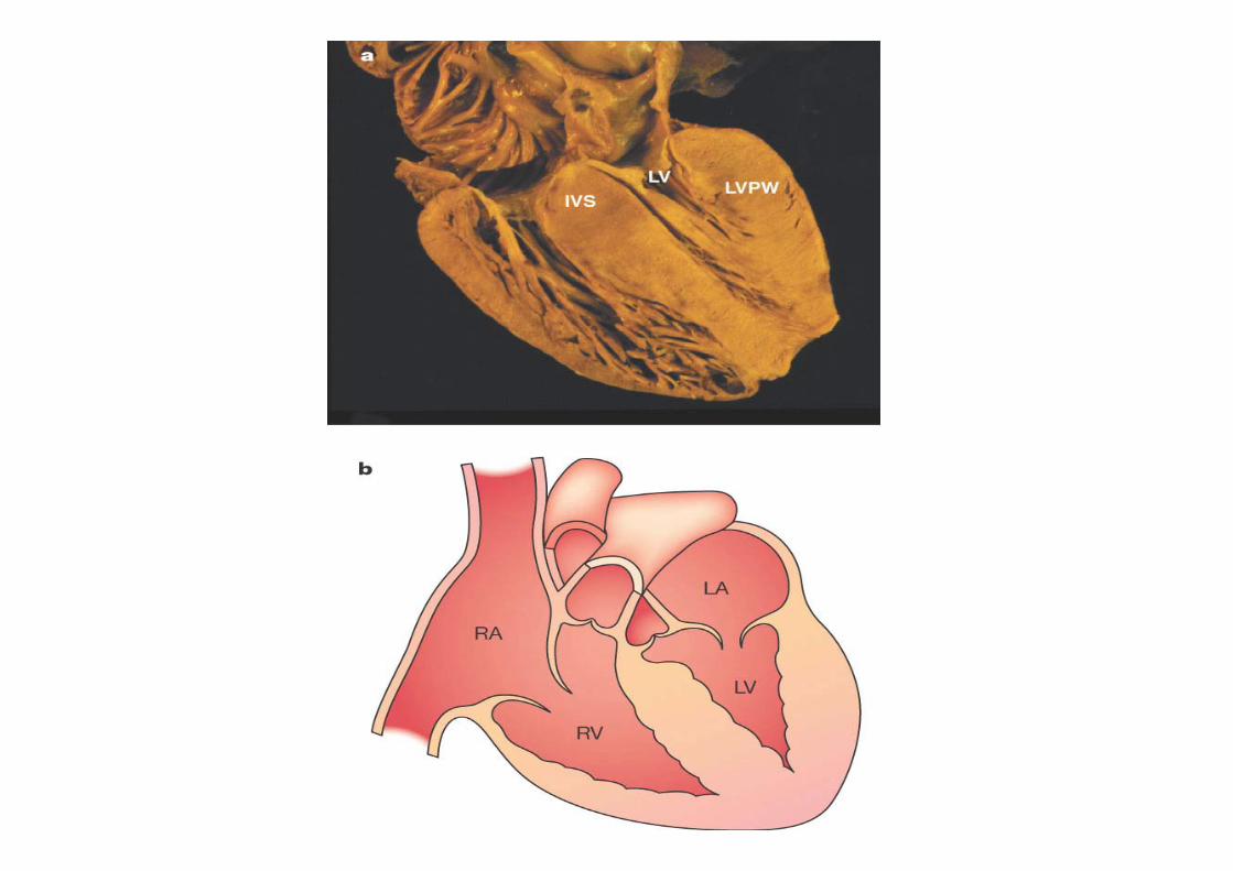
Restrictive Cardiomyopathy
• Primary decrease in ventricular compliance: impaired filling during diastole, systolic function normal
• D/D; constrictive pericarditis or HCM• Associated with radiation fibrosis, amyloidosis,
sarcoidosis, metastatic tumors, inborn errors of metab • Idiopathic
Morphology
• Ventricles normal sized or enlarged; cavities not dilated, myocardium firm
• Biatrial dilation +• M/E: patchy / diffuse interstitial fibrosis• Endomyocardial Bx diagnostic to distinguish from other
restrictive conditions
Other restrictive conditions
• Endomyocardial fibrosis: children and young adults• Loeffler endomyocarditis: assoc with eosinophillia;
release of MBP from eosinophil granules; has large mural thrombi + endomyocardial fibrosis
• Endocardial fibroelastosis: uncommon heart dis; in first 2 yrs of life

Function-pattern
LV ejection fraction
Mech of Heart failure
Causes Indirect Myo dysfuncn
Dilated <40% Systolic dysfn/ contractile
Alcohol, peripartum, myocarditis, hemochromatosis, chronic anemia, adriamycin, sarcoidosis, genetic
IHD, Valvular, HHD, CHD
Hypertrophic 50-80% ↓ compliance/ diastolic dysfunction
Genetic: Friedreich ataxia; storage dis; infants of diabetic mothers
HHD, AS
Restrictive 45-90% ↓ compliance/ diast dysfn
Amyloidosis, radiation induced fibrosis
Pericardial constriction


Myocarditis
• Inflammatory processes of myocardium that result in injury to cardiac myocytes
• Secondary inflammation may be seen in ischemic injury

Major causes of Myocarditis
Infections• Viruses: Coxsackie,
enterovirus, ECHO, influenza, HIV
• Chlamydia• Rickettsia• Bacteria: diphtheria,
Borrelia (Lymes dis), Neisseria
• Fungi: Candida• Protozoa: Chagas dis
(Trypanosoma cruzi), toxoplasma
• Helminths: Trichinosis
Immune-mediated• Post-viral• Post streptococcal• SLE• Drug hypersensitivity:
antibiotics, diuretics, antihypertensive agents
• Transplant rejectionUnknown• Sarcoidosis• Giant cell myocarditis
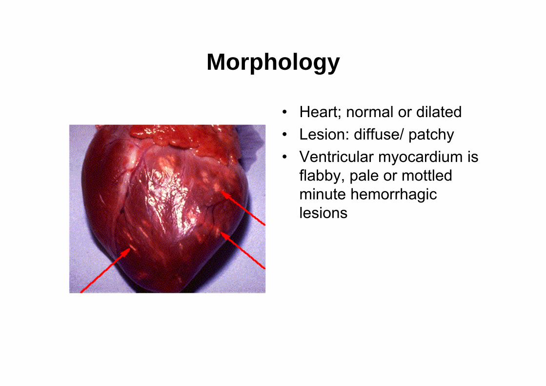
Morphology
• Heart; normal or dilated• Lesion: diffuse/ patchy• Ventricular myocardium is
flabby, pale or mottled minute hemorrhagic lesions

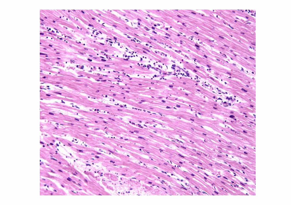
Micro: interstitial inflammatory infiltrate, usually lymphocytic, focal necrosis of myocytes- heal by resolution with no residual changes or heal by progressive fibrosis
• Hypersensitivity myocarditis: perivascular inflammation by lymphos, macrophages, eosinophils
• Giant cell myocarditis: extensive necrosis with multinucleated giant cells (macrophage/ myocyte origin), lymphos, eos, plasma cells, macrophages
• Chagas dis: parasitization of various myocytes by trypanosomes, inflammatory infiltrate of neutros, lymphos, macrophages, eos

Clinical features
• Spectrum: asymptomatic without sequelaeor precipitous onset of heart failure/ arrhythmias with sudden death
• Systolic murmur d/t vent dilation + MR• Nonspecific symptoms of fatigue, dyspnoea, palpitations,
precordial discomfort, fever• Can mimic MI• Later yrs: dilated CMP devlopes


![WELCOME [gmch.gov.in]](https://img.dokumen.tips/doc/110x75/616a5d6311a7b741a351b2cf/welcome-gmchgovin.jpg)


