Embed Size (px)
Citation preview

Cardiac Sarcoidosis
Vera H. Rigolin, MDVice-President, American Society of Echocardiography
Professor of MedicineNorthwestern University
Bluhm Cardiovascular InstituteMedical Director, Echocardiography Laboratory
Northwestern Memorial Hospital

Disclosures: None
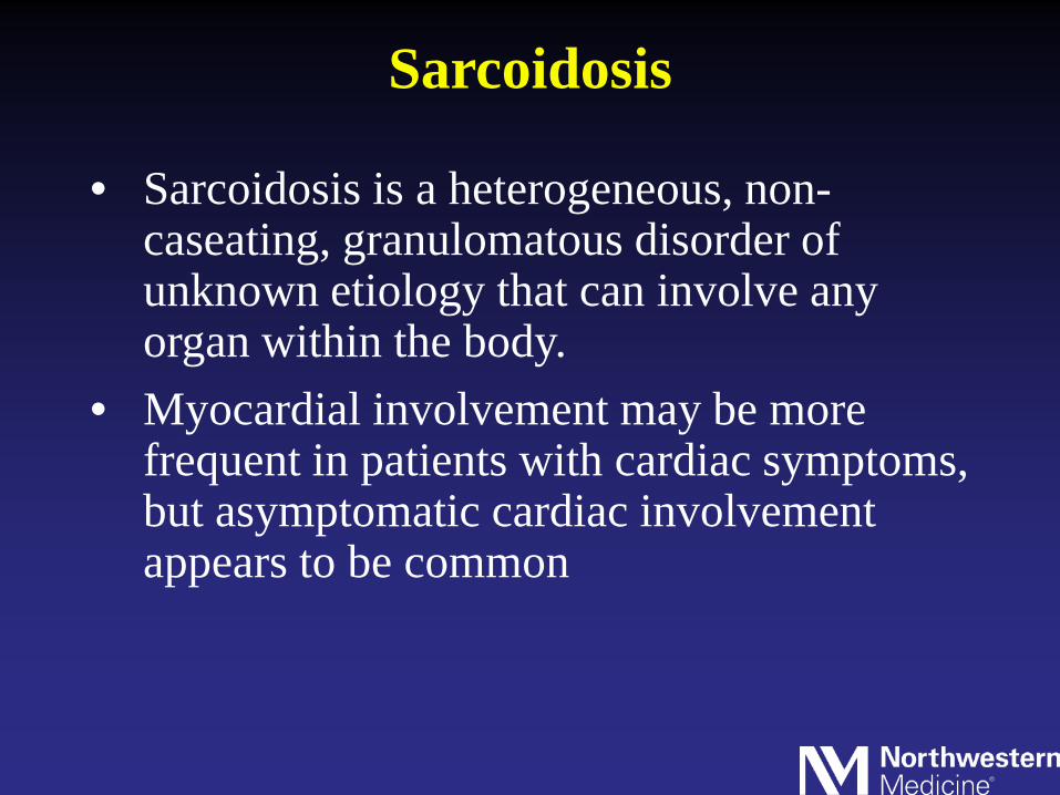
Sarcoidosis
• Sarcoidosis is a heterogeneous, non-caseating, granulomatous disorder of unknown etiology that can involve any organ within the body.
• Myocardial involvement may be more frequent in patients with cardiac symptoms, but asymptomatic cardiac involvement appears to be common


Clinical Manifestations
• Conduction abnormalities (atrioventricularblock or bundle-branch block)
• Tachyarrhythmias• Sudden cardiac death• Coronary infiltration (leading to spasm or
vasculitis)• Cardiomyopathy• Congestive heart failure
• Granulomatous involvement of myocardium• Granulomatous involvement of valves, pap
muscles
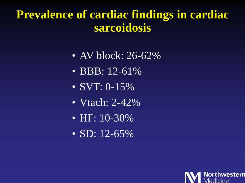
Prevalence of cardiac findings in cardiac sarcoidosis
• AV block: 26-62%• BBB: 12-61%• SVT: 0-15%• Vtach: 2-42%• HF: 10-30%• SD: 12-65%

Echo Findings• Left ventricular dilatation• Septal thinning • Segmental or global hypokinesia of
the left ventricle• Ventricular aneurysms• Valvular regurgitation• Right ventricular dilatation and
hypokinesis.

Echo Findings
• Septum and LV free wall most commonly affected
• Increase in wall thickness simulating LVH or HCM
• Wall motion abnormalities in noncoronary distribution

Speckle Tracking Echocardiography Identifies Patients with Cardiac Sarcoidosis
Sadiya S. Khan, MD; Jason Chodakowski, BS; Jyothy Puthumana, MD; Alex Chicos, MD

Cardiac MRI
• T1 weighted images detect wall motion, hypertrophy, wall thinning
• T2 weighted images/early gad detect edema (inflammation)
• Late gad detects fibrosis/scar
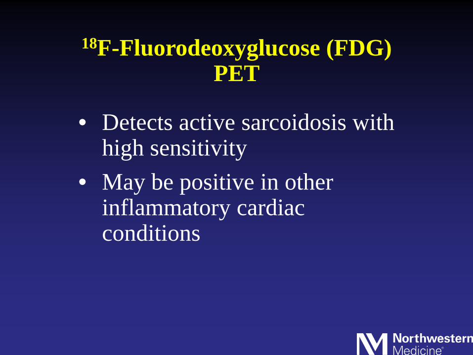
18F-Fluorodeoxyglucose (FDG) PET
• Detects active sarcoidosis with high sensitivity
• May be positive in other inflammatory cardiac conditions


Radionuclide Imaging: Thallium-201
• Focal perfusion deficits may be seen at rest • With exercise, “reverse redistribution” is seen• Fixed defects may represent scar• Gallium-67 can detect active inflammation

Accuracy of Diagnostic Tests
Kim JS et al. Am Heart J 2009;157:9-21.
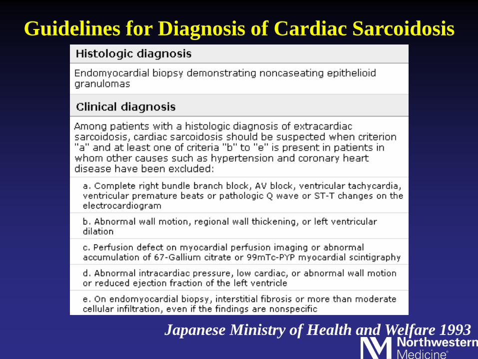
Guidelines for Diagnosis of Cardiac Sarcoidosis
Japanese Ministry of Health and Welfare 1993

2006 Revised Guidelines by the Japanese Society of Sarcoidosisand Other Granulomatous Disorders

Suggested Clinical Algorithm
Kim JS et al. Am Heart J 2009;157:9-21.
Case 1• 48 yr old female who complains of palpitations
for 1 month• She presented to the ER when palpitations
occurred with lightheadedness• Initial rhythm was Afib. She converted to SR. VT
then noted• CT ordered to look for PE: Lymphadenopathy
noted

• Endobronchial biopsy: Sarcoidosis• Endomyocardial biopsy: Negative• Cardiac cath: No CAD

ECG
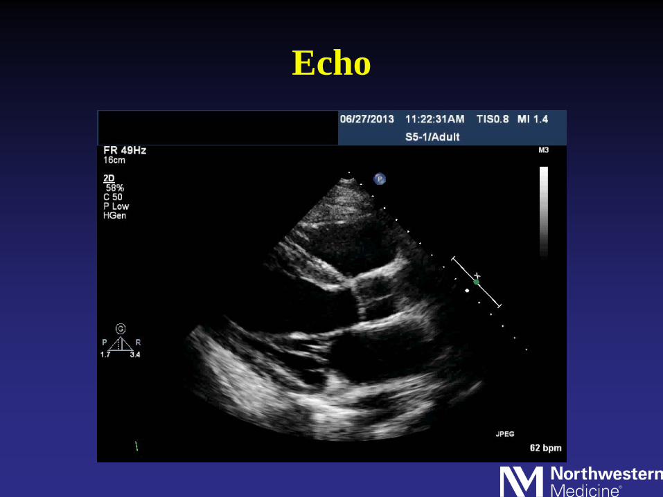
Echo


E/e’= 20


Treatment• Steroids• Antiarrhythmic therapy• ICD• VT ablation (VT noted to come from multiple
foci, including epicardium and paps)
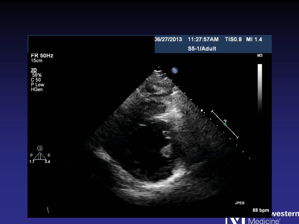
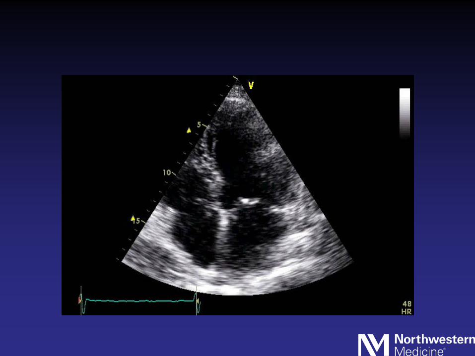
Case 2
• 75 yr old male with a history of pulmonary sarcoidosis
• He complained of SOB and palpitations
• ECG showed Afib• Cardiac cath: No obstructive CAD• Endomyocardial biopsy: Negative

ECG

Echo


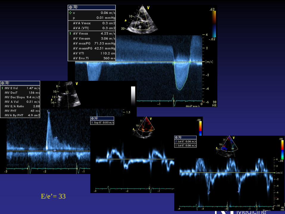
E/e’= 33

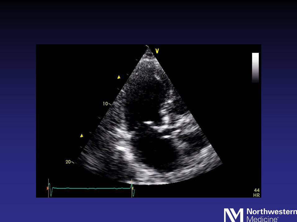


• Patient sent to the OR for AVR• Myocardial biopsy performed in the OR:
Positive for sarcoidosis
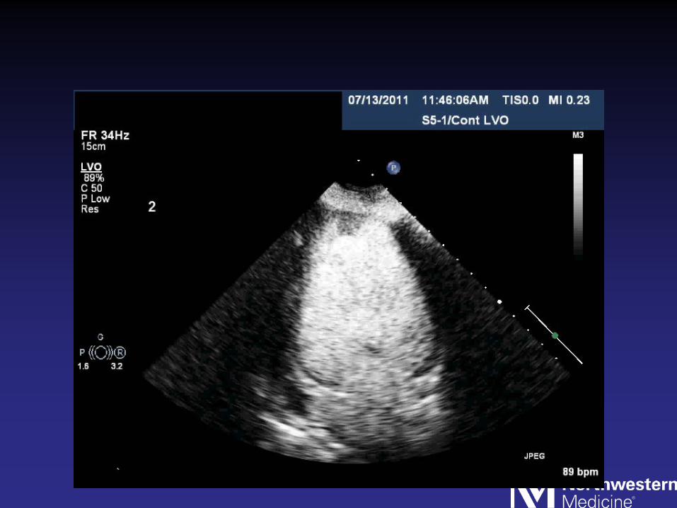
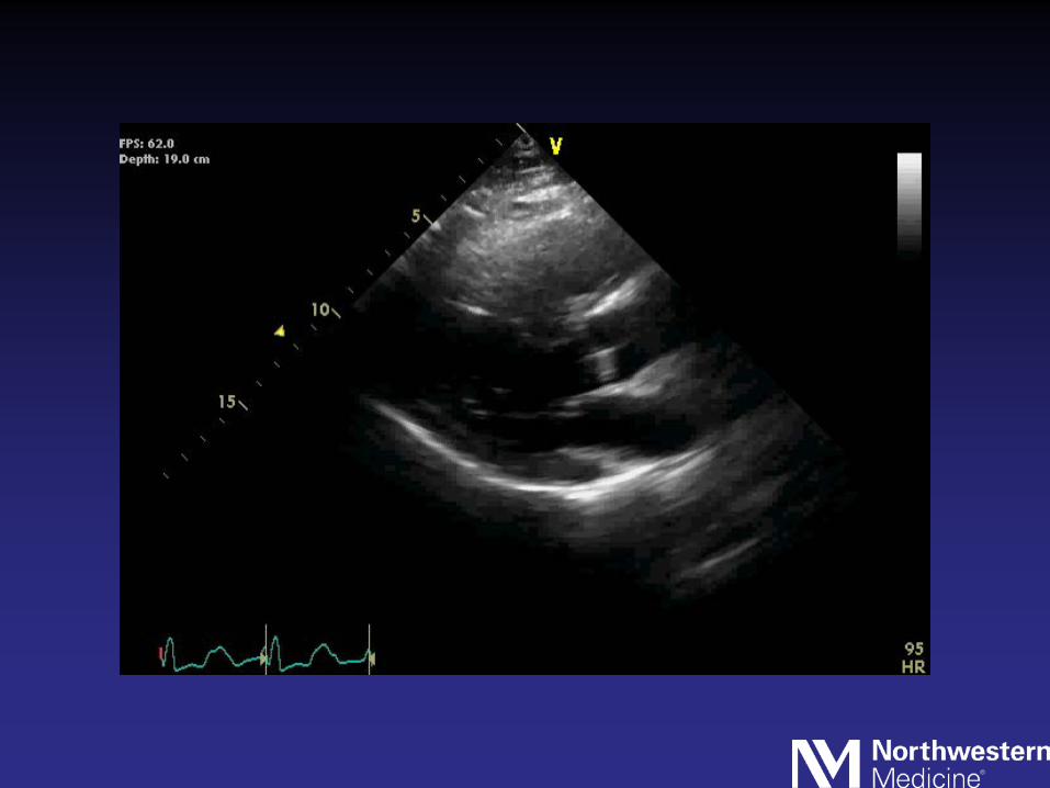
Case 3
• 48 yr old female who developed progressive SOB, LE edema and increasing abdominal girth.
• Admitted to the hospital with decompensated HF

ECG

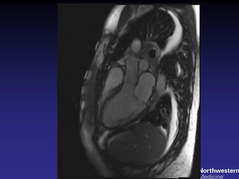
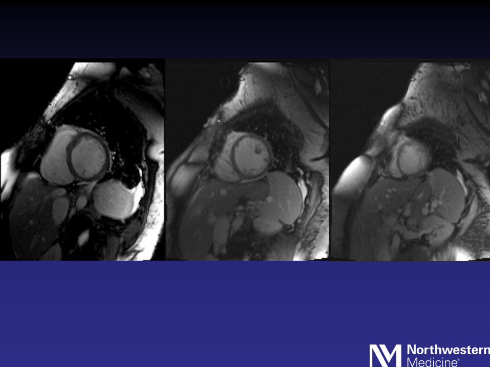
• Cardiac cath: No obstructive CAD• Cardiac MRI: Myocardial
infiltration suspicious for sarcoidosis
• Endomyocardial biospy: +Sarcoidosis


Echo


• Patient had progressive heart failure despite maximal meds
• She ultimately underwent heart transplantation

Case 4
• 56 yr old ER physician with no past medical history who presented with dyspnea and palpitations


Hospital Course
• Endobronchial biopsy pending• ICD/pacer implanted

Thank You