Embed Size (px)
Citation preview

The Cedars-Sinai Heart Center is located at 8700 Beverly Blvd., on the fifth and sixth floors.
From the Santa Monica Freeway (10), exit La Cienega Blvd. Proceed north to the Medical Center, just north of Third Street.
From the Hollywood Freeway (101), exit Highland Ave. Proceed south to Beverly Blvd. Turn right, and proceed to the
Medical Center. At San Vicente Blvd., turn left.
8700 Beverly Blvd.Los Angeles, CA 90048Office: (310) 423-3535Fax: (310) 423-0739
www.cedars-sinai.edu/heartcenter
LEADING THE QUEST FOR HEALTH®
©2008 Cedars-Sinai Health System (01/08)Form # CB0015
Cardiac Patient and Family
Heart Center
Education Handbook
CardiacBro2008_FNL 1/23/08 4:20 PM Page 1

Dedication
The Cardiac Patient and FamilyEducation Handbook is dedicated to Cedars-Sinai heart patients and
their families.
The Cedars-Sinai Heart Center would like to thank the Max Factor Family Foundationfor their generous contribution in making this handbook possible.
Special thanks goes to the Cedars-Sinai Heart Center and the Heart Families Volunteer Program for their hard workand assistance in making this handbook inclusive for all cardiac patients. And most specifically, to the diligent work of the following people:
Fifth Edition Revisions: Judy Beyer, RNGeorge Chaux, MDBernice Coleman, RN, PhD, ACNPStanley Conte, RNBarbara Cowen, LCSW, Heart Families CoordinatorBonnie De Los Santos, RN, MN, CNS, CCRNJeffrey S. Goodman, MD, FACCRichard Gordon, MAWalter Lemankiewicz, RNCristina Luper, RNScot Macdonald, PhD, Sr. Marketing AssociateC. Noel Bairey Merz, MD, FACCMargo Minissian, ACNP-C, MSN, CNSDonna Polk, MDElaine Winters, Heart Families Volunteer Program
January 2008
A Word ofWelcome
Admission to the hospital for a heartcondition can be unsettling or evenalarming for the patient as well as the
family. The purpose of this handbook is tofamiliarize you with medical words, terms,expressions and treatments that are frequentlyused in connection with problems of theheart, in understandable language.
The Cedars-Sinai Heart Center is an internationally recognized leader in thediagnosis and effective treatment of heart disease. Over the last decade there has been many advances in technologies for diagnosis and treatment of heartdisease, giving Cardiology a “high-tech” profile. We will attempt to make you familiar with these new advances.
The health care team at our institution includes doctors, nurse practitioners, clinical nurse specialists, physician assistants, nurses, dietitians, respiratory therapists, pharmacists, physical therapists, and clinical partners. This team is ready to provide individualized treatment to our cardiac patients. This treatment is based upon the condition of the patient and type of heart disease, with the goals of improving quality of life. Working together, we hope to provide the best care for you and make your stay as comfortable as possible.
Eduardo Marbán, MD, PhDDirector, Heart Institute
P.K. Shah, MDChief of Cardiology
Alfredo Trento, MDChief of Cardiothoracic Surgery
CardiacBro2008_FNL 1/23/08 4:20 PM Page 3
Table Of Contents
I. Cedars-Sinai Heart CenterCardiac Care Units...............................................................................5Coronary Intensive Care Unit (CICU) ................................................5Coronary Observation Unit .................................................................5Cardiac Surgical Intensive Care Unit (CSICU) ....................................5Cardiovascular Intervention Center (CVIC) ........................................6Cardiac Electrophysiology Laboratory..................................................6Cardiology Department .......................................................................7Heart Failure Program..........................................................................7Nuclear Cardiology ..............................................................................7Women’s Heart Center.........................................................................7Preventive & Rehabilitative Cardiac Center .........................................8Cardiac Channel: 12 ............................................................................9
II. How May We Help You? (Resources)Nursing Team ....................................................................................10Nurse Manager...................................................................................10Clinical Nurse Specialist.....................................................................10Nurse Practitioner ..............................................................................11Case Manager.....................................................................................11Physician Assistant .............................................................................11Cardiac Surgical Liaison Nurse...........................................................11Clinical Dietitian ...............................................................................11Heart Families....................................................................................12Medical Social Worker .......................................................................12POOCH Program..............................................................................12
III. Diagnostic ProceduresRoom Monitors and Telemetry ..........................................................13Stress Test (Exercise Test)....................................................................13Echocardiogram (ECHO) of the Heart..............................................14Transesophageal Echocardiography (TEE) .........................................15Nuclear Medicine Studies (Heart Scans) ............................................16Cardiac MRI/MRA............................................................................17Cardiac Imaging.................................................................................18Tilt-Table Study .................................................................................20Cardiac Catherization and Angiography (Heart Cath or Angiogram) 20Electrophysiology Studies (EPS).........................................................22Lipid Panel.........................................................................................24
Heart FamiliesProgram
As part of your education, the HeartFamilies volunteers are available toyou. They comprise a very special
group of men and women who themselveshave had a cardiac crisis (including, in somecases, a heart transplant), or have experiencedone in their immediate family. They are specially trained volunteers who have been in your shoes. They have felt the fear and
loneliness and survived. Now, they are here to help you.
Heart Families volunteers provide assistance to cardiac patients undergoing testing and treatment procedures and, when necessary, heart surgery, as well as providing support to patients’ families. Volunteers focus on:
■ Offering emotional support on a person-to-person basis during your time of crisis
■ Helping you understand and better utilize medical and hospital services■ Acting as a liaison with other Medical Center staff, such as nurses
and physicians■ Acquainting you with methods to make home care simpler and easier
In addition to their own personal experience with cardiac problems, Heart Families volunteers undergo extensive training at Cedars-Sinai, working underthe supervision of a licensed clinical social worker. The Heart Families volunteers are further supported by a team consisting of the director of Volunteer Services and representatives from Nursing, Psychiatry, Pediatrics,Medicine and Surgery, and have served in this capacity since 1981.
The Heart Families Program has been honored with the American HospitalAssociation’s Award under the Volunteer Excellence Program. They also travel to other national hospitals to provide guidance to other cardiac programs for developing volunteer programs.
The Heart Families Program is offered free of charge. We look forward to helping you with your cardiac needs and education.
CardiacBro2008_FNL 1/23/08 4:20 PM Page 5

5
Cedars-SinaiHeart Center
What Services Do We Provide?
Cardiac Care UnitsThe Cardiac Care Units are patient care units on the 5th and 6th floors of theNorth Tower. The Coronary Intensive Care Unit and the Cardiac SurgicalIntensive Care Unit are both on the 6th floor of Saperstein Critical Care Tower.The 3rd and 4th floors of Saperstein also admits cardiac patients. A majority ofpatients admitted in these units are: 1) patients who have cardiovascular diseaserequiring medical treatment including coronary artery disease, heart attack(myocardial infarction), chest discomfort (angina), congestive heart failure(CHF), and/or irregular heartbeat (arrhythmia) problems; and/or 2) patients who have had a diagnostic or therapeutic cardiac procedure. The goal of care is not only the return of health but also to navigate the hospitalization course and positively reckon with their disease.
Coronary Intensive Care Unit (CICU)The Coronary Intensive Care Unit (CICU) is a 12-bed unit on the 6th floor,south side of the Saperstein Critical Care Tower. This unit cares for patientswho are acutely ill due to heart attack, chest discomfort, irregular heartbeats,congestive heart failure or any other potentially life threatening disease pertain-ing to the heart or blood vessels and/or patients who have undergone invasivecardiac procedures. This unit is equipped with specialized equipment (rhythmand heart pressure monitoring) and highly qualified health care personnel toprovide care directed towards continuous and intensive monitoring and treat-ment of actual or potentially serious complications from a cardiovascular disease.
Coronary Observation Unit 5 North East and 5 North West are units with cardiac monitors, that care forpatients with cardiac problems who require close monitoring of their heartrhythm and vital signs.
Cardiac Surgical Intensive Care Unit (CSICU)The Cardiac Surgical Intensive Care Unit (CSICU) is a 12-bed unit on the 6thfloor of the Saperstein Critical Care Tower. The majority of patients admitted
IIV. Therapeutic ProceduresCoronary Angioplasty Procedures ......................................................25
■ Balloon Angioplasty (PTCA)■ Laser Angioplasty■ Directional Atherectomy■ Stent Implantation■ Valvuloplasty
Pacemakers.........................................................................................28■ Temporary Pacemakers■ Permanent Pacemakers
Implantable Cardioverter Defibrillator (ICD) ....................................29Radiofrequency Ablation....................................................................30Septal Ablation...................................................................................30Cardiac Surgery..................................................................................31
■ Coronary Artery Bypass Graft (CABG)■ Minimally Invasive Direct Coronary Artery Bypass Grafting (MIDCAB) ■ Transmyocardial Revascularization (TMR)■ Valve Replacement■ Thoracic Aortic Aneurysm■ Heart Transplantation■ Lung Transplantation■ Atrial Fibrillation Center
V. Important Facts We Want You to KnowEarly Warning Signs of Heart Disease ................................................34
■ Symptoms of Heart Attack■ Symptoms of Congestive Heart Failure (CHF)
Risk Factor Control............................................................................35Nutrition and Heart Disease ..............................................................35Lowering Your Cholesterol .................................................................36How to Take a Pulse ..........................................................................38
VI. Basic Anatomy of the Heart .....................................................40
VII. Glossary of Terms ....................................................................41
VIII. Telephone Numbers ...............................................................46
CardiacBro2008_FNL 1/23/08 4:20 PM Page 7
7
Cardiology DepartmentThe Cardiology Department is located on the 5th Floor, Professional Tower. In the Cardiology Department, non-invasive tests (diagnostic procedures) for theheart rhythm and heart function are performed. The Cardiology Department has 2 sections: 1) Electrocardiography section; and 2) Non-invasive section.
In the Electrocardiography section, the following procedures are performed:electrocardiograms, holter monitoring and event monitoring, ambulatory bloodpressure monitoring, pacemaker follow-up, implantable cardiac defibrillator follow-up, and consult services.
In the Non-invasive section, the following procedures are performed:Echocardiograms (2D/Doppler, Transesophageal, Exercise Stress Echo, Pacing,Dobutamine Echocardiogram), Tilt Table Study; and Exercise Stress Testing.
Heart Failure ProgramThe Heart Failure Program specializes in the diagnosis, treatment and on-goingmanagement of heart failure patients. Along with your primary care physician,the treatment team includes a physician who specializes in heart failure andnurses educated in the management of your heart disease. The program pro-vides both inpatient and outpatient services, and offers innovative therapies,through clinical trials of new medications, surgical devices and procedures.
Nuclear CardiologyNuclear Cardiology is located at the North Tower, Lower Level, Room AO41.Nuclear Cardiology performs various non-invasive tests to assess the functionand/or blood flow to the heart muscle. These procedures include: various typesof stress tests (which include Stress Thallium, Adenosine Thallium, IpyridamoleThallium, MIBI Stress, Dobutamine Stress Test, or Dual Isotope Stress); WallMotion Studies; and Technetium Pyrophosphate Scans. These procedures may be scheduled while you’re in the hospital or after you are discharged.
Women’s Heart CenterThe Women’s Heart Center at Cedars-Sinai provides risk assessment, diagnosisand heart disease care specifically for women. The program is designed to help women reduce their chances of heart disease through a preventativeapproach, including state-of-the-art screening and diagnostic testing. Located in the outpatient Noninvasive Cardiac Laboratory, the Women’s Heart Centerprovides convenient access to all of Cedars-Sinai's heart disease diagnostic andtreatment resources.
6
to this unit have undergone a surgical procedure such as open heart surgery (i.e., coronary artery bypass surgery [CABG] or valvular surgery), left ventricular assist device, repair of aortic aneurysm, heart and/or lung transplant,surgery of the blood vessels in the leg, or surgery of the chest. The goal of management is to help restore the patient to an optimal state, as well as provide emotional support for patients and their families during and after heart, chest, blood vessel or transplant surgery.
This unit consists of highly qualified healthcare personnel and is equipped with highly specialized equipment to provide continuous and intensive monitoring and treatment during the acute phase of recoveryfollowing major surgery.
6 North East is a 24-bed monitored unit that cares for stable cardiac surgical patients requiring close monitoring of their heart rhythm.
6 North West is a 32-bed non-monitored cardiovascular surgical floor that cares for stable cardiac-surgical patients or those patients who have undergoneother surgical procedures.
Cardiovascular Intervention Center (CVIC)The Cardiovascular Intervention Center (CVIC), previously known as theCardiac Catheterization Laboratory (Cath Lab), is located on the 6th floor of the Professional Tower. In this laboratory, a wide range of diagnostic andtherapeutic procedures are performed for coronary artery disease, heart musclefunction and/or heart valve dysfunction. These include: coronary angiography,left and right heart catheterization, percutaneous transluminal coronary angioplasty (PTCA) by balloon, directional atherectomy, laser, or intracoronarystent, valvuloplasty, and myocardial biopsies and septal ablation. The staff of the CVIC, physicians, nurses and technicians are knowledgeable in the state-of-the-art technology administered in the CVIC and have experience in identification and treatment of emergency cardiac intervention.
Cardiac Electrophysiology LaboratoryThe Cardiac Electrophysiology Laboratory is also located in the CVIC (Professional Tower). In this laboratory, a wide range of invasive diagnostic and therapeutic procedures for irregular heartbeats are performed. These procedures include: electrophysiology, ablation, arrhythmia mapping, radiofrequency ablation, implantable cardioverter defibrillator (ICD) implantation, and/or pacemaker implantation and adjustments.
CardiacBro2008_FNL 1/23/08 4:20 PM Page 9

9
A Phase 5 program is available for your family or friends who have risk factors,but no current heart condition, and would like to stay that way. Nutrition counseling, support groups, Yoga classes, psychological screening/counseling and regular relaxation classes are available, as are group and individual educa-tion. For further information regarding these programs, contact the CardiacRehabilitation Program at (310) 423-9660.
Cardiac Channel: 12The Cedars-Sinai Medical Center Cardiac Channel (Channel 12) is dedicated to bringing patients, families and friends up-to-date information about heart disease treatment and prevention. Cutting edge research performed at Cedars-Sinai and elsewhere has provided a wealth of information, which can help patients, and physicians successfully manage heart disease. Our programming topics range from cardiac procedure orientation, to treatmentsand lifestyle changes which can reverse heart disease, prevent recurrence andimprove quality of life. For further information about the Cedars-Sinai Cardiac Channel, please call (310) 423-4444 or ext. 34444 or look in the television guide in your room.
8
The Women’s Heart Center is directed by C. Noel Bairey Merz, MD, an internationally recognized cardiologist specializing in heart disease in women.She is one of the country's most respected researchers and takes a leadership role in educating the community and physicians about heart disease prevention. Dr. Bairey Merz is available for consultation to patients of theWomen’s Heart Center. To make an appointment, please call 310-423-9680.
Preventive & Rehabilitative Cardiac CenterThe Preventive and Rehabilitative Cardiac Center offers a wide range of programs designed to restore you to a productive, satisfying life and to prevent worsening of your heart disease. These goals are accomplished through a comprehensive program which utilizes physicians, nurses, exercise physiologists, physical therapists, occupational therapists, dietitians, psychologists and stress management therapists (Yoga) in the hospital and outpatient clinic.
You will be seen initially in the hospital (Phase I), at your physician’s request, for a program which includes education about nutrition, smoking cessation and stress management, as well as exercise designed to assist in the recoveryprocess. There is also special television programming on the heart available at particular times (see the television guide in your room or ask your primarynurse for details). Our goal is to assist you in speedy recovery and provide you with the necessary tools to prevent further heart disease problems.
You can further hasten your recovery and gain behaviors which prevent or reduce coronary artery disease by participating in our outpatient (Phase 2) program. This can begin usually 2 to 4 weeks after your discharge from thehospital, depending on your doctor’s instructions. Located in the MarkGoodson Building, 444 S. San Vicente Blvd., Suite 901, there is ample parkingand a wide range of operational hours to suit your schedule. The programfocuses on your achievement of a full recovery. You will also focus more specifically on food habits, blood cholesterol, stress management and exercise.Individualized nutrition counseling and risk factor assessment are included inthe program. The program is supervised by a cardiologist and physician referral is required.
The Preventive and Rehabilitative Cardiac Center offers many other programsand services designed to assist you in reducing your chances of more heart disease in the future. Maintenance exercise programs (Phases 3A and 3-4) are available to help you maintain your fitness and conditioning, as well as your preventive edge. A recent stress test is typically required for entry intothese programs.
CardiacBro2008_FNL 1/23/08 4:20 PM Page 11

11
Nurse PractitionerThe Nurse Practitioner (NP) is an advanced practice registered nurse who isboard certified in a specialty area of cardiology or cardiac surgery. Under direct supervision of your physician, the NP provides comprehensive healthassessment, diagnosis, treatment, consultation, education and follow-up care for individuals with cardiac needs. The NP works collaboratively with yourphysician(s) along with the entire interdisciplinary team to deliver exceptionalpatient care.
Case ManagersCase Managers are nurses who will follow your progress and assist you along with the social worker in meeting your needs after discharge.
Physician AssistantThe Physician Assistant (PA) is a board-certified health professional who worksclosely with the surgeons before, during and after your cardiothoracic surgery.In the operating room, the PA serves as an assistant, and may harvest conduits,such as the saphenous vein or radial artery, for use in a bypass procedure. Onthe wards, the PA assists with admissions, daily progress notes, physical exams,medications, and ordering diagnostic tests, as well as with discharges and officefollow-ups. PAs continuously coordinate, monitor and provide high qualitymedical care under the supervision of surgeons to improve the quality of yourhospital stay.
Cardiac Surgical Liaison NurseThe Cardiac Surgical Liaison Registered Nurse provides the patient and theirfamilies with individual education, guidance and emotional support throughoutthe surgical process from the initial consultation through after discharge.Educational needs regarding the surgery, hospitalization and future life styleswill be reviewed in detail with each patient and family. They will assist patientsand their families with discharge planning including initiating referrals to cardiac rehabilitation, social services and community resources (agencies) asindicated. In addition, they promote an atmosphere of respect, support andopen effective communication with patients, families, and the entire healthcareteam. Ask your primary nurse to contact the Cardiac Surgical Liaison Nurse for specific information and assistance at any time at (310) 423-3851.
Clinical DietitianThe Clinical Dietitian offers individual and group nutrition counseling by registered dietitians. A dietitian is available to both inpatient and outpatientprograms. While in the hospital, you may ask your doctor to order an individual diet instruction, free of charge. In the outpatient setting, diet
10
How May We Help You?
Not only are you here under the care of your personal physician, but you arealso supported by a team of healthcare professionals who combine their differ-ent skills to provide you with the finest care possible.
Nursing TeamYou will be cared for by a team of nursing personnel including: registered nurses(RNs), advanced practice nurses (APNs), licensed vocational nurses (LVNs),clinical and support partners (CPs). As your condition improves you may bemoved to areas that will best meet your individualized needs. To best prepareyou for recovery, the nursing team will encourage you to participate in your careas much as your condition permits. Please feel free to offer any comments orsuggestions regarding your special needs. We hope to help make your stay aspleasant as possible.
Nurse ManagerThe Nurse Manager is responsible for the coordination and management of activities on designated units. The Nurse Manager is available to you and yourfamily. Should you have any concerns about your hospitalization, ask yournurse to notify the Nurse Manager.
Clinical Nurse SpecialistThe Clinical Nurse Specialist is a registered nurse with a master’s degree in aclinical specialty. As an expert in a field of specialty (i.e. cardiology, cardiac sur-gery), the Clinical Nurse Specialist assists patients and families in difficultiesassociated with serious cardiovascular illness and assists the nursing team in thecare of patients with complex cardiovascular problems. The Clinical NurseSpecialist continually works collaboratively with the various members of thehealth care team (i.e. physicians, nurse managers, nursing team, liaison nurses,social workers, etc.) to achieve a positive patient outcome. You may ask yournurse to contact the Clinical Nurse Specialist to assist you and your family withany concerns you might have (i.e., education regarding your disease or diagnos-tic procedures, medications, discharge planning and psychological support).
II
CardiacBro2008_FNL 1/23/08 4:20 PM Page 13

13
DiagnosticProcedures
It is normal to feel anxious about the testing that is being done. When youhave questions or concerns about any of these tests or procedures, always askyour doctor and/or nurse to explain.
Room Monitors and TelemetryWhile you are in the Intensive CareUnit (ICU) or the Telemetry Unit,Electrocardiographic (EKG) electrodes(sponge pads) will be placed on yourchest. The EKG electrodes will be con-nected to a monitor. The continuouspicture of your heart’s electrical activityhelps plan your treatment. The EKGelectrodes are very sensitive, so don’t befrightened if your movement activatesthe monitor’s alarm. This is a safetyfeature that alerts nurses and physicians regarding any significant changes in the rate or rhythm of your heartbeat. Your physician may also order a Holtermonitor. This monitor is worn by the patient and retains a constant record of your heart rhythm on a tape recorder for 24 hours.
An electrocardiogram (EKG) is likely to be done frequently while you’re in the hospital. This painless test gives an electrical “picture” of how your heart is working. The test requires only that you lie still while the technician putselectrodes (small adhesive pads) on your arms, legs and chest. The test takesabout five minutes.
Stress Test (Exercise Test)A stress test allows us to see how your heart is working during the stress of exercise, and just after exercise. The exercise device or stress test can be performed on a treadmill or a stationary bicycle.
12
counseling may be provided through the Preventive Rehabilitative CardiacCenter (310) 423-9660 or ext. 39660, which requires a fee for service. In both settings, the dietitian can provide nutrition information specific to yourindividual needs.
Heart FamiliesHeart Families is a program of volunteers providing support to cardiac patientsand their families. They have personal experience with cardiac patients, haveundergone extensive training at Cedars-Sinai and work under the supervision of a clinical social worker. They support hospitalized patients on 5 North and 6 North as well as families who are waiting for news of their loved one in the6th floor lobby. The volunteers are liaisons for the families waiting for news ofa heart surgery or about a patient’s status who is undergoing a procedure in thecardiac catheterization lab. They are also accessible to patients and families inthe coronary intensive care units. Ask your primary nurse to contact HeartFamilies at (310) 423-3193 or ext. 33193.
Medical Social WorkerSocial Workers are available to all inpatients, outpatients and their families. The social worker is a professional who can provide emotional support andguidance, assist with discharge planning and post-hospital care and provideresource information.
You and your family members may contact the Social Work Department during office hours, 8 a.m. to 5 p.m., seven days a week at (310) 423-4446 or ext. 34446.
POOCH Visits (Pets Offering Ongoing Care and Healing) The physical and psychological benefits of animals has been well supported in healthcare literature. Pet teams are authorized to visit cardiac patients on 5 North, 6 North and the coronary intensive care units. Volunteers and theirdogs, evaluated and trained, offer patients unconditional love, warmth and an opportunity to be distracted from fears, loneliness and uncertainty oftenassociated with hospitalization. The visits are beneficial not only to patients and families, but to the doctors, nurses and entire medical staff. To request a POOCH team, please contact Volunteer Services at (310) 423-8044 orext. 38044.
III
CardiacBro2008_FNL 1/23/08 4:20 PM Page 15

15
obtain the exact angle desired. You may be asked to turn to your side or brieflyhold your breath as the technician locates the best transducer position.
Preparation: None
2. Dobutamine EchocardiogramWhile the above doppler echocardiogram is being performed you will be given amedication (Dobutamine) through anintravenous solution that will increase your heart rate. The echocardiogram will be performed to evaluate the effect of Dobutamine on your heart. This procedure may take about two hours.
3. Stress EchocardiogramA stress test combined with an echocardio-gram. The echocardiogram will be performed prior to and immediately afterthe exercise portion of the stress test. The purpose of the test is to evaluate theeffect of exercise on your heart.
4. Pacing EchocardiogramPreparation:■ Eat or drink nothing three to four hours before the test.■ Wear comfortable clothing, as you will be laying on a table or bed.
During the dobutamine test or stress echocardiogram, it is important to let the doctor or nurse know if you have any:■ Unusual feeling in the chest, arms, neck or jaw■ Light-headedness or dizziness■ Feeling of your heart beating fast (palpitations)■ Shortness of breath
Transesophageal Echocardiography (TEE)Transesophageal Echocardiography is a minimally invasive procedure used toassess and diagnose abnormalities of heart valves, blood clots and infectious vegetation in the heart, and aneurysms of the heart and aorta (main artery) that are difficult to evaluate by other tests.
The equipment used is the same as in conventional echocardiography exceptthat a probe with an ultrasound transducer is placed into the esophagus via yourmouth (similar to gastroscopy). The transducer sends ultrasound waves from the
14
During the test you are connected to a monitor by EKG electrodes (adhesive pads).A doctor watches and evaluates you very closely. They will make adjustments in the speed and angle of the treadmill or theresistance of the bicycle upon their evaluation of you. Always communicate how you arefeeling throughout the test. After the stresstest is over you will return to a table for apost-exercise EKG and to relax.
Preparation for a stress test:■ Nothing to eat or drink for three to
four hours prior to the test (other thanmedication).
■ Wear good walking shoes (rubber soles, if possible). You may want to bring a robe or something warm.
■ Some medication may be withheld temporarily prior to the stress test.Your doctor will decide this and you will be notified.
■ For certain types of stress tests, you may be asked not to consume caffeinated foods or drinks before the test (i.e. sodas, Mountain Dew,colas, coffee, tea, chocolate).
During the stress test, it is important to let the doctor or nurse know if you:■ Have any severe fatigue, nausea and/or difficulty breathing■ Have any chest pain, jaw or back discomfort■ Experience light-headedness or dizziness■ Are physically unable to continue the test
Echocardiogram (ECHO) of the HeartThese are painless, non-invasive procedures that image the heart’s chambers, valves, and blood flow using high frequency sound waves (ultrasound).
1. Doppler EchocardiogramYou will remove upper body clothing and stockings and lie quietly on a table orbed. A technician will place a small transducer on your chest. The transducersends ultrasound waves over the various heart structures. As the sound waves bounce off the moving heart structures, they are converted to images that can beseen on a television screen, recorded on videotape, and paper. This proceduremay take 45-60 minutes.
It is important that you lie quietly so that the transducer can be positioned to
CardiacBro2008_FNL 1/23/08 4:20 PM Page 17

17
■ For nuclear studies that are combined with a stress test, the followingshould be observed:• Nothing to eat or drink for three to four hours before the test• Wear good walking shoes.
■ If you are pregnant (or suspect that you might be pregnant), let your doctor or nurse know; such tests should not generally be done on pregnant patients
During the nuclear medicine test, it is important to let the doctor or nurseknow if you have:■ Any unusual feelings in the chest, arms, neck, jaw or back■ Severe fatigue■ Lightheadedness or dizziness
Cardiac MRI/MRAYour doctor has recommended you for either magnetic resonance imaging(MRI) or magnetic resonance angiography (MRA) of your chest and heart.These procedures use a magnetic field, radio waves and a computer to createdetailed images of the chest area. The angiography procedure is specificallydesigned to examine the heart and the blood vessels entering your lungs. Ourteam of specialist physicians can use these images to distinguish between differ-ent types of tissue as well as diseased tissue. Our team of physicians, nurses andtechnologists are led by our Chief of Cardiac Imaging and Nuclear Cardiology,Daniel S. Berman, MD, and our Chief of Thoracic Imaging, Peter J. Julien, MD.
Preparation:■ Before Arriving at the S. Mark Taper Foundation Imaging Center:
• You should not have anything to eat or drink three to four hours prior to the exam start time, unless instructed to do so by your physician.
• If you are undergoing MRI for cardiac perfusion, you will be asked to avoid caffeine for 24 hours before the test, and to not take any betablockers, calcium channel blockers, ace inhibitors or angiotensin receptor blockers (ARBs) for the 48 hours before your procedure.
• If you are claustrophobic (fearful of small, enclosed areas) or experiencepain when lying on your back for more than 30 minutes, your referringphysician may prescribe a relaxant or pain medication to help youthrough the exam. The imaging physicians at Cedars-Sinai will not prescribe such medications for you.
• We want to make your waiting time as pleasant as possible. Considerbringing your favorite magazine, book or music player to help you passthe time. You may also bring a CD to listen to during the procedure.
• Please wear comfortable clothing.
16
esophagus over various heart structures, which are converted to images that canbe seen on a television screen, recorded on videotape, and paper.
You will be given intravenous (IV) sedation to make you sleepy. Your bloodpressure and heart rate are monitored during the procedure. The procedure may take about 30 minutes. However, you will be in the examining room about60 to 90 minutes to recover from the sedation.
Preparation:■ Nothing to eat or drink for at least eight hours before the test.■ Wear comfortable clothes. You will be asked to undress and wear a hospital
gown.■ A licensed driver must accompany outpatients. You are not allowed to
drive for 24 hours after being sedated.
Nuclear Medicine Studies (Heart Scans)A cardiac nuclear scan is a special type of imaging procedure in which a smallamount of radioactive material is injected into a vein. A special camera and a computer take pictures of the heart. Sometimes the pictures are taken during a stress test as well. During the procedure you will be connected to a monitorthrough EKG electrodes. The test may be done either in the Nuclear MedicineDepartment or in your room.
The procedure may take two to three hours and sometimes it is necessary totake further pictures of the heart four and 24 hours after the initial picture-taking session.
There are four types of heart scans used:■ Thallium scan (the most common procedure)■ Wall motion test■ Sestamibi■ Technetium Pyrophosphate scan
Preparation for nuclear studies:■ Do not consume any products containing caffeine (which includes coffee,
tea, decaffeinated products, chocolate, cocoa, soda [diet or regular]) 24hours prior to the procedure.
■ The following medications may be withheld prior to a stress test procedurein consultation with your doctor:• Beta-Blockers (Inderal, Lopressor, Sectral, Tenormin, Visken, Corgard, etc.)• Calcium-Channel Blockers (Calan, Cardizem, Isoptin, Procardia)• Nitrates (Isordil, Nitro-bid, etc.)
CardiacBro2008_FNL 1/23/08 4:20 PM Page 19

19
Please tell the doctor or tech about any allergies that you have. The technologistinjects a radiotracer into a vein. A radiotracer is a compound made of a radioac-tive isotope and a pharmaceutical agent. In the radiotracer used for myocardialperfusion SPECT, the pharmaceutical part keeps the tracer in the blood until it is filtered out by the kidneys. The radioactive isotope releases energy, and aspecial camera creates an image from it.
Myocardial perfusion SPECT is used to evaluate damage that might have been caused by a myocardial infarction (heart attack) and to assess the presenceand extent of myocardial ischemia (reduced blood flow due to obstruction inthe vessels).
Coronary Reactivity TestingYour doctor has recommended you for Coronary Reactivity Testing of yourheart arteries. The angiography procedure is specifically designed to examinethe blood vessels in your heart and how they respond to different medications.Our team of specialist physicians can use these images to distinguish differenttypes of blood vessels reactivity dysfunction. Our team of physicians, nursesand technologists are led by our director of the Women’s Heart Center, NoelBairey Merz, MD, and our interventional cardiologist, Saibel Kar, MD.
Women often experience chest symptoms differently than men. The Women’sIschemia Syndrome Evaluation (WISE) study is one of the primary studieschanging the way women’s heart disease is detected and treated. For men, heartdisease often manifests as blockage in the large arteries of the heart. One of themajor discoveries of the WISE study is that many women with chest pain orother symptoms, have microvascular disease, a narrowing of the small arteriesand blood vessels of the heart. Blood flow to the heart is restricted by fattyplaque buildup, but the restriction does not show up in traditional diagnosticexams. Until recently, this led physicians to discount the possibility of heart disease in many female patients. These women often found themselves makingrepeated visits to physicians and hospitals trying to unravel the mysteries of theirsymptoms. The Coronary Reactivity Test is the gold standard for diagnosingcoronary microvascular disease and endothelial wall dysfunction.
The procedure occurs in the catheterization laboratory and patients shouldfollow the same instructions before and after catheterization explained in theCardiac Catheterization and Angiography section on page 20. The physicianwill ask to see you in clinic a week before your procedure. At that time, youwill have a pre-procedure chest X-ray, blood work and an EKG. Patients areasked to avoid caffeine 24 hours before the test and to not take beta blockers,calcium channel blockers, ace inhibitors and angiotensin receptor blockers
18
■ After Arriving• You must tell the technologist, radiology nurse and/or imaging physician
of any allergies you may have, and if you are pregnant or are nursing.• You will be asked to complete a questionnaire, which will help determine
if an MRI is safe for you. People with various implants (usually metallic)or with metal in their bodies (including some tattoos) may have difficultywith an MRI, which utilizes a strong magnetic field. The imaging physician needs to be informed of any of these potential problems.
During the Exam■ You will be positioned on the scanning table head-first with your arms
at your side.■ Coils (special devices to improve image quality) may be placed on or
around your chest. The scanning table will slide your entire body into the magnet.
■ During the scan you will not feel anything, but you will hear intermittenthumming, thumping, clicking and knocking sounds. Earplugs will beprovided to help mask the noise and to allow you to listen to music.
■ In most cases, the imaging physician requests a contrast agent (dye) toimprove the quality of the images.
■ The contrast agent is injected into a vein in the arm, and may cause a cool sensation.
■ As pictures are taken, you must hold very still and, in some cases, hold your breath for up to 25 seconds.
■ The technologist is always able to see and hear you during the exam.■ The MRI exam will take approximately 60 minutes. The MRA exam
will take approximately 30 to 60 minutes.
After Your Exam■ There are no restrictions placed upon you. You may eat or drive as normal.■ Your films will be examined by an imaging physician and a report sent to
your doctor. Your doctor will review the results with you.
To request a copy of your images on a CD or film, or a copy of your report,please call (310) 423-8000.
Cardiac ImagingMyocardial Perfusion SPECTA myocardial perfusion SPECT (Single Photon Emission Computed Tomography)study, also called a cardiac stress-rest test, is used to evaluate the heart’s blood supply. Two sets of images showing blood flow are obtained: the first following a period of rest, and the second following a period of stress (i.e., exercise).
CardiacBro2008_FNL 1/23/08 4:20 PM Page 21

21
Heart catheterization helps in the diagnosis of disease affecting the heart muscle,heart walls, and heart valves, as well as the condition of the coronary arteries thatfeed blood and oxygen to the heart. Angiography involves the injection of con-trast or “dye” into the coronary arteries and sometimes the heart chambers.During contrast injections, X-ray movies are viewed in real time and can later bereviewed as part of your medical record. You may experience a warm sensationthroughout your body when the dye is injected into the main pumping chamber(left ventricle). Let the doctor or nurse know if you experience any chest discom-fort.During the procedure you will see monitors nearby and may be able to see yourEKG and the pictures of the heart and arteries. The total procedure may takeone to two hours. However, the preparation and recovery time may take anadditional two hours. Your family can wait in the 6th floor lobby where your
doctor can meet with them following the procedure.
Preparation for Cardiac Catheterization:■ Your nurse will ask you to sign consent form(s) for the procedure(s).■ Nothing to eat or drink six to eight hours prior to the procedure. (Note:
You may, however, take your cardiac medication, aspirin, or any other medications prescribed by your physician the morning of the procedurewith very little water.)
■ Intravenous line will be started in your room before you leave before youleave for the catheterization laboratory. Medications can be given directlyinto this line as needed during the test.
20
(ARBs) during the 48 hours before your procedure. The exam lasts approxi-mately 60 to 90 minutes, and patients are often released from the outpatientprocedure area that afternoon.
For more information regarding Coronary Reactivity Testing, please contact the Women’s Heart Center at (310) 423-9680 or ext. 39680.
Tilt-Table StudyA Tilt-Table study is a procedure used to evaluate changes in heart rate and blood pressure during changes in body position. The most common indication to perform this test is in patients who have symptoms of temporary loss of consciousness or near fainting.
The nurse prepares you prior to the procedure by starting an intravenous (IV)line and connecting you to a machine that continuously monitors your heartrate and blood pressure. Initially, you will be lying in a flat position for 20-30minutes. Then, the tilt bed will be changed to a near vertical (80 degrees) position for a period of 30 to 45 minutes. During this portion of the verticaltilt position, it is possible that you will experience lightheadedness, dizziness, or a feeling of fainting. It is important that you inform the physician or nurseof any symptoms you may be feeling. Finally, the tilt bed will be placed in ahorizontal position and you will be observed for another 20 to 30 minutes while lying in a flat position.
You may also be given a medication through your IV as part of this test. Thephysician will determine this at the time of the study. During this portion ofthe test your position will be changed from a vertical to a flat position every ten minutes.
Depending on the individual response, the test may last from one to three hours.
Preparation:■ Wear comfortable clothing or your hospital gown■ Nothing to eat or drink for at least eight hours before the test.■ You may continue to take your heart medication.
Cardiac Catheterization and Angiography (Heart Cath or Angiogram)In this test, a small catheter (tube) is put into an artery and/or a vein, in yourleft (groin) and/or in the arm or neck and passed up to the heart under X-rayguidance. Numbing medication is used to minimize any discomfort with theinsertion of the catheter at the insertion site.
AortaCatheter
RightCoronary
Artery
Aorta
Catheterenters arm or leg
LeftCoronary
Artery
CardiacBro2008_FNL 1/23/08 4:20 PM Page 23

23
tions in the chambers of your heart to record signals and stimulate the heart toidentify abnormal rhythms. Once the wires are in place, electrical signals fromyour heart are monitored and recorded. In addition, the doctor may artificiallyincrease your heart rate in an attempt to provoke any abnormal rhythm distur-
bances with medication. The entire study may take from one to three hours.
Preparation:■ Nothing to eat or drink six to eight hours before the test. ■ Intravenous line will be started in your room before you leave before you
leave for the catheterization laboratory. Medications can be given directlyinto this line, as needed, during the test.
■ A small area on your upper legs and neck will be used for the catheter insertion. This area will be clipped and scrubbed with an antiseptic soap.
■ Empty your bladder before going to the study.■ Wear a hospital gown and remove all undergarments.
After Electrophysiologic Study (EPS):■ You must remain in bed and avoid bending the groin area for four hours.■ You must use the urinal or bedpan for elimination.■ The nurse will check your vital signs often.■ You may move your foot or wiggle your toes and you may move your
arms freely.
Discharge Instructions for Outpatients:■ Arrange for someone to drive you home upon your release from the hospital.■ Arrange for a responsible adult to be with you for 12 hours at home.
22
■ Wear a hospital gown, remove all undergarments and empty your bladderbefore you leave your room.
■ Depending on the procedure approach, a small area will be prepped by clipping hair in the area and then cleaning with an alcohol based antisepticsolution for the catheter insertion.
■ Often a relaxing medication is given to you before you leave your room.
After Cardiac Catheterization:■ The sheath (or tube) in your artery will be taken out in the cath lab
(CVIC) before returning to your room or after blood thinning medicationswear off for patients held in Cath Lab Recovery Unit.
■ Avoid bending the groin for four to six hours (complete bed rest; head should be relatively flat). Your physician or nurse will discuss how long you will need to be flat.
■ A closure device (collagen plug or stitch) may be used to seal the puncturesite that may reduce your bed rest time.
■ You must use the urinal or bedpan for elimination.■ If you have to sneeze or cough, apply pressure with your fingers over the
groin or puncture site to reduce risk of bleeding.■ It is not necessary to hold your affected leg stiff (you may move your
foot or wiggle your toes, and move your arms freely). You may moveall unaffected limbs.
■ The nurse will check your vital signs and the insertion site (usuallythe groin) frequently.
Electrophysiologic Studies (EPS)An electrophysiology study is designed to thoroughly assess the conduction system or “electrical wiring” of the heart and to assess abnormal heart beats.This test may be done to assess for abnormal fast heart rhythms that can lead topalpitation, fainting or even cardiac arrest. It is also used to assess the need forpacemaker implantation, or the need for further special procedures.This test involves the insertion of several thin catheters (tubes) or wires into yourgroin, and commonly the right neck. These wires are advanced to various loca-
Neck
Arm
Groin
Sites of Insertion
Electrode Catheters Inside the Heart
CAUTION!!! If you feel:■ Sudden pain at the site; or■ Warm, sticky sensations or sensation of fluid or blood on the affected leg,
NOTIFY THE NURSE IMMEDIATELY!Pressure will be reapplied for as long as needed to stop the bleeding.
CardiacBro2008_FNL 1/23/08 4:20 PM Page 25
25
TherapeuticProcedures
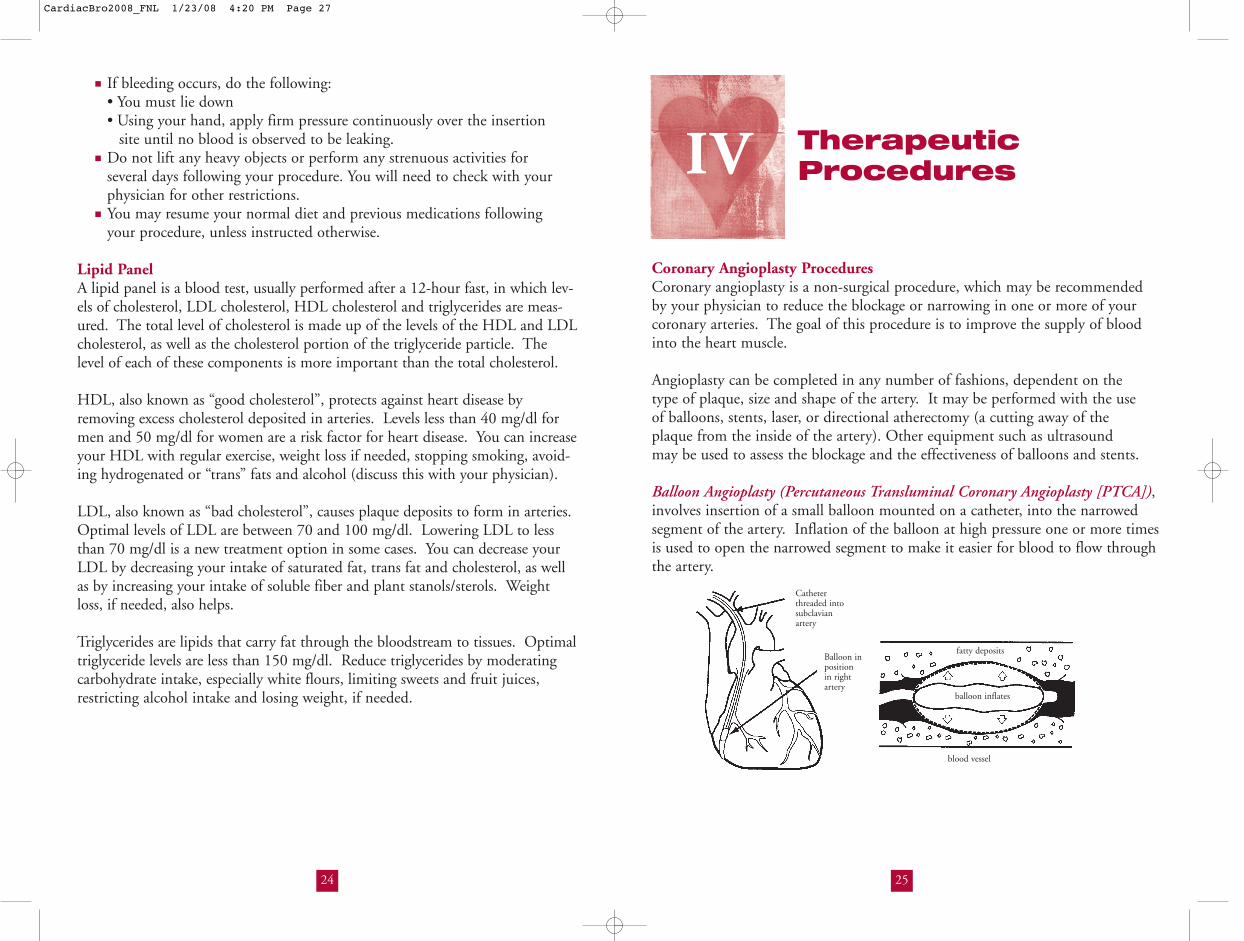
Coronary Angioplasty ProceduresCoronary angioplasty is a non-surgical procedure, which may be recommendedby your physician to reduce the blockage or narrowing in one or more of yourcoronary arteries. The goal of this procedure is to improve the supply of bloodinto the heart muscle.
Angioplasty can be completed in any number of fashions, dependent on the type of plaque, size and shape of the artery. It may be performed with the use of balloons, stents, laser, or directional atherectomy (a cutting away of the plaque from the inside of the artery). Other equipment such as ultrasound may be used to assess the blockage and the effectiveness of balloons and stents.
Balloon Angioplasty (Percutaneous Transluminal Coronary Angioplasty [PTCA]),involves insertion of a small balloon mounted on a catheter, into the narrowedsegment of the artery. Inflation of the balloon at high pressure one or more timesis used to open the narrowed segment to make it easier for blood to flow throughthe artery.
24
■ If bleeding occurs, do the following:• You must lie down• Using your hand, apply firm pressure continuously over the insertion
site until no blood is observed to be leaking.■ Do not lift any heavy objects or perform any strenuous activities for
several days following your procedure. You will need to check with yourphysician for other restrictions.
■ You may resume your normal diet and previous medications following your procedure, unless instructed otherwise.
Lipid PanelA lipid panel is a blood test, usually performed after a 12-hour fast, in which lev-els of cholesterol, LDL cholesterol, HDL cholesterol and triglycerides are meas-ured. The total level of cholesterol is made up of the levels of the HDL and LDLcholesterol, as well as the cholesterol portion of the triglyceride particle. Thelevel of each of these components is more important than the total cholesterol.
HDL, also known as “good cholesterol”, protects against heart disease by removing excess cholesterol deposited in arteries. Levels less than 40 mg/dl formen and 50 mg/dl for women are a risk factor for heart disease. You can increaseyour HDL with regular exercise, weight loss if needed, stopping smoking, avoid-ing hydrogenated or “trans” fats and alcohol (discuss this with your physician).
LDL, also known as “bad cholesterol”, causes plaque deposits to form in arteries.Optimal levels of LDL are between 70 and 100 mg/dl. Lowering LDL to lessthan 70 mg/dl is a new treatment option in some cases. You can decrease yourLDL by decreasing your intake of saturated fat, trans fat and cholesterol, as wellas by increasing your intake of soluble fiber and plant stanols/sterols. Weightloss, if needed, also helps.
Triglycerides are lipids that carry fat through the bloodstream to tissues. Optimaltriglyceride levels are less than 150 mg/dl. Reduce triglycerides by moderatingcarbohydrate intake, especially white flours, limiting sweets and fruit juices,restricting alcohol intake and losing weight, if needed.
IV
Catheterthreaded intosubclavianartery
Balloon inpositionin rightartery
blood vessel
fatty deposits
balloon inflates
CardiacBro2008_FNL 1/23/08 4:20 PM Page 27

27
After Coronary Interventional Procedures:■ Most likely you will go to a monitored (telemetry) unit overnight.■ The catheters are usually removed shortly after the procedure.■ Catheters are removed from your groin and hand pressure is applied to
the puncture site(s) to prevent bleeding.■ A vascular hemostatic device (VHD) may be used. This is an invasive
method of sealing the large puncture in the artery. It allows the puncture site to close faster and stabilize the artery so you can walk sooner.
■ TOTAL BED REST is needed after the procedure. If a VHD is used, you may need to lie still for 2 – 4 hours before getting up.
■ If your doctor does not use VHD, you may be required to lie still for 4 – 6 hours before getting up. The most important thing to remember isNOT TO BEND the leg at the puncture site.
■ You must use the urinal or bedpan for elimination until the doctor has determined you can get up.
■ If you sneeze or cough, apply pressure with your fingers over the puncture site.■ The nurse will check your vital signs and puncture site(s) frequently.
After Discharge from the Hospital:1.Activity
■ Minimize activity for two days after discharge.■ Do not lift heavy objects for five to seven days; check with your doctor.■ Do not bend from the waist. Avoid strenuous exercise. Exercise is
considered strenuous if you sweat.■ You may drive the day after you come home.■ Avoid straining during bowel movements.■ Talk to your doctor about when to return to work.
2.Groin Care■ You may shower the day after discharge. However, you should avoid tub
baths, swimming and the use of Jacuzzi or sauna until the puncture site has completely healed.
■ You may remove the Band-Aid or dressing from the site the next day andwash with soap and water. Pat dry.
26
Laser Angioplasty(Excimer LaserCoronary Angioplasty[ELCA]) involves usingthe insertion of a laseremitting catheter intothe narrowed segmentof the artery. Once thelaser energy is turnedon, the plaque is vapor-ized, thus reducing theblockage of the artery.
Directional CoronaryAtherectomy involvesthe use of a small sharpblade housed inside acatheter which is placedagainst the plaqueallowing the doctor tocut and remove part ofthe plaque from thewall of the artery.
Stent Implantation, a thin metal scaffoldingmounted on a ballooncatheter, is advancedinto a narrowed coro-nary artery. This stentis permanently implanted in the coronary artery after the balloon catheter isinflated. An expanded stent acts as a scaffold within the artery to keep it open.
Valvuloplasty is a non-surgical technique for increasing flow of blood through narrowed or tight heart valves using dilation catheters. Thisprocedure is similar to PTCA. Catheters areinserted from a femoral approach (from the arteryin the groin) and advanced to the narrowed valveusing fluoroscopic (X-ray) guidance. A dilationcatheter is inflated to increase the heart valve opening and improve blood flow.
0.038" guidewire in descending aorta
Aortic valve
Atrial septum
LV apex
Valvuloplasty balloon in mitral valve
CAUTION!!! If you feel:■ Sudden pain at the site; or■ Warm, sticky sensations or sensation of fluid or blood on the affected leg,
NOTIFY THE NURSE IMMEDIATELY!Pressure will be reapplied for as long as needed to stop the bleeding.
Catheter threaded intosubclavian artery
Balloon inpositionin rightartery
blood vessel
fatty deposits
laser energy
Catheter threaded intosubclavian artery
Balloon inpositionin rightartery
atherectomy catheterwith cutter
blood vesselfatty deposits
Catheter threaded intosubclavian artery
Balloon inpositionin rightartery
blood vessel
fatty deposits
stent
inflatedballoon
CardiacBro2008_FNL 1/23/08 4:20 PM Page 29

29
signals back and forth between the heart and the pacemaker. The pacemaker isthen attached to the wire and placed under the skin in a pocket-like area.
ACTIVITIES YOU CAN DO AND WHEN YOU SHOULD BE CAREFULAt first, many people are overly concerned about their pacemaker. However, as they become more confident in themselves and the pacemaker system, they feel comfortable resuming a normal lifestyle.
■ You may wear comfortable clothing.■ You may operate any of the electrical devices in your home.■ You may drive or ride in an automobile, tractor or boat.■ You may not go through magnetic resonance imaging (MRI) machines.
Before you go for any test, tell your doctor you have a pacemaker implant.Cardiac pacemaker is contraindicated for MRI studies.
■ Airports: Sometimes the metal case of the pacemaker will trigger the security-screening device in airports. If this happens, simply show your pacemaker identification card to the security guard (the function of yourpacemaker is not affected by these airport devices).
■ Regular appointments with your doctor or visits to a pacemaker clinic may be necessary to evaluate you and your pacemaker.
Note: Your pacemaker function needs to be checked every one to threemonths.
Implantable Cardioverter Defibrillator (ICD)The ICD is an electronic device which is designed to stop rapid abnormal heartrhythms. Patients who have suffered cardiac arrest or have life-threateningarrhythmias (rapid abnormal heart rhythms) uncontrolled by the usual methods(e.g. medications, etc.) may receive this device.
The ICD terminates rapid abnormal heart rhythms bypacing the heart faster than the abnormal rate(called overdrive pacing) or delivering one orseveral shocks directly to the heart. Theseshocks may or may not be felt depending onthe amount of energy required to restore yourheart to normal rhythm.
The ICD is implanted under the skin in eitherside of the chest region. It is connected to leads (wires), which are used for pacing,shocking and sensing. The ICD is implanted in the operating room setting whileyou are asleep. Several kinds of surgical approaches are available and your physicianwill discuss the various options.
28
PacemakerA pacemaker is an artificial device placed inside or outside the body to regulate yourheartbeat. As you perform daily activities, the pacemaker sends and receives electricalsignals to and from the heart to regulate your heartbeat to meet your body’s needs.
1. Temporary PacemakerWhile in the hospital, you may have a temporary pacemaker to stabilize your heart-beat. The temporary pacemaker is placed outside of the body. This allows yourdoctor to regulate the number of electrical signals going to the heart through a pace-maker wire.
■ External Non-Invasive PacemakerA temporary pacemaker that involves application of adhesive electrode pads on the chest and connected to a pacemakermachine via a pacemaker wire. Electrical signals between the heart and the pacemakermachine are exchanged via these electrodepads applied on the chest.
■ Transvenous PacemakerA temporary pacemaker that involves insertion of a pacemaker wire into a major vein and advanced until it reaches the heart.One end of the pacemaker wiretouches the heart muscle whilethe other end is located outsidethe body to be connected toa pacemaker box. The pro-cedure is done under localanesthesia.
2. Permanent PacemakerA permanent pacemaker is usually placed in thebody through a small incision in the upperchest or abdomen close to a major vein.This procedure is done under local anes-thesia. The area will be clipped andscrubbed with an antiseptic agent. The vein is opened and the pacemaker wire isadvanced into the heart. The wire musttouch the heart so that it can carry electrical
Wire enters body
Battery isoutside ofbody so thata doctor canadjust thetemporarypacemaker
battery
wire
vein
Permanent pacemakers are placedcompletely inside the body.
Anterior(front)
pacemaker electrode
Posterior (back)pacemaker electrode
EKG electrode
CardiacBro2008_FNL 1/23/08 4:20 PM Page 31

31
patients who suffer from a heart condition called hyper-trophic obstructive cardiomyopathy (HOCM).Hypertrophic obstructive cardiomyopathy is an inheritedcondition that causes excessive muscle growth in theinterventricular septum. This limits blood flow from theleft ventricle to the aorta, the main valve of the heart,which means the heart must work harder to move theblood. The procedure helps to make the interventricularseptum muscle smaller so the heart works better.
Cardiac SurgeryThere are many options for the surgical approach to bypass and valve surgeries.They range from the traditional sternotomy incision down the middle of the chest,to a partial sternotomy where only a portion of the sternum is cut through, tocompletely minimally invasive or robotic approaches. You should discuss theoptions available to you with your surgeon.
1. Coronary Artery Bypass Graft (CABG) SurgeryBypass surgery is recommended for some people with narrowing in one or more ofthe coronary arteries. Surgery involves removing an artery from the internal chest
wall, the forearm or a vein from the leg, dependingon which procedure is medically appropriate forthe individual. These are then surgically attachedto supply blood to the area beyond the blockage ina coronary artery.
2. Minimally Invasive Direct Coronary Artery Bypass Grafting (MIDCAB) and Beating Heart SurgeriesThe minimally invasive direct coronary artery bypass or MIDCAB procedure is asurgical approach. This is done through the making of smaller incisions on thechest, usually between the ribs. The MIDCAB procedure can be performed on single or multiple blocked coronary arteries. Both traditional sternotomy andMIDCAB procedures can be done with or without the use of a “bypass machine”.When done without the use of a “bypass machine”, it is called a “beating heart sur-gery”. There are many factors that go into determining which approach and optionis best for the individual patient. Please discuss these options with the surgeon.
30
Preparation for ICD Implant:■ Nothing to eat or drink six to eight hours before surgery.■ Intravenous line will be started in your room before surgery.■ Medications can be given directly into this line as needed during the surgery.■ Empty your bladder before going to surgery.■ Wear a hospital gown.
After ICD Implant:■ You may go to a monitored (telemetry) care area.■ The nurse may check your vital signs frequently.■ You may have incisional pain. You may ask your nurse for pain medication.
After discharge, patients are followed up every few months to check device status as well as perform several non-invasive tests.
Radiofrequency AblationRadiofrequency ablation is a non-surgical procedure designed to locate abnormalconduction pathways in the heart and eliminate them by delivering burning orfreezing energy to that area. The procedure is performed in the CardiacCatheterization Laboratory and can be done on an inpatient or outpatient basis.The radiofrequency ablation procedure is similar to a cardiac catheterization inthat catheters (small thin wires) are placed into different areas of the heart byentering through the groin, and commonly the neck. Once the abnormal path-way is found, energy is applied to that area through the catheter. Several appli-cations of radiofrequency or cryo energy (a freezing technique to burn tissue)may be needed to eliminate any abnormal heart rhythm disturbances. You maybe asleep during the entire procedure, which can take from two to eight hours.
After Radiofrequency Ablation:■ All catheters and sheaths are removed before you go back to your room.■ You may go to a monitored (telemetry) care area.■ You will be asked to lie flat in bed, keeping your legs straight for four hours.■ The nurse will check your vital signs and access sites (groin) frequently.■ You may be discharged the following morning, which will be determined by
your physician.
Septal AblationThis is a new, non-surgical procedure that requires injecting pure alcoholthrough catheters into the arteries that feed the interventricular septum (themuscle that separates the two chambers of the heart) creating a mild heart attackthat thins the thickened heart muscle. This procedure is done in the CardiacCatheterization Laboratory under local anesthesia. This procedure is beneficial for
Aorta
Saphenousvein graft
Internalmammeryarterygraft
Common sites for bypass grafts
Chest incision
Saphenous vein
Radial artery
CardiacBro2008_FNL 1/23/08 4:20 PM Page 33

3. Transmyocardial Revascularization (TMR)Transmyocardial revascularization (TMR) is a procedure designed to relievesevere angina or chest pain in patients who are not candidates for bypass surgeryor angioplasty. Sometimes TMR is used with traditional heart surgeries. A laserdevice is used to develop pathways within the heart muscle necessary to supplythe heart with oxygen-rich blood.
4. Valve SurgeryIt is sometimes necessary to repair or replace a heart valvethat is no longer functioning properly. Often the valve hasbecome damaged or scarred by birth defects, rheumaticfever or infection. When the heart valves do not open orclose properly, the heart has to pump harder to get bloodto the body. Over time this can weaken the heart andcause pain, shortness of breath, dizziness or other symp-toms. When medication cannot correct these problems,heart valve surgery is often recommended. Some conditionsallow for robotic or minimally invasive valve surgery. This is done by makingseveral small porthole incisions on the chest and/or a small incision on the rightside of the chest. Some heart valves can be repaired or replaced with this tech-nique. Please discuss the possibility of this option with your surgeon and cardi-ologist.
5. Thoracic Aortic Aneurysm SurgeryA thoracic aortic aneurysm is an abnormal dilation in a weakened wall of the aorta in the chest. If left untreated, an aneurysm will lead to a variety of life-
threatening conditions, such as aortic dissection or rupture.Due to the potential risks, timely diagnosis and treatment iscrucial. Thoracic aneurysms may occur in the aortic root,ascending aorta, arch or descending aorta. Surgery maybecome necessary to replace the weakened aorta with agraft. In addition, endovascular stent grafting is anotherpotential procedure available for individuals with distal archand descending aortic aneurysms.
6. Heart TransplantationOnce a heart has reached advanced heart failure, the Heart Transplantation Program provides a continuum of comprehensive treatments and surgical care,individually tailored to each patient. Treatments include surgical therapies, such as heart transplantation, heart-lung transplantation, left ventricular assist deviceimplantation and automatic implantable cardioverter defibrillator insertion. The program was also the first in California to implant the portable artificial
33
heart (HeartMate VAD). For patients with advanced heart failure, the use of a ventricular assist device (VAD) can be used as a life-saving treatment option.Such devices act as a “bridge” for patients awaiting heart transplantation or theVAD can provide a new alternative which can extend the life expectancy of end-stage heart failure patients. In addition, the program provides detailed follow-up care that entails endomycardial biopsies and management of immunosuppressive therapy.
7. Lung TransplantationCedars-Sinai Medical Center has been a national leader in lung transplantationwith the longest running program in the western United States, established in 1988. Since that time, the lung transplant team at Cedars-Sinai has completed more than 150 transplants with excellent outcomes. Lung transplantation is a good treatment option for many individuals with end-stage lung disease who have exhausted all other medical and surgical treatment options without significant improvement. The conditions for whichlung transplantation is typically considered include severe emphysema, cystic fibrosis, pulmonary fibrosis and pulmonary hypertension. This treatmentoption has traditionally been restricted to younger individuals who are healthyexcept for their lung disease but Cedars-Sinai has been able to expand the benefits of lung transplantation to older individuals with other health conditions that would have precluded lung transplantation at most other centers. In spite of this, the results of lung transplantation at Cedars-Sinai have been comparable to the results reported by most centers around the country. In addition, the lung transplant program is an integral part of theWomen’s Guild Lung Institute which provides comprehensive services for individuals with all forms of advanced lung disease.
8. Atrial Fibrillation CenterAtrial fibrillation is the most common clinically encountered arrhythmia (irregular heart rhythm) and can lead to dizziness, fainting and stroke. It is most common in older people with heart disease or after cardiac surgery but can also occur in otherwise young and healthy individuals. Here at Cedars-Sinai Medical Center, our world class team of experts are at the cutting edge of the many treatment options for atrial fibrillation. Our comprehensive staff utilizes state of the art options including: evaluation and treatment of the underlying causes, medical treatment options, catheter based ablation using state of the art technology and the newest minimally invasive surgical approaches.
32
Artificialheart valvein aortic position
CardiacBro2008_FNL 1/23/08 4:20 PM Page 35

35
■ Swelling of the lower extremities (i.e., legs and ankles )■ Inability to sleep except when propped up on two or more pillows■ Shortness of breath (may be continuous, with exertion, or may awaken
you at night)■ Frequent dry, hacking cough without sputum (especially when lying down)■ General fatigue
These symptoms of heart failure occur when the heart fails to pump blood effectively and blood backs up in the heart and other organs.
Risk Factor ControlTo control your risk factors for developing heart disease, most doctors recommendthe following changes:
■ Stop smoking■ Maintain or achieve a healthy body weight■ Eat less saturated fat, trans fat and cholesterol, and more vegetables,
fruits, whole grains and fish■ Lower your blood pressure■ Find a way to manage your stress■ Exercise■ Keep your blood sugar under control■ Normalize your blood lipids (HDL, LDL and triglycerides)■ Get enough rest■ Visit your doctor regularly■ Regularly take your medications as prescribed by your physician.
(Do not stop taking prescribed medications without discussing it with your doctor).
Nutrition and Heart DiseaseEating more vegetables, fruits, whole grains and fish while lowering your intake of saturated fat, trans fat and cholesterol are important aspects of yourheart healthy eating plan. The transition to a healthier eating style can reducecholesterol, triglycerides, and blood pressure, improve insulin resistance, and aid in weight management. To help you implement the “Therapeutic LifestyleChanges” outlined on the following page, professional assistance from aRegistered Dietitian is highly recommended and can be obtained on an outpatient basis by calling the Preventive and Rehabilitative Cardiac Center at (310) 423-9660 or the Outpatient Nutrition Counseling Center at(310) 423-3444.
34
Important FactsWe Want You To Know
Early Warning Signs of Heart Disease
1. Symptoms of a Heart Attack: (MI = Myocardial Infarction)■ Prolonged discomfort or a sensation of pressure in the chest■ Aching, burning tightness or squeezing■ A feeling of indigestion, fullness or heaviness ■ Choking feeling■ Sudden shortness of breath
These symptoms may spread to the upper body, abdomen, chest, back, jaw, and shoulder blades or to one or both arms.
Other symptoms may include one or more of the following:■ Sweating (cold or hot)■ Nausea and/or vomiting■ Dizziness■ Palpitations■ Loss of consciousness
The symptoms of a heart attack may vary in severity, quality and location. Therefore, it is important to describe symptoms to your doctor and nurse as accurately as possible.
2. Symptoms of Congestive Heart Failure (CHF)■ A sudden weight gain (2-5 pounds in 1 – 4 days ); weigh yourself daily
wearing the same clothes and at the same time each day
V
If the above symptoms are new or last longer than 15 minutes, stop whatever you are doing and sit or lie down.
CALL (911) EMERGENCY IMMEDIATELY!
CardiacBro2008_FNL 1/23/08 4:20 PM Page 37
3736
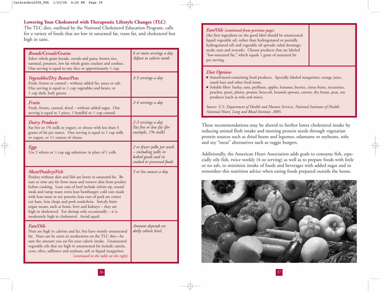
Fats/Oils (continued from previous page)(the first ingredient on the good label should be unsaturatedliquid vegetable oil, rather than hydrogenated or partiallyhydrogenated oil) and vegetable oil spreads; salad dressings;seeds; nuts and avocado. Choose products that are labeled“low-saturated fat,” which equals 1 gram of saturated fat per serving.
Diet Options■ Stanol/sterol-containing food products. Specially labeled margarines, orange juice,
snack bars and other food items.■ Soluble fiber: barley, oats, psyllium, apples, bananas, berries, citrus fruits, nectarines,
peaches, pears, plums, prunes, broccoli, brussels sprouts, carrots, dry beans, peas, soyproducts (such as tofu and miso).
Source: U.S. Department of Health and Human Services, National Institutes of Health,National Heart, Lung and Blood Institute, 2005.
Lowering Your Cholesterol with Therapeutic Lifestyle Changes (TLC)The TLC diet, outlined by the National Cholesterol Education Program, callsfor a variety of foods that are low in saturated fat, trans fat, and cholesterol buthigh in taste.
These recommendations may be altered to further lower cholesterol intake byreducing animal flesh intake and meeting protein needs through vegetarian protein sources such as dried beans and legumes, edamame or soybeans, tofuand soy “meat” alternatives such as veggie burgers.
Additionally, the American Heart Association adds goals to consume fish, espe-cially oily fish, twice weekly (4 oz serving) as well as to prepare foods with littleor no salt, to minimize intake of foods and beverages with added sugar and toremember this nutrition advice when eating foods prepared outside the home.
6 or more servings a day Adjust to calorie needs
3-5 servings a day
2-4 servings a day
2-3 servings a dayFat free or low fat (for example, 1% milk)
2 or fewer yolks per week– including yolks inbaked goods and incooked or processed foods
5 or less ounces a day.
Amount depends on daily calorie level.
Breads/Cereals/GrainsSelect whole grain breads, cereals and pasta, brown rice, oatmeal, potatoes, low fat whole grain crackers and cookies.One serving is equal to one slice or approximately 1⁄2 cup.
Vegetables/Dry Beans/PeasFresh, frozen or canned – without added fat, sauce or salt. One serving is equal to 1⁄2 cup vegetables and beans, or 1 cup dark, leafy greens.
FruitsFresh, frozen, canned, dried – without added sugar. Oneserving is equal to 1 piece, 1 handful or 1⁄2 cup canned.
Dairy ProductsFat free or 1% milk or yogurt, or cheese with less than 3 grams of fat per ounce. One serving is equal to 1 cup milk or yogurt, or 11⁄2 ounces of cheese.
EggsUse 2 whites or 1⁄4 cup egg substitute in place of 1 yolk.
Meat/Poultry/FishPoultry without skin and fish are lower in saturated fat. Besure to trim any fat from meat and remove skin from poultrybefore cooking. Lean cuts of beef include sirloin tip, roundsteak and rump roast; extra lean hamburger; cold cuts madewith lean meat or soy protein; lean cuts of pork are center cut ham, loin chops and pork tenderloin. Strictly limit organ meats, such as brain, liver and kidneys – they are high in cholesterol. Eat shrimp only occasionally – it is moderately high in cholesterol. Avoid squid.
Fats/OilsNuts are high in calories and fat, but have mostly unsaturatedfat. Nuts can be eaten in moderation on the TLC diet—besure the amount you eat fits your calorie intake. Unsaturatedvegetable oils that are high in unsaturated fat include canola,corn, olive, safflower and soybean; soft or liquid margarines
(continued in the table on the right)
CardiacBro2008_FNL 1/23/08 4:20 PM Page 39

Cou
ntin
g th
e pu
lse
Rec
ordi
ng y
our
puls
e
Get a watch with a second hand and place it so you can easily see it.
● RELAX! so your heart rests. It’s best to count a resting pulse.
● Find your pulse and just FEEL THE BEATING for a while.
● Now – start COUNTING WITH ZERO AND COUNTEACH BEAT YOU FEEL FOR ONE FULL MINUTE.
Got the number? THAT’S YOUR PULSE!!!
To practice : Try counting your nurse’s pulse or someone in yourfamily. You will find that everyone’s pulse is a little different.
● Be sure to write down in a log the NUMBER of your pulse, and if it was regular or not.
● If your pulse is slower than _________ or fasterthan__________ for one full minute, DON’T take your medicine. Check with your doctor first.
COMMENTS
38
How much your heart beats EACH MINUTE depends on many things:
● whether you are resting or exercising● medications you take● the condition of your heart and body
Often when your dosage of medicine needs to be changed or whenthere is a change in your heart, the pulse will change. It may be,FAST or SLOW, REGULAR or IRREGULAR.
Learning to count your pulse helps you know when to take your medication, and when to call the doctor.
During my hospitalization:My pulse at rest ______________My pulse after an activity, such as walking
There are three places you can try:The WRIST - just below the bottom of the thumb (We will use the wrist.)
Place your first two or threefingers on the radial artery at the wrist. Can you feel the beating of the pulse? Now take your fingers awayand try to find it again. Feel it? Is the beating regularor irregular?
Note: If you are having trouble, try using your other wrist, or try someone else’s first
Inside of the ELBOW At the bottom of the NECK
Why
lear
n to
cou
nt y
our
puls
e?Fi
ndin
g yo
ur p
ulse
How to Take a Pulse
CardiacBro2008_FNL 1/23/08 4:20 PM Page 41
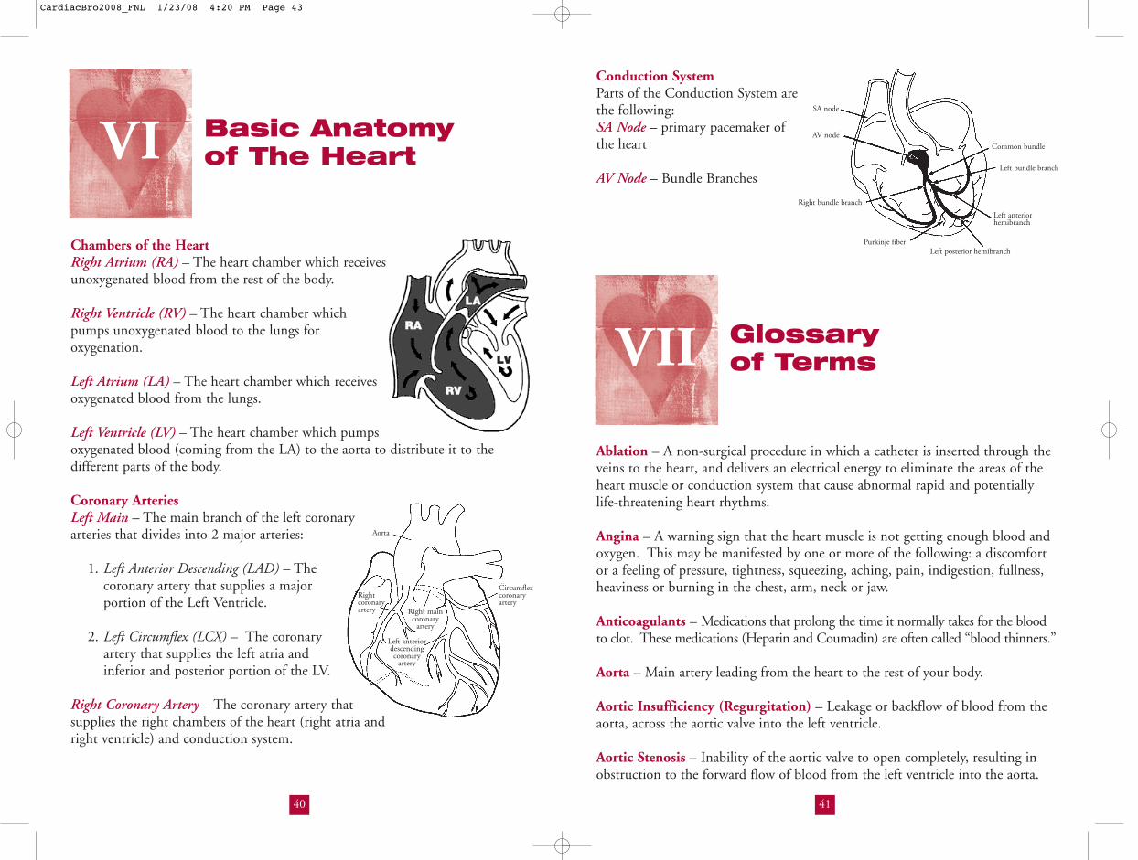
41
Conduction SystemParts of the Conduction System arethe following:SA Node – primary pacemaker ofthe heart
AV Node – Bundle Branches
Glossary of Terms
Ablation – A non-surgical procedure in which a catheter is inserted through theveins to the heart, and delivers an electrical energy to eliminate the areas of theheart muscle or conduction system that cause abnormal rapid and potentially life-threatening heart rhythms.
Angina – A warning sign that the heart muscle is not getting enough blood andoxygen. This may be manifested by one or more of the following: a discomfort or a feeling of pressure, tightness, squeezing, aching, pain, indigestion, fullness,heaviness or burning in the chest, arm, neck or jaw.
Anticoagulants – Medications that prolong the time it normally takes for the bloodto clot. These medications (Heparin and Coumadin) are often called “blood thinners.”
Aorta – Main artery leading from the heart to the rest of your body.
Aortic Insufficiency (Regurgitation) – Leakage or backflow of blood from theaorta, across the aortic valve into the left ventricle.
Aortic Stenosis – Inability of the aortic valve to open completely, resulting inobstruction to the forward flow of blood from the left ventricle into the aorta.
40
Basic Anatomyof The Heart
Chambers of the HeartRight Atrium (RA) – The heart chamber which receives unoxygenated blood from the rest of the body.
Right Ventricle (RV) – The heart chamber whichpumps unoxygenated blood to the lungs for oxygenation.
Left Atrium (LA) – The heart chamber which receivesoxygenated blood from the lungs.
Left Ventricle (LV) – The heart chamber which pumps oxygenated blood (coming from the LA) to the aorta to distribute it to the different parts of the body.
Coronary ArteriesLeft Main – The main branch of the left coronary arteries that divides into 2 major arteries:
1. Left Anterior Descending (LAD) – The coronary artery that supplies a major portion of the Left Ventricle.
2. Left Circumflex (LCX) – The coronaryartery that supplies the left atria and inferior and posterior portion of the LV.
Right Coronary Artery – The coronary artery thatsupplies the right chambers of the heart (right atria andright ventricle) and conduction system.
SA node
AV node
Right bundle branch
Purkinje fiberLeft posterior hemibranch
Common bundle
Left bundle branch
Left anterior hemibranch
VI
Aorta
Rightcoronaryartery Right main
coronary artery
Left anterior descendingcoronary
artery
Circumflexcoronary artery
RRAA
RRVV
LLAA
LLVV VII
CardiacBro2008_FNL 1/23/08 4:20 PM Page 43

43
High-Density Lipoproteins (HDL) – A special type of cholesterol that, whenhigh, decreases the incidence of fatty deposits within arterial wall (decreasingrisk for coronary atherosclerosis). Called “good cholesterol.”
Triglycerides – Another type of fat which, when elevated, increases the risk for atherosclerosis.
Congestive Heart Failure – A condition in which the heart muscle is unable to effectively pump out blood to the rest of the body which results in increase accumulation of blood in the heart chambers and the lungs.
Coronary Arteries – Vessels originating from the main artery (aorta), which runson the surface of the heart, supplying blood to the heart muscle from the aorta.
Coronary Arteriography – X-ray procedure in which dye is injected to outlinethe coronary arteries, allowing the physician to see blockages or abnormal nar-rowing in the artery.
Coronary Artery Disease (CAD) – A condition in which a coronary artery isclogged by a buildup of cholesterol and fatty deposits. Clogged arteries maypotentially decrease delivery of oxygen-rich blood supply to the heart muscleand predispose to development of heart attacks (myocardial infarction).
Coronary Artery Bypass Graft (CABG) – Open heart surgery used to bringnew blood supply to the heart muscle by connecting pieces of a vein or artery toreroute blood flow around the obstructed arteries.
Coronary Thrombosis – A blood clot in an artery which blocks the supply ofblood to the heart muscle.
Diuretics – A medication commonly called a “water pill” that helps the bodyget rid of excess salt and fluid.
Echocardiogram – A procedure which uses sound waves to: 1) evaluate thestructures and functions of the heart valves and heart muscle; and 2) measurethe size of the heart’s pumping chambers (ventricles).
Electrophysiological Study (EPS) – A procedure in which a catheter is insertedthrough the veins to the heart to study the electrical activity of the heart and to identify absence or presence of abnormal heart rhythms. If abnormal heartrhythms are present, the specific site of origin is identified and appropriate treatment is instituted.
42
Arrhythmias – Refers to irregularities in heart rhythm with the heart beating either too fast (tachycardia) or too slow (bradycardia). The arrhythmias may reduce the pumping ability of the heart.
Arteries – Vessels branching from the aorta which carry oxygen-rich blood to different parts of the body.
Arteriosclerosis – Commonly called “hardening of the arteries.” A condition inwhich the arterial wall becomes thick and hard, losing its elasticity.
Atherosclerosis – Narrowing or blockage of arteries (blood vessels) caused bybuild-up of fatty plaque made up of cholesterol and other materials within theartery wall.
Blood Pressure – A measurement of the force that moving blood puts on theartery wall. It consists of two measurements: the systolic pressure, and the diastolic pressure.
Diastolic Pressure – Pressure left in the arteries in between heartbeats. The “lower number” in a blood pressure reading.
Systolic Pressure – Peak pressure in the arteries when the heart pumps out the blood to the body. The “top number” in a blood pressure reading.
Cardiomyopathy – Enlargement and weakening of the heart muscle which may have resulted from any one of various causes, i.e. long-standing heart disease, viral infections, toxic effects of alcohol or unknown causes. A dilated heart does not pump as well as it should.
Catheter – A small plastic tube used to inject liquid (dye) into the coronary artery at the time of coronary angiogram or angioplasty/stent.
Cholesterol – A waxy substance with fat-like properties present in animal tis-sues and, thus, in foods of animal origin such as meat, eggs and dairy products.Saturated fats will also increase cholesterol levels. In addition to food sources, cholesterol is manufactured by the body. Cholesterol in excess is stored in the fatty deposits in the arterial wall in atherosclerosis.
Low-Density Lipoproteins (LDL) – Special type of cholesterol that, when high, increases the incidence of fatty deposits within arterial wall (increasingrisk for coronary atherosclerosis). Called “bad cholesterol.”
CardiacBro2008_FNL 1/23/08 4:20 PM Page 45
4544
Fats - Monounsaturated Fats – Aid in lowering blood cholesterol levels. They areliquid at room temperature and originate only from plant and vegetablesources. Examples: olive oil, canola oil, peanut oil, and avocado.
Polyunsaturated Fats – Aid in lowering of blood cholesterol levels. They areusually soft at room temperature and originate from plant and vegetablesources. Examples: cottonseed, soybean, corn and safflower oils.
Saturated Fats – Raise blood cholesterol levels. Generally, they are solid atroom temperature. They originate primarily from from animal sources, suchas beef, veal, lamb, poultry, milk, butter, cheese and lard.
High Blood Pressure (Hypertension) – A greater-than-normal pressure ofblood against the walls of the arteries. It may not make you sick, but over time it may cause wear and tear on the artery walls and may damage the heart,brain, eyes and kidneys. The causes of high blood pressure are not completelyunderstood. Stress, high sodium diet, obesity, and smoking may contribute toor aggravate high blood pressure. In some cases the tendency for high bloodpressure runs in the family.
Implantable Cardioverter Defibrillator (ICD) – A special type of electronicdevice placed inside the body to automatically terminate life-threatening abnormal heart rhythms.
Invasive Procedures – Procedures which involve puncture or incision of the skin or insertion of an instrument into the body.
Ischemia – Blood supply to the heart muscle is insufficient to maintain normal function of an organ.
Mitral Insufficiency (Regurgitation) – Inability of the mitral valve to close completely, resulting in leakage or backflow of blood into the top left (atria) chamber and the lungs.
Mitral Stenosis – Obstruction of the passage of blood across the mitral valve due to narrowing of the opening, resulting in a decreased forward flow of blood and accumulation of blood into the top left (atria) chamber and the lungs.
Myocardial Infarction – Heart muscle damage brought about by a prolonged or persistent abrupt blockage of the blood vessels that supply the heart muscle(coronary arteries). Also known as a heart attack.
Pacemaker – A device placed in the body to help regulate your heart rhythm.This device sends out electrical signals to keep the heart beating at a speed appropriate for your body’s needs.
Pacemaker-Cardioverter Defibrillator (PCD) – A special type of electronicdevice placed inside the body to automatically detect and terminate life-threat-ening abnormal heart rhythm and at the same time regulate the speed of theheartbeat to meet your body’s needs.
Percutaneous Transluminal Coronary Angioplasty (PTCA) – A procedure used to widen the narrowing in your coronary artery using a small balloon that is inflated within the narrowed section of the coronary artery.
Plaque – Thickened portion of the artery containing cholesterol that blocks the arteries.
Resting Electrocardiogram – Recording of the heart’s electrical activity at rest.
Sheath – A small catheter or tube which allows for access into a vein or arteryduring catheterization, angiogram and angioplasty procedures and electrophysio-logical study procedures. This tube is usually removed immediately after mostof these procedures except for an angioplasty procedure in which it may be leftin place for several hours.
Stress Electrocardiogram – Recording of the heart’s electrical activity while the patient exercises.
Transesophageal Echocardiogram – An invasive type of echocardiographic procedure wherein a probe with an ultrasound transducer is placed into the esophagus via the mouth.
Thrombolysis – A process of dissolving clots that clog arteries.
Vasodilators – Medications used to increase the size (dilatation) of a blood vessel.
Valvuloplasty – A non-surgical procedure to loosen up narrowed or tight heart valves.
CardiacBro2008_FNL 1/23/08 4:20 PM Page 47

Cedars-Sinai Heart Center Outpatient ServicesCardiac Rehabilitation 423-9660Diabetic Education (DOTEC) 423-3870General Information 423-5000LifeLine Program 423-5468Nutrition Counseling Center 423-3444Patient and Community Education 423-5177Physical Medicine 423-3148Pulmonary Rehabilitation 423-9566Senior Care Program 423-9581Center Strutters Walk for Fitness Program
A Collaborative Program with Beverly Center 423-9581
Community Associations:American Heart Association (AHA) (800) AHA-USA1American Lung Association
(ALA Smoking Cessation Program) (323) 935-5864
Important Websiteswww.cedars-sinai.eduwww.HealthyDiningFinder.comwww.nhlbi.nih.gov (National Heart, Lung and Blood institute)www.americanheart.org (American Heart Association)
Review the AHA guidelines for healthier eating to protect your heart.Includes a good selection of recipes, including those low in salt.
www.eatright.org (American Dietetic Association) Click on Food and Nutrition Information to view nutrition fact sheets,including those for healthy aging. Some information is restricted to ADA members only.
Personal Contacts (e.g., physicians, nurses . . .)
4746
TelephoneNumbers
Cedars-Sinai Heart Center Inpatient ServicesBlood Donor Facility (310) 423-5346Cardiothoracic Surgery 423-3851Chaplaincy 423-5550Evaluation Center 423-4860Heart Failure Program 423-2431Heart Families 423-3193Hospice and Palliative Care 423-9520Hospitality Center 423-4444Medical Social Work 423-4446Non-Invasive Echo Lab 423-4861Pacemaker/ICD Clinic 423-2726
Cardiac Patient Care CentersMain Hospital
5NE: 5001-5032 423-45815NW: 5101-5132 423-4585Volunteer Information Desk - 5th Floor Lobby 423-55616ICU: 6002-6016 423-60386NE: 6018-6032 423-46516NW: 6101-6132 423-4655Volunteer Information Desk - 6th Floor Lobby 423-5661
Saperstein Critical Care Tower3N40 to 3N68 423-38663S10 to 3S38 423-38673rd Floor Lobby 423-71434N40 to 4N68 423-44264S10 to 4S38 423-44274th Floor Lobby 423-7142Plaza Lobby 423-7133Saperstein: 6 South CICU 6S10-6S32 423-6247Saperstein: 6 North CSICU 6S46-6S68 423-6276Saperstein Lobby 6th Floor 423-7148
VIII
CardiacBro2008_FNL 1/23/08 4:20 PM Page 49

Questions for Doctors and Nurses
Tests & Results During Hospitalization
Medications When I Go Home
Other
48
CardiacBro2008_FNL 1/23/08 4:20 PM Page 51