Embed Size (px)
Citation preview

8/17/2011
1
Stanford UniversityStanford University
Medical CenterMedical Center
Frandics Chan, M.D., Ph.D.Frandics Chan, M.D., Ph.D.
Lucile Packard Lucile Packard
Children’s HospitalChildren’s Hospital
Cardiac MRI Sequences and
Protocols
Traditional Protocol Model
for Tomographic Imaging
× →
Techniques Orientations
Interpretation
SUMC Department of RadiologyLPCH
Challenges in Cardiac Imaging
• Large number techniques
T1/DIR MRA Coronary MRA ssfp cine
Perfusion Delayed Enhance Phase Contrast Tagging
SUMC Department of RadiologyLPCH
Challenges in Cardiac Imaging
• Infinite number of imaging planes
SUMC Department of RadiologyLPCH
Objectives
• To know the basic types of clinically
used cardiac MRI sequences
• To understand how cardiac MRI records
the moving heart
• To understand trade-off in noise and
performance
• To learn how to set up standard cardiac
planes and a cardiac function protocol
SUMC Department of RadiologyLPCH
Basic Cardiac MRI Sequences
8/17/2011
2
SUMC Department of RadiologyLPCH
Basic Cardiac MRI Sequences
• Bright blood
• Cardiac gated
• Cardiac cine
• Breath-hold
• Non-contrast
• Names
– Fiesta (GE)
– True Fisp (Siemens)
Balanced-SSFP
SUMC Department of RadiologyLPCH
Basic Cardiac MRI Sequences
• “Grey” blood
• Cardiac gated
• Cardiac cine
• Breath-hold
• Non-contrast
• Names
– mtag (Stanford)
– Tagged fastcine (GE)
Myocardial tagging
SUMC Department of RadiologyLPCH
Basic Cardiac MRI Sequences
• Dark blood
• Cardiac gated
• Single cardiac phase
• Breath-hold
• Non-contrast
• Names
– DIRFSE (GE)
– HASTE (Siemens)Double IR
SUMC Department of RadiologyLPCH
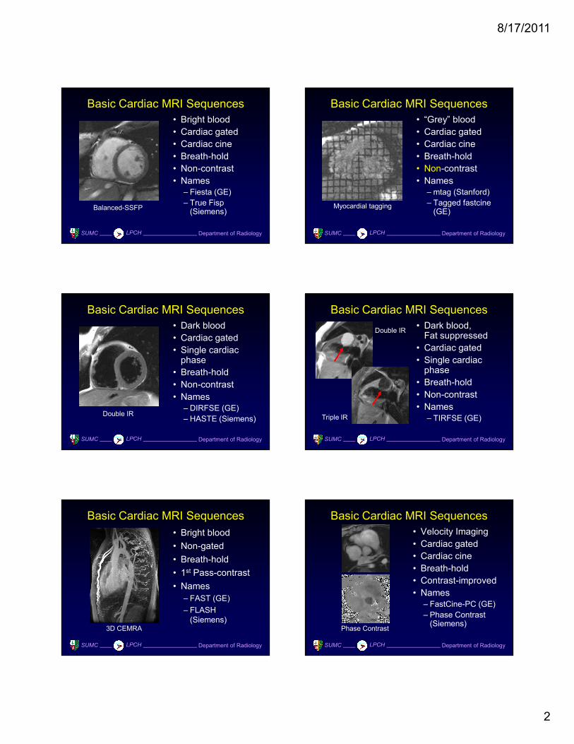
Basic Cardiac MRI Sequences
• Dark blood,Fat suppressed
• Cardiac gated
• Single cardiac phase
• Breath-hold
• Non-contrast
• Names
– TIRFSE (GE)
Double IR
Triple IR
SUMC Department of RadiologyLPCH
Basic Cardiac MRI Sequences
• Bright blood
• Non-gated
• Breath-hold
• 1st Pass-contrast
• Names
– FAST (GE)
– FLASH
(Siemens)3D CEMRA
SUMC Department of RadiologyLPCH
Basic Cardiac MRI Sequences
• Velocity Imaging
• Cardiac gated
• Cardiac cine
• Breath-hold
• Contrast-improved
• Names
– FastCine-PC (GE)
– Phase Contrast (Siemens)
Phase Contrast
8/17/2011
3
SUMC Department of RadiologyLPCH
Basic Cardiac MRI Sequences
• Bright-blood
• Cardiac gated
• Single cardiac
phase
• Navigator echo
• Non-contrast
• Names
– MSLAB (GE)Coronary MRA
SUMC Department of RadiologyLPCH
Basic Cardiac MRI Sequences
• Dark myocardium
• Cardiac gated
• Single cardiac
phase
• Breath-hold
• Post-contrast
• Names– IrPFSE / MDE (GE)
– TFLASH (Siemens)Delay-enhancement
SUMC Department of RadiologyLPCH
Basic Cardiac MRI Sequences• Dark myocardium
• Cardiac gated
• Single cardiac phase
• Breath-hold
• 1st Pass-contrast
• Multiple contrast phases
• Names
– IrP-EPI/GRE (GE)Myocardial Perfusion
SUMC Department of RadiologyLPCH
How to record a moving heart?
SUMC Department of RadiologyLPCH
Fast, Real-Time Imaging
• Bright blood
• Non-gated
• Breath-hold optional
• Non-contrast
• Low spatial and temporal res.
• Names
– MR Echo (GE)Real-time imaging
SUMC Department of RadiologyLPCH
Gated cine
• Almost all cardiac sequences assume
periodic, repeating cardiac motion
• Each RR-interval records part of the k-
space information
• To build up multiple frames of k-space
information requires multiple heart beats
• The method of dividing up the k-space
is called segmented k-space.

8/17/2011
4
SUMC Department of RadiologyLPCH
Conventional Cine Acquisition
SUMC Department of RadiologyLPCH
Conventional Cine Acquisition
Ph 1 Ph 2 Ph 3 Ph N
SUMC Department of RadiologyLPCH
Conventional Cine Acquisition
Ph 1 Ph 2 Ph 3 Ph N
1TR
SUMC Department of RadiologyLPCH
Conventional Cine Acquisition
Ph 1 Ph 2 Ph 3 Ph N
SUMC Department of RadiologyLPCH
Conventional Cine Acquisition
Ph 1 Ph 2 Ph 3 Ph N
SUMC Department of RadiologyLPCH
Conventional Cine Acquisition
Ph 1 Ph 2 Ph 3 Ph N

8/17/2011
5
SUMC Department of RadiologyLPCH
Conventional Cine Acquisition
Ph 1 Ph 2 Ph 3 Ph N
SUMC Department of RadiologyLPCH
Conventional Cine Acquisition
Ph 1 Ph 2 Ph 3 Ph N
SUMC Department of RadiologyLPCH
Conventional Cine Acquisition
Ph 1 Ph 2 Ph 3 Ph N
SUMC Department of RadiologyLPCH
Conventional Cine Acquisition
Ph 1 Ph 2 Ph 3 Ph N
SUMC Department of RadiologyLPCH
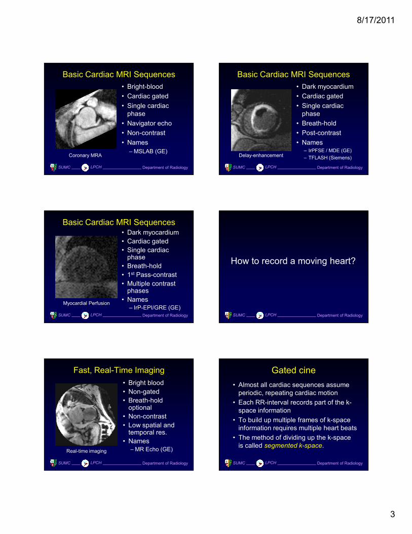
Questions
• How long does this scan take?
– Phase encodes x RR-interval
• At 60 bpm, 192 lines, how long?
– 3 minutes 12 seconds
• What is the temporal resolution?
– TR
• In real terms?
– ~ 5 ms
SUMC Department of RadiologyLPCH
Conventional Cardiac cine
• Best possible
temporal res.
• Long scan time
• Respiratory
motion
• Means of control
– Resp. comp.
– Resp. gating
– Breath-hold

8/17/2011
6
SUMC Department of RadiologyLPCH
Problem: How to shorten scan time?
Solution: Acquire more than one phase
encoding line per heart beat.
SUMC Department of RadiologyLPCH
Segmented K-Space
Ph 1 Ph 2 Ph 3 Ph N
4 vps
SUMC Department of RadiologyLPCH
Segmented K-Space
Ph 1 Ph 2 Ph 3 Ph N
SUMC Department of RadiologyLPCH
Segmented K-Space
Ph 1 Ph 2 Ph 3 Ph N
SUMC Department of RadiologyLPCH
Segmented K-Space
Ph 1 Ph 2 Ph 3 Ph N
SUMC Department of RadiologyLPCH
GE: View per segment (vps) =
number of lines per heart beat
8/17/2011
7
SUMC Department of RadiologyLPCH
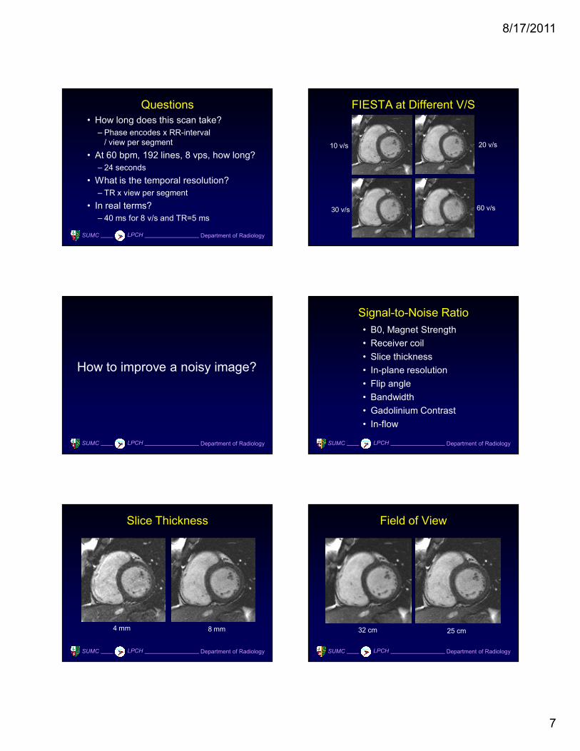
Questions
• How long does this scan take?
– Phase encodes x RR-interval
/ view per segment
• At 60 bpm, 192 lines, 8 vps, how long?
– 24 seconds
• What is the temporal resolution?
– TR x view per segment
• In real terms?
– 40 ms for 8 v/s and TR=5 ms
FIESTA at Different V/S
10 v/s 20 v/s
30 v/s 60 v/s
SUMC Department of RadiologyLPCH
How to improve a noisy image?
SUMC Department of RadiologyLPCH
Signal-to-Noise Ratio
• B0, Magnet Strength
• Receiver coil
• Slice thickness
• In-plane resolution
• Flip angle
• Bandwidth
• Gadolinium Contrast
• In-flow
SUMC Department of RadiologyLPCH
Slice Thickness
4 mm 8 mm
SUMC Department of RadiologyLPCH
Field of View
32 cm 25 cm

8/17/2011
8
SUMC Department of RadiologyLPCH
Matrix Size
128 x 128 224 x 224
SUMC Department of RadiologyLPCH
Flip Angle
10 degree 30 degree 50 degree
SUMC Department of RadiologyLPCH
Contrast
Non-contrast Contrast
SUMC Department of RadiologyLPCH
In-Flow (FastCine)
10 degree 50 degree
SUMC Department of RadiologyLPCH
How to do a cardiac function
MRI study?
SUMC Department of RadiologyLPCH
Optimal MRI Protocol• Every image taken must serve a diagnostic
goal.
• The number of sequences and breath-holds
should be minimized.
• Fast sequences and parallel imaging
should be used whenever possible, but …
• Sequences should be grouped according to
contrast usage.
• Oblique planes should be prescribed in the
least number of intermediate steps.

8/17/2011
9
Protocol: Cardiac Function
• Pre-contrast • 3-plane SSFP localizer
• Optional axial localizer
• LAX SSFP cine localizer
• SAX SSFP cine
• 4ch SSFP cine
• LV3ch SSFP cine
• LV2ch SSFP cine
• RV3ch SSFP cine
• RV2ch SSFP cine
• RVOT SSFP cine
• Ao long SSFP cine localizer
• PA long SSFP cine localizer
• Ao PC cine
• PA PC cine
• TV PC cine
• MV PC cine
SUMC Department of RadiologyLPCH
Protocol: Viability
• Pre-contrast
• Post-contrast
• 3-plane SSFP localizer
• Optional axial localizer
• Loc LAX SSFP cine
• SAX SSFP cine
• 4ch SSFP cine
• LV3ch SSFP cine
• LV2ch SSFP cine
• Cine IR
• 4ch Test for TI
• SAX IR at TI
• LV3ch, LV2ch IR at TI
SUMC Department of RadiologyLPCH
Protocol: TOF
• Pre-contrast
• 1st Pass
• Post-contrast
• 3-plane SSFP localizer
• Optional axial localizer
• LAX SSFP cine localizer
• SAX SSFP cine
• 4ch, LV-LAX, RV-LAX SSFP cine
• RVOT SSFP cine
• Ao long SSFP cine localizer
• PA long SSFP cine localizer
• CE MRA
• Ao PC cine
• PA sup, PA sub PC cine
• TV sup PC cine
SUMC Department of RadiologyLPCH
Coronal Localizer
• Options: GRE, single phase SSFP, SSFSE,
T1-SE, or double-IR
GRE SSFSE
SUMC Department of RadiologyLPCH
Axial Localizer
• ECG-gated, Breath-hold
GRE Single phase SSFP
SUMC Department of RadiologyLPCH
Oblique Sagittal Cine
• ECG-gated, Breath-hold
Single phase SSFP SSFP cine

8/17/2011
10
SUMC Department of RadiologyLPCH
Short Axis Cine
• Scan from base to apex
SSFP cine
SSFP cine
SUMC Department of RadiologyLPCH
Ventricular Volumes / Mass
SUMC Department of RadiologyLPCH
SAX Valve Plane
Netter
MV
TV
AV
PV
SUMC Department of RadiologyLPCH
4-Chamber View (HLAX)
Netter
SUMC Department of RadiologyLPCH
4-Chamber View (HLAX)
SUMC Department of RadiologyLPCH
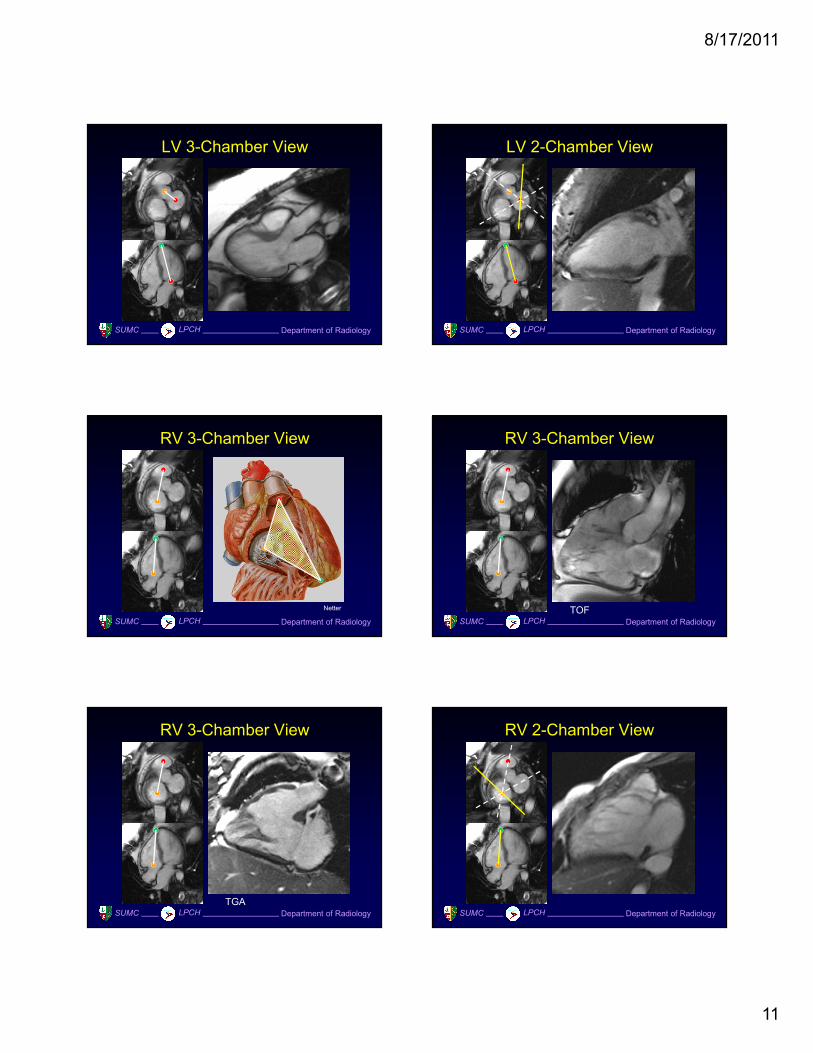
LV 3-Chamber View
8/17/2011
11
SUMC Department of RadiologyLPCH
LV 3-Chamber View
SUMC Department of RadiologyLPCH
LV 2-Chamber View
SUMC Department of RadiologyLPCH
RV 3-Chamber View
Netter
SUMC Department of RadiologyLPCH
RV 3-Chamber View
TOF
SUMC Department of RadiologyLPCH
RV 3-Chamber View
TGASUMC Department of RadiologyLPCH
RV 2-Chamber View

8/17/2011
12
SUMC Department of RadiologyLPCH
PA Longitudinal Localizer
Axial localizer SSFP cine valve
SUMC Department of RadiologyLPCH
RVOT View
Axial localizer SSFP cine
SUMC Department of RadiologyLPCH
Aorta Longitudinal Localizer 1
Coronal localizer SSFP cine
SUMC Department of RadiologyLPCH
Aorta Longitudinal Localizer 2
Coronal localizer SSFP cine
SUMC Department of RadiologyLPCH
Aortic Flow Quantification
SSFP cine diastole Phase contrast cine
SUMC Department of RadiologyLPCH
Aortic Stenosis Velocity
SSFP cine diastole Phase contrast cine
8/17/2011
13
SUMC Department of RadiologyLPCH
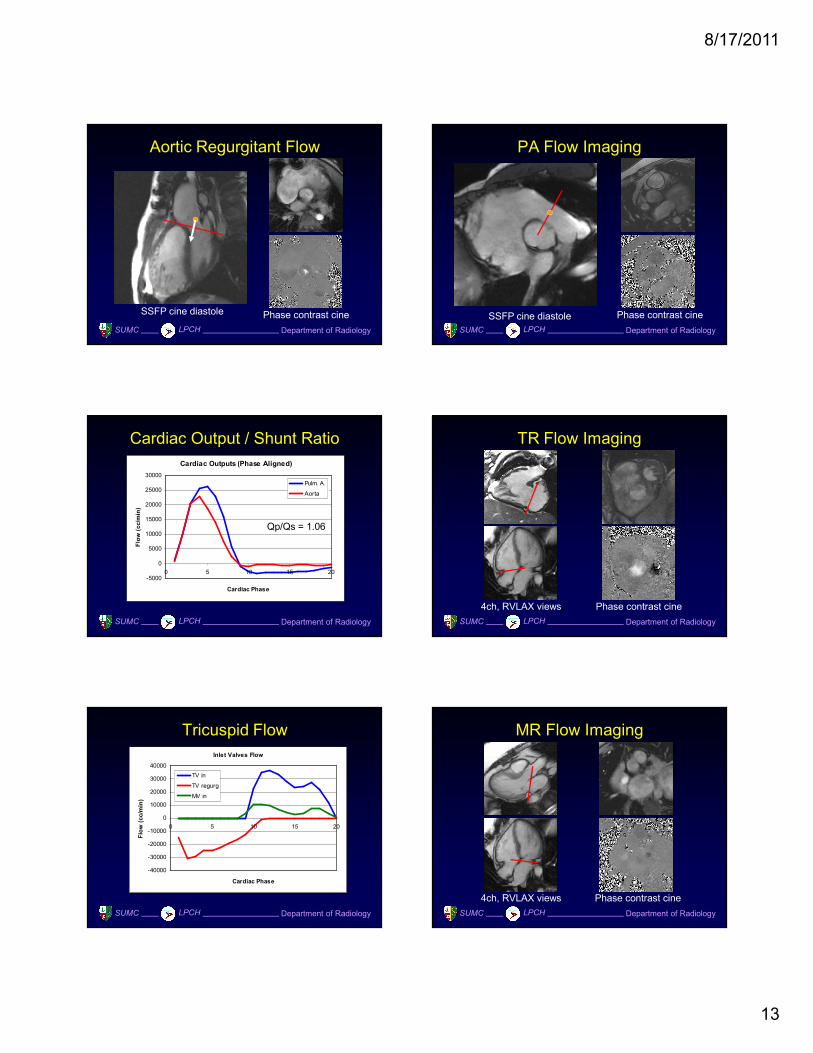
Aortic Regurgitant Flow
SSFP cine diastole Phase contrast cine
SUMC Department of RadiologyLPCH
PA Flow Imaging
SSFP cine diastole Phase contrast cine
SUMC Department of RadiologyLPCH
Cardiac Output / Shunt Ratio
Cardiac Outputs (Phase Aligned)
-5000
0
5000
10000
15000
20000
25000
30000
0 5 10 15 20
Cardiac Phase
Flow (cc/min)
Pulm. A.
Aorta
Qp/Qs = 1.06
SUMC Department of RadiologyLPCH
TR Flow Imaging
4ch, RVLAX views Phase contrast cine
SUMC Department of RadiologyLPCH
Tricuspid Flow
Inlet Valves Flow
-40000
-30000
-20000
-10000
0
10000
20000
30000
40000
0 5 10 15 20
Cardiac Phase
Flow (cc/min)
TV in
TV regurg
MV in
SUMC Department of RadiologyLPCH
MR Flow Imaging
4ch, RVLAX views Phase contrast cine

8/17/2011
14
SUMC Department of RadiologyLPCH
Summary
• The practice of cardiac MRI is an
integration of
– Clinical knowledge
– Technical knowledge
– Patience and interest
• Like the making of a violin virtuoso, it is
– 99% practice
– 1% gift
SUMC Department of RadiologyLPCH