Embed Size (px)
Citation preview
RESEARCH ARTICLE
Carcinoma cells induce lumen filling and EMT in epithelial cellsthrough soluble E-cadherin-mediated activation of EGFRPratima U. Patil1,2, Julia D’Ambrosio2, Landon J. Inge3, Robert W. Mason2 and Ayyappan K. Rajasekaran1,4,*
ABSTRACTIn epithelial cancers, carcinoma cells coexist with normal cells.Although it is known that the tumor microenvironment (TME) plays apivotal role in cancer progression, it is not completely understood howthe tumor influences adjacent normal epithelial cells. In this study, athree-dimensional co-culture system comprising non-transformedepithelial cells (MDCK) and transformed carcinoma cells (MSV-MDCK) was used to demonstrate that carcinoma cells sequentiallyinduce preneoplastic lumen filling and epithelial–mesenchymaltransition (EMT) in epithelial cysts. MMP-9 secreted by carcinomacells cleaves cellular E-cadherin (encoded by CDH1) from epithelialcells to generate soluble E-cadherin (sE-cad), a pro-oncogenicprotein. We show that sE-cad induces EGFR activation, resulting inlumen filling in MDCK cysts. Long-term sE-cad treatment inducedEMT. sE-cad caused lumen filling by induction of the ERK signalingpathway and triggered EMT through the sustained activation of theAKT pathway. Although it is known that sE-cad induces MMP-9release and consequent EGFR activation in tumor cells, our results,for the first time, demonstrate that carcinoma cells can induce sE-cadshedding in adjacent epithelial cells, which leads to EGFR activationand the eventual transdifferentiation of the normal epithelial cells.
KEY WORDS: EGFR, MMP-9, Soluble E-cadherin,Transdifferentiation
INTRODUCTIONBidirectional communication between tumor cells and themicroenvironment plays a crucial role in driving tumorprogression. The tumor microenvironment (TME) is comprised ofa heterogeneous population of cells, including stromal cells,adjacent normal cells, fibroblasts, infiltrating immune cells andangiogenic vascular cells, and secretions from these cells such asgrowth factors, cytokines and extracellular matrix (ECM) (Hanahanand Coussens, 2012). During the initial stages of carcinogenesis, thesurrounding normal epithelial cells and other microenvironmentcomponents are anti-tumorigenic as they block transformed cellproliferation and survival (Hogan et al., 2011; Kajita et al., 2010;Toillon et al., 2002). However, once tumor cells multiply andoverride this self-defense mechanism, they modify and cause theevolution of the non-transformed cell types to participate in tumorprogression (Ivers et al., 2014). For instance, normal prostateepithelial cells can induce intraepithelial neoplasia in mice when
co-injected with carcinoma-associated fibroblasts (CAFs) but notwhen co-injected with normal fibroblasts, suggesting alteration offibroblasts by tumor cells (Olumi et al., 1999). Similarly CAFs frombreast cancer tissue induce EMT and enhance the metastaticpotential of both premalignant and malignant breast epithelial cells,unlike normal fibroblasts, which suppress metastasis (Orimo et al.,2005). Although some aspects of the TME, such as ECMremodeling and tumor angiogenesis are well studied, theinteraction between tumor cells and the adjacent epithelium ispoorly understood.
Epithelial cells in the tissues reorganize themselves to formtubular networks and cyst-like spheroids (Zegers et al., 2003).Spherical epithelial cysts are the building blocks of glandular organsand are characterized by a hollow lumen with apical–basal polarity(Bryant and Mostov, 2008). An intact, hollow lumen formed byselective apoptosis of centrally located cells in the cyst is crucial forproper control and function of epithelial tissues (Debnath et al.,2002). However, abnormal pathophysiological conditions causelesions in the lumen, resulting in the formation of multiple lumensand, finally, filling of the luminal space by cells. Filling of the lumenis a salient feature of early pre-invasive stages of epithelial cancers(Debnath and Brugge, 2005). Several carcinomas such as breast,kidney and prostate carcinomas display lumen filling duringtheir early stages (Debnath and Brugge, 2005). Lumen filling isfollowed by epithelial to mesenchymal transition (EMT), atransdifferentiation process, which facilitates invasion and themetastatic potential of carcinoma cells (Tiwari et al., 2012).
A key event observed during epithelial-derived carcinogenesis isthe downregulation of E-cadherin (also known as CDH1), a Ca2+
-dependent cell adhesion molecule located in the adherens junctionand along the basolateral surface of epithelial cells (Gumbiner et al.,1988; Takeichi, 1991). One of the mechanisms bywhich tumor cellsdownregulate E-cadherin is by the proteolytic cleavage of itsextracellular domain. Matrix metalloproteinase (MMP)-9, one ofthe proteases present in the extracellular environment, cleaves theectodomain of membrane-bound E-cadherin at the MMP cleavagesite between amino acids Leu581 and Ser582 to generate a 80-kDafragment referred to as soluble E-cadherin (sE-cad) (DeWever et al.,2007; Maretzky et al., 2005). MMP-9 levels are elevated in severalcancers, and this enzyme appears to be involved in tumor cellinitiation and progression, metastatic ability and genetic instability(Farina and Mackay, 2014).
Significantly elevated sE-cad levels have been observed inthe sera and urine of cancer patients diagnosed with a variety ofcancers. Increased circulating levels of sE-cad are indicative ofhistopathological grade, metastasis recurrence and poor prognosis(Chan et al., 2003; DeWever et al., 2007; Katayama et al., 1994). Inhuman skin, breast, prostate and ovarian cancer cells, thepathophysiological consequences of sE-cad activity includeenhanced tumor cell migration and invasion, induction of pro-invasive MMPs and increased cell signaling, all of which ultimatelyReceived 28 April 2015; Accepted 13 October 2015
1Department of Biological Sciences, University of Delaware, Newark, DE 19716,USA. 2Nemours Center for Childhood Cancer Research, Department of BiomedicalResearch, Alfred I. duPont Hospital for Children, Wilmington, DE 19803, USA.3Thoracic and Esophageal disease, Norton Thoracic Institute, St. Joseph’s Hospitaland Medical Center, Phoenix, AZ 85013, USA. 4Therapy Architects, LLC, 2700,Silverside Road, Wilmington, DE 19810, USA.
*Author for correspondence ([email protected])
4366
© 2015. Published by The Company of Biologists Ltd | Journal of Cell Science (2015) 128, 4366-4379 doi:10.1242/jcs.173518
Journal
ofCe
llScience

promote tumor progression (Brouxhon et al., 2007, 2014a, 2013b;David and Rajasekaran, 2012; Noe et al., 2001; Symowicz et al.,2007; Zuo et al., 2011). We have previously demonstrated that sE-cad acts as a survival factor and prevents apoptosis in normalepithelial cells (Inge et al., 2011). However, it is not known whetherthe sE-cad present in the TME impacts normal tissue architecture.In tumor cells, sE-cad mediates its pro-oncogenic effects
predominantly by activating the epidermal growth factor receptor(EGFR) pathway. In MCF-7 breast cancer cells sE-cad complexeswith the HER2 and HER3 receptors (also known as ERBB2 andERBB3, respectively) of the EGFR family, resulting in enhancedtumor cell migration and invasion (Najy et al., 2008). In squamouscell carcinoma cells, sE-cad contributes to skin carcinogenesisthrough association with HER1–HER3 and insulin-like growthfactor-1 receptor (IGF-1R), resulting in activation of thedownstream MAPK–PI3K–AKT–mTOR pathway [i.e. thepathway mediated by mitogen-activated protein kinases,phosphatidylinositol 3-kinase (PI3K), AKT and mammalian targetof rapamycin (mTOR)] and inhibitor of apoptosis signaling(Brouxhon et al., 2014a). An E-cadherin-ectodomain-specificmonoclonal antibody, Decma-1, has been found to suppresstumor growth by downregulation of levels of EGFR familymembers and downregulation of components of the MAPK–PI3K–AKT–mTOR pathways resulting in apoptosis (Brouxhonet al., 2013a). However, it is not known whether elevated circulatinglevels of sE-cad can also deregulate cell signaling events in normalepithelial cells.In this study, we addressed the question of how tumor cells
interact and alter adjacent normal epithelial cells. Using a three-dimensional (3D) co-culture system comprised of non-transformedMadin–Darby canine kidney (MDCK) epithelial cells to representnormal epithelial cells and Moloney sarcoma virus transformedMDCK (MSV-MDCK) as carcinoma cells, we demonstrate thatcarcinoma cells sequentially induce pre-neoplastic lumen fillingand EMT in MDCK cysts. We show that carcinoma cells secreteMMP-9, which cleaves E-cadherin from the basolateral surface ofMDCK cells to generate sE-cad. sE-cad in turn induces lumen
filling and EMT through activation of EGFR and its downstreamERK and AKT pathways. These studies demonstrate that carcinomacells utilize normal epithelial cells to generate a pro-oncogenicpeptide, sE-cad, which facilitates transdifferentiation of normalepithelial cells.
RESULTSCo-culture of carcinoma cells with MDCK cysts disruptsluminal architectureTo investigate how invasive carcinoma cells influence adjacentnormal epithelial cells, MDCK cysts (red) were co-cultured withMSV-MDCK cells (green). After 72 h, MDCK cells formedpolarized cysts with hollow lumens (Fig. 1A). At this time pointMSV-MDCK cells were added and co-culture established. At 4 and8 h after addition of these cells the MDCK cysts were deformed andrevealed multiple lumens (Fig. 1B,C), whereas after 24 h of co-culture the lumen was filled with cells (Fig. 1D). Quantificationrevealed that in co-culture conditions, 80% of the cysts showed afilled lumen (Fig. 1G). Direct contact of MDCK cysts throughextension of filopodia-like structures from MSV-MDCK cells wereobvious in some cases (Fig. 1D, arrow). Interestingly, MDCK cyststhat were not in direct contact withMSV-MDCK cells also showed afilled lumen suggesting that a soluble component produced in co-culture is involved in the induction of lumen filling in MDCK cysts.Consistent with this hypothesis, conditioned medium collectedfrom MSV-MDCK cells induced lumen filling in ∼80% of MDCKcysts (Fig. 1F,H). These results indicate that carcinoma cells disruptepithelial luminal architecture by secreting a soluble factor.
MSV-MDCKcells induce sheddingof sE-cad fromMDCKcystsin an MMP-9-dependent mannerMMP-9 secreted by invasive carcinoma cells has been shown tofacilitate invasion by modification of the ECM (Farina and Mackay,2014). Therefore, we measured the levels of MMPs in conditionedmedium derived from co-culture. MDCK cysts in co-culture showeda 2.7-fold increase in total MMP-9 levels compared to cysts grownalone (Fig. 2A). Quantification of active MMP-9 using gelatin
Fig. 1. Co-culture of carcinoma cells with MDCKcysts disrupt luminal architecture. MDCK–RFPcysts (red) were formed by culturing the cells for 72 hin Matrigel™ (A). MSV-MDCK–GFP cells (green)were then added in co-cultures and imaged after 4 h(B), 8 h (C) and 24 h (D). The arrow in D points to afilopodium from an MSV-MDCK cell contacting acyst. Control cysts were cultured alone for anadditional 24 h (E). MDCK–RFP cysts were treatedwith conditioned medium (CM) from MSV-MDCKcells for 24 h (F). Lumen-filled cysts in threeindependent experiments were counted andcompared to controls after 24 h in the co-culture (G)or after exposure to the conditioned medium fromtumor cells for 24 h (H). Results are mean±s.e.m.**P<0.005 (Student’s t-test).
4367
RESEARCH ARTICLE Journal of Cell Science (2015) 128, 4366-4379 doi:10.1242/jcs.173518
Journal
ofCe
llScience

Fig. 2. Conditioned medium from co-culture contains active MMP-9, which is crucial for lumen filling and mediates sE-cad shedding in MDCK cysts.(A) Representative immunoblot from three independent experiments showing MMP-9 levels in the supernatant from MDCK 3D cultures. Quantification data areshown underneath the blot and are expressed as mean±s.e.m. from three independent experiments. (B) Representative zymogram from three independentexperiments showing MMP-9 activity. Lane 1, MDCK control; lane 2, co-culture; lane 3, co-culture+10 µM MMP-9 inhibitor. The intensity of MMP-9 bandsnormalized to control is shown. (C) Confocal images showing co-culture in presence and absence of 10 µM MMP-9 inhibitor after 24 h treatment. Note theabsence of lumen filling with MMP-9 inhibition (right panel). (D) Quantification of lumen filling in MDCK cysts in the presence and absence of MMP-9 inhibitor froman average of three independent experiments. Results are mean±s.e.m. ***P<0.0001 (Student’s t-test). (E) Representative immunoblot from two independentexperiments showing E-cadherin levels in MDCK andMSV-MDCK cells. β-actin is used as a loading control. (F) Diagrammatic representation of the Transwell co-culture system. (G) Top panel, zymogram showing MMP-9 activity in the conditioned medium from the bottom chamber of the Transwell. A dose-dependentincrease in MMP-9 levels with increasing number of MSV-MDCK cells in the bottom chamber was observed. 10 µM of MMP-9 inhibitor was used to block MMP-9activity in the bottom chamber. Bottom panel, immunoblot showing sE-cad levels in the conditioned medium (CM) in the bottom chamber of the Transwell.(H) Immunoblot showing increased sE-cad levels in the co-culture conditionedmedium. Note that in the presence of MMP-9 inhibitor, sE-cad levels were reduced.(I) Immunoblot showing sE-cad levels in the bottom chamber of a Transwell assay consisting of MCF10A and MDA-MB435S cell lines. Quantification data areshown as mean±s.e.m. from three experiments underneath the blots for A,G,H and I.
4368
RESEARCH ARTICLE Journal of Cell Science (2015) 128, 4366-4379 doi:10.1242/jcs.173518
Journal
ofCe
llScience

zymography revealed a 2.3-fold increase in the co-cultureconditioned medium. Addition of CAS-1177749, an MMP-9inhibitor, substantially reduced the level of active MMP-9(Fig. 2B), but the total MMP-9 protein level was not altered(Fig. 2A). Treatment of MDCK–MSV-MDCK co-cultures withMMP-9 inhibitor prevented lumen filling (Fig. 2C). Quantificationof these results indicated that MMP-9 inhibitor treatment waseffective in blocking lumen filling in ∼70% of the cysts in co-culture (Fig. 2D).MMP-9 cleaves the E-cadherin extracellular domain to produce a
soluble fragment known as sE-cad (Symowicz et al., 2007).Immunoblot analysis revealed that MSV-MDCK cells lackE-cadherin expression (Fig. 2E; Behrens et al., 1989) and,therefore, these cells are unlikely to be a major source of sE-cad.This led us to hypothesize that under co-culture conditions, MMP-9produced by MSV-MDCK cells cleaves cell surface E-cadherinfrom MDCK cells to generate sE-cad.Two independent assays were used to demonstrate that sE-cad is
produced from MDCK cells. First, MDCK cells were grown ontranswell filters to form polarized monolayers with functional tightjunctions [transepithelial resistance (TER) >250 Ω/cm2]. MSV-MDCK cells were co-cultured in the lower chamber, facing thebasolateral side of the polarizedMDCKmonolayer (Fig. 2F). ActiveMMP-9 and sE-cad levels in the media from the apical andbasolateral chambers were determined using gelatin zymographyand immunoblot analyses, respectively. MMP-9 and sE-cad werenot detected in the conditioned medium collected from the apicalchamber. MMP-9 activity in the basolateral chamber was six timeshigher in conditioned medium from co-cultures compared toMDCK cells grown alone. There was a dose-dependent increasein MMP-9 levels with increasing numbers of MSV-MDCK cells inthe bottom chamber. In the presence of an inhibitor of MMP-9,activity was substantially reduced (Fig. 2G). Immunoblot analysisrevealed a 2-fold increase of sE-cad levels in co-culture conditionedmedium (Fig. 2G, bottom panel). We then evaluated sE-cadshedding using our 3D-culture system. In this assay, MSV-MDCKcells were co-cultured with MDCK cysts, and the levels of sE-cad inthe conditioned medium were compared to that from MDCK cystsgrown alone. There was a 1.9-fold increase in sE-cad levels in theconditioned medium ofMDCK cysts challenged withMSV-MDCKcells compared to that of control. MMP-9 inhibition reduced sE-cadlevels by 75% (Fig. 2H). Taken together, these results demonstratethat MSV-MDCK cells induce MMP-9-mediated sE-cad sheddingfrom the basolateral surface of MDCK cells.To demonstrate that tumor-cell-secreted MMPs induce sE-cad
shedding in other epithelial cells, a transwell co-culture assayconsisting of human breast epithelial cells, MCF10A and MDA-MB-435S (E-cadherin-negative invasive human breast cancer cells)were used. The data demonstrate that MMP released from MDA-MB43S cleaved E-cadherin fromMCF10A cells to generate sE-cad.Compared to MCF10A cells grown alone, co-culture conditionedmedium from the basal chamber revealed a 2.2-fold increase in sE-cad levels (Fig. 2I). This processing was blocked by Marimastat, abroad spectrum MMP inhibitor. These data confirm that MMPsreleased from human tumor cells generate sE-cad from humanepithelial cells as observed in the MDCK co-culture system.
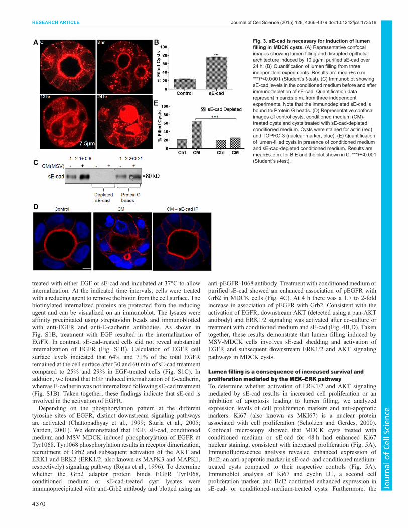
sE-cad is necessary to induce lumen filling in MDCK cystsLumen filling might be induced by multiple factors present in theconditioned medium. To determine whether sE-cad is crucial for theinduction of lumen filling, two independent assays were performed.First, we tested the effect of purified sE-cad on lumen filling. As
shown in Fig. 3A,B, recombinant purified sE-cad exogenouslyadded to MDCK cysts induced lumen filling. Next, to demonstratethat sE-cad is involved in the induction of lumen filling induced byconditioned medium, a sE-cad immunodepletion assay was utilized.In this assay, sE-cad was removed from the co-culture conditionedmedium by immunoprecipitation using an antibody against theextracellular domain of E-cadherin. sE-cad depletion in theconditioned medium was confirmed by immunoblotting(Fig. 3C). sE-cad-depleted conditioned medium failed to inducelumen filling in the MDCK cysts, whereas untreated conditionedmedium induced lumen filling in 80% of the cysts (Fig. 3D,E).These results demonstrate that sE-cad is a crucial soluble factorinvolved in lumen filling in MDCK cysts. It also indicates thatlumen filling induced by MSV-MDCK cells is mediated by sE-cad.
MSV-MDCK cells mediate lumen filling through activation ofEGFRActivation of the EGFR has been implicated in sE-cad-mediatedproliferation, migration and invasion effects (Brouxhon et al.,2007, 2013a; Inge et al., 2011; Maretzky et al., 2005; Najy et al.,2008; Noe et al., 2001). We therefore hypothesized that theinduction of lumen filling by MSV-MDCK cell co-culture ismediated by the activation of EGFR signaling in MDCK cysts. Thephosphorylation of EGFR in MDCK cysts following co-culturewith MSV-MDCK cells or conditioned medium was visualizedusing immunofluorescence and confocal microscopy. As shown inFig. 4A, co-culture of MDCK cysts with MSV-MDCK cells andconditioned medium disrupted luminal architecture and enhancedEGFR phosphorylation at the Tyr1068 which could be blocked bythe EGFR inhibitor CL-387,785. Stimulation with EGF alsoresulted in EGFR phosphorylation and disruption of the hollowluminal architecture in MDCK cysts. Consistent with theimmunofluorescence data, a quantifiable immunoblot analysisshowed a 1.5- and 2-fold increase in the phosphorylation ofEGFR in the co-culture and conditioned medium conditions,respectively. This phosphorylation was blocked by the EGFRinhibitor (Fig. 4B,D). Addition of purified sE-cad to MDCK cystsinduced a 3-fold increase in EGFR phosphorylation, which was alsoinhibited by CL-387,785 (Fig. 4A,B,D). Immunoblot analysis didnot reveal substantial changes in the phosphorylation of other EGFRtyrosine residues at 1173, 1045 and 1086.
sE-cad is reported to associate with the HER2–HER3 receptor ofthe EGFR family in breast cancer cells and also interacts with EGFRin squamous skin cancer cells (Brouxhon et al., 2007; Najy et al.,2008). To investigate whether sE-cad acts as a ligand for EGFRin MDCK epithelial cells, co-immunoprecipitation analysis wasdone to detect association between sE-cad and phosphorylatedEGFR (pEGFR) in sE-cad-treated MDCK cells (Fig. S1A). Therecombinant sE-cad used has a Myc tag at its N-terminus. To preventpulling down cellular E-cadherin along with sE-cad, anti-Mycantibody was used. Co-immunoprecipitation analysis showed anassociation between phosphorylated EGFR and sE-cad in MDCKcells, indicating that sE-cad binds to theEGF receptor inMDCKcells.
Furthermore, the mechanism by which sE-cad activates EGFRwas investigated using a cell surface biotinylation assay. Cellsurface localization of EGFR is crucial for its activation.Internalization of EGFR following ligand binding functions as anegative regulatory mechanism, and prolonged membrane signalingof EGFR is associated with oncogenesis (Tomas et al., 2014).Therefore, cell surface levels of EGFR were compared followingEGF and sE-cad treatment. In this assay, a cleavable biotin was usedfor cell surface biotinylation. Following biotinylation, cells were
4369
RESEARCH ARTICLE Journal of Cell Science (2015) 128, 4366-4379 doi:10.1242/jcs.173518
Journal
ofCe
llScience
treated with either EGF or sE-cad and incubated at 37°C to allowinternalization. At the indicated time intervals, cells were treatedwith a reducing agent to remove the biotin from the cell surface. Thebiotinylated internalized proteins are protected from the reducingagent and can be visualized on an immunoblot. The lysates wereaffinity precipitated using streptavidin beads and immunoblottedwith anti-EGFR and anti-E-cadherin antibodies. As shown inFig. S1B, treatment with EGF resulted in the internalization ofEGFR. In contrast, sE-cad-treated cells did not reveal substantialinternalization of EGFR (Fig. S1B). Calculation of EGFR cellsurface levels indicated that 64% and 71% of the total EGFRremained at the cell surface after 30 and 60 min of sE-cad treatmentcompared to 25% and 29% in EGF-treated cells (Fig. S1C). Inaddition, we found that EGF induced internalization of E-cadherin,whereas E-cadherin was not internalized following sE-cad treatment(Fig. S1B). Taken together, these findings indicate that sE-cad isinvolved in the activation of EGFR.Depending on the phosphorylation pattern at the different
tyrosine sites of EGFR, distinct downstream signaling pathwaysare activated (Chattopadhyay et al., 1999; Sturla et al., 2005;Yarden, 2001). We demonstrated that EGF, sE-cad, conditionedmedium and MSV-MDCK induced phosphorylation of EGFR atTyr1068. Tyr1068 phosphorylation results in receptor dimerization,recruitment of Grb2 and subsequent activation of the AKT andERK1 and ERK2 (ERK1/2, also known as MAPK3 and MAPK1,respectively) signaling pathway (Rojas et al., 1996). To determinewhether the Grb2 adaptor protein binds EGFR Tyr1068,conditioned medium or sE-cad-treated cyst lysates wereimmunoprecipitated with anti-Grb2 antibody and blotted using an
anti-pEGFR-1068 antibody. Treatment with conditioned medium orpurified sE-cad showed an enhanced association of pEGFR withGrb2 in MDCK cells (Fig. 4C). At 4 h there was a 1.7 to 2-foldincrease in association of pEGFR with Grb2. Consistent with theactivation of EGFR, downstream AKT (detected using a pan-AKTantibody) and ERK1/2 signaling was activated after co-culture ortreatment with conditioned medium and sE-cad (Fig. 4B,D). Takentogether, these results demonstrate that lumen filling induced byMSV-MDCK cells involves sE-cad shedding and activation ofEGFR and subsequent downstream ERK1/2 and AKT signalingpathways in MDCK cysts.
Lumen filling is a consequence of increased survival andproliferation mediated by the MEK–ERK pathwayTo determine whether activation of ERK1/2 and AKT signalingmediated by sE-cad results in increased cell proliferation or aninhibition of apoptosis leading to lumen filling, we analyzedexpression levels of cell proliferation markers and anti-apoptoticmarkers. Ki67 (also known as MKI67) is a nuclear proteinassociated with cell proliferation (Scholzen and Gerdes, 2000).Confocal microscopy showed that MDCK cysts treated withconditioned medium or sE-cad for 48 h had enhanced Ki67nuclear staining, consistent with increased proliferation (Fig. 5A).Immunofluorescence analysis revealed enhanced expression ofBcl2, an anti-apoptotic marker in sE-cad- and conditioned medium-treated cysts compared to their respective controls (Fig. 5A).Immunoblot analysis of Ki67 and cyclin D1, a second cellproliferation marker, and Bcl2 confirmed enhanced expression insE-cad- or conditioned-medium-treated cysts. Furthermore, the
Fig. 3. sE-cad is necessary for induction of lumenfilling in MDCK cysts. (A) Representative confocalimages showing lumen filling and disrupted epithelialarchitecture induced by 10 μg/ml purified sE-cad over24 h. (B) Quantification of lumen filling from threeindependent experiments. Results are mean±s.e.m.***P<0.0001 (Student’s t-test). (C) Immunoblot showingsE-cad levels in the conditioned medium before and afterimmunodepletion of sE-cad. Quantification datarepresent mean±s.e.m. from three independentexperiments. Note that the immunodepleted sE-cad isbound to Protein G beads. (D) Representative confocalimages of control cysts, conditioned medium (CM)-treated cysts and cysts treated with sE-cad-depletedconditioned medium. Cysts were stained for actin (red)and TOPRO-3 (nuclear marker, blue). (E) Quantificationof lumen-filled cysts in presence of conditioned mediumand sE-cad-depleted conditioned medium. Results aremean±s.e.m. for B,E and the blot shown in C. ***P<0.001(Student’s t-test).
4370
RESEARCH ARTICLE Journal of Cell Science (2015) 128, 4366-4379 doi:10.1242/jcs.173518
Journal
ofCe
llScience

Fig. 4. See next page for legend.
4371
RESEARCH ARTICLE Journal of Cell Science (2015) 128, 4366-4379 doi:10.1242/jcs.173518
Journal
ofCe
llScience

EGFR kinase inhibitor CL-387,785 reduced the levels of cellproliferation and anti-apoptotic markers in the sE-cad- andconditioned medium-treated MDCK cysts (Fig. 5B). These datasuggest that both increased cell proliferation and anti-apoptoticmechanisms are involved in lumen filling induced by sE-cad.In order to determine the role of downstream ERK1/2 and AKT
pathways in lumen filling, conditionedmedium or sE-cad-treated cysts
were also treated with specific inhibitors of each pathway. Treatmentwith the inhibitor U0126 against the MEK family proteins (upstreamregulators of ERK proteins) suppressed the expression of Ki67 andBcl2 as revealed by immunofluorescence analysis. Furthermore,lumen filling in conditioned medium or sE-cad-treated cysts wasreducedby60–70% in presence ofU0126, suggesting thatERK1/2 hasa key role in sE-cad-mediated lumen filling at 24 h. Interestingly,inhibition of PI3K–AKT pathway by LY294002 led to only a 25%reduction in lumen filling compared to conditioned medium and sE-cad-treated cysts at 24 h (Fig. S2A–D). The concentration of U0126and LY294002 (1 µM) used in the immunofluorescence analysiseffectively blocked ERK1/2 and AKT phosphorylation, respectively(Fig. S2E). These results suggest that lumen filling is predominantlydriven by the MEK–ERK pathway.
sE-cad and conditioned medium treatment induced EMT inMDCK cystsCysts in co-culture for more than 48 h showed strikingmorphological changes. At 96 h, control cysts remained spherical,whereas conditioned medium and the sE-cad treatment led to moreelongated cells emanating from the cysts (Fig. 6A). This observationled us to hypothesize that sE-cad and conditioned medium can
Fig. 4. MSV-MDCK cells induce lumen filling in MDCK cells by sE-cad-mediated activation of EGFR and downstreamAKTand ERK1/2 pathwaysin MDCK cysts. (A) Representative immunofluorescence images showingincreased pEGFR expression in MDCK cysts treated with MSV-MDCK cells,conditioned medium (CM), sE-cad, EGF with or without CL-387,785 (CL).Immunofluorescence shows actin (red), E-cadherin (blue) and pEGFR(green). (B) Representative immunoblot from three independent experimentsshowing pEGFR, total EGFR, phosphoryalted AKT (pAKT), phosphorylatedERK1/2 (pERK1/2) and total AKT and ERK1/2 levels in MDCK 3D cyst lysatesco-cultured with MSV-MDCK cells, conditioned medium, sE-cad and EGF for4 h. 1 µM CL-387,785, an EGFR kinase inhibitor was used to block EGFRactivation for 4 h where indicated. EGF treatment was for 15 min. (C) Co-immunoprecipitation of pEGFR (Tyr1068) with Grb2 in MDCK 3D cysts treatedwith conditioned medium or sE-cad at 2 h and 4 h. (D) Graphs representquantification data as mean±s.e.m. from three independent experiments.*P<0.05, **P<0.005 (Student’s t-test).
Fig. 5. Lumen filling is a consequence of reducedapoptosis and increased proliferation.(A) Immunofluorescence images showing increased Ki67and Bcl2 expression in MDCK cysts treated with conditionedmedium (CM) and sE-cad for 48 h. Images were obtainedfrom staining cysts with anti-Bcl2 antibody (green),phalloidin–Alexa-Fluor-546 (actin, red) and Ki67 (blue).(B) Representative immunoblot showing Ki67, Bcl2 andcyclin D1 expression inMDCK cysts treated with sE-cad andconditioned medium. 1 µM CL-387,785 (CL) EGFR inhibitorwas used where indicated. Quantification data representmean±s.d. from two independent experiments.
4372
RESEARCH ARTICLE Journal of Cell Science (2015) 128, 4366-4379 doi:10.1242/jcs.173518
Journal
ofCe
llScience

induce EMT in the normal epithelial cysts. To confirm EMT, wetested for the presence of multiple EMT markers in the cysts byperforming immunofluorescence and immunoblot analyses. Asshown in Fig. 6B, the expression of N-cadherin (also known asCDH2), MMP-9 and fibronectin were enhanced in these cells. Thepresence of stress fibers is a well-established marker for cellsundergoing EMT, and is essential for their invasive behavior.Strikingly, enhanced actin stress fibers were detected in the cyststreated with sE-cad and conditioned medium (Fig. 6B). z-stacksshowing expression of fibronectin, N-cadherin and actin stressfibers in control, sE-cad- and conditioned-medium-treated MDCKcysts for 96 h are shown in Movies 1, 2 and 3 respectively. Theseresults reveal that long-term sE-cad treatment induces EMT inMDCK cysts. It is also important to note that cysts exhibiting anEMT-like phenotype are larger than control cysts.The PI3K–AKT axis is frequently activated in human cancer and
is a key contributor in the induction of EMT (Larue and Bellacosa,
2005). Treatment with the PI3K and AKT inhibitor LY294002effectively blocked EMT in MDCK cells, with cysts retainingluminal morphology. By contrast, treatment with the MEK inhibitorU0126 did not block EMT in long-term sE-cad- or conditioned-medium-treated cells, as revealed by the expression of fibronectin,N-cadherin and stress fibers using confocal microscopy(Fig. 7A–D). Immunoblot analysis also showed an increase infibronectin and N-cadherin levels upon sE-cad and conditionedmedium treatment, which was blocked in the presence of 1 μM ofthe PI3K and AKT inhibitor LY294002, but not in the presence of1 μM MEK inhibitor U0126 (Fig. 7E,F). Long-term inhibitionwith LY294002 resulted in the inhibition of lumen filling andEMT, possibly by blocking both the PI3K-mediated AKT andERK pathways. By contrast, long-term treatment with U0126enhanced the expression of EMT markers in the lumen-filled cysts(Fig. 7A,B); this is because MEK inhibition by U0126 is known toincrease AKT activation (Aksamitiene et al., 2010), thereby further
Fig. 6. sE-cad and conditioned medium induces an EMT-likephenotype inMDCK cysts. (A) Phase-contrast images of controlMDCK cysts and cysts treated with conditioned medium (CM) andsE-cad for 96 h. (B) Immunofluorescence staining with differentEMT markers. Representative merged confocal images showingactin (red), fibronectin (green), N-cadherin (green) and MMP-9(blue).
4373
RESEARCH ARTICLE Journal of Cell Science (2015) 128, 4366-4379 doi:10.1242/jcs.173518
Journal
ofCe
llScience

confirming the role of AKT in driving EMT inMDCK cysts. This isconsistent with the immunofluorescence data, which indicates thatAKT inhibition blocked EMTmore effectively than theMEK–ERKpathway. In addition, long-term treatment with sE-cad revealeddistinct activation patterns for ERK and AKT; sE-cad induced ERKactivation diminished from 48 to 96 h, whereas the AKTphosphorylation was sustained over 96 h as determined by
immunoblot analysis (Fig. S3). These results further authenticatethat sustained activation of AKT is involved in the induction ofEMT.
DISCUSSIONNormal epithelial cells co-exist with carcinoma cells in solid tumors.Although it is well known that the TME contributes to metastatic
Fig. 7. AKT is involved in the induction EMT in MDCK cysts. (A,B) Immunofluorescence showing EMT expression in cysts treated with sE-cad (A) andconditioned medium (CM; B) in presence of inhibitors at 96 h. U0126 and LY294002 were used at 1 µM each. Representative confocal images obtained fromstaining cysts with anti-fibronectin antibody (green), anti-N-cadherin antibody (blue), phalloidin–Alexa-Fluor-546 (for actin, red) are shown. (C,D) Quantification ofcysts displaying an EMT phenotype after 96 h with sE-cad treatment (C) or conditioned medium treatment (D) in presence of inhibitors. *P<0.05, ***P<0.001(Student’s t-test). (E,F) Representative immunoblots from two independent experiments showing fibronectin and N-cadherin levels in sE-cad-treated cysts(E) and conditioned-medium-treated cysts (F) in the presence of inhibitors. U0126 and LY294002 were used at 1 µM each. β-actin was used as a loading control.Quantification data represent mean±s.d. from two independent experiments.
4374
RESEARCH ARTICLE Journal of Cell Science (2015) 128, 4366-4379 doi:10.1242/jcs.173518
Journal
ofCe
llScience

dormancy, tumor stability, and progression to invasive andmetastaticcarcinoma (Taylor et al., 2014), it is not known how carcinoma cellsinteractwith normal epithelial cells in the TME. In this study, we useda 3D co-culture system comprisingMDCK andMSV-MDCK cells toaddress whether carcinoma cells influence adjacent normal epithelialcells. There aremany new findings reported here: (1) the developmentof a co-culture system to study normal and cancer cell interactions; (2)the sequential induction of lumen filling andEMT in vitro; (3) that sE-cad acts as a crucial soluble factor involved in the induction of lumenfilling and EMT; (4) that carcinoma cells use normal cells as a sourcefor the production of sE-cad; and (5), although, involvement ofMMP-9 to cleave E-cadherin, and a role for EGFR in the induction ERK1/2and AKT signaling pathways has been shown in many cancer cells,we demonstrate that the same well-established oncogenic signalingpathway is utilized by carcinomacells to induce transdifferentiation ofnormal cells in the vicinity (Fig. 8). Clinically, these studies suggestthat elevated levels of sE-cad present in the sera from cancer patientsmight have a pathological role in the transdifferentiation of normalepithelial cells, thereby facilitating invasion and metastatic potential.Filling of the luminal space and multiple lumen lesions are
distinctive features of early pre-invasive stages of carcinomadevelopment (Debnath and Brugge, 2005). Several carcinomassuch as breast, kidney and prostate carcinomas display lumen fillingand architectural disorder during their early stages, before theyinvade the basement membrane and disseminate (Debnath andBrugge, 2005). The molecular mechanisms that occur during theinitial lumen-filling stage of tumorigenesis are poorly understood. A3D co-culture system comprising epithelial cysts and carcinomacells is reminiscent of normal epithelial tissue co-existing withtumor cells in vivo. This novel 3D assay allowed us to demonstratethat carcinoma cells disrupt epithelial architecture by inducing pre-neoplastic lumen filling. Interference with the key regulators of theapico-basal polarity and tight junction assembly, such as theCrumbs3 and PAR complexes, β1 integrin, RhoA and the Na+/K+-ATPase β-subunit generates multiple lumina or no lumen inepithelial cysts (Barwe et al., 2013; Martin-Belmonte et al., 2007;Schluter et al., 2009; Shin et al., 2005; Straight et al., 2004; Yu et al.,2008). However, it is not well known how these markers are affected
during carcinogenesis. Our results indicate that carcinoma cells inthe vicinity of normal tissue might come in direct physical contactwith the normal epithelial cells through filopodia-like projections,or alternatively they might secrete soluble factors that then disruptsluminal architecture. Data obtained using conditioned mediastrongly suggest that secretion of soluble factors is the primarymechanism by which carcinoma cells induce normal celltransformation. Recent studies have also demonstrated thatcarcinoma cells induce oncogenic transformation in the adjacentnormal epithelial cells using exosomes to force adjacent normalcells to participate in cancer progression (Melo et al., 2014).
A key soluble factor identified in our assay is MMP-9. AlthoughMMP-9 is implicated in the shedding of E-cadherin in several cancercell lines (Grabowska and Day, 2012), its potential to impact normalcells in the context of the tumor microenvironment has not beenexamined. Our 3D co-culture system enabled us to determine thatMMP-9 released by carcinoma cells acts on E-cadherin on thebasolateral surface of adjacent epithelial cells to release sE-cad.Inhibition of MMP-9 abolished sE-cad shedding and consequentlyblocked the lumen-filling phenotype. Lack of E-cadherin expressionon MSV-MDCK cells confirms that the source of sE-cad present inthe conditioned medium is primarily from MDCK cells. A transwellco-culture assay involving MCF10A, an immortalized human breastepithelial cell line, with MDA-MB435S, a breast carcinoma cell linelacking E-cadherin expression also revealed MMP-mediated sE-cadshedding from normal cells. This corroborates the finding that tumorcells induce sE-cad shedding from adjacent normal epithelial cells.Although activeMMP-2was also observed inour zymogram, specificinhibition of MMP-9 suggests that MMP-9 plays a prominent role inthe production of sE-cad in this co-culture system. However, wecannot rule out the involvement of other MMPs or soluble factorspresent in the conditioned medium, which might act upstream ofMMP-9 and be involved in its activation and the production of sE-cad.
Two complementary approaches were utilized to conclude thatsE-cad is essential for the induction of lumen filling and EMT in 3Dculture. Exogenously added sE-cad induced lumen filling, whereas,immunodepletion of sE-cad from the conditioned medium blockedthe lumen-filling phenotype, indicating that sE-cad is the crucialfactor of the conditioned medium that is involved in the induction oflumen filling. Although both purified sE-cad and conditionedmedium induced lumen filling, conditioned medium under co-culture conditionsmight contain additional factors that are not presentin the purified sE-cad. These additional factors might be involved instabilizing or enhancing the effect of sE-cad in conditioned medium.In fact, in co-culture, with conditioned medium the sE-cad levelswere 1 µg/ml, which were sufficient to induce lumen filling,indicating that additional factors are likely to be involved. sE-cadhas been shown to influence tumor cell proliferation, migration andinvasion (Brouxhon et al., 2007; Maretzky et al., 2005; Najy et al.,2008; Noe et al., 2001; Symowicz et al., 2007; Zuo et al., 2011).Here, we show that sE-cad disrupted luminal architecture of fullyformed cysts, which was previously unknown. This situation isreminiscent of early tumorigenesis, suggesting that sE-cad in theTME induces a preneoplastic lumen-filling phenotype in adjacentnormal epithelial cells.
In polarized epithelial cells, E-cadherin is localized at theadherens junction and along the basolateral region. In our transwellco-culture assay MDCK cells were in the upper chamber and MSV-MDCK cells were in the lower chamber. MMP-9 and sE-cad wereonly detected in the lower chamber, indicating that the MMP-9produced by the MSV-MDCK cells cleaved E-cadherin on thebasolateral membrane of MDCK cells. MMP-9 inhibition in the
Fig. 8. Proposed model for sequential lumen filling and EMT induced bycarcinoma cells in MDCK cysts. See text for details.
4375
RESEARCH ARTICLE Journal of Cell Science (2015) 128, 4366-4379 doi:10.1242/jcs.173518
Journal
ofCe
llScience

lower chamber abrogated sE-cad shedding further confirming thisresult. Therefore, it is likely that proteases cleave basolaterallylocalized E-cadherin (David and Rajasekaran, 2012). Additionalstudies are required to determine whether E-cadherin in adherensjunctions is processed by MMP-9.We demonstrated that the sE-cad-mediated loss of 3D architecture
and lumen filling inMDCK cysts is driven by activation of the EGFRand its downstream ERK1/2 and AKT signaling pathways. Weshowed that co-culturewithMSV-MDCK cells, conditionedmediumor sE-cad induced lumen filling in an EGFR-dependent manner.Activation of EGFR increased cell proliferation as well as reducingapoptosis, thereby resulting in lumen filling, consistent with previousreports (Reginato et al., 2005).It has been shown that sE-cad acts as a ligand to the EGFR family
of receptors, leading to activation of oncogenic signaling insquamous cell carcinoma cells as well as breast cancer cells(Brouxhon et al., 2014a, 2013a,b; Najy et al., 2008). It should benoted that the cell lines used in these studies were tumor-derivedcells. Our co-immunoprecipitation assay reveals that purified sE-cadbinds to pEGFR (Tyr1068) in normal epithelial cells. Although thisassay is not quantitative, it reveals an association of sE-cad withEGFR. Thus, it seems likely that sE-cad also functions as an EGFRligand in normal epithelial cells. Interestingly, our cell surfaceinternalization assay reveals that, unlike EGF, internalization ofEGFR in the presence of sE-cad is substantially reduced.Endocytosis of EGFR, following ligand binding, functions as acrucial negative regulator of EGFR signaling, particularly EGFR-induced ERK1/2 and AKT signaling (Sousa et al., 2012; Tomaset al., 2014). Interestingly, EGFR mutants present in non-small celllung cancer display defects in endocytotic regulation, leading topersistent signaling from the plasma membrane (Shtiegman et al.,2007). The differential rate of receptor internalization induced byEGF and sE-cad might be due to faster recycling of EGFR to themembrane upon sE-cad activation thereby resulting in increasedstabilization of EGFR at the cell surface. However, additionalexperiments are needed to validate this notion. Thus, reduction ofEGFR internalization in the presence of sE-cad might be one of themechanisms of EGFR activation in our co-culture model.Previous reports from our laboratory have demonstrated that sE-
cad retains the ability to interact with E-cadherin, and thatE-cadherin is necessary for the anti-apoptotic effect of sE-cad inMDCK cells (Inge et al., 2011). E-cadherin homophilic adhesionbetween adjacent cells transiently activates the EGFR and itsdownstream PI3K–AKT and ERK1/2 signaling cascades, therebyinducing cell survival and differentiation (Pece and Gutkind, 2000).sE-cad binding to E-cadherin might mimic E-cadherin homophilicbinding, resulting in the activation of the EGFR signaling cascade.However, given that the sE-cad–E-cadherin interaction does notresult in junction formation and cell adhesion, the subsequent EGFRtransactivation might trigger atypical cellular pathways, such asproliferation and survival. Taken together, sE-cad-mediated lumenfilling might be a consequence of both sE-cad–E-cad as well as sE-cad–EGFR interactions.The occurrence of EMT in two-dimensional (2D) cultures is well
established. For example, TGFβ induces EMT (Heldin et al., 2009;Kim et al., 2004). However, induction of EMT in 3D epithelialstructures has not been shown previously. We demonstrated thatlong-term treatment of MDCK cysts with sE-cad or conditionedmedium induced transdifferentiation of the cysts into a disorganizedmotile mesenchymal phenotype. Cells with a mesenchymalmorphology were observed emanating from the cysts with aconsequent loss of cyst morphology. These mesenchymal cells were
highly motile. Consistent with their morphology, the mesenchymalmarkers N-cadherin, fibronectin and actin stress fibers wereupregulated. MMP-9 expression was also elevated in these cells.These results indicate that sE-cad produced either directly orindirectly by carcinoma cells can induce EMT in adjacent normalepithelial cells. The trigger that induces EMT following pre-neoplastic lumen filling is not known. Although both AKT andERK1/2 pathways are activated, inhibition of AKT rather than theERK1/2 pathway abrogated EMT. Inhibition of ERK1/2 pathwayprevented lumen filling effect at 24 h; however, long-term treatmentwith the MEK inhibitor U0126 resulted in induction of EMTmarkers and loss of hollow lumen. We hypothesize that theinhibition of the ERK1/2 pathway for 96 h induces compensatoryactivation of the PI3K–AKT pathway, which drives induction ofEMT and disruption of hollow lumen at 96 h. Constitutively activeAKT has been reported to drive EMT within 72–96 h (Grille et al.,2003). Here, we have shown the strength of the ERK1/2 signalingreduced after 48 h, whereas AKT phosphorylation was elevated at72 and 96 h, suggesting that sustained activation of AKT drivesinduction of EMT in MDCK cysts.
We demonstrated that cancer cells can utilize the surroundingnormal epithelial cells as accomplices to produce pro-oncogenicfactors. This findingmight have important clinical correlations, as ourresults suggest that elevated sE-cad levels observed in sera fromcancerpatients are derived from both tumor cells and adjacent normal cells.Carcinoma cells induced shedding of cell bound E-cadherin fromadjacent normal tissue and this might influence transdifferentiationof the compact epithelial tissue into a disorganized group ofmesenchymal cells. Elevated sE-cad levels might also have pro-tumorigenic effects on other components of the TME, such as thevarious stromal cell types. Thus, accumulation of sE-cad in themicroenvironment might have additive or synergistic effects onthe pro-oncogenic TME since it can alter several components of theTME. Disruption of normal epithelial cell function could enhancebasal extrusion of tumor cells (Slattum and Rosenblatt, 2014), andthereby accelerate metastasis of the tumor cells.
Blocking sE-cad using a monoclonal antibody (mAb) has beenshown to be an effective anti-tumor strategy in mouse models ofbreast carcinoma and squamous skin carcinoma. Anti-sE-cad mAbtreatment attenuates activation of multiple receptor tyrosine kinasessuch as HER1, HER2 and IGF-1R, and leads to activation ofapoptotic pathways and inhibition of proliferative pathways,ultimately attenuating tumor growth in mice (Brouxhon et al.,2014a, 2013a). Although a more detailed analysis examining thetoxicity and specificity of anti-sE-cad mAb therapy is necessary,antibody-mediated elimination of sE-cad could act synergisticallywith current EGFR-based therapies to improve efficacy in thetreatment of a range of carcinomas.
MATERIALS AND METHODSCell linesMadin–Darby canine kidney cells (MDCK) and MD-MB-435 S werepurchased from the American Type Culture Collection (Manassas, VA) andwere maintained in Dulbecco’s modified Eagle’s medium (DMEM) with1 g/l sodium bicarbonate, 10% fetal bovine serum, 1 mM L-glutamine,100 U/ml penicillin and 100 μg/ml streptomycin. MCF-10A cells were alsoobtained from the American Type Culture Collection and maintained inDMEM with F12 (Gibco-BRL) supplemented with 5% donor horse serum,20 ng/ml EGF, 10 µg/ml insulin, 100 ng/ml cholera toxin, 0.5 µg/mlhydrocortisone, 50 U/ml penicillin and 50 µg/ml streptomycin. TheMoloney-Sarcoma-virus-transformed MDCK cell line (MSV-MDCK) hasbeen described previously (Rajasekaran et al., 2001). Cell lines expressingGFP and RFP were generated by transfection. MDCK epithelial cells stably
4376
RESEARCH ARTICLE Journal of Cell Science (2015) 128, 4366-4379 doi:10.1242/jcs.173518
Journal
ofCe
llScience

expressing RFP–actin were used for live microscopy experiments. MSV-MDCK cells expressing GFP were generated as described previously(Barwe et al., 2005).
Antibodies and reagentsPrimary antibodies used in this study for immunoblotting andimmunostaining were against: E-cadherin (Decma-1; Sigma-Aldrich),MMP-9 (Santa Cruz Biotechnology), Ki67 and N-cadherin (Abcam),fibronectin and β-catenin (BD Transduction Laboratories), smooth muscleactin (Santa Cruz Biotechnology), total EGFR (Fitzgerald Industries),pEGFR (Tyr1068), phosphorylated AKT (Ser473), phosphorylated p44/42MAPK (ERK1/2), Grb2, total AKT, total MAPK, cyclin D1 and Bcl2 (CellSignaling Technology). The horseradish peroxidase (HRP)-conjugated anti-mouse-IgG, rabbit-IgG and rat-IgG secondary antibodies were obtainedfrom Cell Signaling Technology. AlexaFluor-488-conjugated anti-mouse-IgG, Alexa-Fluor-633-conjugated anti-rabbit-IgG, Alexa-Fluor-546-conjugated phalloidin and TOPRO-3 were purchased from MolecularProbes. FITC- and Texas-Red-labeled, affinity-purified secondaryantibodies were obtained from Jackson ImmunoResearch Laboratories.
Growth-factor-reducedMatrigel™ (CorningDiscovery Labware)was usedfor 3D culture. Protein-free and serum-free UltraDOMA™ medium (Lonza)was used for conditionedmediumexperiments. Corning cell recovery solutionwas used to harvest 3D cultures from the Matrigel™ matrix.
The broad spectrumMMP inhibitorMarimastat, andU0126 andLY294002were purchased from Tocris (Minneapolis, MN). Recombinant human EGFfrom Peprotech, EGFR Inhibitor CL-387,785 and MMP-9 Inhibitor I(CAS1177749-58-4) were purchased from EMD Millipore Chemicals.
3D Matrigel™ culturesMDCK 3D cultures were grown and maintained in Matrigel™ as previouslydescribed (O’Brien et al., 2001, 2006). Briefly, MDCK cells weretrypsinized and suspended at a final concentration of 15,000 cells/ml in2% growth-factor-reduced Matrigel™. The cell–Matrigel™ mixture (4500cells per well in 300 µl) was layered onto Matrigel™-coated chambers,3.5 µl/well, (LAB-TEK II Chambered Coverglass, Nalgene NuncInternational) and incubated at 37°C, 5% CO2 and 95% humidity. Cellswere monitored daily for cyst formation. Medium was replenished every2 days. MDCK cysts grow in 72–96 h. For sE-cad experiments, cysts werecultured with 10 µg/ml of recombinant purified sE-cad in serum-freemedium for the indicated time points. For experiments requiring MSV-MDCK co-culture, MSV-MDCK cells (3000 cells per 200 µl) were overlaidonto acini on day 3 of culture and monitored at the indicated time points.Conditioned medium is growth medium from MSV-MDCK cells after 48 hculture. Conditioned medium was centrifuged to remove floating cells andother debris. EGF was used at a concentration of 1 ng/µl wherever indicated.
Purification of sE-cadsE-cad was purified from the sE-cad-overexpressing HEK-293T cells usingNi-NTA resin as described previously (Inge et al., 2011). Briefly, sE-cad-overexpressing HEK-293T cells were generated by stably expressing thepSEC-Tag2-SECAD vector into HEK-293T cells and stable clones wereselected with Zeocin (Invitrogen). sE-cad-HEK-293T cells were grown inUltra-DOMA-PF (Cambrex, Rutherford, NJ) medium for 48 h. 100 µg/mlof Zeocin antibiotic was added to induce sE-cad expression into theextracellular media. The medium was collected and centrifuged to removecells and debris. The cell-free growth medium from 48 h was mixed withNi-NTA resin overnight in a spinner flask to allow the His-tagged sE-cad tobind to the Ni-NTA resin. Resin was then loaded into a column. The resinwas washed with 2× column volumes of phosphate buffer (160 mMphosphate, 4 MNaCl, 5 mM Imidazole, pH 7.4), and 5× column volumes ofphosphate buffer containing 10 mM imidazole to remove any nonspecificprotein. sE-cad was eluted with 5× column volumes of phosphate buffercontaining 250 mM imidazole. Fractions (1 ml) were collected and thepurity of each fraction was determined by SDS-PAGE. sE-cad-containingfractions were pooled, concentrated using centriplus centrifuge filters(Millipore) and dialyzed against sterile PBS at 4°C. The concentration of thepurified sE-cad was determined using a Bio-Rad protein DC kit (Hercules,
CA). The purified, concentrated sE-cad was aliquoted and stored at −80°Cuntil further use.
Immunofluorescence of 3D Matrigel™ culturesCells grown inMatrigel™were prepared and immunostained as follows: 3DMDCK cultures were fixed with 4% paraformaldehyde for 15 min at roomtemperature, washed three times with phosphate-buffered saline (PBS), thenpermeabilized and blocked with PBS, 0.7% fish skin gelatin and 10%saponin (PFS) for 30 min at room temperature. The fixed and permeabilizedcysts were stained for the following proteins: β-catenin, actin, and pEGFR,Ki67, Bcl2, N-cadherin and fibronectin by incubating the antibodies at1:400 dilution in PFS overnight at 4°C. Staining was detected using Alexa-Fluor-488,546- and 633-conjugated secondary antibodies at 1:400 dilutionin PFS and added to cultures for 2 h at room temperature. TOPRO-3 at1:1000 dilution was used to stain nuclei. Cells and cysts were then washedwith PBS three times and used for imaging.
Confocal microscopy and quantification of 3D cystsImages were captured using a Leica TCS SP5 scanning confocal microscope(Leica Microsystems) using a 63×/1.4 NA oil immersion objective lens. Thecysts were assessed for the presence of cells within the lumen. The number ofcysts with clear or filled lumens were counted across at least ten differentrandom fields and expressed as a percentage of total number of cysts. At least100 cysts were examined per experimental group. All images were capturedusing the same laser intensity, and gain and offset settings.
Generating 3D cell lysates and conditioned medium from 3Dcultures3D cyst protein lysates were prepared by recovering cultured cells from theMatrigel™ basement matrix using a cell recovery solution (Corning LifeSciences Product #354253) following the manufacturer’s instructions.The harvested cysts were then lysed as described previously (Tushir andD’Souza-Schorey, 2007). Briefly, chilled RIPA buffer (50 mM Tris-HCl pH7.4, 1%NP-40, 0.5%Na-deoxycholate, 0.1%SDS, 150 mMNaCl and 2 mMEDTA) supplemented with 1× protease inhibitor cocktail and 1%phenylmethylsulfonyl fluoride was added to the harvested cyst pellet. Thecells in RIPA buffer were incubated on ice for 15 min followed by sonicationfor 15 min at 4°C. Lysates were cleared by centrifugation at 16,000 g for15 min at 4°C. Supernatants were collected and the protein concentration wasmeasured using Bio-Rad DC reagent as per the manufacturer’s instructions.For detection of proteins (shed sE-cad and MMP-9) in the conditionedmedium, the cystswere grown inUltraDOMA-PF to prevent interference fromalbumin and other serum proteins present in the complete medium.Conditioned medium was collected and concentrated using Amiconultracentrifugation filter devices (EMD, Millipore). Equal amounts ofconcentrated medium were then loaded onto SDS-polyacrylamide gels andanalyzed by immunoblotting or gelatin zymography.
ImmunoblottingSDS-PAGE was used to separate proteins in 3D cyst lysates. Separatedproteins were transferred from the gel onto a nitrocellulose membrane.For immunoblotting, the membranes were blocked in 5% non-fat milk inTBS with 0.1% Tween 20 (TBS-T), and then probed with primaryantibodies at a dilution of 1:1000 and incubated overnight at 4°C. Themembranes were further probed with HRP-conjugated anti-rabbit-IgG,-mouse-IgG or -rat-IgG secondary antibodies diluted 1:2000 in 5% non-fat milk in TBS-T and incubated for 1 h at room temperature. Fordetection of Ki67 protein (molecular mass, 345–395 kDa), 3–8% Trisacetate (NuPage Novex) gradient gels were used. Antibody binding wasvisualized using enhanced chemiluminescence (ECL) and ECL prime(GE Healthcare). ImageJ software was utilized for immunoblotquantification and image analysis.
Gelatin zymographyGelatin zymography was performed as described previously, withmodifications (Lu et al., 2004). Samples were mixed with zymogramsample buffer and loaded onto 8% polyacrylamide gels containing 0.1%
4377
RESEARCH ARTICLE Journal of Cell Science (2015) 128, 4366-4379 doi:10.1242/jcs.173518
Journal
ofCe
llScience

gelatin. The gels were run in zymogram running buffer (pH 8.3) withoutSDS, and then incubated in 1× zymogram renaturation solution for 30 min atroom temperature, followed by overnight incubation at room temperature inthe zymogram development solution. All the buffers for zymography werepurchased from Bio-Rad (Hercules, CA) and used as per the manufacturers’instruction. The gels were stained with Coomassie Brilliant Blue R250 for2 h at room temperature, and destained until the gelatinolytic activities weredetected as clear bands against the blue background.
Transwell co-culture assayMDCK cells were seeded onto 0.4-μm pore size Transwell filters (CorningLife Sciences) and cultured in a six-well plate. MSV-MDCK carcinomacells were seeded at increasing densities ranging from 3000–10,000 cells perwell in a separate six-well plate. MDCK polarized monolayers (TER>250 ohms/cm2) on Transwell filters were then transferred onto the MSV-MDCK cells. Co-cultures in the presence and absence of MMP-9 inhibitorwere maintained for 48 h in Ultra-DOMA-PF. Growth medium from thelower (basolateral side) and upper (apical side) chambers was used foranalyzing MMP-9 and sE-cad levels. MMP-9 activity in the conditionedmedium was analyzed using gelatin zymography.
Co-immunoprecipation and immunodepletionImmunoprecipitation assays were carried out by harvesting cysts fromMatrigel™ matrix using cell recovery solution (Corning Lifesciences) andpreparing cell lysates using RIPA buffer as described above. Antibodiesagainst pEGFR (Tyr1068), Grb-2, E-cadherin and Myc-tag were pre-coupled to protein G/A agarose beads with rabbit anti-mouse-IgG antibody(RAM) for 4 h and incubated overnight with 1mg of total protein lysate. Thebeads were washed and the samples eluted using 4× sample buffer. Thesamples were separated by SDS-PAGE and immunoblotted to visualizeprecipitated proteins.
To deplete sE-cad from the co-culture conditioned medium, theconditioned medium was collected and cleared of cells by centrifugation.The conditioned mediumwas then incubated overnight with anti-E-cadherinantibody coupled to Protein-A–agarose beads. Depletion of sE-cad fromconditioned medium was confirmed using SDS-PAGE of conditionedmedium samples and the Protein-A–agarose beads. The sE-cad-depletedconditioned medium was then added back to newMDCK cysts to determineits effect on lumen filling.
Biotinylation internalization assayMDCK cell surface proteins were treated with 0.5 mg/ml cleavable sulfo-NHS-SS-biotin (Pierce) for 30 min at 4°C. After quenching the excessbiotin with 50 mM NH4Cl in PBS containing 0.1 mM CaCl2 and 1 mMMgCl2, the cells were stimulated with 10 μg/ml sE-cad or 10 ng/ml of EGFfor 30 min and 60 min at 37°C. For controls, untreated plates were eitherreduced on ice (reduced) or not reduced. Following treatment, theremaining biotin on the cell surface was stripped with the membrane-impermeant reducing reagent 50 mM MesNa in 100 mM Tris-HCl pH 8.6(containing 100 mM NaCl and 2.5 mM CaCl2) at 4°C for 30 min. Cellswere lysed in 0.5 ml of lysis buffer (10 mM Tris-HCl, 1% Triton X-100,1 mM EGTA, 1 mM PMSF, 5 mg/ml each of antipain, leupeptin andpepstatin). Protein (500 µg) from each lysate was used for precipitation(16 h at 4°C) with 30 µl of Ultralink streptavidin beads (Pierce) and theprecipitates were probed with anti-EGFR and anti-E-cadherin antibody. Tocalculate the amount of cell surface EGFR, the levels from EGF and sE-CAD-treated samples were calculated with respect to non-reduced controls(100%).
Statistical analysisResults are expressed as mean±s.e.m. and as mean±s.d., as indicated.Statistical analysis between experimental groups was done using Student’st-test and two-way ANOVA. P<0.05 was considered significant.
AcknowledgementsWe thank Dr Anne Muesch, Albert Einstein Medical College, NY for providingMDCK-RFP cells.
Competing interestsThe authors declare no competing or financial interests.
Author contributionsP. U. Patil and A. K. Rajasekaran conceived and designed the work, and withJ.D’Ambrosio, developed the methodology. P. U. Patil, J. D’Ambrosio, L. J. Inge andA. K. Rajasekaran acquired data. P. U. Patil, L. J. Inge and A. K. Rajasekarananalyzed and interpreted data (eg. statistical analysis, biostatistics, computationalanalysis). P. U. Patil, R. W. Mason and A. K. Rajasekaran wrote, review and/orrevised the manuscript. R. W. Mason and A. K. Rajasekaran contributed toadministrative, technical and material support (ie. reporting or organizing data,constructing databases). A. K. Rajasekaran supervised the study.
FundingThis work was supported by funds from the National Institutes of Health (NIH) [grantnumber DK56216]; the Nemours Research Programs; and the Centers ofBiomedical Research Excellence (COBRE) [grant number P20GM103464].Deposited in PMC for release after 12 months.
Supplementary informationSupplementary information available online athttp://jcs.biologists.org/lookup/suppl/doi:10.1242/jcs.173518/-/DC1
ReferencesAksamitiene,E.,Kholodenko,B.N.,Kolch,W.,Hoek,J.B. andKiyatkin,A. (2010).
PI3K/Akt-sensitive MEK-independent compensatory circuit of ERK activation inER-positive PI3K-mutant T47D breast cancer cells. Cell Signal. 22, 1369-1378.
Barwe, S. P., Anilkumar, G., Moon, S. Y., Zheng, Y., Whitelegge, J. P.,Rajasekaran, S. A. and Rajasekaran, A. K. (2005). Novel role for Na,K-ATPasein phosphatidylinositol 3-kinase signaling and suppression of cell motility. Mol.Biol. Cell 16, 1082-1094.
Barwe, S. P., Skay, A., McSpadden, R., Huynh, T. P., Langhans, S. A., Inge, L. J.and Rajasekaran, A. K. (2013). Na,K-ATPase beta-subunit cis homo-oligomerization is necessary for epithelial lumen formation in mammalian cells.J. Cell Sci. 125, 5711-5720.
Behrens, J., Mareel, M. M., Van Roy, F. M. and Birchmeier, W. (1989). Dissectingtumor cell invasion: epithelial cells acquire invasive properties after the loss ofuvomorulin-mediated cell-cell adhesion. J. Cell Biol. 108, 2435-2447.
Brouxhon, S., Kyrkanides, S., O’Banion, M. K., Johnson, R., Pearce, D. A.,Centola, G. M., Miller, J.-n. N., McGrath, K. H., Erdle, B., Scott, G. et al. (2007).Sequential down-regulation of E-cadherin with squamous cell carcinomaprogression: loss of E-cadherin via a prostaglandin E2-EP2 dependentposttranslational mechanism. Cancer Res. 67, 7654-7664.
Brouxhon, S. M., Kyrkanides, S., Teng, X., O’Banion, M. K., Clarke, R., Byers, S.and Ma, L. (2013a). Soluble-E-cadherin activates HER and IAP family membersin HER2+ and TNBC human breast cancers. Mol. Carcinogen. 53, 893-906.
Brouxhon, S. M., Kyrkanides, S., Teng, X., Raja, V., O’Banion, M. K., Clarke, R.,Byers, S., Silberfeld, A., Tornos, C. and Ma, L. (2013b). Monoclonal antibodyagainst the ectodomain of E-cadherin (DECMA-1) suppresses breastcarcinogenesis: involvement of the HER/PI3K/Akt/mTOR and IAP pathways.Clin. Cancer Res. 19, 3234-3246.
Brouxhon, S.M., Kyrkanides, S., Teng, X., Athar, M., Ghazizadeh, S., Simon,M.,O’Banion, M. K. and Ma, L. (2014a). Soluble E-cadherin: a critical oncogenemodulating receptor tyrosine kinases, MAPK and PI3K/Akt/mTOR signaling.Oncogene 33, 225-235.
Brouxhon, S. M., Kyrkanides, S., Raja, V., Silberfeld, A., Teng, X., Trochesset, D.,Cohen, J. and Ma, L. (2014b). Ectodomain-specific E-cadherin antibodysuppresses skin SCC growth and reduces tumor grade: A multitargeted therapymodulatingRTKsand thePTEN–p53–MDM2axis.Mol.CancerTher.13, 1791-802.
Bryant, D. M. and Mostov, K. E. (2008). From cells to organs: building polarizedtissue. Nat. Rev. Mol. Cell Biol. 9, 887-901.
Chan, A. O.-O., Chu, K.-M., Lam, S.-K., Wong, B. C.-Y., Kwok, K.-F., Law, S., Ko,S., Hui, W.-M., Yueng, Y.-H. and Wong, J. (2003). Soluble E-cadherin is anindependent pretherapeutic factor for long-term survival in gastric cancer. J. Clin.Oncol. 21, 2288-2293.
Chattopadhyay, A., Vecchi, M., Ji, Q.-s., Mernaugh, R. andCarpenter, G. (1999).The role of individual SH2 domains in mediating association of phospholipaseC-gamma1 with the activated EGF receptor. J. Biol. Chem. 274, 26091-26097.
David, J. M. and Rajasekaran, A. K. (2012). Dishonorable discharge: theoncogenic roles of cleaved E-cadherin fragments. Cancer Res. 72, 2917-2923.
De Wever, O., Derycke, L., Hendrix, A., De Meerleer, G., Godeau, F., Depypere,H. and Bracke, M. (2007). Soluble cadherins as cancer biomarkers. Clin. Exp.Metastasis 24, 685-697.
Debnath, J. and Brugge, J. S. (2005). Modelling glandular epithelial cancers inthree-dimensional cultures. Nat. Rev. Cancer 5, 675-688.
Debnath, J., Mills, K. R., Collins, N. L., Reginato, M. J., Muthuswamy, S. K. andBrugge, J. S. (2002). The role of apoptosis in creating and maintaining luminalspace within normal and oncogene-expressing mammary acini. Cell 111, 29-40.
4378
RESEARCH ARTICLE Journal of Cell Science (2015) 128, 4366-4379 doi:10.1242/jcs.173518
Journal
ofCe
llScience

Farina, A. R. and Mackay, A. R. (2014). Gelatinase B/MMP-9 in tumourpathogenesis and progression. Cancers 6, 240-296.
Grabowska, M. M. and Day, M. L. (2012). Soluble E-cadherin: more than asymptom of disease. Front. Biosci. (Landmark Ed) 17, 1948-1964.
Grille, S. J., Bellacosa, A., Upson, J., Klein-Szanto, A. J., van Roy, F., Lee-Kwon,W., Donowitz, M., Tsichlis, P. N. and Larue, L. (2003). The protein kinaseAkt induces epithelial mesenchymal transition and promotes enhanced motilityand invasiveness of squamous cell carcinoma lines. Cancer Res. 63, 2172-2178.
Gumbiner, B., Stevenson, B. and Grimaldi, A. (1988). The role of the celladhesion molecule uvomorulin in the formation and maintenance of the epithelialjunctional complex. J. Cell Biol. 107, 1575-1587.
Hanahan, D. and Coussens, L. M. (2012). Accessories to the crime: functions ofcells recruited to the tumor microenvironment. Cancer Cell 21, 309-322.
Heldin, C.-H., Landstrom, M. and Moustakas, A. (2009). Mechanism of TGF-betasignaling to growth arrest, apoptosis, and epithelial–mesenchymal transition.Curr. Opin. Cell Biol. 21, 166-176.
Hogan, C., Kajita, M., Lawrenson, K. and Fujita, Y. (2011). Interactions betweennormal and transformed epithelial cells: their contributions to tumourigenesis.Int. J. Biochem. Cell Biol. 43, 496-503.
Inge, L. J., Barwe, S. P., D’Ambrosio, J., Gopal, J., Lu, K., Ryazantsev, S.,Rajasekaran, S. A. and Rajasekaran, A. K. (2011). Soluble E-cadherinpromotes cell survival by activating epidermal growth factor receptor. Exp. CellRes. 317, 838-848.
Ivers, L. P., Cummings, B., Owolabi, F., Welzel, K., Klinger, R., Saitoh, S.,O’Connor, D., Fujita, Y., Scholz, D. and Itasaki, N. (2014). Dynamic andinfluential interaction of cancer cells with normal epithelial cells in 3D culture.Cancer Cell Int. 14, 108.
Kajita, M., Hogan, C., Harris, A. R., Dupre-Crochet, S., Itasaki, N., Kawakami, K.,Charras, G., Tada, M. and Fujita, Y. (2010). Interaction with surrounding normalepithelial cells influences signalling pathways and behaviour of Src-transformedcells. J. Cell Sci. 123, 171-180.
Katayama, M., Hirai, S., Kamihagi, K., Nakagawa, K., Yasumoto, M. and Kato, I.(1994). Soluble E-cadherin fragments increased in circulation of cancer patients.Br. J. Cancer 69, 580-585.
Kim, E. S., Kim, M. S. and Moon, A. (2004). TGF-beta-induced upregulation ofMMP-2 and MMP-9 depends on p38 MAPK, but not ERK signaling in MCF10Ahuman breast epithelial cells. Int. J. Oncol. 25, 1375-1382.
Larue, L. and Bellacosa, A. (2005). Epithelial–mesenchymal transition indevelopment and cancer: role of phosphatidylinositol 3′ kinase/AKT pathways.Oncogene 24, 7443-7454.
Lu, K. V., Jong, K. A., Rajasekaran, A. K., Cloughesy, T. F. and Mischel, P. S.(2004). Upregulation of tissue inhibitor of metalloproteinases (TIMP)-2 promotesmatrix metalloproteinase (MMP)-2 activation and cell invasion in a humanglioblastoma cell line. Lab. Invest. 84, 8-20.
Maretzky, T., Reiss, K., Ludwig, A., Buchholz, J., Scholz, F., Proksch, E., deStrooper, B., Hartmann, D. and Saftig, P. (2005). ADAM10mediates E-cadherinshedding and regulates epithelial cell-cell adhesion, migration, and beta-catenintranslocation. Proc. Natl. Acad. Sci. USA 102, 9182-9187.
Martin-Belmonte, F., Gassama, A., Datta, A., Yu, W., Rescher, U., Gerke, V. andMostov, K. (2007). PTEN-mediated apical segregation of phosphoinositidescontrols epithelial morphogenesis through Cdc42. Cell 128, 383-397.
Melo, S. A., Sugimoto, H., O’Connell, J. T., Kato, N., Villanueva, A., Vidal, A.,Qiu, L., Vitkin, E., Perelman, L. T., Melo, C. A. et al. (2014). Cancer exosomesperform cell-independent microRNA biogenesis and promote tumorigenesis.Cancer Cell 26, 707-721.
Najy, A. J., Day, K. C. and Day, M. L. (2008). The ectodomain shedding of E-cadherin by ADAM15 supports ErbB receptor activation. J. Biol. Chem. 283,18393-18401.
Noe, V., Fingleton, B., Jacobs, K., Crawford, H. C., Vermeulen, S., Steelant, W.,Bruyneel, E., Matrisian, L. M. and Mareel, M. (2001). Release of an invasionpromoter E-cadherin fragment by matrilysin and stromelysin-1. J. Cell Sci. 114,111-118.
O’Brien, L. E., Jou, T.-S., Pollack, A. L., Zhang, Q., Hansen, S. H., Yurchenco, P.and Mostov, K. E. (2001). Rac1 orientates epithelial apical polarity througheffects on basolateral laminin assembly. Nat. Cell Biol. 3, 831-838.
O’Brien, L. E., Yu, W., Tang, K., Jou, T.-S., Zegers, M. M. P. and Mostov, K. E.(2006). Morphological and biochemical analysis of Rac1 in three-dimensionalepithelial cell cultures. Methods Enzymol. 406, 676-691.
Olumi, A. F., Grossfeld, G. D., Hayward, S. W., Carroll, P. R., Tlsty, T. D. andCunha, G. R. (1999). Carcinoma-associated fibroblasts direct tumor progressionof initiated human prostatic epithelium. Cancer Res. 59, 5002-5011.
Orimo, A., Gupta, P. B., Sgroi, D. C., Arenzana-Seisdedos, F., Delaunay, T.,Naeem, R., Carey, V. J., Richardson, A. L. andWeinberg, R. A. (2005). Stromal
fibroblasts present in invasive human breast carcinomas promote tumor growthand angiogenesis through elevated SDF-1/CXCL12 secretion.Cell 121, 335-348.
Pece, S. and Gutkind, J. S. (2000). Signaling from E-cadherins to the MAPKpathway by the recruitment and activation of epidermal growth factor receptorsupon cell-cell contact formation. J. Biol. Chem. 275, 41227-41233.
Rajasekaran, S. A., Palmer, L. G., Quan, K., Harper, J. F., Ball, W. J., Jr., Bander,N. H., Soler, A. P. and Rajasekaran, A. K. (2001). Na,K-ATPase beta-subunit isrequired for epithelial polarization, suppression of invasion, and cell motility. Mol.Biol. Cell 12, 279-295.
Reginato, M. J., Mills, K. R., Becker, E. B. E., Lynch, D. K., Bonni, A.,Muthuswamy, S. K. and Brugge, J. S. (2005). Bim regulation of lumen formationin cultured mammary epithelial acini is targeted by oncogenes.Mol. Cell. Biol. 25,4591-4601.
Rojas, M., Yao, S. and Lin, Y.-Z. (1996). Controlling epidermal growth factor (EGF)-stimulated Ras activation in intact cells by a cell-permeable peptide mimickingphosphorylated EGF receptor. J. Biol. Chem. 271, 27456-27461.
Schluter, M. A., Pfarr, C. S., Pieczynski, J., Whiteman, E. L., Hurd, T. W., Fan, S.,Liu, C.-J. and Margolis, B. (2009). Trafficking of Crumbs3 during cytokinesis iscrucial for lumen formation. Mol. Biol. Cell 20, 4652-4663.
Scholzen, T. and Gerdes, J. (2000). The Ki-67 protein: from the known and theunknown. J. Cell Physiol. 182, 311-322.
Shin, K., Straight, S. and Margolis, B. (2005). PATJ regulates tight junctionformation and polarity in mammalian epithelial cells. J. Cell Biol. 168, 705-711.
Shtiegman, K., Kochupurakkal, B. S., Zwang, Y., Pines, G., Starr, A., Vexler, A.,Citri, A., Katz, M., Lavi, S., Ben-Basat, Y. et al. (2007). Defective ubiquitinylationof EGFR mutants of lung cancer confers prolonged signaling. Oncogene 26,6968-6978.
Slattum, G. M. and Rosenblatt, J. (2014). Tumour cell invasion: an emerging rolefor basal epithelial cell extrusion. Nat. Rev. Cancer 14, 495-501.
Sousa, L. P., Lax, I., Shen, H., Ferguson, S. M., De Camilli, P. and Schlessinger,J. (2012). Suppression of EGFR endocytosis by dynamin depletion reveals thatEGFR signaling occurs primarily at the plasma membrane. Proc. Natl. Acad. Sci.USA 109, 4419-4424.
Straight, S. W., Shin, K., Fogg, V. C., Fan, S., Liu, C.-J., Roh, M. and Margolis, B.(2004). Loss of PALS1 expression leads to tight junction and polarity defects.Mol.Biol. Cell 15, 1981-1990.
Sturla, L.-M., Amorino, G., Alexander, M. S., Mikkelsen, R. B., Valerie, K. andSchmidt-Ullrichr, R. K. (2005). Requirement of Tyr-992 and Tyr-1173 inphosphorylation of the epidermal growth factor receptor by ionizing radiationand modulation by SHP2. J. Biol. Chem. 280, 14597-14604.
Symowicz, J., Adley, B. P., Gleason, K. J., Johnson, J. J., Ghosh, S., Fishman,D. A., Hudson, L. G. and Stack, M. S. (2007). Engagement of collagen-bindingintegrins promotes matrix metalloproteinase-9-dependent E-cadherinectodomain shedding in ovarian carcinoma cells. Cancer Res. 67, 2030-2039.
Takeichi, M. (1991). Cadherin cell adhesion receptors as a morphogeneticregulator. Science 251, 1451-1455.
Taylor, D. P., Clark, A., Wheeler, S. andWells, A. (2014) Hepatic nonparenchymalcells drive metastatic breast cancer outgrowth and partial epithelial tomesenchymal transition. Breast Cancer Res. Treat. 144, 551-560.
Tiwari, N., Gheldof, A., Tatari, M. and Christofori, G. (2012). EMT as the ultimatesurvival mechanism of cancer cells. Semin. Cancer Biol. 22, 194-207.
Toillon, R.-A., Chopin, V., Jouy, N., Fauquette, W., Boilly, B. and Le Bourhis, X.(2002). Normal breast epithelial cells induce p53-dependent apoptosis and p53-independent cell cycle arrest of breast cancer cells. Breast Cancer Res. Treat. 71,269-280.
Tomas, A., Futter, C. E. and Eden, E. R. (2014). EGF receptor trafficking:consequences for signaling and cancer. Trends Cell Biol. 24, 26-34.
Tushir, J. S. and D’Souza-Schorey, C. (2007). ARF6-dependent activation of ERKand Rac1 modulates epithelial tubule development. EMBO J. 26, 1806-1819.
Yarden, Y. (2001). The EGFR family and its ligands in human cancer: signallingmechanisms and therapeutic opportunities. Eur. J. Cancer 37 Suppl. 4, 3-8.
Yu, W., Shewan, A. M., Brakeman, P., Eastburn, D. J., Datta, A., Bryant, D. M.,Fan, Q.-W., Weiss, W. A., Zegers, M. M. P. and Mostov, K. E. (2008).Involvement of RhoA, ROCK I and myosin II in inverted orientation of epithelialpolarity. EMBO Rep. 9, 923-929.
Zegers, M. M. P., O’Brien, L. E., Yu, W., Datta, A. and Mostov, K. E. (2003).Epithelial polarity and tubulogenesis in vitro. Trends Cell Biol. 13, 169-176.
Zuo, J.-H., Zhu, W., Li, M.-Y., Li, X.-H., Yi, H., Zeng, G.-Q., Wan, X.-X., He, Q.-Y.,Li, J.-H., Qu, J.-Q. et al. (2011). Activation of EGFR promotes squamouscarcinoma SCC10A cell migration and invasion via inducing EMT-like phenotypechange and MMP-9-mediated degradation of E-cadherin. J. Cell Biochem. 112,2508-2517.
4379
RESEARCH ARTICLE Journal of Cell Science (2015) 128, 4366-4379 doi:10.1242/jcs.173518
Journal
ofCe
llScience