Embed Size (px)
Citation preview

HAL Id: hal-01170726https://hal.archives-ouvertes.fr/hal-01170726
Submitted on 16 Jan 2019
HAL is a multi-disciplinary open accessarchive for the deposit and dissemination of sci-entific research documents, whether they are pub-lished or not. The documents may come fromteaching and research institutions in France orabroad, or from public or private research centers.
L’archive ouverte pluridisciplinaire HAL, estdestinée au dépôt et à la diffusion de documentsscientifiques de niveau recherche, publiés ou non,émanant des établissements d’enseignement et derecherche français ou étrangers, des laboratoirespublics ou privés.
Carbohydrate-Decorated PCL Fibers for SpecificProtein Adhesion
Anica Lancuški, Frederic Bossard, Seastien Fort
To cite this version:Anica Lancuški, Frederic Bossard, Seastien Fort. Carbohydrate-Decorated PCL Fibers for Spe-cific Protein Adhesion. Biomacromolecules, American Chemical Society, 2013, 14 (6), pp.1877-1884.�10.1021/bm400263d�. �hal-01170726�

1
Carbohydrate-Decorated PCL Fibers for Specific Protein
Adhesion
Anica Lancuški,1,2 Frédéric Bossard2, Sébastien Fort,1*
1Centre de Recherches sur les Macromolécules Végétales, UPR, CNRS, 5301, BP53,
38041 Grenoble Cedex 9, France†
2Laboratoire Rhéologie et Procédés, Université Joseph-Fourier – Grenoble Institut National
Polytechnique, 1301 rue de la piscine, 38041 Grenoble Cedex 9, France
† Affiliated with Université de Grenoble, member of Institut de Chimie Moléculaire de
Grenoble and member of the Polynat Carnot Institute
ABSTRACT: Ultra-fine biocompatible fibers decorated with carbohydrates were
prepared by electrospinning. Both bulk- and surface- modification approaches have been
devised and compared in terms of practicability and grafting density along the fibrous
mats. On one hand, bulk-functionalized fibers were prepared by electrospinning of native
and galactose-modified PCL polymers. The size and morphology of the resulting fibers
was strongly influenced by the sugar-PCL content as observed by electron microscopy.
Successful surface modification was evidenced by water contact angle measurements, but a
rather low carbohydrate density was attained, as indicated by a colorimetric quantification.
On the other hand, efficient and versatile surface-glycosylation was achieved after
modification of azido-functionalized electrospun fibers by CuAAC click-chemistry.
Homogeneous ultra-fine PCL fibers, decorated with azide functions, have been made
highly hydrophilic upon coupling with propargyl-mannose and propargyl-galactose
derivatives. Specific adhesion of lectins further attested good bioavailability of the

2
carbohydrate surface-residues, suggesting interesting perspectives of the latter approach in
the development of bioactive materials for tissue engineering.
KEYWORDS: electrospinning, fibers, polycaprolactone, click chemistry, carbohydrates,
lectins
1. INTRODUCTION
Electrospun nonwoven scaffolds represent advantageous materials for medical and
bioengineering purposes due to their high surface area and small diameter fibers.1,2 As a
consequence, electrospun fibers are often employed for wound dressing,3 drug delivery,4
sutures or tissue engineering.5–7 The application diversity of these fibrous mats is, however,
often conditioned by their initial physico-chemical properties. Poly(-caprolactone) (PCL),
as biodegradable and biocompatible polymer with low cytotoxicity, has been widely
adopted as synthetic biopolymer for medical applications.8–10 Modification of PCL-based
materials have also been reported in order to improve their hydrophilic properties and to
achieve a friendly interface for living cells. Recent works emphasized that surface
modification of electrospun fibers with chemical functions or biomolecules strongly
influences protein binding and, therefore, cell-material interactions.11,12 Wet chemical
methods, due to their simplicity and availability, have been often adopted for PCL fibers’
surface modification. Mobarakeh et al.6 reported the surface modification of PCL fibers
with Matrigel™ (a soluble sterile extract rich in laminin, collagen IV, fibronectin and
heparin sulphate proteoglycans) by partial alkaline hydrolysis of the scaffold and
subsequent covalent amide bond formation. Alternatively, gelatin-functionalized PCL film
surfaces were developed by a “grafting-from” polymerization approach. Such modification
required prior chemical activation of the PCL chains by aminolysis.10 Click chemistry has

3
recently received significant attention to modify material surfaces, films13 or fibers14, in
order to generate a specific functionality. For example, cellulose surface modification has
been efficiently achieved by means of thiol-ene reaction15 or azide-alkyne cycloaddition16
under heterogeneous conditions. Xu et al.17 highlight click chemistry as a convenient
method for the synthesis of saccharide-terminated poly(-caprolactone)s as potential drug
carriers.
Carbohydrates perform numerous roles in living organisms. They serve for the storage
of energy, as structural components, but they are also involved in diverse cellular
processes, enabling communication, proliferation, differentiation. Functionalization of
polymers such as polyolefins with sugars has been explored periodically as a possible way
to improve their biodegradability.18 Over time, carbohydrate-conjugated polymers have
attracted attention for their biomedical applications. R. Gentsch et al.19 investigated the
surface functionalization of PCL/PPfpMA fibers with monosaccharides and showed that
these functionalized fibers trigger specific interactions with antigen-presenting cells, e.g.,
macrophages. K.-N. Chua et al.20 demonstrated that galactose-conjugated nanofiber
meshes promote cell-substrate interaction, suggesting potential scaffold application in liver
tissue engineering. Equally, sugar-conjugated polymers were employed for immobilization
of proteins,21 as cell’s surface mimics,22 cell adhesion as well as for many pharmacological
and biomedical applications.23
Yet, only recently, attention has been paid to overcome the non-specific protein
adsorption on electrospun fibers.12,24 A significant step towards specific protein adsorption
using biofunctionalized polymeric fibers was made by D. Grafahrend and coworkers. They
highlighted the importance of the polymer choice for electrospinning as well as the choice
of active species at the fibrous surface.24-28 In the present study, we have investigated a
versatile approach for surface functionalization of electrospun fibrous mats that would

4
allow efficient conjugation of carbohydrates, proteins and other biomolecules towards
specific protein adhesion. Two different strategies have been devised. In a first approach,
bulk fiber’s glycosylation has been carried by electrospinning carbohydrate-modified -
poly(-caprolactone)-diol Mn 2000 g mol-1 (PCL2-Gal) and native high molecular weight
poly(-caprolactone) Mn 70000-90000 g mol-1 (PCL80). The second strategy relied on the
surface functionalization by click chemistry of recently reported electrospun azido-fibers.29
Hydrophilicity of the resulting fibers as well as bioavailability of the carbohydrates were
evaluated by contact angle measurements and enzyme-linked lectin assays, respectively. A
debate followed about the optimal path for obtaining carbohydrates-decorated ultra-fine
fibers by comparing the bulk- and the surface- functionalization processes.
2. EXPERIMENTAL SECTION
Materials. Poly(-caprolactone) (PCL80) Mn 70000-90000 g mol-1, copper(II) sulfate
pentahydrate (CuSO4·5H2O), sodium ascorbate, calcium chloride (CaCl2), magnesium
chloride (MgCl2), manganese chloride (MnCl2), TWEEN 20, phosphate buffered saline
(PBS) 10x concentrate (pH=7.2-7.6 at 1:10 dilution), -galactosidase (Aspergillus
oryzae), 3-amino-9-ethylcarbazole (AEC) chromogen kit, Concanavalin A-peroxidase
conjugate (HRP-ConA), Arachis hypogaea-peroxidase conjugate (HRP-PNA) and all
organic solvents were purchased from Sigma Aldrich and used without further
purification. -Poly(-caprolactone)-diol (PCL2), Mn 2000 g mol-1 (Sigma), was
recrystallized from diethyl ether prior to use.
Electrospinning. Electrospinning process was performed with a horizontal setup – a 5 mL
syringe was filled with polymer solution slightly above the overlap concentration and
placed on the syringe pump with the blunt 21-gouge needle attached. Flow rate was
controlled by a syringe pump (KD Scientific series 200, USA) in the range from 0.01 to

5
0.03 mL/min. Fibers were collected directly on aluminum foil. The distance between
needle tip and collector was fixed at 15 cm. Applied voltage (dual high voltage power
supply, ±30 kV, iseq GMBH Germany) ranged from 11 to 15 kV. All experiments were
done at room temperature. The relative humidity noted was between 30 and 55%. For the
bulk-functionalization purposes, PCL2-Gal was blended with PCL80 in ratio 20:80 and
40:60 in dichloromethane/methanol (DCM/MeOH 4/1 v/v) solvent mixture and
electrospun. For the surface-functionalization, f-PCL-N3 -20, -40 and -60 were prepared as
reported previously.29 Briefly, PCL80 and PCL2-N3 were blended, using the same solvent
mixture, in order to obtain 20, 40 or 60 wt% of PCL2-N3 in the blend and electrospun.
Synthesis of -Galactoside-poly(-caprolactone) (PCL2-Gal) and its
Electrospinning with PCL80. -Azide-poly(-caprolactone) (PCL2-N3) was prepared as
reported previously in the literature.29 PCL2-N3 (1.5 g, 0.72 mmol) was then involved into
click reaction with propargyl--D-galactoside (see Supporting Information for its detailed
synthesis) (1.57 g, 7.2 mmol, 5 equiv. per azide group) in tetrahydrofuran/water (1/1 v/v)
solvent mixture (100 mL) at 40 °C for 48 h in the presence of CuSO4·5H2O (0.36 g, 1.44
mmol, 1 equiv. per azide group) and sodium ascorbate (0.28 g, 1.44 mmol, 1 equiv. per
azide group). Reaction mixture was concentrated in rotavapor, dissolved in 2 mL of N,N-
dimethylformamide (DMF) and precipitated in 40 mL of toluene. The solid was filtrated
and dried under vacuum to provide PCL2-Gal (1.6 g) in 94% yield. PCL2-Gal was then
blended with PCL80 in 20:80 and 40:60 w/w ratio in DCM/MeOH (4/1 v/v) solvent mixture
and electrospun. Resulting fibers: f-PCL20-GalB and f-PCL40-GalB stand for a blend of
bulk-functionalized PCL2-Gal and PCL80 polymers in ratios 20/80 and 40/60, respectively.
Surface-Grafting of Monosaccharides onto the f-PCL-N3 Fibers Using Heterogeneous
Click Chemistry. Monosaccharides, propargyl--D-mannoside (see Supporting
Information for detailed synthesis) and propargyl--D-galactoside were conjugated onto
6
the surface of f-PCL-N3 -20, -40, and -60 fibers using CuAAC click chemistry coupling.
Resulting fibers were labeled as f-PCL20-GalS, f-PCL40-GalS, and f-PCL60-GalS for
galactose surface-functionalized, while f-PCL20-ManS, f-PCL40-ManS, and f-PCL60-ManS
correspond to the mannose surface-functionalized fibers from f-PCL-N3 -20, -40, and -60
fibrous mats, respectively. Click reaction between propargyl-monosaccharides and azido-
fibers in heterogeneous phase is described on the example of f-PCL20-GalS preparation. f-
PCL-N3-20 fibers (20 mg) were put in a microcentrifuge tube containing 4 mL of distilled
water, and then 54.7 µL (10 equiv. per azide group on the surface, as estimated by the
ninhydrin assay29) of 0.1 M aqueous solution of propargyl--D-galactoside, 21.9 µL
CuSO4∙5H2O in distilled water (0.1 M, 4 equiv. per azide group) and 21.9 µL of sodium
ascorbate in distilled water (0.1 M, 4 equiv. per azide group) were added. Reaction mixture
was stirred for 24 h at room temperature and then fibers were thoroughly washed with
distilled water. f-PCL40-GalS and f-PCL60-GalS were prepared from f-PCL-N3-40 and f-
PCL-N3-60, respectively, following the same procedure while keeping the same molar
ratio. f-PCL20-ManS, f-PCL40-ManS and f-PCL60-ManS functionalized fibers were prepared
similarly as f-PCL-GalS fibers by replacing propargyl--D-galactoside with propargyl--D-
mannoside.
Characterization. Infrared Spectroscopy. ATR-FTIR spectra of the fibers were recorded
in the transmission mode on a Perkin-Elmer 1720X FTIR instrument using single
reflection diamond ATR.
NMR Spectroscopy. 1H and 13C NMR spectra were obtained with a Bruker AVANCE 400
MHz with 5mm QNP probe at 298 K.
Viscosity. Viscosity measurements of polymer solutions were done using HAAK MARS
III controlled-stress rheometer equipped with cone-plate geometry (titan cone,
characterized by a diameter of 60 mm, 1° angle and 53 µm gap). Flow measurements were

7
performed at 10 °C and an anti-evaporation system was used to reduce the solvent
evaporation.
Electron-Microscopy Measurement. Field Emission Scanning Electron Microscope
(FESEM ZEISS ULTRA55) was used for observing the morphology of the fibers at 1 kV
accelerating voltage, 5 mm of working distance and at magnifications of 500, 1000 and
2000 times. All samples were sputter coated with Pt of 1 nm thickness. Average fiber
diameters of the electrospun fibers, were obtained as a mean value of 150 different
diameters measured by ImageJ software.
Water Contact Angle (WCA). WCA measurements were done in the sessile-drop mode at
20 °C using Dataphysics Intruments Gmb goniometer. Nonwoven fibrous meshes were
fixed onto an object slide using adhesive tapes at the sides of the sample. The volume of
the applied droplet is 1 µL. The resulting value of each measurement represents the
average value of the left and the right contact angle. The images of the water droplet on
electrospun fiber meshes and the corresponding contact angle were recorded from droplet
deposition onto the fibers until its stabilization.
Dynamic Light Scattering (DLS). DLS measurements were performed using a ALV/CGS-
8F goniometer, equipped with a linearly polarized He/Ne laser (=632.8 nm, P=35 mW)
and an ALV multiple τ correlator with a 125 ns initial sampling time. The unfiltered
mixtures were measured at 25 °C for a typical counting time of 200 s at a scattering angle
of 90°. The size distributions were obtained with the CONTIN analysis of the
autocorrelation functions and particularly with the Stokes-Einstein equation as detailed
elsewhere.30,31 The viscosity and refractive index of the DCM/MeOH 4/1 solvent mixture
alone are calculated to be 0.466 cP and 1.398, respectively.32-34
Carbohydrate quantification. Fiber surface carbohydrates were quantified by the Dubois
assay.35 This assay was carried out under heterogeneous conditions directly on the fibers

8
for surface-functionalized ones. For the bulk-modified fibers, Dubois assay was carried out
on the supernatant after enzymatic hydrolysis of the surface carbohydrates.
Quantification of the surface carbohydrates on f-PCL-GalS and f-PCL-ManS. Sugar-
decorated fibers (2 mg) were dispersed in 100 µL of distilled water and 100 µL of phenol
solution (5 w/v %) was added followed by addition of 1000 µL of 96% of sulfuric acid.
The solution was vigorously agitated for 15 min at room temperature and absorbance at
490 nm was measured with UVIKON 810 UV-vis spectrophotometer. The concentration of
sugar was determined by reference to a calibration curve with galactose as a standard for f-
PCL-GalS fibers and mannose as a standard for f-PCL-ManS fibers.
Quantification of surface carbohydrates on f-PCL-GalB. f-PCL-GalB fibers (4 mg) were
put in a microcentrifuge tube containing 500 µL acetate buffer (0.2 M, pH=4.50) and
carbohydrate groups were hydrolyzed by a large excess of -galactosidase from
Aspergillus oryzae (Sigma) (25 µL, 200 units/mL in PBS 1:10 dilution), at 30 °C with
gentle stirring. Quantification of carbohydrates released from the fiber surface was
achieved by standard Dubois assay at different times (4, 24, 48 and 72 h) on 50 µL of
supernatant.
Enzyme-Linked Lectin Assay (ELLA). ELLA test was done to determine the presence and
bioavailability of the carbohydrates on the fiber surface. Horseradish peroxidase-labeled
lectin (HRP) from Canavalia ensiformis (HRP-ConA, mannose-binding lectin) was
dissolved in PBS (1:10 dilution) in order to obtain 250 mg/mL solution. Horseradish
peroxidase-labeled lectin from Arachis hypogaea (HRP-PNA, galactoside-binding lectin)
was dissolved in PBS (1:10) to obtain 200 mg/mL solution. Carbohydrate bulk- and
surface-functionalized fibers, f-PCL-GalB and f-PCL-GalS as well as native fibers, f-PCL80,
were placed separately in screw-capped test tubes and left for 5 min at room temperature in
PBS (1:10 dilution, pH=7.2-7.6) containing 2 v/v% of TWEEN 20 for blocking extra

9
binding sites. The fibers were washed in PBS (1:10 dilution) and then put in a 1 mL of
fresh PBS (1:10 dilution) containing 25 µL of HRP-PNA solution (200 µg/mL), with 1
mM MnCl2, 1 mM MgCl2 and 1 mM CaCl2 for 16 h at 20 °C. Fibers were then thoroughly
washed with PBS (1:10 dilution) and then put in 4 mL of deionized water where 2 drops of
(2.5 M, pH=5.0) acetate buffer, 1 drop of AEC Chromogen Sigma (3-amino-9-
ethylcarbazole), a perceptible peroxidase substrate, provides red chromogen deposition on
lectin-modified surfaces) and 1 drop of 3% hydrogen peroxide were added. After 10-15
minutes, the reddish coloration of the f-PCL-GalB and f-PCL-GalS fibers was observed.
Similar procedure was followed for f-PCL-ManS fibers with the difference of the lectin
used. For -mannose recognition, 25 µL of HRP-ConA solution (200 µg/mL) was used.
3. RESULTS AND DISCUSSION
The present study was focused on the functionalization of electrospun fibers with
carbohydrates, by bulk and surface modification approaches (Scheme 1), and on the
evaluation of their protein adhesion properties. Bulk glycosylated fibers were envisaged by
electrospinning of galactosylated poly(-caprolactone) (PCL2-Gal) and bare PCL80 while
surface-modified fibers were considered by click chemistry conjugation of the sugar groups
onto electrospun azido-PCL fibers.

10
Scheme 1. Schematic representation of the bulk- and surface-functionalization processes
towards carbohydrate-decorated fibers
3.1. Bulk Functionalization of PCL Electrospun Fibers
Prior to electrospinning, PCL2 chains were activated at both ends by azido groups as
recently reported in the literature29 and functionalized with galactosyl ligands through
copper-catalyzed Huisgen cycloaddition. Successful formation of PCL2-Gal was confirmed
by mass spectrometry and by 1H NMR with characteristic signals of the triazolyl group and
of the sugar anomeric proton at 8.03 and 4.81 ppm respectively (Figure 1).

11
8 7 6 5 4 3 2 1 0
c
OO
O
O
O
9
2
O
OH
HOOH
O
OH
N
NN
H2O
ba
c
b
a
ppm
Figure 1. 1H spectrum of the PCL2-Gal in DMSO-d6
Electrospinning, in dichloromethane/methanol (DCM/MeOH 4/1 v/v), of 20:80 and
40:60 (wt/wt) mixtures of PCL2-Gal and PCL80 respectively afforded f-PCL20-GalB and f-
PCL40-GalB fibrous mats. DCM is a good solvent for poly(-caprolactone); volatile and
thus advantageous for electrospinning purposes. However, in order to increase the
conductivity of the electrospinning solution and solubility of monosaccharides, a small
amount of conducting solvent (herein methanol) is usually used.36
Field Emission Scanning Electron Microscopy (FE-SEM) observations showed rather
interesting fiber-diameter trend (Figure 2, A-C). While the average diameter of electrospun
PCL80 fiber was 591 nm, it increased to 1.1 µm for f-PCL20-GalB and reached 2.4 µm for f-
PCL40-GalB fibers. Concurrently, the increasing content of PCL2-Gal reduced the ability to
electrospin the blend and led to heterogeneous fiber diameters (Figure 2D). Water contact
angles (WCA), represented as insets in Figure 2, showed significant decrease from 130° to
90°, for f-PCL80 and f-PCL20-GalB fibers, respectively. These findings support the presence
of galactose groups on the surface of the fibers. Unfortunately, f-PCL40-GalB fibers did not

12
allow precise contact angle measurements because of inhomogeneous layer deposition
resulting from a poor electrospinnability. Nevertheless, surface carbohydrates of bulk-
functionalized fibers were further quantified by colorimetric assay. f-PCL20-GalB and f-
PCL40-GalB fibers were exposed to enzymatic treatment with a -galactosidase
(Aspergillus oryzae) and the concentration of sugar released in the supernatant was
determined by the Dubois assay. Surprisingly, a rather small amount (2-3 wt%) of the
initial galactose content was found to be present on the fiber’s surface. To exclude possible
degradation of carbohydrates during the electrospinning process, a total sugar analysis in
the fibers has been carried out and confirmed that about 97% of the galactose initially
introduced was confined in the fiber’s core.
Figure 2. (A-C) FE-SEM images of Pt-coated: (A) f-PCL80 non-derived fibers, (B) f-PCL20-
GalB and (C) f-PCL40-GalB fibers and (D) graphical representation of their fiber diameter
distributions. The insets A and B represent the water sessile drops onto the PCL80 and f-
PCL20-GalB fibers, respectively.
The modest surface functionalization of f-PCL-GalB fibers as well as their large
diameter distributions opened the question whether this trend is related to inter-, intra-
molecular interactions or rather polymer-solvent interactions? To interpret these results,
dynamic light scattering (DLS) and viscosity measurements were carried out. On the
Figure 3, representing the DLS size distributions, objects with a radius of few tens of
A B C D

13
micrometers are observed for the solvent mixture DCM/MeOH 4/1 alone. It alludes to the
emulsion character of the two partially miscible solvents, with MeOH droplets dispersed in
the DCM medium. Upon addition of PCL2-Gal in the solvent mixture, an additional pic at
Rh350 nm is observed, indicating the formation of PCL2-Gal aggregates. The DLS size
distribution shown on the Figure 3 is mass-weighted; thus larger objects seem to be more
present in the solution. When considering the number of particles in solution (see
Supporting Information), one could realize that polymers aggregates are in fact one million
times more abundant than MeOH droplets.
1 10 100 1000 10000 100000
0
1
Rh= 350 nm
Am
plitu
de [
a.u
.]
Hydrodynamic radius (Rh) [nm]
PCL2-Gal in DCM/MeOH 4/1
DCM/MeOH 4/1
Figure 3. Size distribution of DCM/MeOH 4/1 solvent mixture alone (----) and with
PCL2-Gal dissolved in it (―), at 90°
Viscosity measurements were performed on polymer solutions of PCL80 (8 wt%) and
on blends PCL80/PCL2, PCL80/PCL2-N3 and PCL80/PCL2-Gal at ratio of 80:20 and total
polymer concentration of 10 wt%. Figure 4 shows the viscosity of polymer solutions as a
function of the shear rate. PCL80 and blends of PCL80/PCL2 and PCL80/PCL2-N3 are
Newtonian in the shear-rate range explored. As expected, the viscosity of blends is higher
than the pure PCL80. The slight decrease of PCL80/PCL2-N3 viscosity compared to that of
PCL80/PCL2 could be assigned to a decrease of the density of intermolecular hydrogen

14
bonding induced by the presence of N3 groups. However, the flow behavior of the sugar-
conjugated PCL80/PCL2-Gal solution is extremely different, exhibiting a significant shear-
thinning behavior at low shear rates.
10-2
10-1
100
101
10-1
100
0, P
a.s
PCL80
PCL80
/PCL2
PCL80
/PCL2-N
3
PCL80
/PCL2-Gal
Figure 4. Zero-shear viscosity/shear rate profiles for polymer mixtures of 8 wt % PCL80
and: (■) without PCL2, () 2 wt % of PCL2,() 2 wt % PCL2-N3 and () 2 wt % of
PCL2-Gal
Accordingly, it was hypothesized that the amphiphilic structure of PCL2-Gal might lead
to the formation of a transient network of PLC chains. The presence of inter-chain
complexes could then induce the steric interactions responsible for the significant increase
in the solution viscosity and the polymer fibers’ diameter, as already observed by Yu et
al.37 with a mixture of phosphatidyl choline (PC) surfactant and polyvinylpyrrolidone
(PVP) in chloroform. Indeed, in the solvent system of DCM (polar, aprotic, good solvent
for PCL) – MeOH (polar, protic, non-solvent for PCL but good solvent for galactose)
(4/1), galactosyl units of PCL2-Gal might tend to aggregate inside the methanol micro-
emulsions, forming aggregated galactose domains in MeOH and PCL chain domains in

15
DCM, as schematically represented in the Figure 5. Gentsch et al.38 already observed the
aggregation phenomenon of the low-molecular-weight peptide-PLLA amphiphile in
chlorinated solvents. As a consequence, most of the galactose content is certainly confined
in the core of the fibers to minimize interaction with DMC thus explaining their irregular
morphologies and micrometric diameters as observed by FE-SEM. Enzyme hydrolysis and
water-contact-angle tests also go in behalf of such hypothesis, confirming that the sugar
density on the fiber’s surface is rather limited.
Figure 5. Schematic representation of the PCL2-Gal/PCL80 polymer organization in the
DCM/MeOH 4/1 solvent mixture
In order to reduce or avoid the carbohydrate confinement in the fibers during
electrospinning, alternative solvent systems should be employed. However few of the usual
solvents combine i) a good solubility of both the carbohydrate and the PCL parts and ii) a
good processing regarding electrospinning. Hence, an alternative approach to draw highly
surface-decorated PCL fibers was further investigated by means of selective surface
modification.

16
3.2. Surface-Functionalization of the f-PCL-N3 Fibers
In a recent work we reported a simple and elegant way to obtain ultra-fine surface-
decorated azido-fibers.29 Briefly, using a classical electrospinning setup, a blend of azide-
functionalized low-molecular-weight PCL2-N3 and non-derived PCL80 in DCM/MeOH 4/1
solvent mixture was electrospun. The high electric field induced migration of the azide
group to the surface of the fibers. These findings opened exciting perspectives towards
versatile surface functionalization of the fibers by click chemistry.
Herein, we investigated the effectiveness of such surface functionalization with
monosaccharides – galactose (Gal) and mannose (Man) – to access to new biomaterials
with protein adhesion capacities. f-PCL-N3 -20, -40, and -60 fibers were functionalized by
heterogeneous click chemistry with propargyl--D-mannoside and propargyl--D-
galactoside affording f-PCL20-ManS, f-PCL40-ManS and f-PCL60-ManS, and f-PCL20-GalS,
f-PCL40-GalS and f-PCL60-GalS, respectively. ATR-FTIR spectra of the azido fiber f-PCL-
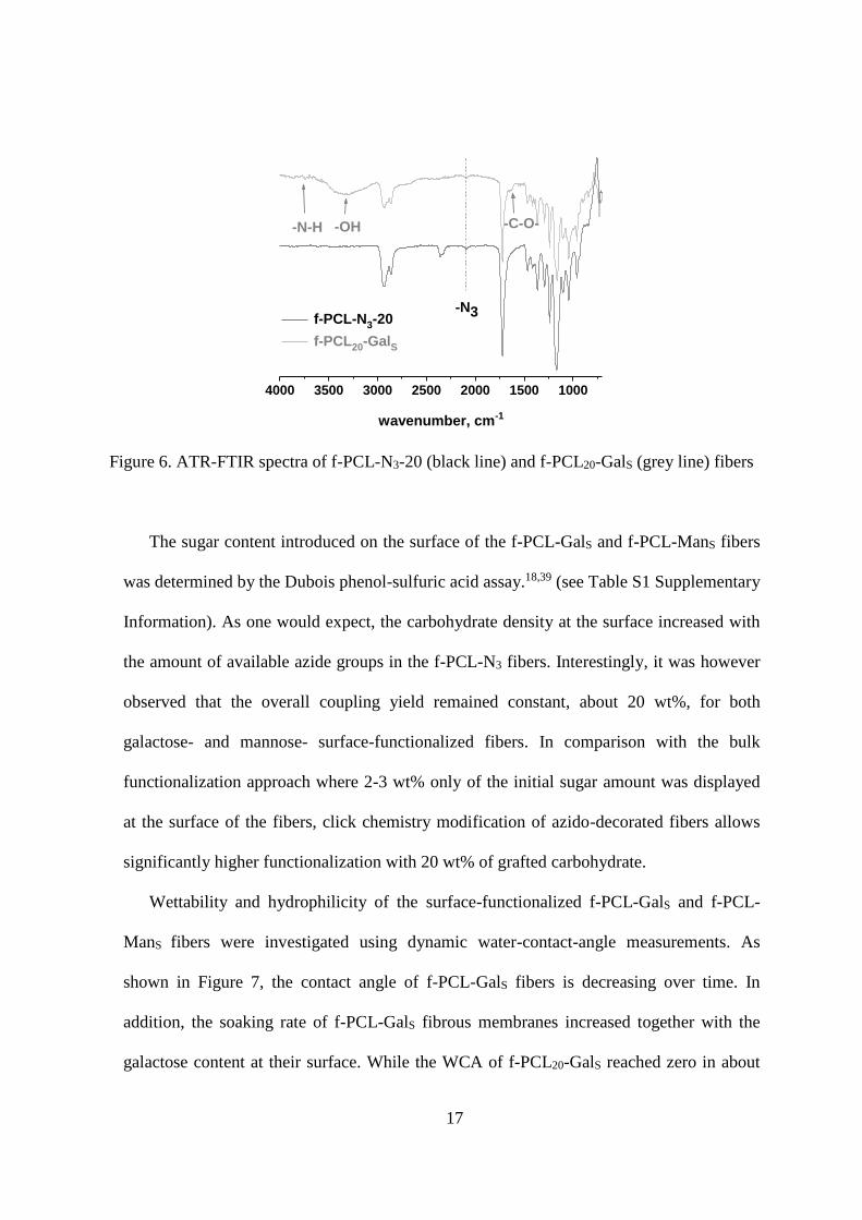
N3-20 and its galactosylated counterpart f-PCL20-GalS are illustrated in Figure 6. The
presence of sugars on the fibers is evidenced by the large peak at 3300 cm-1 assigned to
stretching vibrations of hydroxyl groups as well as vibrations of -N-H at 3744 cm-1. A peak
at ~1642 cm-1, corresponding to -C-O- stretching groups of monosaccharide, absent in
native PCL80, was also noted. The characteristic peak of azide groups, at 2100 cm-1,
indicates some remaining azido groups on the surface as well as inside the fibers after the
sugar coupling.
17
4000 3500 3000 2500 2000 1500 1000
-N3
-C-O--OH-N-H
wavenumber, cm-1
f-PCL-N3-20
f-PCL20-GalS
Figure 6. ATR-FTIR spectra of f-PCL-N3-20 (black line) and f-PCL20-GalS (grey line) fibers
The sugar content introduced on the surface of the f-PCL-GalS and f-PCL-ManS fibers
was determined by the Dubois phenol-sulfuric acid assay.18,39 (see Table S1 Supplementary
Information). As one would expect, the carbohydrate density at the surface increased with
the amount of available azide groups in the f-PCL-N3 fibers. Interestingly, it was however
observed that the overall coupling yield remained constant, about 20 wt%, for both
galactose- and mannose- surface-functionalized fibers. In comparison with the bulk
functionalization approach where 2-3 wt% only of the initial sugar amount was displayed
at the surface of the fibers, click chemistry modification of azido-decorated fibers allows
significantly higher functionalization with 20 wt% of grafted carbohydrate.
Wettability and hydrophilicity of the surface-functionalized f-PCL-GalS and f-PCL-
ManS fibers were investigated using dynamic water-contact-angle measurements. As
shown in Figure 7, the contact angle of f-PCL-GalS fibers is decreasing over time. In
addition, the soaking rate of f-PCL-GalS fibrous membranes increased together with the
galactose content at their surface. While the WCA of f-PCL20-GalS reached zero in about
18
60 s, f-PCL60-GalS fibers’ WCA attained 0° value in less than 3 s. The gradual difference in
the soaking rate was more accentuated for f-PCL-ManS samples where complete
wettability was reached in 40, 12 and 7 s for f-PCL20-ManS, f-PCL40-ManS and f-PCL60-
ManS fibers, respectively. These results, again support the efficient glycosylation of the
fiber’s surface. In addition, this glycosylation allowed to turn a hydrophobic material into a
highly hydrophilic one. This point is very important in perspective of biological
applications since one could expect hydrophilic materials to be more biocompatible and to
favor interactions with proteins and cells.
0 10 20 30 40 50 60
0
50
100
150
200 f-PCL
20-Gal
S
f-PCL40
-GalS
f-PCL60
-GalS
f-PCL80
wa
ter
co
nta
ct
an
gle
(°)
drop age (s)
0 5 10 15 20 25 30 35
0
50
100
150
200
wa
ter
co
nta
ct
an
gle
(°)
drop age (s)
f-PCL20
-ManS
f-PCL40
-ManS
f-PCL60
-ManS
f-PCL80
Figure 7. Dynamic water contact angles of (A): (□) f-PCL20-GalS, (◊) f-PCL40-GalS, and (○) f-
PCL60-GalS and (B): (□) f-PCL20-ManS, (◊) f-PCL40-ManS, and (○) f-PCL60-ManS surface-
functionalized fibers together with (■) f-PCL80 as a reference
Bioavailability of carbohydrates was next investigated by enzyme-linked lectin assay
(ELLA). Galactose and mannose-decorated fibers were respectively exposed to solutions
of Concanavalin A-peroxidase conjugate (HRP-ConA) and Arachis hypogaea-peroxidase
conjugate (HRP-PNA). Otman et al.40 reported specific recognition of -D-mannose at the
surface of polymeric nanoparticles by ConA, demonstrating that mannose groups,
conjugated to the poly(-caprolactone), could bind the lectin. In this study, upon staining
B A

19
with the AEC chromogenic substrate, both f-PCL-GalS and f-PCL-ManS fibrous mats
exhibited a positive coloration in presence of HPR-PNA and HRP-ConA lectins
respectively (Figure 8). f-PCL-ManS fibers showed a very intense red color while f-PCL-
GalS staining was less pronounced (see Figure 8). Such a difference between ConA and
PNA binding efficiencies is not unexpected and Wu et al.41 previously observed that PNA
lectin binds more strongly to lactose than to galactose, suggesting that this lectin needs a
longer arm-spacer for a better carbohydrate recognition. Nonetheless, the control samples
with non-functionalized f-PCL-N3 fibers showed no significant coloration under the same
treatment, demonstrating that ELLA labeling is highly carbohydrate-specific.
Figure 8. Image (from left to right) of ELLA assays on: (A) f-PCL20-ManS, f-PCL40-
ManS, and f-PCL60-ManS (B) f-PCL20-GalS, f-PCL40-GalS and f-PCL60-GalS fibers. For each
sample, top line corresponds to the positive test samples while bottom line matches the
control samples.
Comparing the collected results of bulk- and surface-functionalized fibrous scaffolds,
we could stress several important points. First of all, electrospun fibers derived from PCL2-
Gal and PCL2-N3 present quite different diameters and morphologies. f-PCL-N3, exhibited
A
B

20
a regular diameter of 600 nm whatever the azide group content while the diameter of f-
PCL-GalB fibers was rather irregular and increased with the galactose content in the
electrospinning blend. In addition, bulk-functionalized f-PCL20-GalB fibers had a rather
small sugar content at their surface (~3 wt%) compared with the surface-functionalized f-
PCL20-GalS and f-PCL20-ManS fibers (~20 wt%). Such a difference might be explained by
the different solubility of PCL2-N3 and PCL2-Gal in the solvent system used for
electrospinning. Indeed, PCL2-N3 is hydrophobic and totally soluble in DCM/MeOH. On
the contrary, PCL2-Gal is amphiphilic and forms aggregates in the same solvent system as
evidenced by DLS. The polymer probably adopts a conformation in which the sugar is
confined in the core of the aggregates to minimize interactions with the solvent.
Accordingly, the galactose residues are predominantly entrapped in the core of the fiber
and very few are available at the surface for protein binding.
On the contrary, f-PCL-N3 electrospun fibers presented high density of azide groups at
their surface as a response to the high electric field applied. Functionalization by click
chemistry has further allowed the glycosylation of 20 wt% of sugar at the fiber surface and
transformed hydrophobic PCL fibers into hydrophilic and bioactive PCL scaffolds. Despite
the effectiveness of surface functionalization by CuAAC, the use of copper catalysts can be
an obstacle for medical applications. To overcome this drawback, strained alkynes might
be used to realize copper-free click conjugation.42
4. CONCLUSIONS
Bulk- and surface-glycosylation of electrospun fibers were explored and their effectiveness
was compared. Electrospun blend of PCL2-Gal and PCL80 resulted in poorly surface-
decorated fibers with moderately increased hydrophilicity. Yet, surface functionalization by
click chemistry of f-PCL-N3 ultra-fine fibers with galactose and mannose moieties was

21
rewarded with successful sugar conjugation and high hydrophilicity of functionalized fibers.
Enzyme-linked lectin assays of the fibrous mats confirmed the ability of carbohydrate to
interact with specific lectins, indicating the biological potential of these scaffolds in tissue-
engineering.
SUPPORTING INFORMATION
Additional information about synthesis of propargyl--D-galactose and propargyl--D-
mannose accompanied with their NMR and MALDI-TOF mass spectroscopy, NMR and
MALDI-TOF analyses of PCL2-Gal, as well as tabulation of the Dubois colorimetric results
of surface-functionalized f-PCL-GalS and f-PCL-ManS fibers. This material is available free
of charge via the Internet at http://pubs.acs.org
Corresponding Author
Sébastien Fort*
Centre de Recherches sur les Macromolécules Végétales, UPR CNRS 5301, BP53,
38041 Grenoble Cedex 9, France.
E-mail: [email protected]
Author Contributions
All authors have given approval to the final version of the manuscript.
Funding Sources
This research was supported by the grant of Institut Carnot “Polynat” and the MNERT PhD
grant N° 2010/A8 to Anica Lancuški.

22
AKNOWLEDGEMENTS
The authors gratefully acknowledge Rachel Martin for her expert help and technical
assistance in FESEM microscopy, Dr. Bernard Priem for helpful discussions on ELLA tests,
Stéphanie Boullanger her technical assistance in mass spectrometry analysis, Dr. Christophe
Travelet for his technical assistance in dynamic light scattering measurements and laboratory
NanoBio and Hugues Bonnet for providing us facilities for contact angle measurements. We
also thank the Institute Carnot “Polynat” for financial support and the MNERT for PhD grant
N° 2010/A8 to Anica Lancuški.
ABBREVIATIONS
PCL80 poly(-caprolactone) Mn 80 000 g/mol; PCL2 - diol-poly(-caprolactone) Mn 2
000 g/mol; PCL2-N3 -azido-poly(-caprolactone); PCL2-Gal -galactoside-poly(-
caprolactone); f-PCL80 poly(-caprolactone) fibers; f-PCL-N3-X fibers containing X=20, 40
and 60 wt% of PCL-N3 and (100-X) wt% of PCL80; f-PCLX-GalB bulk-functionalized fibers
containing X=20 and 40 wt% of PCL2-Gal and (100-X) wt% of PCL80; f-PCLX-GalS surface-
functionalized f-PCL-N3-X fibers with propargyl--D-galactose; f-PCLX-ManS surface-
functionalized f-PCL-N3-X fibers with propargyl--D-mannose.
REFERENCES
1. Sell, S. A.; Wolfe, P.S.; Garg, K.; McCool, J.M.; Rodriguez, I. A.; Bowlin, G. L.
Polymers 2010, 2, 522–553.
2. Barnes, C. P.; Sell, S. A.; Boland, E. D.; Simpson, D. G.; Bowlin, G. L. Adv. Drug Deliv.
Rev. 2007, 59, 1413–1433.
3. Lalani, R.; Liu, L. Biomacromolecules 2012, 13, 1853–1863.

23
4. Mickova, A.; Buzgo, M.; Benada, O.; Rampichova, M.; Fisar, Z.; Filova, E.; Tesarova,
M.; Lukas, D.; Amler, E. Biomacromolecules 2012, 13, 952–962.
5. Shalumon, K. T.; Anulekha, K. H.; Chennazhi, K. P.; Tamura, H.; Nair, S. V.; Jayakumar,
R. Int. J. Biol. Macromol. 2011, 48, 571–576.
6. Ghasemi-Mobarakeh, L.; Prabhakaran, M. P.; Morshed, M.; Nasr-Esfahani, M. H.;
Ramakrishna, S. Mater. Sci. Eng., C 2010, 30, 1129–1136.
7. Seyednejad, H.; Ji, W.; Yang, F.; van Nostrum, C. F.; Vermonden, T.; van den Beucken,
J. J. J. P.; Dhert, W. J. A.; Hennick, W. E.; Jansen, J. A. Biomacromolecules 2012, 13,
3650–3660.
8. Xie, J.; Michael, P. L.; Zhong, S.; Ma, B.; MacEwan, M. R.; Lim, C. T. J. Biomed. Mater.
Res., Part A 2012, 100A, 929–938.
9. Chung, A. S.; Hwang, H. S.; Das, D.; Zuk, P.; McAllister, D. R.; Wu, B. M. J. Biomed.
Mater. Res., Part B 2012, 100B, 274–284.
10. Yuan, W.; Li, C.; Zhao, C.; Sui, C.; Yang, W.-T.; Xu, F.-J.; Jie, M. Adv. Funct. Mater.
2012, 22, 1835–1842.
11. Choong, C.; Foord, J. S.; Griffiths, J.-P.; Parker, E. M.; Baiwen, L.; Bora, M.; Moloney,
M. G. New J. Chem. 2012, 36, 1187-1200.
12. Grafahrend, D.; Calvet, J. L.; Klinkhammer, K.; Salber, J.; Dalton, P. D.; Moller, M.;
Klee, D. Biotechnol. Bioeng. 2008, 101, 609–621.
13. Fleischmann, S.; Hinrichs, K.; Oertel, U.; Reichelt, S.; Eichhorn, K.-J.; Voit, B.
Macromol. Rapid Commun. 2008, 29, 1177–1185.
14. Zheng, J.; Liu, K.; Reneker, D. H.; Becker, M. L. J. Am. Chem. Soc. 2012, 134, 17274–
17277.
15. Zhao, G.-L.; Hafrén, J.; Deiana, L.; Córdova, A. Macromol. Rapid Commun. 2010, 31,
740–744.

24
16. Krouit, M.; Bras, J.; Belgacem, M. N. Eur. Polym. J. 2008, 44, 4074–4081.
17. Xu, N.; Lu, F.-Z.; Du, F.-S.; Li, Z.-C. Macromol. Chem. Phys. 2007, 208, 730–738.
18. Galgali, P.; Puntambekar, U.; Gokhale, D.; Varma, A. Carbohydr. Polym. 2004, 55, 393–
399.
19. Gentsch, R.; Pippig, F.; Nilles, K.; Theato, P.; Kikkeri, R.; Maglinao, M.; Lepenies, B.;
Seeberger, P.H.; Broner, H.G. Macromolecules 2010, 43, 9239–9247.
20. Chua, K.-N.; Lim, W.-S.; Zhang, P.; Lu, H.; Wen, J.; Ramakrishna, S.; Leong, K. W.;
Mao, H.-Q. Biomaterials 2005, 26, 2537–2547.
21. Kim, T. G.; Park, T. G. Biotechnology Progress 2006, 22, 1108–1113.
22. Fraser, C.; Grubbs, R. H. Macromolecules 1995, 28, 7248–7255.
23. Shukla, R. K.; Tiwari, A. Carbohydr. Polym. 2012, 88, 399–416.
24. Grafahrend, D.; Heffels, K.-H.; Beer, M.V.; Gasteier, P.; Moller, M.; Boehm, G.; Dalton,
P. D.; Groll, J. Nature Mater. 2011, 10, 67–73.
25. Grafahrend, D.; Calvet, J. L.; Salber, J; Dalton P.D.; Moeller M.; Klee, D. J. Mater. Sci.
Mater. Med. 2007, 19, 1479-1484.
26. Klinkhaller, K.; Bockelmann, J.; Simitzis, C.; Brook, G. A.; Grafahrend, D.; Groll, J.;
Moller, M.; Mey, J.; Klee, D. J. Mater. Sci. Mater. Med. 2010, 21, 2637-2651.
27. Grafahrend, D.; Heffels, K.-H.; Moller, M.; Klee, D.; Groll, J. Macromol. Biosci. 2010,
10, 1022-1027.
28. Losel, R.; Grafahrend, D.; Moller, M.; Klee, D. Macromol. Biosci. 2010, 10, 1177-1183.
29. Lancuški, A.; Fort, S.; Bossard, F. ACS Appl. Mater. Interfaces 2012, 4, 6499–6504.
30. Provencher, S. W. Macromol. Chem. Phys. 1979, 180, 201–209.
31. Dal Bo, A. G. ; Soldi, V. ; Giacomelli, F. C.; Travelet, C.; Jean, B.; Pignot-Paintrand, I.;
Borsali, R.; Fort, S. Langmuir 2012, 28, 1418−1426.
32. Heller, W. J. Phys. Chem. 1965, 69, 1123–1129.
33. Sharma, S.; Patel, P. B.; Patel, R. S.; Vora, J. J. E-J. Chem. 2007, 4, 343–349.

25
34. Mehra, R. Proc. Indian Acad. Sci. (Chem. Sci.) 2003, 115, 147–154.
35. Dubois, M.; Gilles, K. A.; Hamilton, J. K.; Rebers, P. A.; Smith, F. Anal. Chem. 1956, 28,
350–356.
36. Liu, Y.; Ma, G.; Fang, D.; Xu, J.; Zhang, H.; Nie, J. Carbohydr. Polym. 2011, 83, 1011–
1015.
37. Yu, D.-G.; Branford-White, C.; Williams, G. R.; Bligh, S. W. A.; White, K.; Zhu, L.-M.;
Chatterton, N. P. Soft Matter 2011, 7, 8239-8247.
38. Gentsch, R.; Pippig, F.; Schmidt, S.; Cernoch, P.; Polleux, J.; Borner, H.G.
Macromolecules 2011, 44, 453–461.
39. Bech, L.; Lepoittevin, B.; El Achhab, A.; Lepleux, E.; Teule-Gay, L.; Boisse-Laporte, C.;
Roger, P. Langmuir 2007, 23, 10348–10352.
40. Otman, O.; Boullanger, P.; Drockenmuller, E.; Hamaide, T. Beilstein J. Org. Chem. 2010,
6, 1-7.
41. Wu, P.; Chen, X.; Hu, N.; Tam, U.C.; Blixt, O.; Zettl, A.; Bertozzi, C.R. Angew. Chem.
Int. Ed. 2008, 47, 5022–5025.
42. Codelli, J. A.; Baskin, J. M.; Agard, N. J.; Bertozzi, C. R. J. Am. Chem. Soc. 2008, 130,
11486-11493.
26
Table of contents: