Embed Size (px)
Citation preview

126 Biochimica et Bmphy~ica Acta, 715 (1982) 126 136 Elsevier Biomedical Press
BBA 21078
CARBOHYDRATE COMPOSITIONAL EFFECTS ON TISSUE DISTRIBUTION OF CHICKEN RIBOFLAVIN-BINDING PROTEIN
M A R K S. MILLER, R I C H A R D C. B R UC H * and H A R O L D B. WHITE, III **
Department of Chemistry, UniversiO' of Delaware, Newark, DE 19711 (U.S.A.)
(Received September 15th, 1981 )
Key words: Carbol[vdrate composttion," Glyeoprotein clearance," Riboflacm-hinding protein; (Chicken)
Riboflavin-binding proteins (RBP) purified from chicken egg white, yolk and the serum of laying hens differ in their carbohydrate compositions reflecting tissue-specific modifications of a single gene product. All three are complex glycoproteins having more than twice as many N-acetylglucosamine residues (> 12) as mannose residues (approx. 6). Egg white RBP is distinctive in having only one sialic acid and two galactose residues. Serum RBP contains approx, five sialic acid and seven galactose residues. In addition there is one residue of fucose. The carbohydrate composition of yolk RBP indicates the hydrolysis, respectively, of one, one, two and 3 residues of sialic acid, fucose, galactose, and N-acetylglucosamine from its precursor, serum RBP. The effect of these differing levels of glycosylation on plasma clearance, ovarian uptake and tissue distribution of 12s I-labeled riboflavin-binding proteins in laying hens were compared. 2 h after intravenous injection, 19% of the egg white RBP, 29% of the yolk RBP, and 37% of the serum RBP remained in circulation. The kinetics of plasma clearance was distinctly biphasic for each of the radioiodinated proteins. The initial rapid-turnover component (tl/2 = 13 min) ranged from 27% of the serum RBP sample to 48% of the egg white RBP sample. The remaining slow-turnover components were cleared with half-lives of 81 min (egg white RBP), 101 min (yolk RBP), and 121 rain (serum RBP). 16 h after injection, only 4% of the egg white RBP was deposited in the yolk of developing oocytes while about 12% of the serum RBP and yolk RBP was deposited. This highly significant difference is apparently due to preferential, carbohydrate-dependent clearance of egg white RBP by the liver rather than preferential uptake of serum and yolk RBP by the ovarian follicle. We find no evidence for carbohydrate-directed uptake of riboflavin-binding protein by the ovarian follicle.
Introduction
Riboflavin-binding protein (RBP) is one of several nutrient-binding proteins found in eggs of birds and other vertebrates. Using a mutant strain of chickens which are unable to deposit riboflavin in their eggs and which excrete large quantities of riboflavin in their urine (riboflavinuria), Winter et al. [1] demonstrated that RBP is necessary for the
* Present address: Department of Pathology, Hahnemann Medical College, Philadelphia, PA 19102 U.S.A.
** To whom correspondence should be addressed. Abbreviations: RBP, riboflavin-binding protein: SDS, sodium dodecyl sulfate.
transfer of riboflavin to the egg. Hammer et al. [2] found that within the egg, radioactively labeled RBP was transferred specifically to yolk following intravenous injection into laying hens. Like several other nutrient-binding proteins found in egg yolk such as biotin-binding protein [3], transferrin [4], and phosvitin [5], yolk RBP is glycoslyated [6]. The occurrence of these glycoproteins suggested that their oligosaccharide moieties might contain important recognition features for their specific uptake into the yolk [7,8]. This possibility is in accordance with the role of carbohydrates in cellu- lar recognition of glycoproteins which has become apparent within recent years (for review, see
0304-4165/82/0000-0000/$02.75 ~" 1982 Elsevier Biomedical Press

Ref. 9). Miller et al. [10,11] have examined this possible function by modifying the carbohydrate moieties of egg white RBP and yolk RBP and comparing the amounts of native and modified proteins which were transferred to yolk following injection into the bloodstream of laying hens. They found an indication that sialic acid may be im- portant for ovarian recognition and uptake of RBP.
RBP has been isolated from three locations: egg white [12,13], egg yolk [11,14], and the blood of the laying hen [15,16,37]. Since Winter et al. [1] found that RBP is missing from all three of these locations in the mutant chickens homozygous for the riboflavinuria trait, egg white, yolk, and serum RBP must be products of the same gene. Similari- ties in molecular weight [6,12] and amino acid composition [6,13] have also indicated the identity of the peptide component of these proteins. How- ever, the carbohydrate compositions of egg white RBP [10,17] and yolk RBP [6,11] are quite differ- ent. We find the carbohydrate composition of serum RBP to differ from both egg white and yolk RBP. In the experiments reported in this paper, we utilized these three naturally occurring riboflavin- binding proteins, each with its distinct carbo- hydrate component, to test the carbohydrate re- cognition hypothesis.
Materials and Methods
Experimental animals and source of proteins. The animals used in these experiments were mature female Single Comb White Leghorn chickens (Gal- lus domesticus) obtained from Dover Poultry Prod- ucts Inc., Baltimore, MD. They were maintained for at least 2 weeks at the facilities of the Univer- sity of Delaware, College of Agriculture, and fed a diet of layer feed ad lib. Only those hens which were actively laying at the time of the experiments were used for our studies. Egg white RBP, pre- pared from egg whites by the method of Farrell et al. [13], was donated by Robert Guyer from the Pennsylvania State University. Yolk RBP was pre- pared from fresh White Leghorn eggs obtained from Red Bird Egg Farm, Bear, DE, by methods described previously [11]. Serum RBP was purified from laying hen plasma obtained from Dover Poultry Products Inc., Baltimore, MD, by the
127
method described below. Protein-bound riboflavin was not removed from these proteins during the purification procedures. Chicken serum albumin was purchased from the United States Biochemical Corp., Cleveland, OH.
Isolation of serum riboflavin-binding protein. Serum RBP was purified to apparent homogeneity in four steps: salt precipitation; two ion-exchange steps, one batchwise and one column; and a gel filtration step. Fresh blood from laying hens was treated with 0.5 M sodium citrate, pH 5.5 (107o, v /v) to prevent coagulation. Following centrifuga- tion for 10 min at 5000 × g, the citrated plasma was retained. A typical preparation started with approx. 201 citrated plasma. The precipitate form- ing at 4 5 - 85% saturation (NH4)2SO 4 was pre- pared. This material, which floated following centrifugation for l h at 16000×g was resus- pended in a minimum volume of distilled water and dialyzed overnight against running water. The pH of the retentate was adjusted to 4.3 with 1.0 M acetic acid and insoluble material was removed by centrifugation.
Serum RBP was adsorbed to DEAE-cellulose by batchwise addition of 200 g of Whatman DE-52, which had been equilibrated with 0.1 M sodium acetate buffer (pH 4.3). Following washing on a Buchner funnel with the same buffer, the cellulose was packed into a 2.5 cm diameter column, and washing was continued until A280 returned to baseline. Serum RBP was eluted with 1 M NaC1 in the same buffer and all fractions with A450 greater than 0.1 were pooled. Following dialysis, the solu- tion was applied to a 2 .5×60 cm column of DEAE-cellulose in 0.1 M sodium acetate buffer (pH 4.3) and eluted with a linear 0-1 M NaC1 gradient in the same buffer. All fractions with A450 greater than 0.1 were pooled, dialyzed and lyophi- lized.
The sample was dissolved in a small volume of 25 mM sodium acetate buffer (pH 4.3) and chro- matographed on a 2.5 × 80 cm column of Bio-Gel P-100 in the same buffer. All fractions with A4s o greater than 0.1 were pooled, dialyzed and lyophi- lized. Riboflavin-binding protein was assayed by fluorescence titration [10]. Protein was determined by the method of Lowry et al. [18]. Homogeneity of the protein was established by SDS- polyacrylamide gel electrophoresis [19].

128
Carbohydrate analytical methods. Gas-liquid chromatographic analysis of neutral and amino sugars was performed by the method of Porter [20], using meso-inositol (ICN Nutritional Bio- chemical Co., Cleveland, OH) as internal standard. Glucosamine was measured as peracetylated 2,5- anhydromannitol following nitrous acid deamina- tion, while neutral sugars were measured as the alditol acetates in samples not treated with nitrous acid [20]. Chloroform solutions of the reduced acetylated sugars were injected into a Perkin-Elmer 3920 gas chromatograph equipped with a silanized glass column (4 mm i.d. × 1.8 m) of 3% OV-225 on 100/120 mesh Gas ChromQ (Applied Science, State College, PA). Chromatography was per- formed isothermally at a column temperature of 210°C with a nitrogen carrier flowrate of 40 ml/min.
N-Acetylneuraminic acid was determined by the thiobarbituric acid reaction [21] following hydroly- sis in 25 mM H2SO 4 at 80°C for 1 h [22]. Gluco- samine was also determined colorimetrically by the method of Elson and Morgan as described by Winzler [23] following hydrolysis for 4 h at 100°C in 4 N HCI in vacuo.
Plasma clearance times. Plasma clearance stud- ies were performed by intravenous injection of 125I-labeled protein samples. Proteins were labeled with ~25I by the solid state lactoperoxidase method [24]. l mg protein was added to 250 ~1 0.1 M sodium phosphate buffer (pH 7.0) with 0.9% NaC1, 1 mCi Na 125I (New England Nuclear, Boston, MA), and 50 ~1 washed lactoperoxidase-agarose beads (Worthington Biochemical Corp., Freehold, N J). The reaction was started by addition of 20 ~1 0.1 mM H202 followed by two more 15 FI addi- tions of 0.1 mM H202 at 10 min intervals. After 30 min, the lactoperoxidase was inhibited by addi- tion of 10 /~1 2.5M NaN 3. NaI was added as carrier (0.5 ml of 2 m g / m l in 0.1 M phosphate- buffered saline, pH 7.0) and free radioiodide was separated from the labeled protein by chromatog- raphy on Sephadex G-25 (1 × 20 cm) eluted with 0.9% NaC1. The specific activities of the iodinated proteins were typically 50-100 FCi/mg. At this level of radioactivity, about 1 out of every 1000 RBP molecules is iodinated. Over 90% of the radioactivity was precipitable with 15% trichloro- acetic acid.
Following injection of 100 /.tg of freshly iodinated egg white, yolk or serum RBP in l0 mM phosphate-buffered saline (pH 7.0) into the wing vein of an actively laying hen, samples were taken from the opposite wing vein at 2min and at 10 min intervals thereafter up to 2 h and placed into heparinized capillary tubes. The capillary tubes were centrifuged in a hematocrit centrifuge and the radioactivity of duplicate aliquots of plasma was determined using a Tracor Analytic Gamma Counter, Model 1185.
Uptake of riboflavin-binding proteins by develop- ing oocytes. Actively laying hens were injected in the wing vein with 30 #g of freshly iodinated egg white, yolk or serum RBP. The birds were killed 16h later by cervical dislocation and all ovarian follicles 3 mm in diameter and larger were re- moved and placed into ice-cold 0.9% NaC1. The outer vascular epithelium was stripped off and the follicles were weighed. Follicles weighing less than 0.5 g were pooled; the others were handled indi- vidually. Follicles were ruptured and mixed with an equal volume of 0.5 M NaC1, 0.1 M KI (those weighing less than 10 g were mixed with 10 ml of the salt solution) and their contents were homoge- nized with a loose Dounce homogenizer pestle. In addition, if an egg was present in the oviduct, it was also removed, the yolk was washed to remove egg white and treated the same as the follicles. Radioactivity representing free and protein-bound iodine was determined on duplicate 200 ~1 aliquots of these samples. Another duplicate set of 200 ~1 aliquots was mixed with 1 ml 15% trichloroacetic acid and the precipitates, recovered by centrifuga, tion, were washed with another 1 ml 15% trichloro- acetic acid to remove any residual free radioiodide. Protein-bound radioactivity was determined on these precipitates.
Tissue distribution. Laying hens were given in- travenous injections of 20-30 ~g of 125I-labeled egg white, yolk, or serum RBP, or 50 /tg of 12sI- labeled chicken serum albumin. Initial blood sam- ples were taken from the opposite wing vein at 2 min following injection and placed into heparinized microfuge tubes. The birds were killed after 1 h by cervical dislocation and final blood samples were taken by cardiac puncture. The fol- lowing organs were excised within 15 min and placed into ice-cold 10 mM phosphate-buffered

saline (pH 7.0): liver, gall bladder, kidneys, lungs, spleen, ovary and all associated follicles, oviduct, uterus, and small intestine. The gall bladder was opened and the bile collected. The oviduct was divided into the magnum and isthmus regions, and each region was weighed separately. The intestine was cleaned of its contents prior to weighing. The ovary was divided arbitrarily into those follicles greater than 4 mm in diameter and the rest of the ovary, and those two portions were weighed sep- arately. The other organs were cleaned of extra- neous connective tissue and weighed.
Portions of each organ were homogenized with a Waring blender in 4 volumes of ice-cold 10 mM phosphate-buffered saline (pH 7.0). For the small intestine, approximately equal portions of duodenum, jejunum, and ileum were mixed prior to homogenization. For the large ovarian follicles, one representative follicle weighing approx. 5 g was chosen for homogenization. Radioactivity was determined on duplicate 200 ~1 aliquots of each homogenate and 50/~1 aliquots of plasma and bile both before and after precipitation with 15% tri- chloroacetic acid.
129
Results
Purification of serum riboflavin-binding protein Although there are methods available for the
large-scale purification of egg white RBP [12,13] and yolk RBP [6,11], those which have been re- ported for serum RBP have been for 'purification of small quantities (< 5 mg) of protein [15,16,37]. The method which we have devised is suitable for preparation of large quantities (> 250 rag) of ho- mogeneous serum RBP. Throughout the purifica- tion process, the brilliant yellow color of protein- bound riboflavin, which has a strong absorbance at 450 nm [25], was used to identify serum RBP. The procedure, which is derived from several other published procedures for purification of RBP [ 11,13], is simple and purification may be achieved within 4 to 5 days.
A summary of the purification of serum RBP is presented in Table I. Although the first step did not result in a large purification of serum RBP, it was necessary to reduce the total volume of the plasma to a size that could be handled more easily.
TABLE I
PURIFICATION SUMMARY OF SERUM RIBOFLAVIN-BINDING PROTEIN
Total protein was determined by the method of Lowry et al. [18] using standard holo-yolk RBP as reference protein. Serum RBP was assayed by fluorescence titration followed by acid-release of bound riboflavin [10]. Total serum RBP is based on binding of 1 mol riboflavin (376 g /mol) per mol riboflavin-binding protein (36000 g/mol). Specific activity represents ratio of riboflavin-binding protein to total protein.
Step Total Serum Specific -Fold Recovery protein (g) RBP (g) activity purification (%)
Citrated plasma (20 1) I 140 1.02 8.95- 10 -4 _ 100
45%-85% ( N H 4 ) 2 8 0 4
precipitate 226 0.438 1,94.10 3 2.17 42.9
DEAE-cellulose batch treatment 0.816 0.343 0.420 469 33.6
DEAE-cellulose column chromatography 0.467 0.298 0.638 713 29.2
Bio-Gel P-100 column chromatography 0.270 0.273 1.01 1 130 26.8

130
a b c d e - 3 . - - 3
- - - , , ~
i
- - c o
w
I00
80
60
4 0
> -
I - - ~>
20 o ,,-,,,
n,- a Nm0 (.9
" ' 8
u. 6 o
+ 0 20 40 60 80 ~"~ " ~ ~ T I M E ( minutes )
IO0 120
Fig. I. SDS-polyacrylamide gel electrophoresis of serum RBP during its purification from laying hen plasma. Serum RBP was purified from citrated plasma (a) in four steps: 45%-85% (NH4)2SO 4 precipitation (b): batch treatment with DEAE-cellulose (c): NaC1 gradient elution from a DEAE-cellulose column (d): and gel filtration on Bio-Gel P-100 (e).
Fig. 2. Plasma clearance of 125I-labeled serum RBP (,~), yolk RBP ([]), and egg white RBP (©). The upper series of curves represents the percent of injected radioactivity present in the plasma over a 2 h time course based on a plasma sample taken 2 min after injection (100%). Each value is the mean of individual values taken from six different birds at identical time points. The half-lives of the terminal (slow) components of these curves were obtained by linear regression analysis of the 60-120 rain values. The difference between the actual data points and the extrapolated values of the slow component yielded the lower series of curves. These lines represent the initial rapid components of the clearance curves. Their half-lives were also obtained by linear regression analysis. Serum (A): tm/2 rapid: 12.5 rain (r=0.993), tl/2 slow: 121 min (r=0.993). Yolk (E3): q/2 rapid: 13.4 rain (r=0.994), q/2 slow: 101 rain (r-0.988). White (O): tl/2 rapid: 13.0 min (r-0.995), tl/2 slow: 80.9 min (r-0.995).
T h e D E A E b a t c h t r e a t m e n t and c o l u m n steps,
wh ich re l ied on the low isoelec t r ic po in t o f s e r u m
R B P ( p I < 4.0 [26]), were very e f fec t ive in separa t -
ing s e rum R B P f r o m the o the r p l a s m a p ro te ins
wh ich in genera l have m u c h h igher i soe lec t r ic
poin ts . In the pu r i f i c a t i on of yo lk a n d egg whi te
R B P , the D E A E c o l u m n step resul ts in c o m p l e t e
p u r i f i c a t i o n of the pro te ins . H o w e v e r , s e r u m R B P
at this s tage still c o n t a i n e d s o m e h igher m o l e c u l a r
w e i g h t c o n t a m i n a n t s , as r e v e a l e d by S D S - p o l y a c r y l a m i d e gel e lec t rophores i s , which were
c o m p l e t e l y r e m o v e d by gel f i l t ra t ion on B io -Ge l
P-100 (Fig. l) .

Chomical comparison of riboflavin-binding proteins The molecular weights of serum, yolk, and egg
white RBP, estimated by comparison of mobilities of duplicate samples with those of standard pro- teins (Bio-Rad, low molecular weight standard mixture) following electrophoresis on SDS-10% acrylamide gels, were as follows: serum RBP, 36000-+ 1500; Yolk RBP, 35600 ± 1200; and egg white RBP, 35000-+ 900. The proteins were elec- trophoresed not only individually but also in mix- tures containing pairs or all three of the proteins. If there were significant differences in the molecu- lar weights of the three species, the mixtures would have been separable by SDS-polyacrylamide gel electrophoresis or at least have resulted in broad- ening of the protein bands. We found the molecu- lar weights of the individual proteins to be indis- tinguishable from those of the mixtures (36000-+ 1500). Furthermore the width of each individual protein band, measured at half of the peak height, did not significantly differ from those of the mix- tures.
The carbohydrate compositions of egg white, yolk, and serum RBP are presented in Table II. In determining the number of residues of carbohydrate per mol of protein, the molecular weights of all three proteins were assumed to be 36000, the molecular weight of the mixture as determined above. The compositions of each of the three proteins differ from the others. Serum
131
RBP has the most complex composition. In addi- tion to containing more galactose, N-acetyl- glucosamine, and sialic acid than either egg white or yolk RBP, serum RBP contains one residue of fucose, a sugar found only in trace quantities on both egg white and yolk RBP.
Physiological studies The clearance of 125I-labeled serum, yolk, and
egg white RBP from the plasma of laying hens is shown in Fig. 2. Qualitatively, it is apparent from these curves that the clearance of egg white RBP proceeds more rapidly than that of either yolk or serum RBP, and that serum RBP has the slowest clearance of the three proteins. Quantitatively, the amounts of the proteins remaining in circulation after 2h may be used for comparison of the turnover of the three species of RBP. Thus, the amount of egg white RBP remaining after 2 h (18.8 -+ 6.2%) was significantly less (P < 0.01) than that of yolk RBP (28.5 + 3.4%), which in turn was significantly less (P<0.01) than that of serum RBP (37.3 -+ 5.9%).
By applying the mathematical methods of com- partmental analysis described by Jacquez [27] the curves were resolved into two components. The half-lives of the slow-turnover components were obtained by linear regression analysis of the pooled 60-120 min data points from six animals for each protein. The correlation values for each were close
TABLE II
CARBOHYDRATE COMPOSITIONS OF RIBOFLAVIN-BINDING PROTEINS FROM EGG WHITE, YOLK AND SERUM OF LAYING HENS
All values are based on 36 000 g per mol RBP and were corrected for protein concentration by the method of Lowry et al. [18] using holo-yolk RBP, dried 19 h in vacuo at 55°C, as reference protein. The values for mannose, galactose, and fncose were determined by gas chromatography and represent the mean and range of two analyses. The values for N-acetylglucosamine were determined both by gas chromatography and colorimetry and represent the mean* S.D. of four determinations, two by each method. N-Acetylneuraminic acid was determined colorimetrically and the values presented are the mean± S.D. of four analyses.
Carbohydrate Residues per mol RBP
Egg white RBP Yolk RBP Serum RBP
Mannosc 6.23 + 0.07 5.87 ±- 0.01 5,84 ± 0.19 Galactose 2.14+0.04 4.93 ± 0. I 1 6.84 +0.10 N-Acetylglucosamine 14.9 +1.2 12.0 ±2.3 15.6 --+2.5 N-Acetylneuraminic acid 1.14 + 0.51 4.23 t0 .14 5.37 -+0.82 Fucose < 0.1 0.20 + 0.06 1.13 + 0.05

132
to unity (r ~> 0.99). The slow-turnover components of the clearance curves from the individual birds had standards deviations of 11.7 min (egg white RBP), 25.1 min (yolk RBP), and 24.0 min (serum RBP). Even with these large standard deviations, the slow-turnover clearance rates of all three pro- teins differed at the P < 0.1 level. The half-lives of the rapid-turnover components of the clearance curves were calculated from the difference between the actual data points and the regression line calculated for the slow-turnover components. In contrast to the slow-turnover components, the rapid-turnover components of the clearance curves had similar half-lives. The amounts of the rapid- turnover species, calculated by extrapolation of the rapid component curves to 2 min (Fig. 2), com- prised approx. 48% of egg white RBP, 36% of yolk RBP, and 27% of serum RBP.
We examined the contribution of free radio- iodide to the clearance curves in several experi- ments by precipitating the plasma with 15% tri- chloroacetic acid and determining both free and protein-bound radioactivity. At the initial 2 min time point, over 93% of the radioactivity was pro- tein bound. The level of free radioiodide, which
i
Serum RBP
~ 40
< o ~30
~-20
_z
I
0 5 llO ll5 0
i i i
Yolk RBP
i I 5 I0 115
r i
Egg White RBP
,'0 ,~ 20 WEIGHT OF FOLLICLE (g)
Fig. 3. Incorporation of 125I-labeled serum, yolk, and egg white RBP into developing oocytes. Laying hens were given in- travenous injections of tracer quantities (30 #g) of the labeled proteins and all oocytes greater than 3 mm in diameter were removed 16 h later. The total protein-bound radioactivity in each oocyte, expressed as the percentage of injected protein- bound radioactivity, is plotted as a function of follicular weight for each of five determinations for serum and yolk RBP and four determinations for egg white RBP. The total percentages of injected RBP deposited in all oocytes from each bird (mean ±S.D.) are as follows: serum RBP, 11.4-+2.1%: yolk RBP, 12.5 + 1.7%; and egg white RBP, 4.3 + 1.2%.
represents a balance between clearance from circu- lation and addition of radioiodide to circulation following degradation of J25I-labeled RBP in tis- sues such as liver, remained fairly constant throughout the experiment. Thus, the half-lives calculated from acid precipitated plasma were not significantly different from those calculated from whole plasma.
In a separate series of experiments we injected Na ~25I intravenously into two chickens and found that free radioiodide is cleared rapidly from circu- lation with an initial half-life of approx. 5.0 min.
The protein-bound ]25I content of all oocytes recovered from hens 16 h after injection with 125I- labeled serum, yolk, or egg white RBP is shown in Fig. 3. The amount of protein transferred to the oocyte increased with the weight of the oocyte, except for the very large oocytes and those which were found in the oviduct (weight of oocyte greater than 16-17 g), which had decreased uptake. These curves revealed considerable individual variation among the hens within each group injected with the same protein. However, the total amounts of each protein transferred to developing oocytes showed very little variation within each group. The total percentages of serum or yolk RBP trans- ferred to developing oocytes were similar to each other but they were both significantly greater (P < 0.0005) than that of egg white RBP by a factor of 2.7 to 2.9.
In addition to measuring protein-bound i25I within each follicle, we measured the total radioac- tivity before trichloroacetic acid precipitation. The difference between the two numbers is an indica- tion of the rate of degradation of the iodinated proteins at other locations within the chicken since free iodide accumulates quite readily within the yolk [28]. The fractions ( f ) of the total radioactiv- ity within the oocytes precipitable with trichloro- acetic acid were similar following injections of serum RBP ( f = 0.45 ± 0.05) or yolk RBP ( f = 0.50±0.10), but were much less following injec- tion of egg white RBP ( f = 0.13 ± 0.04), indicating considerably more extra-ovarian degradation of the latter protein with concomitant release of free iodide.
The distribution of radioactivity within several other tissues of the laying hen 1 h following injec- tion of 125I-labeled serum, yolk, or egg white RBP

133
70 , 5ot 5O
I O - 7, ,x
8 - t~
6 - 7, ~ ~ - 4 - ,/ ~
/ /
o o I smo F, Pin X re 8
~ 6
~ 4 z Q
Follicles
Liver Kidney Spleen
Magnum Isthmus Uterus
Ovary Intestine Bile
Fig. 4. Tissue distribution of 1251-labeled serum (N) , yolk (l~), and egg white (k~) RBP, and chicken serum albumin ([]). Laying hens were given intravenous injections of tracer quanti- ties of the labeled protein and the organs were removed I h later. Portions of each organ were homogenized and radioactiv- ity was determined on aliquots of each homogenate before and after trichloroacetic acid precipitation. The chicken serum al- bumin control values are the average of three determinations. The plasma, liver, and kidney values for RBP represent three to five determinations each; the other values represent one to two determinations each. The lower portion of each bar represents protein-bound radioactivity. The upper portion represents free radioiodide.
is shown in Fig. 4. We used the distribution of 125I-labeled chicken serum albumin, also shown in Fig. 4, as a control for these experiments. The specific uptake of chicken serum albumin by these tissues within the time span of these studies is assumed to be minimal, and thus the amount of ~25 I-labeled chicken serum albumin present in each organ is an indication of both the total blood volume of that organ and the amount of protein which was transferred nonspecifically to the ex- travascular compartment. The difference between the amounts of 125I-labeled serum, yolk, or egg white RBP present in a tissue and that of ~25I-
INTESTINE LIVER KIDNEY LUMEN TISSUE
",,\ / / / ">.,. ,, i V . ,
S R B P / Rf
SRB~ )' Rf / "-.,
PLASMA
FOLLICLE/ YOLK EGG WHITE ~ TISSUE
J I ! ~.PR, SRBP R,--~YRBP R, I *RBP R ' ~ - " ' * - ' ~
/ I t3°%) I \ ] I WRRP ~ i "~wRBP~- °'°"
(70%) | 1 OVARY ~ O V I D U C T
~ EGG
Fig. 5. Model of riboflavin-binding protein transport and metabolism in the laying hen. Riboflavin (Rf) is eliminated from circulation unless bound in a flavoprotein complex. The fate of this complex is discussed in the text. Other abbrevia- tions used in this figure are as follows: SRBP, serum RBP; YRBP, yolk RBP: WRBP, egg white RBP; a.a., amino acids.
labeled chicken serum albumin is due to specific uptake by that tissue. We measured both protein- bound and total radioactivity within each tissue. The difference between these two measurements reflects the amount of non-protein-bound radio- iodide either arising from degradation of the pro- tein within the particular tissue or transported via the plasma from other tissues.
The total radioactivity found within the liver following injection of each of the three species of RBP was greater than that found after injection of the control. In the case of egg white RBP, there was more protein-bound radioactivity as well as total radioactivity when compared with the con- trol. The large quantity of non-protein-bound ra- dioiodide in all three cases indicates active catabo- lism of RBP in the liver. The catabolic rate of egg white RBP based on the amount of free radio- iodide was 2-3-times that of either yolk or serum RBP. The rapid catabolism of egg white RBP in the liver is also indicated by the presence of free radioiodide in the bile, which occurred after injec-
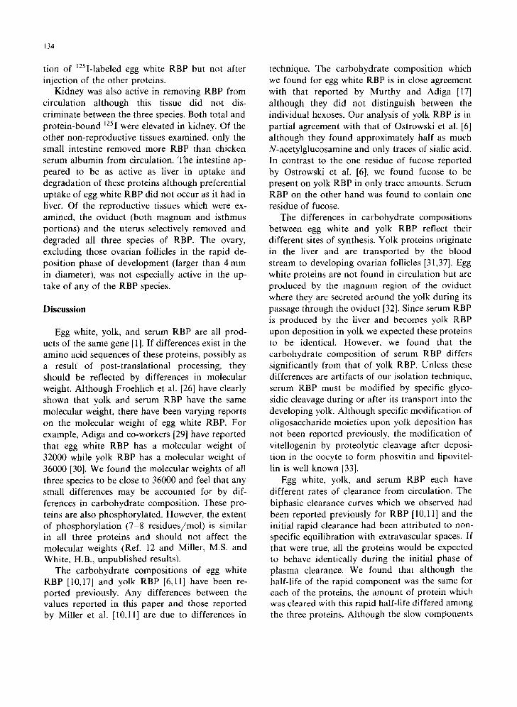
134
tion of 125I-labeled egg white RBP but not after injection of the other proteins.
Kidney was also active in removing RBP from circulation although this tissue did not dis- criminate between the three species. Both total and protein-bound t25I were elevated in kidney. Of the other non-reproductive tissues examined, only the small intestine removed more RBP than chicken serum albumin from circulation. The intestine ap- peared to be as active as liver in uptake and degradation of these proteins although preferential uptake of egg white RBP did not occur as it had in liver. Of the reproductive tissues which were ex- amined, the oviduct (both magnum and isthmus portions) and the uterus selectively removed and degraded all three species of RBP. The ovary, excluding those ovarian follicles in the rapid de- position phase of development (larger than 4 mm in diameter), was not especially active in the up- take of any of the RBP species.
Discussion
Egg white, yolk, and serum RBP are all prod- ucts of the same gene [1]. If differences exist in the amino acid sequences of these proteins, possibly as a resuff of post-translational processing, they should be reflected by differences in molecular weight. Although Froehlich et al. [26] have clearly shown that yolk and serum RBP have the same molecular weight, there have been varying reports on the molecular weight of egg white RBP. For example, Adiga and co-workers [29] have reported that egg white RBP has a molecular weight of 32000 while yolk RBP has a molecular weight of 36000 [30]. We found the molecular weights of all three species to be close to 36000 and feel that any small differences may be accounted for by dif- ferences in carbohydrate composition. These pro- teins are also phosphorylated. However, the extent of phosphorylation (7-8 residues/mol) is similar in all three proteins and should not affect the molecular weights (Ref. 12 and Miller, M.S. and White, H.B., unpublished results).
The carbohydrate compositions of egg white RBP [10,17] and yolk RBP [6,11] have been re- ported previously. Any differences between the values reported in this paper and those reported by Miller et al. [10,11] are due to differences in
technique. The carbohydrate composition which we found for egg white RBP is in close agreement with that reported by Murthy and Adiga [17] although they did not distinguish between the individual hexoses. Our analysis of yolk RBP is in partial agreement with that of Ostrowski et al. [6] although they found approximately half as much N-acetylglucosamine and only traces of sialic acid. In contrast to the one residue of fucose reported by Ostrowski et al. [6], we found fucose to be present on yolk RBP in only trace amounts. Serum RBP on the other hand was found to contain one residue of fucose.
The differences in carbohydrate compositions between egg white and yolk RBP reflect their different sites of synthesis. Yolk proteins originate in the liver and are transported by the blood stream to developing ovarian follicles [31,37]. Egg white proteins are not found in circulation but are produced by the magnum region of the oviduct where they are secreted around the yolk during its passage through the oviduct [32]. Since serum RBP is produced by the liver and becomes yolk RBP upon deposition in yolk we expected these proteins to be identical. However, we found that the carbohydrate composition of serum RBP differs significantly from that of yolk RBP. Unless these differences are artifacts of our isolation technique, serum RBP must be modified by specific glyco- sidic cleavage during or after its transport into the developing yolk. Although specific modification of oligosaccharide moieties upon yolk deposition has not been reported previously, the modification of vitellogenin by proteolytic cleavage after deposi- tion in the oocyte to form phosvitin and lipovitel- lin is well known [33].
Egg white, yolk, and serum RBP each have different rates of clearance from circulation. The biphasic clearance curves which we observed had been reported previously for RBP [10,11] and the initial rapid clearance had been attributed to non- specific equilibration with extravascular spaces. If that were true, all the proteins would be expected to behave identically during the initial phase of plasma clearance. We found that although the half-life of the rapid component was the same for each of the proteins, the amount of protein which was cleared with this rapid half-life differed among the three proteins. Although the slow components

of the clearance curves are significantly different, it is the amount of the rapid-turnover species which leads to the large differences in the amount of protein remaining in circulation 2 h after injec- tion.
Differences in RBP carbohydrate compositions influence the amounts of these proteins which eventually reach the ovarian follicles, but these differences are primarily a consequence of dif- ferential plasma clearance by tissues other than ovary. Egg white RBP has the shortest plasma half-life, the highest liver clearance, and conse- quently the lowest ovarian uptake. Carbohydrate differences in yolk and serum RBP do not affect their uptake by developing oocytes even though their plasma clearance rates are different. Since the oligosaccharide component of RBP does not seem to have a major role in ovarian recognition and transport, we are currently examining the possible role of phosphate residues in these processes.
The uptake and degradation of riboflavin- binding proteins by tissues other than the liver or ovary suggest important additional roles for this protein in transport and salvage of riboflavin in the laying hen. The possible roles of RBP in riboflavin metabolism in the laying hen are il- lustrated in Fig. 5. Serum RBP is synthesized in the liver [26] and complexes with riboflavin in plasma to form the holoprotein. If riboflavin, which is absorbed from the lumen of the small intestine, is not trapped by complexing with serum RBP, it is excreted by the kidney [34]. Some of the holo- serum RBP is removed from circulation by ovarian follicles in the rapid deposition phase and trans- ported into the developing oocytes. Upon modifi- cation of its oligosaccharide moieties, holo-serum RBP is transformed into holo-yolk RBP. Another portion of the holo-serum RBP is removed by the magnum of the oviduct. This tissue is actively synthesizing all egg white proteins and probably removes many proteins indiscrimanately from the plasma as a source for its amino acid pool. Within the magnum, holo-serum RBP is catabolized with the subsequent release of riboflavin. This ribofla- vin can then be bound by egg white RBP which has been synthesized de novo within the secretory cells of the magnum. Unlike yolk RBP, egg white RBP is never fully saturated with its ligand [35],
135
and the level of saturation of egg white RBP probably reflects the availability of holo-serum RBP.
Additional sites of removal of serum RBP from circulation include liver, kidney, and intestine. Al- though the liver site is more specific for egg white RBP, a species not normally found in circulation, it does seem to remove significant quantities of serum RBP. Upon degradation of the protein, serum RBP would release its bound riboflavin, making it available for use by liver flavoenzymes. Although serum RBP has been shown to have no role in intestinal absorption of riboflavin [36], its specific uptake by intestinal tissue may indicate a role in preferential binding of riboflavin im- mediately after its absorption. A similar role can be postulated for serum RBP in kidney, although here it would function to rescue any free riboflavin before it is excreted. Thus serum RBP effectively guarantees that no riboflavin is lost by the laying hen, a role which becomes very important under conditions of riboflavin-poor diet, so that each egg receives a sufficient quantity of this essential nutri- ent to assure viability.
Acknowledgements
We wish to thank Robert Alphin for manage- ment of the birds used in these experiments. This work was supported by National Science Founda- tion Grant PCM-7920683. H.B.W. is the recipient of U.S. Public Health Service Research Career Development Award AM-00152.
References
1 Winter, W.P., Buss, E.G., Clagett, C.O. and Boucher, R.V. (1967) Comp. Biochem. Physiol. 22, 897-906
2 Hammer, C., McDonald, K., Saylor, E.M., Buss, E.G. and Clagett, C.O. (1971) Poultry Sci. 50, 938-944
3 Meslar, H.W., Camper, S.A. and White, H.B, IlL (1978) J. Biol. Chem. 253, 6979-6982
4 Williams, J. (1968) Biochem. J. 108, 57-67 5 Shainkin, R. and Perlmann, G. (1971) Arch. Biochem.
Biophys. 145, 693-700 6 Ostrowski, W., Zak, Z. and Krawczyk, A. (1968) Acta
Biochim. Polonica 15, 241-260 7 Miller, M.S. (1976) Ph.D. Thesis, The Pennsylvania State
University, University Park, PA 8 Muniyappa, K. and Adiga, P.R. (1979) Biochem. J. 177,
887-894

136
9 Neufeld, E.F. and Ashwell, G. (1980) in The Biochemistry of Glycoproteins and Proteoglycans (Lennarz, W.J., ed.), pp. 241-266, Plenum Press, New York
10 Miller, M.S., Buss, E.G. and Clagett, C.O. (1981) Comp. Biochem. Physiol. 69B, 681-686
I1 Miller, M.S., Buss, E.G. and Clagett, C.O. (1981) Biochim. Biophys. Acta 677, 225-233
12 Rhodes, M.B., Bennett, N. and Feeney, R.E. (1959) J. Biol. Chem. 234, 2054-2060
13 Farrell, H.M., Jr., Mallette, M.F., Buss, E.G. and Clagett, C.O. (1969) Biochim. Biophys. Acta 194, 433-442
14 Ostrowski, W., Skarzynski, B. and Zak, Z. (1962) Biochim. Biophys. Acta 59, 515-517
15 Merrill, A.H. and McCormick, D.B. (1978) Anal. Biochem. 89, 87-102
16 Murthy, U.S. and Adiga, P.R. (1978) Biochem. J. 170, 331-335
17 Murthy, U.S. and Adiga, P.R. (1977) Indian J. Biochem. Biophys. 14, 118-124
18 Lowry, O.H., Rosebrough, N.J., Farr, A.L. and Randall, R.J. (1951) J. Biol. Chem. 193,265-275
19 Weber, K. and Osborn, M. (1969) J. Biol. Chem. 244, 4406- 4412
20 Porter, W.H. (1975) Anal. Biochem. 63, 27-43 21 Warren, L. (1959) J. Biol. Chem. 234, 1971-1975 22 Spiro, R.G. (1966) Methods Enzymol. 8, 3-52 23 Winzler, R.J. (1955) in Methods of Biochemical Analysis,
(Glick, D., ed.), Vol. 2, pp. 279-311, Interscience, New York
24 David, G.S. and Reisfeld, R.A. (1974) Biochemistry 13, 1014-1021
25 Nishikimi, M. and Yagi, K. (1969) J. Biochem. (Japan) 66, 427-429
26 Froehlich, J.A., Merrill, A.H., Jr., Clagett, C.O. and Mc- Cormick, D.B. (1980) Comp. Biochem. Physiol. 66B, 397- 401
27 Jacquez, J.A. (1972) Compartmental Analysis in Biology and Medicine, pp. 102-120, Elsevier Publishing Company, Amsterdam
28 Schjeide, O.A. and Prahlad, K.V. (1977) Poultry Sci. 56, 1036-1038
29 Muniyappa, K. and Adiga, P.R. (1980) Biochim. Biophys. Acta 623, 339-347
30 Murthy, U.S., Sreekrishna, K. and Adiga, P.R. (1979) Anal. Biochem. 92, 345-350
31 Schjeide, O.A., Wilkens, M., McCandless, R.G., Munn, R., Peterson, M. and Carlsen, E. (1963) Am. Zoolog. 3, 167-184
32 Mandeles, S. and Ducay, E.D. (1962) J. Biol. Chem. 237, 3196-3199
33 Christmann, J.L., Grayson, M.J. and Huang, R.C.C. (1977) Biochemistry 16, 3250-3256
34 Cowan, J.W., Boucher, R.V. and Buss, E.G. (1966) Poultry Sci. 45, 538-541
35 Feeney, R.E. and Allison, R.G. (1969) Evolutionary Bio- chemistry of Proteins, pp. 66-67, Wiley-Interscience, New York
36 Cowan, J.W., Boucher, R.V. and Buss, E.G. (1964) Pouhry Sci. 43, 172-174
37 Blum, J.-C. (1967) Le M~tabolisme de la Riboflavine chez la Poule pondeuse, F. Hoffman-LaRoche et Cie., Paris