Embed Size (px)
Citation preview
Colloids and Surfaces
A: Physicochemical and Engineering Aspects 209 (2002) 83–104
Capillary mechanisms in membrane emulsification:oil-in-water emulsions stabilized by Tween 20 and milk
proteins
N.C. Christov, D.N. Ganchev, N.D. Vassileva, N.D. Denkov, K.D. Danov,P.A. Kralchevsky *
Laboratory of Chemical Physics & Engineering, Faculty of Chemistry, Uni�ersity of Sofia, 1 James Bourchier A�e.,Sofia 1164, Bulgaria
Received 10 December 2001; accepted 22 March 2002
Abstract
We investigate the process of membrane emulsification in the presence of the nonionic surfactant Tween 20, andthe milk proteins Na-caseinate and beta-lactoglobulin (BLG). Our goal is to examine the factors which control thedrop-size distribution in the formed emulsions. The drops are produced at the outer surface of a cylindricalmicroporous glass membrane, so that the process of their formation and detachment can be directly observed by anoptical microscope. In the case of 2 wt.% aqueous solution of Tween 20 we obtain a relatively fine and monodisperseoil-in-water emulsion with a mean drop diameter about three times that of the pore. The microscopic observationsshow that in this case the oil drops intensively pop out of separate pores. In contrast, for the lower concentrationsof Tween 20, as well as for the investigated solutions of Na-caseinate and BLG, we observe that the membrane iscovered by a layer of growing attached emulsion drops, which are polydisperse, with a relatively large mean drop size.This fact can be explained with a greater dynamic contact angle solid-water-oil. In such a case, after a drop protrudesfrom an opening, it does not immediately detach, but instead, the contact area drop/membrane expands over severalpore openings. The smaller drop size in the emulsions stabilized by BLG, in comparison with those stabilized byNa-caseinate, is related to the circumstance that BLG adsorbs faster at the oil-water interface than Na-caseinate. Inthe investigated emulsions we did not observe any pronounced coalescence of oil drops. Hence, the generation oflarger and polydisperse oil drops in some of the studied solutions is attributed mostly to the effect of expansion ofthe drop contact line and formation of hydrophobized domains on the membrane surface. Therefore, any factor,which leads to decrease of the dynamic three-phase contact angle, and thus prevents the contact-line expansion,facilitates the production of fine and monodisperse emulsions. © 2002 Elsevier Science B.V. All rights reserved.
Keywords: Membrane emulsification; Oil-in-water emulsions; Dynamic interfacial tension; Milk proteins; Kinetic barrier toadsorption
www.elsevier.com/locate/colsurfa
* Corresponding author. Tel.: +359-2-962-5310; fax: +359-2-962-5643.E-mail address: [email protected] (P.A. Kralchevsky).
0927-7757/02/$ - see front matter © 2002 Elsevier Science B.V. All rights reserved.
PII: S0927 -7757 (02 )00167 -X

N.C. Christo� et al. / Colloids and Surfaces A: Physicochem. Eng. Aspects 209 (2002) 83–10484
1. Introduction
1.1. Studies on membrane emulsification
The method of membrane emulsification, pro-posed by Nakashima et al. [1,2], has found aconsiderable development and many applicationsduring the last decade [3–39]. The influence ofdifferent factors on the process of emulsificationby microporous membranes has been investigatedin the works by Kandori et al. [3,4,6], Asano et al.[8,9,17,33,34], Schubert and Schroder [19–21],Williams et al. [22–24], and Yuyama et al. [37–39]. The method has been applied in many fields,in which monodisperse emulsions are needed. Anexample is the application in food industry forproduction of oil-in-water (O/W) emulsions:dressings, artificial milk, cream liqueurs, as well asfor preparation of some water-in-oil (W/O) emul-sions: margarine and low-fat spreads [5–10,17,32–35]. Another application of this methodis for fabrication of monodisperse colloidal parti-cles: silica-hydrogel and polymer microspheres;porous and cross-linked polymer particles; micro-spheres containing carbon black for toners, etc.[4,11,12,18,25–31,38,39]. A third field of utiliza-tion is for obtaining multiple emulsions and mi-cro-capsules, which have found applications inpharmacy and chemotherapy [13–16]. A recentreview can be found in [36]. Closely related to themembrane emulsification is the method employingcapillary tubes and micro-channels to producemonodisperse emulsions [40–45]. Studies on thedetachment of emulsion drops from the tip of asingle capillary have been also carried out [46–48].
1.2. Factors affecting the emulsion drop size
A key problem of membrane emulsification isto explain and predict the dependence of the meandrop diameter, ddrop, on the experimental parame-ters: mean pore diameter, dpore, applied cross flowin the continuous phase, flux of the disperse phasealong the pores, viscosity of the oil and waterphases, interfacial tension and kinetics of surfac-tant adsorption, etc. (Here and hereafter we call‘disperse’ the phase from which the drops are
made, despite the fact that this phase is continu-ous before the drop detachment from the mem-brane.) The values of the ratio ddrop/dpore, reportedin different experimental works, vary in the rangefrom 2 to 10 [36]; the reasons for this variationhave not yet been well understood. Below webriefly consider the major factors affecting theratio ddrop/dpore.
The experiment shows that the presence ofcrossflow (stirring) in the continuous phase de-creases the size of the oil drops, which are detach-ing from the emulsification membrane[20,21,23,32,35,36,47]. This fact can be explainedwith the action of a hydrodynamic drag force,which helps the emulsion drops to brake away[23,47,49]. Ito et al. [47] have obtained a semiem-pirical theoretical expression, which agrees verywell with the experimental dependence of the dropvolume on the applied shear rate (characterizedby the Weber number) in the case, when there isno surfactant dissolved in either of the two liquidphases. The approach by Ito et al. [47] uses thediameter of the drops, formed in the absence ofcrossflow, as an input parameter. In the latter case(no cross flow), the experiment shows that ddrop/dpore�3�0.5. On the other hand, values ddrop/dpore�3 are typical for experiments, which arecarried out in the presence of sufficiently intensivecrossflow, supposedly the disperse phase does notwet the membrane [2]. (If the disperse phase wetsthe membrane, one obtains emulsions with ddrop/dpore�3.5 even in the presence of considerablestirring; this issue is discussed in details in Section6 below.)
The flux of the disperse phase along the poresof the emulsification membrane can be varied bycontrolling the pressure difference applied acrossthe membrane. The experiments show that typi-cally an increase of the transmembrane flow (or ofthe applied pressure) results in a greater meandrop size and in a higher polydispersity of theformed drops [9,19,21,32,37]. Moreover, one candistinguish two regimes of transmembrane flow:(i) fixed flow rate and (ii) fixed pressure. Theformer regime takes place in the emulsificationsetups using a bunch of capillaries or micro-chan-nels, where the disperse phase is usually suppliedby a pump [40–45]. The second regime is typical

N.C. Christo� et al. / Colloids and Surfaces A: Physicochem. Eng. Aspects 209 (2002) 83–104 85
for the standard emulsification setups, in whichthe disperse phase is pushed across the membraneby nitrogen gas from a bottle [2,5,9,14,20]. In thiscase the gas plays the role of a buffer which keepsconstant the applied pressure difference across themembrane; on the other hand, the flow rate alonga given pore may oscillate when drops grow anddetach at its orifice.
The oil–water interfacial tension, �, is recog-nized to be the major retention force, that is theforce which keeps the drops attached to the mem-brane surface [19,21,46]. Greater � is expected tocause the production of larger emulsion drops.Complications arise from the fact that, as a rule,a surfactant (emulsifier) is dissolved in the contin-uous phase to stabilize the produced emulsionagainst drop coalescence. Since the surfactant hasa finite rate of adsorption at the oil–water inter-face [50], the coverage of the drop surface withadsorbed surfactant molecules decreases (and thedynamic value of � increases) when the frequencyof drop release from the pores grows. The lattereffect could explain, at least in part, the aforemen-tioned rise in the drop size with the increase of thetransmembrane pressure.
Another factor, related to the surfactant ad-sorption, is the role of coalescence for establish-ment of the drop-size distribution. The lowcoverage of the drop surfaces with adsorbed sur-factant leads (i) to a larger interfacial tension and(ii) to a drop coalescence in the formed emulsion.The latter two effects have the same consequence:appearance of larger drops. It should be clarifiedwhich effect [(i) or (ii), or both] is more importantfor the observed formation of larger drops (ofdiameter greater than three times the pore diame-ter). It is possible to have coalescence of emulsiondrops at the membrane surface (during their for-mation) and later, in the bulk of the formedemulsion [19]. To our surprise, it turns out thatneither of the aforementioned effects is decisivefor the appearance of larger drops (ddrop/dpore�4)in the emulsions studied by us (in Section 6 wedescribe the actual mechanism of large drop for-mation, which involves an expansion of the con-tact area drop/membrane over several poreopenings).
Last, but not least, the membrane hydrophilic-ity/hydrophobicity also influences the drop sizeand monodispersity. In the first experiments byNakashima et al. [2] it became clear that themembrane should be initially soaked and wet bythe continuous phase for fine and monodisperseemulsion to be produced. The effect of membranewettability was noticed by several authors[2,8,17,37]; some of them characterized the usedmembranes with the respective solid–water–oilcontact angle. It has been established experimen-tally that more hydrophilic membranes producefiner O/W emulsions with a better monodisper-sity, and vice versa [2,37].
1.3. Aim and structure of the paper
Our paper is aimed at investigating the relativeimportance of several factors for the occurrenceof the membrane emulsification with O/W foodemulsions. With such emulsions, the greatest dif-ferences between the size distributions of the pro-duced drops have been registered, depending onthe emulsifier type and concentration[8,17,32,34,36]. In contrast with the precedingstudies, in which the emulsion drops have beenformed at the inner surface of a tubular mem-brane, we employ a special setup for producingthe drops at the outer membrane surface (Fig. 1).In the former regime of emulsification, the mem-brane could sustain larger applied transmembranepressures, but one cannot observe the actual pro-
Fig. 1. Sketch of the used membrane emulsification method:the oil phase is supplied under pressure in a tubular micropo-rous glass membrane; the emulsion drops appear at the outersurface of the membrane, which is immersed in the waterphase.

N.C. Christo� et al. / Colloids and Surfaces A: Physicochem. Eng. Aspects 209 (2002) 83–10486
cess of drop formation at the microporous sur-face, which is hidden inside the non-transparenttube. In contrast, the setup sketched in Fig. 1enables one to observe directly the processes atthe membrane surface and to relate the distribu-tion of the produced emulsion drops with theelementary acts of drop formation anddetachment.
In this paper we focus our attention (i) at therole of the surfactant adsorption and dynamicinterfacial tension and (ii) at the effect of mem-brane hydrophilicity on the drop size distribution.For that reason, in the present study we do notapply a cross flow in the continuous phase (suchflow would also influence the drop size). As men-tioned above, the approach by Ito et al. [47],allows one to determine the mean drop diameterfor a given crossflow rate, if the drop diameter inthe absence of stirring is known. In this way, theproblem is reduced to determining the mean dropdiameter, and the factors influencing it, in theabsence of crossflow (stirring). The latter problemis the subject of the present study.
The paper is organized as follows. In the nextsection we describe the used methods and materi-als. In Section 3 we report results for productionof O/W emulsions by microporous membranesusing Tween 20 as emulsifier; we present andinterrelate experimental drop-size distributionsand photographs of drop formation at the mem-brane surface. Section 4 and Section 5 containsimilar data, but for the milk proteins Na-ca-seinate and �-lactoglobulin (BLG). Finally, inSection 6, we discuss and interpret the experimen-tal results for the process of membrane emulsifica-tion in an attempt to identify the underlyingcapillary mechanisms.
2. Methods and materials
2.1. Membrane emulsification apparatus andprocedure
In our experiments we used a Microkit mem-brane emulsification module (SPG Technology,Miyazaki, Japan), which works with tubular typeShirasu porous glass membranes of outer diame-
ter 10 mm, thickness 1 mm and working area ofapproximately 3 cm2. Membranes of different av-erage pore diameter are available: from 1 to 20�m. According to the manufacturer, the porosity(the surface fraction of the pores) is about 50%.The working pressure difference is up to 105 Pa.As the membranes are made of hydrophilic glass,they are suitable for producing O/W emulsions,which is the type of emulsions investigated by us.(After hydrophobization, the membranes can beused also for generating W/O emulsions.) Allexperiments were carried out at temperature T=23�2 °C.
As mentioned above, one of our aims is toobserve the emulsion drops in the moment oftheir detachment from the membrane. Therefore,to produce O/W emulsions, the oil phase is sup-plied inside the tubular membrane, and the oildrops are released in the outer aqueous phase(Fig. 1); this is the so called ‘batch method’ [6].The higher pressure in the oil is provided by a gas(N2) bottle. We do not apply a cross flow in theouter aqueous phase. The only directed motion ofthe formed oil droplets occurs under the action ofthe buoyancy force. The membrane and the waterphase are placed in a cuvette from optical glass(Hellma GmbH, Mullheim, Germany), which en-ables one to directly observe the emulsificationprocess at the membrane surface. For that pur-pose, we used a CCD camera with an attachedlong-focus objective: CTL-6 (Tokyo ElectronicIndustry Co., Japan) with magnification ×6 andworking distance 39 mm.
After the end of every emulsification experi-ment, the used membrane has been recovered bymeans of the following procedure: (i) soaking in 2wt.% solution of AOT in ethanol for 2 h; (ii)rinsing and soaking in pure water; (iii) immersionfor 1 h in water solution of 2 M HCl at 70 °C;(iv) rinsing and soaking in pure water for 30 min.Our practice shows that this procedure fully re-covers the initial hydrophilic state of the SPGmembranes, which can be used in subsequentexperiments.
In the beginning of each experiment, the tubu-lar membrane is first fixed at the holder of theemulsification setup and immersed in the aqueoussurfactant solution. Then, the oily phase is sup-

N.C. Christo� et al. / Colloids and Surfaces A: Physicochem. Eng. Aspects 209 (2002) 83–104 87
plied under pressure. We start with an initialpressure that is not sufficiently high for the oil todisplace the aqueous phase from the pores of themembrane, and consequently, emulsification isnot observed. Next, the pressure is gradually in-creased, until the emulsification begins at a certainthreshold (critical) pressure, Pcr [2]. The workingpressure, P, during the emulsification experimentis afterwards fixed at a value, which is slightly(with less than 10%) greater than Pcr.
The critical pressure can be estimated from theequation Pcr= (4�ow cos �)/dpore, where �ow is theoil–water interfacial tension and � is the mem-brane–water–oil contact angle (measured acrosswater) [2]. Since �ow and � depend on the bulksurfactant concentration, the interfacial age (fre-quency of drop release), and the pore diameter, inour experiments Pcr, as well as the flux of oilacross the membrane, was different for the differ-ent used solutions and membranes. As alreadymentioned, the fixed parameter in our experi-ments was P/Pcr�1.
In other experiments, we varied the transmem-brane pressure, P, in a certain range above Pcr.The formed emulsions were markedly more poly-disperse, in agreement with the results of otherauthors [9,19,21,32,37], and in consonance withthe hydrodynamic theory of drop detachment[51].
2.2. Determination of the drop size distribution
Representative samples of the produced emul-sions were observed in transmitted light by micro-scope Nikon Optiphot 2 with objectives ×100and ×50. The specimens were prepared by plac-ing a small portion of the emulsion in a cytomet-ric glass cell. The resolution of the used opticalmicroscope is 0.8–1 �m; consequently, this is thelower limit for precise measurements of drop di-ameters in our experiments. The microscopic pic-tures were recorded by means of a high resolutionCCD camera (Sony) and digital memory VCR(Panasonic WV-5490). The images were processedusing a semi-automatic image analysis software,operating with Targa+graphic board (True Vi-sion, USA). To determine the size distribution ina given emulsion, the diameters of several hun-
dred droplets (at least 300) were measured andrecorded. To decrease the probability for system-atic errors, the measurements were performedwith, at least, six different randomly chosenframes. All droplets on the frames were taken intoaccount, even the smallest ones which were stillvisible. The collected data were used to obtain thedrop size distribution in the form of histograms orline-and-scatter plots (see below).
2.3. Materials
As oil phase in our experiments we used hex-adecane (C16H34), product of Sigma (without ad-ditional purification), and soybean oil. The latterwas purified by passing through a glass columnfilled with the chromatographic adsorbentFlorisil.
In some experiments, as aqueous phase we usedsolutions of the nonionic surfactant Tween 20(polyoxyethylene-20 sorbitan monolaurate),product of Merck. No electrolyte was added. Inother experiments, we used water solutions ofproteins, such as �-lactoglobulin (BLG) andsodium caseinate (both of them products ofSigma), as well as mixed solutions of BLG andTween 20. All protein solutions contained also 0.1g l−1 NaN3 for antibacterial protection.
3. Results for solutions of Tween 20
3.1. Emulsions in 2 wt.% solutions of Tween 20
Fig. 2 shows the size distribution of hexadecanedrops formed by membrane emulsification (porediameters dpore=1.0 and 3.2 �m) in a 2 wt.%aqueous solution of Tween 20. The distribution inFig. 2(a) has two peaks, the major one being atdrop diameter d1=3.0�0.2 �m. The distributionin Fig. 2(b) has a well pronounced single peak atddrop=9.8�0.1 �m, that is at ddrop/dpore�3.06.The same value of the ratio ddrop/dpore�3 hasbeen reported in other membrane-emulsificationexperiments [2,5,6,14,19,35,36]. The hydrody-namic theory of drop detachment [51] really pre-dicts ddrop/dpore�3 for transmembrane pressureequal to the critical one (for commencement of

N.C. Christo� et al. / Colloids and Surfaces A: Physicochem. Eng. Aspects 209 (2002) 83–10488
Fig. 2. Size distribution of hexadecane drops formed by mem-brane emulsification in 2 wt.% water solution of Tween 20; thewater and oil phases are pre-equilibrated; diameters of 300drops are measured: (a) pore diameter 1.0 �m; (b) porediameter 3.2 �m; the curve is a Gaussian fit, see Eq. (1).
2(a)), indicates the presence of a weak drop coa-lescence. Most probably, the drops have coalescedin the produced emulsion: due to the buoyancyforce the drops emerge to form a cream in theupper part of the cuvette. There, the drops rest ina close contact with each other and some of themcould coalesce. The absence of a secondary peakin Fig. 2(b) can be explained with the fact that thesample for drop-size measurement has been takenimmediately after the production of the emulsion.In particular, Fig. 2(b) demonstrates that it ispossible to obtain emulsions with ddrop/dpore�3and with a narrow drop-size distribution underunstirred conditions. Our experiments indicatethat such results can be obtained if the followingtwo necessary conditions are satisfied: (i) Thetransmembrane pressure is equal, or slightly (withless than 10%) greater, than the critical(threshold) pressure for drop production, and (ii)the continuous (aqueous) phase wets well themembrane. In addition, if stirring is applied, onecan achieve ddrop/dpore�3, see e.g. [47].
3.2. Effect of pre-equilibration of oil �ersus waterphases
It is worthwhile noting that Tween 20 is solublenot only in the aqueous, but also in the oilyphase. The data in Fig. 2 are obtained with hex-adecane, which was pre-equilibrated with the sur-factant solution. If the two phases are notequilibrated, one may expect a lower surfactantadsorption at the oil–water interface, which im-plies higher interfacial tension and easier coales-cence of the emulsion drops upon collision [52].Moreover, some dynamic phenomena, like thecyclic dimpling [53] and osmotic swelling [54],have been observed with emulsion films fromnon-equilibrated phases. To check whether thepre-equilibration of the oil and water phases isimportant for the membrane emulsification, inFig. 3 we compare the drop-size distributionsobtained with pre-equilibrated and non-equili-brated phases. (The distribution for pre-equili-brated phases is the same as in Fig. 2(a)). Onesees that the fraction of the larger drops is slightlygreater in the case of non-equilibrated emulsions.For statistical analysis of the drop-size distribu-
emulsification) and in the absence of cross flow inthe continuous phase.
The secondary peak in Fig. 2(a), which appearsat d2�3.8 �m, can be explained as a result ofdrop coalescence. Indeed, if two drops of volumeV1 coalesce, the resulting bigger drop will havevolume V2=2V1, which implies d2=21/3d1. Withd1=3 �m, this really yields d2�3.8 �m, as exper-imentally observed. The fact that the secondarypeak is much smaller than the major one (Fig.

N.C. Christo� et al. / Colloids and Surfaces A: Physicochem. Eng. Aspects 209 (2002) 83–104 89
Fig. 3. Comparison of the size distributions obtained bymembrane emulsification (pore diameter 1 �m) using pre-equi-librated and non-equilibrated hexadecane and water phases; 2wt.% Tween 20 were initially dissolved in the water phase; foreach distribution diameters of 300 drops are measured; thecurves are Gaussian fits, see Table 1.
The values of R in Table 1 show that the sizedistributions in both types of emulsions are fittedwell by a Gaussian curve. The Gaussian characterof the distribution is, most probably, due to therandom, essentially non-equilibrium, character ofthe drop detachment (by a necking instability),and to the non-uniformity in the pore sizes. Thevalues of dm, b and N0 are slightly greater for thecase of non-equilibrated phases. We could at-tribute the difference between the two size-distri-butions to the effect of a weak coalescence in theemulsion produced from non-equilibrated phases,or to the effect of contact-line expansion, which isdiscussed in Section 6 below. The obtained smalldifferences (Table 1) indicate that the effect ofpre-equilibration turns out to be insignificant, atleast for the investigated system. In contrast, asdemonstrated in the next section, the variation ofsurfactant concentration may have a dramaticeffect.
3.3. Effect of the concentration of Tween 20
Fig. 4 shows experimental size distributions ofhexadecane drops formed by membrane emulsifi-cation (pore size 1.0 �m) for three concentrations,0.02, 0.2 and 2 wt.% Tween 20. For Tween 20 wehave CMC�2.8×10⁻⁵ M, that is the abovethree working concentrations correspond to �6,60 and 600 times the CMC, respectively. The oiland water phases have been pre-equilibrated. Foreach concentration, the diameters of 300 dropshave been measured.
It is seen in Fig. 4 that the peak at ddrop�3 �mis predominant for 2 wt.%, whereas at the lowerconcentrations, 0.02 and 0.2 wt.%, the fraction ofthe larger drops prevails, although the peak at 3�m is still present. To find the reasons for thedifferences between the obtained drop-size distri-
tion, we fitted the data points with a 4-parameterGaussian curve:
N(d)=N0+a exp�
−(d−dm)2
2b2
n(1)
where N denotes the number of drops; d is thedrop diameter, dm is its most probable value,corresponding to the maximum of the Gaussiancurve (Fig. 3), b is the half-width of the peak, N0
characterizes the background at which the peakappears, and a is a scaling parameter. We deter-mined the parameters of the best fit using acommercially available software (Sigma Plot 6.0,SPSS Inc.) The parameters, corresponding to thetwo curves in Fig. 3, are listed in Table 1. Themultiple correlation coefficient, R, is a measure ofhow well the regression curve describes the data(0�R�1). R values near 1 indicate that themodel curve agrees well with the experimentalpoints.
Table 1Parameters of the Gaussian fits in Fig. 3 drawn by means of Eq. (1)
dm (�m)State of the oil and water phases b (�m) RN0
0.983.19�0.03Non-equilibrated 5.3�1.40.39�0.033.04�0.02 0.34�0.03 4.1�2.4 0.99Pre-equilibrated

N.C. Christo� et al. / Colloids and Surfaces A: Physicochem. Eng. Aspects 209 (2002) 83–10490
Fig. 4. Comparison of size distributions of hexadecane dropsformed by membrane emulsification (pore diameter 1 �m) forthree concentrations (a) 0.02 wt.%, (b) 0.2 wt.% and (c) 2 wt.%Tween 20; for each distribution diameters of 300 drops aremeasured.
Fig. 5. Photos of the outer surface of a microporous glassmembrane taken during the process of emulsification; thedrops are of hexadecane in water solutions: (a) 0.02 wt.%Tween 20, pore size 3.2 �m; (b) 2 wt.% Tween 20, pore size10.4 �m.
butions, we carried out direct observations of thedrops, at the moment of their formation andrelease from the membrane surface.
Typical video-frames of the membrane surface,taken during the process of emulsification of hex-adecane in solutions of Tween 20 at concentra-
N.C. Christo� et al. / Colloids and Surfaces A: Physicochem. Eng. Aspects 209 (2002) 83–104 91
tions 0.02 and 2 wt.% are shown in Fig. 5(a) and(b), respectively. To produce drops of greater size,which are better visible by optical microscope, weused membranes with larger pores, viz. dpore=3.2and 10.4 �m.
At concentration 0.02 wt.% Tween 20, we ob-served simultaneous formation of small oil drops(ddrop/dpore�3) and larger oil drops (ddrop/dpore�3) at the membrane surface (Fig. 5(a)), which is inagreement with the size distribution shown in Fig.4(a). The small drops are quickly emitted frommembrane pores; the dynamics of their detach-ment resembles the release of gas bubbles in themaximum bubble pressure method [55,56]. In con-trast, the larger drops are observed to stay andgrow attached to the membrane surface for rela-tively longer periods of time, from several secondsto dozens of seconds. A closer examination of thevideo-records shows that a larger drop originatesfrom a small drop, whose ejection from the poreis accompanied by expansion of its three-phasecontact line. Thus a larger drop is formed at-tached to the membrane. In fact, the whole mem-brane surface is covered with a layer of largerdrops, which stay attached, gradually increase insize, and eventually detach. The smaller drops areemitted from pores situated in-between the largerdrops.
At concentration 2 wt.% only small drops(ddrop/dpore�3) are formed, which are swiftlyejected from the pores (Fig. 5(b)). This observa-tion is consonant with the size distribution in Fig.3 and Fig. 4(c). We occasionally observed detach-ment of larger drops from few isolated smalldomains on the membrane surface, but suchlarger drops seem to be an exclusion, which con-tribute to the background of the drop-size distri-bution (see Fig. 3 and Table 1). The domains ofquestion are most probably hydrophobized spotson the membrane surface, see Section 6 for amore detailed discussion.
We examined carefully the video-records toidentify acts of coalescence of oil drops at themembrane surface. For the lower concentration,0.02 wt.% Tween 20, we observed only a few actsof coalescence; for the higher concentration, 2wt.% Tween 20, we could not observe any. (Asalready mentioned, the secondary peak in Fig.
2(a) can be attributed to a weak coalescence in theproduced emulsion cream.) In general, the coales-cence of emulsion drops in these experiments is avery rare phenomenon. On the other hand, asexplained above, the formation of larger drops(ddrop/dpore�3), which is typical for 0.02 wt.%Tween 20 (Fig. 5(a)), occurs through expansion ofthe contact line of forming attached drops; seeSection 6 for further explanations.
4. Membrane emulsification with milk proteins
4.1. Experiments with Na-caseinate
We applied membrane emulsification (pore size1.0 �m) to form emulsions from hexadecane inaqueous solutions of Na-caseinate without addedelectrolyte. At Na-caseinate concentration 0.01wt.%, it was impossible to produce emulsion;instead, we observed the formation of a cream ofmillimeter-sized drops adhering to the membrane.In 3–4 min, the drops in this adherent creamcoalesced and formed an oil layer covering themembrane. This is the only case with pronouncedcoalescence observed in our experiments.
At ten times larger concentration, 0.1 wt.%Na-caseinate, it was possible to produce emulsion,which contained large drops with a relativelybroad size-distribution: the drop diameters rangedfrom 3 to 300 �m. At higher concentrations, 1and 3 wt.% Na-caseinate, the produced emulsionshad slightly smaller drops and narrower size dis-tributions, see Fig. 6. The respective mean dropdiameters, ddrop�33 and 31 �m, are close to eachother, see Table 2. The half-widths of the respec-tive Gaussian fits are relatively large, b�7 and 11�m. Despite some scattering of the data, reflectedby the higher values of N0 (cf. Table 1), the sizedistributions are fitted well by Gaussian curves,which is evidenced by the value R=0.97 in Table2.
The above results rise the question why theratio ddrop/dpore is about 10 times greater for Na-caseinate in comparison with 2 wt % Tween 20(cf. Figs. 3 and 6). To get some additional infor-mation, we carried out direct microscopic obser-vations of the membrane emulsification process

N.C. Christo� et al. / Colloids and Surfaces A: Physicochem. Eng. Aspects 209 (2002) 83–10492
Fig. 6. Size distributions of hexadecane drops formed bymembrane emulsification (pore diameter 1 �m) in water solu-tions containing 1 and 3 wt.% Na-Caseinate; for each distribu-tion diameters of 300 drops are measured; the curves areGaussian fits, see Table 2.
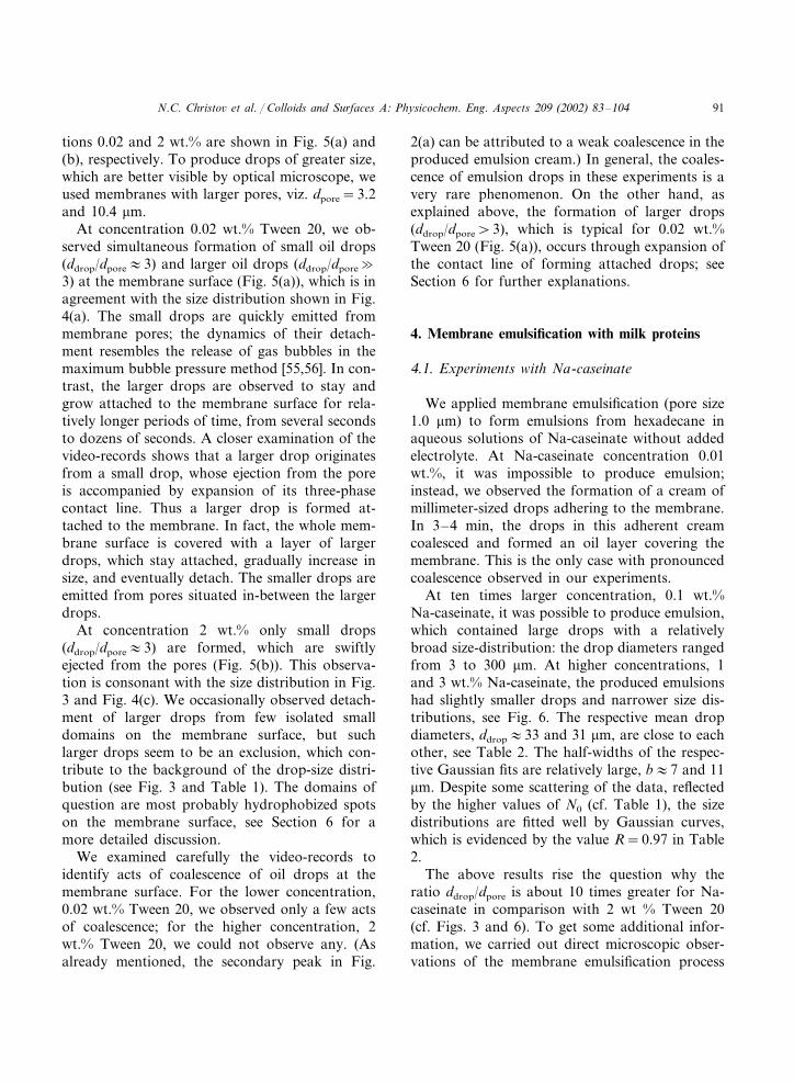
Na-caseinate is that for BLG the average dropsize is considerably smaller. This can be seen alsoin Fig. 8, where the drop size distributions inemulsions, formed by membrane emulsification(dpore=1.0 and 2.1 �m) in solutions containing 1wt.% BLG and 1 wt.% Na-Caseinate, are com-pared. One sees that in the case of BLG the mostprobable diameter and the half-width of theGaussian curve (Table 3) are several times smallerin comparison with those for Na-caseinate. Inparticular, for dpore=1.0 �m we obtain dm�6versus 33 �m and b�1 versus 6 �m. The situationis similar for the membrane with dpore=2.1 �m(Fig. 8(b) and Table 3). The drop size distribu-tions for both BLG and Na-caseinate are fittedwell by Gaussian curves.
We conducted comparative experiments onemulsification by a common rotor-stator homoge-nizer Ultra Turrax T25 (Janke & Kunkel GmbH& Co.). Emulsions containing 30 vol.% of oil wereprepared by stirring the oil–protein solution mix-ture for 3 min at 20 500 rpm. The results for 1wt.% BLG solutions are compared in Fig. 9 andTable 3 with those for emulsions produced bymembrane emulsification, pore size 1 �m. Onesees that in this specific case, the emulsion pro-duced by Ultra Turrax has smaller mean dropsize. The relative width of the Gaussian peak iscomparable: b/dm=0.25 and 0.26 for membraneof 1 �m pores and Ultra Turrax. On the otherhand, the background, N0, is higher in the case ofUltra-Turrax, in comparison with the membraneemulsification (Table 3). The microscopic obser-vations showed that the membrane-producedemulsion does not contain drops of diameterssmaller than dpore, whereas the emulsion obtainedby Ultra Turrax contains even sub-�m drops; inthe latter case the Gaussian peak appears on thebackground of drops of various size.
for solutions containing Na-caseinate, see Fig.7(a). We observed the formation of a layer ofrelatively large oil drops adherent to the mem-brane surface. The major difference with Fig. 5(a)is that the small drops (ddrop/dpore�3) are missing.The adherent layer of oil drops (Fig. 7(a)) isdynamic: the drops grow, detach and new dropsare formed on their place. At concentrations 1and 3 wt.% Na-caseinate, we did not observe anypronounced coalescence of oil drops in the adher-ent layer. The results of these experiments arediscussed and interpreted in Section 6.
4.2. �-Lactoglobulin in comparison withNa-caseinate
At concentration 1 wt.% �-lactoglobulin (BLG)we also observed the presence of transiently ad-herent oil drops at the membrane surface (Fig.7(b)). The main difference in comparison with
Table 2Parameters of the Gaussian fits in Fig. 6 drawn by means of Eq. (1)
dm (�m) b (�m)Concentration of Na-Caseinate N0 R
32.9�0.5 0.971 wt.% 5.2�1.26.8�0.530.9�0.8 11.5�1.0 3.6�1.4 0.973 wt.%
N.C. Christo� et al. / Colloids and Surfaces A: Physicochem. Eng. Aspects 209 (2002) 83–104 93
4.3. Dynamic interfacial tension and adsorptionrate
One of the reasons for the larger emulsiondrops formed in Na-caseinate solutions, in com-parison with the BLG solutions (see Fig. 8), canbe the difference between the rates of adsorptionof these two proteins at the oil–water interface.To check this hypothesis, we carried out measure-ments of dynamic interfacial tension by means ofthe fast formed drop (FFD) technique [57,58]. Adrop with fresh oil–water interface is produced at
Fig. 7. Photos of the outer surface of a microporous glassmembrane taken during the process of emulsification; thedrops are of hexadecane in water solutions: (a) 1 wt.% Na-ca-seinate, pore size 2.1 �m; (b) 1 wt.% BLG, pore size 10.4 �m.
Fig. 8. Size distributions of hexadecane drops produced bymembrane emulsification in solutions containing 1 wt.% Na-caseinate or 1 wt.% BLG. (a) Pore diameter 1 �m, sizedistributions for 413 drops with BLG and for 300 drops withNa-caseinate; (b) pore diameter 2.1 �m, size distribution for600 drops. The curves are Gaussian fits, see Table 3.

N.C. Christo� et al. / Colloids and Surfaces A: Physicochem. Eng. Aspects 209 (2002) 83–10494
Table 3Parameters of the Gaussian fits in Figs. 8 and 9 drawn by means of Eq. (1)
b (�m) N0 Rdm (�m)Protein concentration
Pore diameter 1.0 �m1.4�0.1 4.1�2.0 0.981 wt.% BLG 5.8�0.16.8�0.5 5.2�1.2 0.9732.9�0.51 wt.% Na-caseinate
Pore diameter 2.1 �m6.1�0.8 3.5�4.41 wt.% BLG 0.9115.9�0.7
12.3�0.8 3.0�1.6 0.9750.0�0.71 wt.% Na-caseinate
Emulsion obtained by Ultra Turrax1 wt.% BLG 0.7�0.12.7�0.1 11�5 0.96
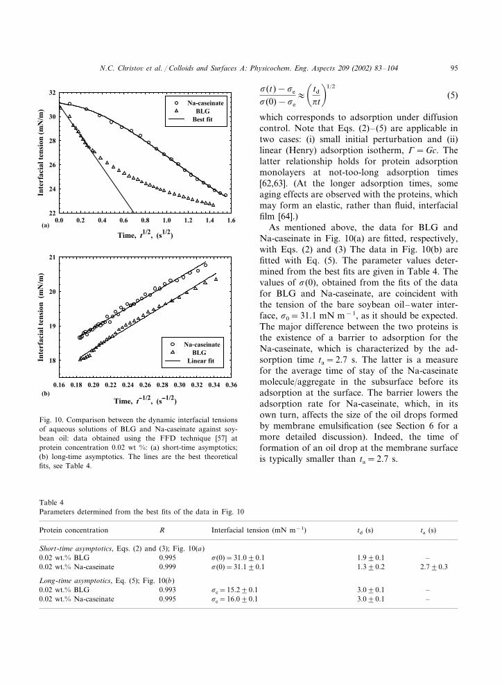
the tip of a capillary by a sudden breaking of a jetfrom the aqueous solution, which flows out of thecapillary into the oil phase. The protein (thesurfactant) adsorbs at the immobile curved sur-face of the formed drop, and consequently, theinterfacial tension �, and the pressure inside thedrop, decrease with time, t. The pressure is regis-tered by means of a piezo-transducer, whose elec-tric output can be converted in terms of interfacialtension, �, by using the Laplace equation of capil-larity. In this way, the data for the dynamicinterfacial tension �(t) in Fig. 10 have been ob-tained. The capillary pressure has been recordedevery 0.005 s, which gives a large number ofexperimental points and provides a good statisticsand time-resolution.
Fig. 10(a) shows the plots of � versus t1/2
during the first 2.5 s of the adsorption process.The data for BLG, at the earlier times, wellcomply with the dependence
�(t)−�e
�(0)−�e
�1−2� t
�td
�1/2
(2)
which represents the short-time asymptotics of theinterfacial tension in the case of adsorption underdiffusion control [50]; �e is the solution’s equi-librium interfacial tension, while �(0) is the ten-sion of the bare oil–water interface; td is thediffusion relaxation time. In contrast, the shorttime behavior of �(t) for Na-caseinate (Fig. 10(a))is typical for adsorption under mixed barrier-dif-fusion control; in such a case, the theoreticaldependence �(t) is described by the expression[59]
�(t)−�e
�(0)−�e
=2�
�td
ta
�1/2��
0
exp(− t�2/ta)(�2−1)2+ td�2/ta
d� (3)
where ta is the adsorption relaxation time whichaccounts for the existence of a barrier to adsorp-tion. In general, one has [59]
td=G2
D, ta=
Gka
, G����
�c�
e
(4)
D is the diffusivity of the adsorbing surfactant; ka
is the kinetic rate constant of adsorption [59–61];c and � are, respectively, the surfactant concen-tration and adsorption.
Fig. 10(b) shows the long-time asymptotics of�(t) (for 8� t�30 s) plotted as � versus t−1/2.One sees that the data agree well with the theoret-ical dependence
Fig. 9. Comparison of the size distributions of oil dropsproduced by membrane emulsification (1 �m pore diameter)and by a rotor-stator homogeniser Ultra Turrax in watersolutions containing 1 wt.% BLG; the curves are Gaussian fits,see Table 3.
N.C. Christo� et al. / Colloids and Surfaces A: Physicochem. Eng. Aspects 209 (2002) 83–104 95
Fig. 10. Comparison between the dynamic interfacial tensionsof aqueous solutions of BLG and Na-caseinate against soy-bean oil: data obtained using the FFD technique [57] atprotein concentration 0.02 wt %: (a) short-time asymptotics;(b) long-time asymptotics. The lines are the best theoreticalfits, see Table 4.
�(t)−�e
�(0)−�e
��td
�t�1/2
(5)
which corresponds to adsorption under diffusioncontrol. Note that Eqs. (2)– (5) are applicable intwo cases: (i) small initial perturbation and (ii)linear (Henry) adsorption isotherm, �=Gc. Thelatter relationship holds for protein adsorptionmonolayers at not-too-long adsorption times[62,63]. (At the longer adsorption times, someaging effects are observed with the proteins, whichmay form an elastic, rather than fluid, interfacialfilm [64].)
As mentioned above, the data for BLG andNa-caseinate in Fig. 10(a) are fitted, respectively,with Eqs. (2) and (3) The data in Fig. 10(b) arefitted with Eq. (5). The parameter values deter-mined from the best fits are given in Table 4. Thevalues of �(0), obtained from the fits of the datafor BLG and Na-caseinate, are coincident withthe tension of the bare soybean oil–water inter-face, �0=31.1 mN m−1, as it should be expected.The major difference between the two proteins isthe existence of a barrier to adsorption for theNa-caseinate, which is characterized by the ad-sorption time ta=2.7 s. The latter is a measurefor the average time of stay of the Na-caseinatemolecule/aggregate in the subsurface before itsadsorption at the surface. The barrier lowers theadsorption rate for Na-caseinate, which, in itsown turn, affects the size of the oil drops formedby membrane emulsification (see Section 6 for amore detailed discussion). Indeed, the time offormation of an oil drop at the membrane surfaceis typically smaller than ta=2.7 s.
Table 4Parameters determined from the best fits of the data in Fig. 10
Interfacial tension (mN m−1)RProtein concentration ta (s)td (s)
Short-time asymptotics, Eqs. (2) and (3); Fig. 10(a)0.995 �(0)=31.0�0.1 1.9�0.1 –0.02 wt.% BLG
2.7�0.30.999 1.3�0.20.02 wt.% Na-caseinate �(0)=31.1�0.1
Long-time asymptotics, Eq. (5); Fig. 10(b)0.993 –0.02 wt.% BLG 3.0�0.1�e=15.2�0.1
–3.0�0.1�e=16.0�0.10.9950.02 wt.% Na-caseinate

N.C. Christo� et al. / Colloids and Surfaces A: Physicochem. Eng. Aspects 209 (2002) 83–10496
It is known that in aqueous solutions bothBLG and Na-caseinate form polydisperse aggre-gates, see e.g. [65,66] and the literature citedtherein. When the protein diffuses toward a newlycreated oil-water interface, the smaller aggregatesreach the phase boundary earlier, while the largeraggregates arrive later. This is a possible explana-tion why the characteristic diffusion time, td=G2/D, see Table 4, is smaller for the short-timeasymptotics and larger for the long-time asymp-totics (D is greater for the smaller aggregateswhich affect the short-time asymptotics, whereasD is smaller for the larger aggregates whose trans-port influences the long-time asymptotics). Notealso, that the values of �e in Table 4, determinedfrom the long-time asymptotics (from the inter-cepts of the lines in Fig. 10(b)), correspond to afluid protein adsorption layer, before the occur-rence of structural changes in this layer.
5. �–Lactoglobulin and Tween 20
We carried out additional experiments in whichthe aqueous phase was again 1 wt.% BLG with0.1 g dm−3 sodium azide (NaN3), but we addedalso a certain amount of the nonionic surfactantTween 20. It is believed that the molecules ofTween 20 (and of other low-molecular-weight sur-factants) adsorb at vacant holes in the interfacialprotein network. Thereafter, the surfactant dis-connects this network and causes a displacementof the protein molecules [67,68]. The latter arefound to form aggregates at the oil–water inter-face; this action of Tween 20 has been called the‘orogenic effect’ [69,70]. Our purpose is to seewhether the addition of Tween 20 to the BLGsolution would influence the membraneemulsification.
Fig. 11 compares the size distributions of soy-bean oil drops formed by membrane emulsifica-tion (pore diameter 1 �m) in aqueous solutions of1 wt.% BLG with and without Tween 20. Theparameters of the Gaussian fits in Fig. 11 arelisted in Table 5. One sees that the addition ofTween 20 slightly decreases the mean drop diame-ter, 5.1 versus 5.8 �m, but simultaneously it in-creases the distribution’s half-width: 1.8 versus 1.4
Fig. 11. Comparison of size distributions of soybean oil dropsproduced by membrane emulsification (pore diameter 1 �m) insolutions containing 1 wt.% BLG without Tween 20 and with0.2 wt.% Tween 20; for each distribution diameters of 413drops are measured; the curves are Gaussian fits, see Table 5.
�m. We may conclude that the addition of Tween20 has a weak influence on the emulsions pro-duced by a microporous membrane. We couldhypothesize that the reason for the weak effect ofTween 20 is that the drop size is influenced mostlyby the adsorption of BLG at the surface of theglass membrane (rather than by the orogenic ef-fects), see Section 6 for details.
To examine the effect of the type of oil, in Fig.12 we compare the experimental size distributionsof soybean-oil and hexadecane drops, obtainedwith the same emulsification membrane (dpore=2.1 �m) at transmembrane pressure, �P=60 kPa.The aqueous phase is also the same: 1 wt.%BLG+0.02 wt.% Tween 20+0.1 g l−1 NaN3.
Table 5Parameters of the Gaussian fits in Figs. 11 and 12 drawn bymeans of Eq. (1)
dm (�m) N0b (�m) RSystem
Effect of Tween 20 (Fig. 11; dpore=1.0 �m)No Tween 20 5.8�0.1 1.4�0.1 4.1�2.0 0.98
0.975.1�0.1 6.6�3.00.2 wt.% Tween 1.8�0.120
Effect of the type of oil (Fig. 12; dpore=2.1 �m)17.1�0.5Soybean oil 2.5�2.16.5�0.4 0.97
Hexadecane 14.7�0.4 4.5�0.4 2.7�2.2 0.97

N.C. Christo� et al. / Colloids and Surfaces A: Physicochem. Eng. Aspects 209 (2002) 83–104 97
Fig. 12. Comparison of size distributions of soybean oil andhexadecane drops, obtained by membrane emulsification (porediameter is 2 �m; transmembrane pressure, �P=60 kPa); theaqueous phase is 1 wt.% BLG+0.02 wt.% Tween 20. For eachdistribution the diameters of 300 drops are measured; thecurves are Gaussian fits, see Table 5.
Regime A: almost monodisperse emulsiondrops are produced by separate pores. Thedrops are released quickly, similarly to the bub-bles in the case of the known maximum-bubble-pressure method [55,56]. Drops, which stayattached to the membrane surface, are not ob-served. In this regime, as a rule ddrop/dpore�3,see e.g. Table 1, and Figs. 2 and 3. Such is thecase of O/W emulsions stabilized by Tween 20(at higher concentrations), by ionic surfactantslike sodium dodecyl-sulfate (SDS), and by someegg yolk emulsifiers; similar results have beenobtained also with some W/O emulsions; see[2,5,6,14,19,35,36].Regime B: larger, ddrop/dpore4, and relativelypolydisperse emulsion drops are formed. Thesurface of the microporous membrane is cov-ered with a layer of emulsion droplets, whichare attached to the membrane, grow with time,and eventually break away. The contact areabetween an attached drop and the membranemay span several pores at the membrane sur-face. This regime is observed with Tween 20 atthe lower concentrations (Fig. 4(a) and (b) andFig. 5(a)), as well as for the investigated solu-tions of Na-caseinate and BLG (see Figs. 6–8and Tables 3 and 5). ddrop/dpore4 has beenfound for milk proteins and diluted solutions ofTween 20 [8,17,32,34,36].To get some additional information about the
occurrence of the membrane emulsification inregimes A and B, we carried out microscopicobservations with a model system: a glass capil-lary connected to a Hamilton syringe, which sup-plies the oil phase. A hydrophilic capillary withinner and outer diameters, respectively, 65 and250 �m, is used. In the beginning of each experi-ment, the capillary was filled with the aqueousphase (solution of an emulsifier) and, next, the oilphase (hexadecane) was supplied by the syringe.Thus oil drops were released in the water phaseone after another. Typical photos are shown inFig. 13.
For solutions which provide membrane emul-sification in Regime A, we observed the presenceof water, wetting the inner wall of the capillary inthe vicinity of its orifice (Fig. 13(a)). The flux ofoil could not remove this water from the capillary
The viscosities of the two oils differ by more than17 times: 3 mPa s for hexadecane and above 50mPa s for the soybean oil. Nevertheless, the dif-ference between the produced emulsions is small(Fig. 12 and Table 5). Still, the more viscoussoybean oil produces slightly bigger drops, meandiameter 17.1 versus 14.7 �m, with a slightlybroader size distribution, standard deviation 6.5versus 4.5 �m. The found weak effect of the oilviscosity is consonant with the results by Schroder[21].
6. Discussion and data interpretation
6.1. Two distinct regimes of emulsification
Our direct microscopic observations of the for-mation and detachment of emulsion drops at thesurface of a SPG membrane showed that tworegimes of emulsification can be distinguished,depending on the emulsifier type andconcentration:

N.C. Christo� et al. / Colloids and Surfaces A: Physicochem. Eng. Aspects 209 (2002) 83–10498
channel. In other words, there is a lasting hy-drophilization of the capillary inner wall by thesolution of emulsifier. In contrast, for solutions,for which the membrane emulsification occurs inregime B, we did not observe penetration of waterinside the capillary, see Fig. 13(b). Although thecapillary was initially filled with the aqueous solu-tion, the water phase was completely pushed outby the advancing oil.
We use the photos in Fig. 13 only as an illustra-tion of the effect of hydrophilization of the glasssurface around the orifice of a capillary, whichprobably happens in a similar way for the poreopenings of the SPG membranes. On the otherhand, the size of the drops released by the microp-orous membranes and the glass capillary in Fig.13 is not comparable, because the mechanisms of
Fig. 14. Sketch of the surface of a microporous membrane atthe moment t0 and in a subsequent moment t0+�t duringemulsification. (a) The dynamic contact angle � is small andthe contact line solid-water-oil is fixed at the pore diameter. (b)The angle � is larger and facilitates the contact-line expansionin the course of growth of the drop; the latter may span twoor more pores.
Fig. 13. Photos of a capillary (inner diameter 65 �m) withattached hexadecane droplets; the outer phase is an aqueoussolution of (a) 1 wt.% sodium dodecyl benzene sulfonate+0.012 M NaCl; (b) 1 wt % Na-caseinate.
drop detachment are rather different. In bothcases the breaking of the drops occurs through anecking instability, however, in the former casethe source of drop deformation is the hydrody-namic flow of oil along the pore, whereas in thelatter case the deformation is caused by the buoy-ancy force due to gravity [51].
6.2. Role of the dynamic contact angle
First of all, we note again that the formationand detachment of emulsion drops at the surfaceof a microporous membrane is an essentially dy-namic process: its characteristic time is usually�1 s. Hence, the rate of surfactant adsorption isexpected to play an important role. The adsorp-tion rate depends not only on the type ofemulsifier, but also on its concentration. For ex-ample, the decrease of surfactant concentrationleads to an increase of the characteristic adsorp-
N.C. Christo� et al. / Colloids and Surfaces A: Physicochem. Eng. Aspects 209 (2002) 83–104 99
tion time by several orders of magnitude; see e.g.[61], Chapter 1.
Fig. 14(a) illustrates the case when the contactangle solid–water–oil, denoted by �, is small, thatis the oil does not wet the membrane. In such acase, the expansion of the contact line solid–wa-ter–oil is energetically disadvantageous. For thatreason, the contact line tends to acquire thesmallest possible diameter, which is equal to dpore.In this way, the formation of an oil drop at theorifice of a pore occurs at fixed diameter of thecontact line. The drop detachment happens bynecking, when the drop volume reaches a criticalvalue; the hydrodynamic theory [51] predicts thatin such a case ddrop/dpore�3. In other words, thesituation depicted in Fig. 14(a) corresponds to thecase, when relatively monodisperse emulsiondrops are produced by the microporous mem-brane, that is to regime A (see above).
Fig. 14(b) illustrates the case of larger contactangle �. In this case, the growth of the drop at theorifice of a pore may cause a disbalance of theforces acting per unit length of the contact line,and the latter will begin to expand. Thus, after thedrop pops up from the opening, it does notimmediately detach by necking (as in regime A);instead, its contact line expands and the dropspends longer time attached to the membrane.The contact area drop/membrane could span theorifices of two or more pores, and thus, a growingdroplet can be fed by several pores (Fig. 14(b)).Moreover, the expansion of the contact line canlead to the appearance of a hydrophobized do-main on the membrane surface. Therefore, itturns out that the oil drops can be released byseparate hydrophobized domains rather than bythe individual pores. Since the hydrophobized do-mains are greater than the pores, they releaselarger emulsion drops, as it is in regime B (seeabove). In addition, since the hydrophobized do-mains (unlike the individual pores) are polydis-perse in size, they would produce polydisperseemulsion drops (see e.g. Fig. 6).
In the first manual on membrane emulsifica-tion, Nakashima et al. [2] noted that a fine andmonodisperse emulsion can be produced if themembrane is not wetted by the dispersion phase.In other words, one has to ensure small contact
angle �, which leads to working regime A. Here,we will discuss in more details the capillary mech-anisms influencing the emulsification regimes, andespecially, the effect of the dynamic surface ten-sion and the contact-line expansion. The knownYoung equation, which expresses the force bal-ance per unit length of the solid–water–oil con-tact line, reads
cos �(t)=�so−�sw
�ow(t)(6)
where �so and �sw are the superficial tensions ofthe boundaries solid/oil and solid/water, and�ow(t) is the interfacial tension of the fluidboundary oil/water. As the latter interface quicklyand significantly expands during the emulsifica-tion process, then �ow, and consequently �, willessentially depend on the time of drop formation,t. That is the reason why we term �(t) ‘dynamiccontact angle’.
Now, having in mind Eq. (6), let us try toexplain why the drops formed in the solution of 1wt.% BLG are considerably smaller than those in1 wt.% Na-caseinate, see Fig. 8 and Table 3. Asmentioned above, for both these solutions themembrane emulsification happens in regime B. Inview of the proposed interpretation, the formationof smaller drops in the BLG solutions shouldcorrespond to a smaller angle �. Indeed, one canexpect that
�sw (BLG)� �sw (Na-caseinate) (7)
�ow (BLG)��ow (Na-caseinate) (8)
The relationship (Eq. (7)) follows from the experi-mental fact that BLG (and other globularproteins), unlike Na-caseinate, are found to spon-taneously adsorb at the glass–water interface [66],and, consequently, to decrease the surface freeenergy per unit area, �sw. In addition, Fig. 10(a)shows that the dynamic interfacial tension �ow(t)is smaller for BLG in comparison with Na-ca-seinate, because the latter encounters a kineticbarrier to adsorption at the oil water interface(Section 4.3); for this reason, the relation Eq. (8)holds. Eq. (6) implies that the factors behind Eqs.(7) and (8) act in the same direction, viz. both ofthem lead to a smaller dynamic contact angle �

N.C. Christo� et al. / Colloids and Surfaces A: Physicochem. Eng. Aspects 209 (2002) 83–104100
for BLG in comparison with Na-caseinate. Thelatter fact can explain the difference between thetwo curves in Fig. 8.
Likewise, the dynamic interfacial tension �ow isexpected to be significantly smaller for 2 wt.%Tween 20, in comparison with 0.02 wt. % Tween20, which could imply �(2 wt.%)�(0.02 wt.%),see Eq. (6), and could explain why with the for-mer solution the membrane emulsification hap-pens in regime A, while with the lattersolution— in regime B, see Figs. 4 and 5. Moreprecisely, the coexistence of a peak at ddrop/dpore
� 3 with considerably larger drops in Fig. 4(a)and (b), indicates that for the lower concentra-tions of Tween 20 a fraction of the pores remainshydrophilic, while the others are hydrophobized.Hence, one may conclude that in this specific case,we are dealing with a hybrid of regimes A and B.
6.3. Dri�ing force of contact-line expansion
Finally, let us discuss the contact-line expansion(Fig. 14(b)), taking into account the fact that, as arule, a hysteresis of the contact angle exists on thesolid surface, which is not expected to be molecu-larly smooth. The presence of hysteresis impliesthat the contact line will begin to expand (thewater meniscus will start to recede) when theactual contact angle, �, becomes smaller than acertain critical value, the receding angle �R; seee.g. [61], Chapter 6. For �=�R the forces actingper unit length of the contact line are still bal-anced, that is �ow cos �R+�ow−�ow=0. How-ever, for ���R the latter balance is violated andthe oil begins to spread over the glass. The drivingforce of oil spreading, �f, stems from the disbal-ance of tensions exerted at the contact line, viz.
�f=�ow cos �+ �sw−�ow
=�ow(cos �− cos �R)��ow(1−cos �R) (9)
In regime A (Fig. 14(a)) we have 0����R1and then Eq. (9) predicts that in this regime thedriving force �f will have a vanishingly smallvalue. For example, taking �R=3° and �ow=10mN m−1, from Eq. (9) we estimate �f�0.014mN m−1. In other words, in regime A the hypo-thetical driving force is rather small, and for that
reason it is unable to give rise to a contact-lineexpansion. Indeed, the glass surface is not molec-ularly smooth and contact line will stick to anedge.
In contrast, in regime B (Fig. 14(b)), the reced-ing angle �R can be larger, which ensures a greaterdriving force �f. The larger �f prevents the at-tachment of the contact line to small edges at theglass surface, so spreading of oil on the glass willtake place. In particular, to have �f1 mN m−1
for �ow=10 mN m−1, a receding contact angle�R26° is needed, see Eq. (9). Such, and greater,values of �R are quite realistic, which explains thereason why the forming emulsion drop is notattached to the opening of the feeding pore inregime B, but instead, the drop spans a wider areaof the membrane surface and grows larger in size.
Despite the fact that the above analysis hasbeen done for O/W emulsions, it can be easilyadapted to the case of W/O emulsions. In bothcases, the general recommendation is the same:To produce a fine and monodisperse emulsion(ddrop/dpore�3) by means of a microporous mem-brane, one has to ensure such conditions, that themembrane is not wet by the dispersion phase. Inaddition, if a cross flow (not considered in thepresent article) is applied in the continuous phase,one could achieve ddrop/dpore�3, see [47].
7. Summary and conclusions
In this paper we investigate the process ofmembrane emulsification in the presence of thenonionic surfactant Tween 20, and the milkproteins Na-caseinate and BLG. Our goal is toexamine the factors which control the drop-sizedistribution. The emulsion drops are produced atthe outer surface of a cylindrical microporousglass membrane (Fig. 1), so that the process oftheir formation and detachment can be directlyobserved by optical microscopy.
In the case of 2 wt.% aqueous solution ofTween 20, we obtain relatively monodisperse O/Wemulsion with ddrop/dpore�3, see Fig. 2. The pre-equilibration of the oil and water, with respect tothe distribution of Tween 20 between the twophases, has a weak effect on the drop size (Fig. 3
N.C. Christo� et al. / Colloids and Surfaces A: Physicochem. Eng. Aspects 209 (2002) 83–104 101
and Table 1). The direct microscopic observa-tions show that monodisperse oil drops inten-sively pop out of separate pores (Fig. 5(b)). Themonodispersity and the small drop size of theproduced emulsions can be attributed to the fastkinetics of surfactant adsorption at the oil–wa-ter interface which, in accordance with Eq. (6),ensures a good wetting of the membrane surfaceby the respective surfactant solution under dy-namic conditions (Fig. 14(a)).
In contrast, for the lower concentrations ofTween 20, as well as for the investigated solu-tions of Na-caseinate and BLG, we observe thatthe membrane is covered by a layer of growingattached emulsion drops, which are polydisperse,with a relatively large mean drop size (ddrop/dpore5), see Fig. 4(a) and (b), Fig. 5(a), Figs.6–8, 11 and 12, and Tables 2, 3 and 5. This factcan be attributed to a greater dynamic contactangle solid-water-oil. In such a case, after adrop protrudes from an opening, it does notimmediately detach, but instead, its contact lineexpands and the contact area drop/membranecould span the orifices of two or more pores. Inthis way, a growing droplet can be fed by sev-eral pores (Fig. 14(b)). The contact-line expan-sion can lead to the appearance ofhydrophobized domains on the membrane sur-face. Thus, it turns out that the oil drops arereleased by separate hydrophobized domainsrather than by the individual pores. Correspond-ingly, the size distribution of the formed emul-sion drops reflects the size distribution of thehydrophobic domains.
The smaller average size and half-width of thedrop distribution in the emulsions stabilized byBLG, in comparison with those stabilized byNa-caseinate (Figs. 7 and 8 and Table 3), can beexplained (i) with the spontaneous adsorption ofBLG on the membrane surface and (ii) with thefact that BLG has a faster adsorption kinetics atthe oil-water interface than the Na-caseinate (thelatter encounters a kinetic barrier to adsorption,see Fig. 10 and Table 4). Both these effects leadto diminishing of the dynamic contact anglesolid–water–oil, see Eq. (6), and impede the ex-pansion of the drop contact line. In other exper-iments, we established that the addition of 0.2
wt.% Tween 20 to BLG, and the viscosity of theused oil, have a weak influence on the mem-brane emulsification, see Figs. 11 and 12.
It is worthwhile nothing that in none of theinvestigated emulsions (except the case with 0.01wt.% Na-caseinate) we observed any pronouncedcoalescence of the oil drops, either on the mem-brane surface or in the bulk of the producedemulsion. Hence, the generation of larger andpolydisperse drops in some of the studied solu-tions is attributed mostly to the effect of con-tact-line expansion and formation ofhydrophobized domains on the membrane sur-face. Therefore, any factor which leads to de-crease of the dynamic three-phase contact angle� (Fig. 14), and in this way prevents the con-tact-line expansion, facilitates the production offine and monodisperse emulsions.
Acknowledgements
This study was supported by the Inco-Coper-nicus Project, Number IC15CT980911, of theEuropean Commission. The authors are gratefulto Mr Luben Arnaudov for the dynamic surfacetension measurements and to Professor Ivan B.Ivanov and Dr Theodor Gurkov for the stimu-lating discussions.
References
[1] T. Nakashima, M. Shimizu, Advanced inorganic separa-tive membranes and their developments, Chem. Eng.Symp. Ser. 21 (1988) 93–99.
[2] T. Nakashima, M. Shimizu, M. Kukizaki (Eds.), Mem-brane Emulsification Operational Manual, first ed., De-partment of Chemistry, Industrial Research Institute ofMiyazaki Prefecture, Miyazaki, 1991.
[3] K. Kandori, K. Kishi, T. Ishikawa, Formation mecha-nisms of monodispersed W/O emulsions by SPG filteremulsification method, Colloids Surf. 61 (1991) 269–279.
[4] K. Kandori, K. Kishi, T. Ishikawa, Preparation of uni-form silica hydrogel particles by SPG filter emulsifica-tion method, Colloids Surf. 62 (1992) 259–262.
[5] T. Nakashima, K. Nakamura, M. Kochi, Y. Iwasaki,M. Tomita, Development of membrane emulsificationand its applications to food industries, NipponShokuhin Kogyo Gakkaishi 41 (1994) 70–76.

N.C. Christo� et al. / Colloids and Surfaces A: Physicochem. Eng. Aspects 209 (2002) 83–104102
[6] K. Kandori, Applications of microporous glass mem-branes: membrane emulsification, in: A.G. Gaonkar (Ed.),Food Processing: Recent Developments, Elsevier, Amster-dam, 1995, pp. 113–142.
[7] K. Shiomori, T. Hayashi, T. Hano, Hydrolysis rates ofolive oil by lipase in a monodispersed O/W emulsionsystem using membrane emulsification, J. Ferment. Bio-eng. 80 (1995) 552–559.
[8] R. Katoh, Y. Asano, A. Furuya, M. Tomita, Conditionsfor preparation of O/W food emulsions using a membraneemulsification system, Nippon Shokuhin Kagaku KogakiKaishi 42 (1995) 548–555.
[9] R. Katoh, Y. Asano, A. Furuya, K. Sotoyama, M. Tomita,Preparation of food emulsions using a membrane emulsifi-cation system, J. Membrane Sci. 113 (1996) 131–135.
[10] K. Suzuki, I. Shuto, Y. Hagura, Characteristics of themembrane emulsification method combined with prelimi-nary emulsification for preparing corn oil-in-water emul-sions, Food Sci. Technol. Int. 2 (1996) 43–47.
[11] S. Omi, K. Katami, T. Taguchi, K. Kaneko, M. Iso,Synthesis and applications of porous SPG (Shirasu PorousGlass) microspheres, Macromol. Symp. 92 (1995) 309–320.
[12] H. Yoshizawa, H. Ohta, Y. Hatate, Novel procedure formonodispersed polymeric microspheres with high electrify-ing additive content by particle-shrinking method via SPGmembrane emulsification, J. Chem. Eng. Jpn. 29 (1996)1027–1029.
[13] S. Higashi, M. Shimizu, T. Nakashima, K. Iwata, F.Uchiyama, S. Tateno, S. Tamura, T. Setoguchi, Arterial-injection chemotherapy for hepatocellular carcinoma usingmonodisperse poppy-seed oil microdroplets containing fineaqueous vesicles of epirubicin: initial medical applicationof a membrane-emulsification technique, Cancer 75 (1995)1245–1254.
[14] Y. Mine, M. Shimizu, T. Nakashima, Preparation andstabilization of simple and multiple emulsions using amicroporous glass membrane, Colloids Surf. B 6 (1996)261–268.
[15] H. Kage, H. Kawahara, H. Ogura, Y. Matsuno, Complexcoacervation microencapsulation of mono-disperseddroplets prepared by membrane emulsification, KagakuKogaku Ronbunshu 23 (1997) 652–658.
[16] H. Okochi, M. Nakano, Comparative study of two prepa-ration methods of W/O/W emulsions: stirring and mem-brane emulsification, Chem. Pharmaceut. Bull. 45 (1997)1323–1328.
[17] R. Katoh, Y. Asano, A. Furuya, K. Sotoyama, M. Tomita,S. Okonogi, Methods for preparation of W/O food emul-sions using the membrane immersed with oils and fats,Nippon Shokuhin Kagaku Kogaki Kaishi 44 (1997) 238–242.
[18] Y. Hatate, H. Ohta, Y. Uemura, K. Ijichi, H. Yoshizawa,Preparation of monodispersed polymeric microspheres fortoner particles by the Shirasu porous glass membraneemulsification technique, J. Appl. Polym. Sci. 64 (1997)1107–1113.
[19] V. Schroder, O. Behrend, H. Schubert, Effect of dynamicinterfacial tension on the emulsification process usingmicroporous, ceramic membranes, J. Colloid Interface Sci.202 (1998) 334–340.
[20] V. Schroder, H. Schubert, Production of emulsions usingmicroporous, ceramic membranes, Colloids Surf. A 152(1999) 103–109.
[21] V. Schroder, Herstellen von O� l-in-Wasser-Emulsionen mitmikroporosen Membranen, Ph.D. Thesis, University ofKarlsruhe, Shaker Verlag, Aachen, 1999.
[22] S.J. Peng, R.A. Williams, Controlled production of emul-sions using a crossflow membrane, Part. Part. Syst. Char-act. 15 (1998) 21–25.
[23] S.J. Peng, R.A. Williams, Controlled production of emul-sions using a crossflow membrane. Part I: Droplet forma-tion from a single pore, Trans. IchemE 76 (1998) 894–901.
[24] R.A. Williams, S.J. Peng, D.A. Wheeler, N.C. Morley, D.Taylor, M. Whalley, D.W. Houldsworth, Controlled pro-duction of emulsions using a crossflow membrane. Part II:Industrial scale manufacture, Trans. IchemE 76 (1998)902–910.
[25] Y.-K. Ha, H.J. Lee, J.H. Kim, Large and monodispersedpolymeric microspheres with high butadiene content viamembrane emulsification, Colloids Surf. A 145 (1998)281–284.
[26] S. Nagashima, S. Ando, T. Tsukamoto, H. Ohshima, K.Makino, Preparation of monodisperse poly(acrylamide-co-acrylic acid) hydrogel microspheres by a membrane emul-sification technique and their size-dependent surfaceproperties, Colloids Surf. B 11 (1998) 47–56.
[27] S. Nagashima, M. Koide, S. Ando, K. Makino, T.Tsukamoto, H. Ohshima, Surface properties ofmonodisperse poly(acrylamide-co-acrylic acid) hydrogelmicrospheres prepared by a membrane emulsification tech-nique, Colloids Surf. A 153 (1999) 221–227.
[28] Y.K. Ha, H.S. Song, H.J. Lee, J.H. Kim, Preparation ofcore particles for toner application by membrane emulsifi-cation, Colloids Surf. A 162 (1999) 289–293.
[29] G.H. Ma, M. Nagai, S. Omi, Study on preparation andmorphology of uniform artificial polystyrene-poly(methylmethacrylate) composite microspheres by employing theSPG (Shirasu Porous Glass) membrane emulsificationtechnique, J. Colloid Interface Sci. 214 (1999) 264–282.
[30] G.H. Ma, M. Nagai, S. Omi, Effect of lauryl alcohol onmorphology of uniform polystyrene-poly(methylmethacrylate) composite microspheres prepared by porousglass membrane emulsification technique, J. Colloid Inter-face Sci. 219 (1999) 110–128.
[31] G.H. Ma, M. Nagai, S. Omi, Preparation of uniformpoly(lactide) microspheres by employing the ShirasuPorous Glass (SPG) emulsification technique, ColloidsSurf. A 153 (1999) 383–394.
[32] I. Scherze, K. Marzilger, G. Muschiolik, Emulsificationusing micro porous glass (MPG): surface behavior of milkproteins, Colloids Surf. B 12 (1999) 213–221.
[33] Y. Asano, K. Sotoyama, Viscosity change in oil/water foodemulsions prepared using a membrane emulsification sys-tem, Food Chem. 66 (1999) 327–331.
N.C. Christo� et al. / Colloids and Surfaces A: Physicochem. Eng. Aspects 209 (2002) 83–104 103
[34] K. Sotoyama, Y. Asano, K. Ihara, K. Takahashi, K. Doi,Water/oil emulsions prepared by the membrane emulsifi-cation method and their stability, J. Food Sci. 64 (1999)211–215.
[35] S.M. Joscelyne, G. Tragardh, Food emulsions usingmembrane emulsification: conditions for producing smalldroplets, J. Food Eng. 39 (1999) 59–64.
[36] S.M. Joscelyne, G. Tragardh, Membrane emulsifica-tion—a literature review, J. Membrane Sci. 169 (2000)107–117.
[37] H. Yuyama, T. Watanabe, G.H. Ma, M. Nagai, S. Omi,Preparation and analysis of uniform emulsion dropletsusing SPG membrane emulsification technique, ColloidsSurf. A 168 (2000) 159–174.
[38] H. Yuyama, K. Yamamoto, K. Shirafuji, M. Nagai, G.H.Ma, S. Omi, Preparation of polyurethane-urea (PUU)uniform spheres by SPG membrane emulsification tech-nique, J. Appl. Polym. Sci. 77 (2000) 2237–2245.
[39] H. Yuyama, T. Hashimoto, G.H. Ma, M. Nagai, S. Omi,Mechanism of suspension polymerization of uniformmonomer droplets prepared by glass membrane (ShirasuPorous Glass) emulsification technique, J. Appl. Polym.Sci. 78 (2000) 1025–1043.
[40] T. Kawakatsu, Y. Kikuchi, M. Nakajima, Regular-sizedcell creation in microchannel emulsification by visualmicroprocessing method, J. Am. Oil Chemists Soc. 74(1997) 317–321.
[41] T. Kawakatsu, H. Komori, M. Nakajima, Y. Kikuchi, T.Yonemoto, Production of monodispersed oil-in-wateremulsion using crossflow-type silicon microchannel plate,J. Chem. Eng. Jpn. 32 (1999) 241–244.
[42] J. Tong, M. Nakajima, H. Nabetani, Y. Kikuchi, Surfac-tant effect on production of monodispersed microspheresby microchannel emulsification method, J. SurfactantsDetergents 3 (2000) 285–293.
[43] T. Kawakatsu, G. Tragardh, Y. Kikuchi, M. Nakajima,H. Komori, T. Yonemoto, Effect of microchannel struc-ture on droplet size during crossflow, J. Surfactants De-tergents 3 (2000) 295–302.
[44] S. Sugiura, M. Nakajima, M. Seki, Preparation ofmonodispersed solid lipid microspheres using a mi-crochannel emulsification technique, J. Colloid InterfaceSci. 227 (2000) 95–103.
[45] P.B. Umbanhowar, V. Prasad, D.A. Weitz, Monodisperseemulsion generation via drop break off in a coflowingstream, Langmuir 16 (2000) 347–351.
[46] P.M. Heertjes, L.H. de Nie, H.J. de Vries, Drop forma-tion in liquid-liquid systems. I. Prediction of drop vol-umes at moderate speed of formation, Chem. Eng. Sci. 26(1971) 441–449.
[47] R. Ito, Y. Hirata, K. Inoue, Y. Kitagawa, Formation ofa liquid drop at a single nozzle in a uniform stream, Int.Chem. Eng. 20 (1980) 616–620.
[48] Y. Kawase, J.J. Ulbrecht, Formation of drops and bub-bles in flowing liquids, Ind. Eng. Chem. Process Des.Dev. 20 (1981) 636–640.
[49] M.A. Hubbe, Theory of detachment of colloidal particles
from flat surfaces exposed to flow, Colloids Surf. 12(1984) 151–178.
[50] P. Joos, Dynamic Surface Tension, VSP BV, Amsterdam,1999.
[51] K.D. Danov et al., J. Colloid Interface Sci., manuscript inpreparation.
[52] K.D. Danov, P.A. Kralchevsky, I.B. Ivanov, Dynamicprocesses in surfactant stabilized emulsions, in: J. Sjoblom(Ed.), Encyclopedic Handbook of Emulsion Technology,Marcel Dekker, New York, 2001, pp. 621–659 Chapter26.
[53] O.D. Velev, T.D. Gurkov, R.P. Borwankar, Spontaneouscyclic dimpling in emulsion films due to surfactant masstransfer between the phases, J. Colloid Interface Sci. 159(1993) 497–501.
[54] O.D. Velev, T.D. Gurkov, I.B. Ivanov, R.P. Borwankar,Abnormal thickness and stability of nonequilibrium liq-uid films, Phys. Rev. Lett. 75 (1995) 264–267.
[55] T.S. Horozov, C.D. Dushkin, K.D. Danov, L.N. Arnau-dov, O.D. Velev, A. Mehreteab, G. Broze, Effect of thesurface expansion and wettability of the capillary on thedynamic surface tension measured by the maximum bub-ble pressure method, Colloids Surf. A 113 (1996) 117–126.
[56] N.A. Mishchuk, S.S. Dukhin, V.B. Fainerman, V.I. Ko-valchuk, R. Miller, Hydrodynamic processes in dynamicbubble pressure experiments. Part 5. The adsorption atthe surface of a growing bubble, Colloids Surf. A 192(2001) 157–175.
[57] T. Horozov, L. Arnaudov, A novel fast technique formeasuring dynamic surface and interfacial tension ofsurfactant solutions at constant interfacial area, J. ColloidInterface Sci. 219 (1999) 99–109.
[58] T. Horozov, L. Arnaudov, Adsorption kinetics of somepolyethylene glycol octylphenyl ethers studied by the fastformed drop technique, J. Colloid Interface Sci. 222(2000) 146–155.
[59] K.D. Danov, D.S. Valkovska, P.A. Kralchevsky, Adsorp-tion relaxation for nonionic surfactants under mixed bar-rier-diffusion and micellization-diffusion control, J.Colloid Interface Sci. (2002) in press.
[60] J.F. Baret, Theoretical model for an interface allowing akinetic study of adsorption, J. Colloid Interface Sci. 30(1969) 1–12.
[61] P.A. Kralchevsky, K. Nagayama, Particles at Fluid Inter-faces and Membranes, Elsevier, Amsterdam, 2001.
[62] D.E. Graham, M.C. Phillips, Proteins at liquid interfaces:II. Adsorption isotherms, J. Colloid Interface Sci. 70(1979) 415–426.
[63] T. Sengupta, L. Razumovsky, S. Damodaran, Energeticsof protein– interface interactions and its effect on proteinadsorption, Langmuir 15 (1999) 6991–7001.
[64] J.T. Petkov, T.D. Gurkov, B.E. Campbell, R.P. Bor-wankar, Dilatational and shear elasticity of gel-likeprotein layers on air/water interface, Langmuir 16 (2000)3703–3711.

N.C. Christo� et al. / Colloids and Surfaces A: Physicochem. Eng. Aspects 209 (2002) 83–104104
[65] K. Koczo, A.D. Nikolov, D.T. Wasan, R.P. Borwankar,A. Gonsalves, Layering of sodium caseinate submicellesin thin liquid films–a new stability mechanism in fooddispersions, J. Colloid Interface Sci. 178 (1996) 694–702.
[66] T.D. Dimitrova, F. Leal-Calderon, T.D. Gurkov, B.Campbell, Disjoining pressure vs. thickness isotherms ofthin emulsion films stabilized by proteins, Langmuir 17(2001) 8069–8077.
[67] E. Dickinson, S.R. Euston, C.M. Woskett, Competitiveadsorption of food macromolecules and surfactantsat the oil–water interface, Prog. Colloid Polym. Sci.
82 (1990) 65–75.[68] C.M. Wijmans, E. Dickinson, Brownian dynamics simu-
lation of the displacement of a protein monolayer bycompetitive adsorption, Langmuir 15 (1999) 8344–8348.
[69] A.R. Mackie, A.P. Gunning, P.J. Wilde, V.J. Morris,Orogenic displacement of protein from the oil/water inter-face, Langmuir 16 (2000) 2242–2247.
[70] A.R. Mackie, A.P. Gunning, M.J. Ridout, P.J. Wilde,V.J. Morris, Orogenic displacement in mixed �-lactoglob-ulin/�-casein films at the air/water interface, Langmuir 17(2001) 6593–6598.


![Capillary thermostatting in capillary electrophoresis · Capillary thermostatting in capillary electrophoresis ... 75 µm BF 3 Injection: ... 25-µm id BF 5 capillary. Voltage [kV]](https://img.dokumen.tips/doc/110x75/5c176ff509d3f27a578bf33a/capillary-thermostatting-in-capillary-electrophoresis-capillary-thermostatting.jpg)