Embed Size (px)
Citation preview

Review
Canonical binding arrays as molecular recognition elements in theimmune system: tetrahedral anions and the ester hydrolysis
transition state
Dean J. Tantillo, K.N. Houk*Department of Chemistry and Biochemistry, University of California, Los Angeles, 405 Hilgard Avenue, Los Angeles, CA 90095-1569, USA
Received 5 December 2000; accepted 16 March 2001First published online 8 May 2001
Abstract
The structures, obtained by X-ray crystallography, of thebinding sites of catalytic antibodies raised to bind differentphosphonates are compared. Although the amino acid sequencesdiffer, all exhibit a tetrahedral array of hydrogen bond donors (a`canonical binding array') complementary to the tetrahedralanion, which represents a `transition state epitope' for the basichydrolysis of esters and amides. Antibodies for phosphates,
arsonates, and sulfonates are found also to possess the tetrahedralanion canonical binding array. ß 2001 Elsevier Science Ltd. Allrights reserved.
Keywords: Canonical binding array; Molecular recognition; Immune sys-tem; Tetrahedral anion
1. Introduction
The ability of the immune system to produce proteinsthat selectively bind to almost any foreign antigen is amaz-ing. It is known that the immune system possesses a naiverepertoire of greater than 108 antibody binding sites (de-rived from only 103^104 gene fragments) in the absence ofantigen [1]. When the immune system is challenged by anantigen, the process of a¤nity maturation produces newantibodies that bind to antigens with increased a¤nity andspeci¢city [2].
Following a strategy suggested by Jencks [3], antibodycatalysts [4^11] for many organic reactions have been pro-duced by challenging the immune system with stablemimics [12] of putative transition states. Since the matureantibodies produced in this manner bind to transition stateanalogs (TSAs), catalysis is often achieved through selec-tive stabilization of a transition state over other species on
the reaction's potential energy surface. Even in cases thatdo not strictly conform to this paradigm, transition statecomplementarity is responsible for some fraction of catal-ysis, and recent computational studies on antibody-cata-lyzed reactions have revealed intimate details of antibody^transition state interactions [13^24].
Catalysis requires selective binding of the transitionstate. Modi¢cations of the TSA should cause a changein the binding site and attenuation of the catalyst pro¢-ciency. We have explored the structures of 10 catalyticantibodies for ester and amide hydrolysis and have com-pared these to antibodies that bind phosphonates, arso-nates, and sulfonates. We have found that the immunesystem binds all of these tetrahedral anions by a recurringmotif that we refer to as a `canonical binding array'. Theimplications for production of antibody catalysts are dis-cussed.
2. Results and discussion
2.1. Binding sites for tetrahedral anionic haptens
The majority of all known catalytic antibodies hydro-lyze esters or amides or mediate closely related reactionssuch as transesteri¢cation or ester aminolysis [4^11]. As a
1074-5521 / 01 / $ ^ see front matter ß 2001 Elsevier Science Ltd. All rights reserved.PII: S 1 0 7 4 - 5 5 2 1 ( 0 1 ) 0 0 0 3 5 - 7
Abbreviations: BSA, bovine serum albumin; TSA, transition state ana-log; TSE, transition state epitope; CDR, complementarity determiningregion
* Correspondence: K.N. Houk;E-mail : [email protected]
CHBIOL 100 5-6-01 Cyaan Magenta Geel Zwart
Chemistry & Biology 8 (2001) 535^545
www.elsevier.com/locate/chembiol

result, much of our understanding of how antibodies cat-alyze chemical reactions has been derived from experi-ments on hydrolysis.
X-ray structures of greater than 10 hydrolytic antibodieshave been described in the literature [25^60]. The haptensused to elicit these antibodies are shown in Table 1, alongwith kinetic data on catalysis and some information aboutthe X-ray structures. All of the haptens are aryl or benzylphosphonate or phosphonamidate TSAs and were conju-gated to bovine serum albumin (BSA) or keyhole limpethemocyanin carrier proteins during immunization. Fig. 1shows the binding sites for these antibodies. Hapten bind-ing involves recognition of the anionic phosphonate oxy-gens by several hydrogen bond donating residues and se-questration of the aromatic moiety in a hydrophobicpocket (Fig. 1). It was suggested in 1996 by MacBeathand Hilvert that the combining sites of four of these in-dependently derived antibodies ^ 48G7 [25^29], CNJ206[30^33], 17E8 [34^42], and 43C9 (at the time only available
in a modeled structure based on the sequence) [43^51] ^represent `variations on a theme' which may be general forantibody-catalyzed hydrolysis [61] based on sequence anal-ysis and the observation that many of the same residueswere found to contact bound hapten in 48G7, CNJ206,and 17E8 (Fig. 1). Subsequent structures of the germlineprecursor to 48G7 [25^29] and another antibody from thesame immunization as 17E8, 29G11 [34^42], show similarpatterns of hapten recognition (Fig. 1). In Fig. 1, we em-phasize relative orientations of the four or more hydrogenbond donors observed to contact the anionic oxygens ofthe phosphonate haptens.
At the time of the `variations' proposal [61], the X-raystructure of 43C9 was not available. The structure of thisantibody complexed with para-nitrophenol was, however,reported recently [51]. Although the hapten (a phosphona-midate), substrate (an amide), and mechanism of catalysis(thought to involve the formation of a covalent adduct)di¡er from those of 48G7, CNJ206, 17E8, and their rela-
Table 1Haptens and substrates for structurally characterized hydrolytic antibodiesa
aReferences may be found in the text. PNPOH refers to para-nitrophenol.
CHBIOL 100 5-6-01 Cyaan Magenta Geel Zwart
536 Chemistry & Biology 8/6 (2001) 535^545

tives, the binding site of 43C9 does indeed show a similarpattern of residues involved in hapten recognition (Fig. 1).
Crystal structures of additional hydrolytic antibodies(D2.3, D2.4, and D2.5) [52^55] show that the `variations'proposal can be extended to benzyl phosphonate recogni-tion as well [62]. The pockets in D2.3, D2.4, and D2.5 areslightly more spacious than those of the `variations' anti-bodies ^ most likely due to the fact that they were elicitedagainst benzyl rather than aryl phosphonate haptens ^ andhapten recognition is mediated by water molecules in sev-eral places. Nonetheless, antibodies D2.3, D2.4, and D2.5do possess binding sites which are similar to those ob-served for CNJ206, 48G7 and 17E8 (Fig. 1).
The similarity of these binding sites can be assessed in
several ways: in terms of the binding surface presented bythe antibody and in terms of the actual residues that con-tact the bound hapten. Fig. 2 provides a comparison ofcombining site residues from the former perspective. It isclear from this sort of comparison that combining siteselicited against similar phosphonate haptens containmany similar ^ often identical ^ residues at particular po-sitions. All of these antibodies have His35H at site a and atyrosine residue at site c (Figs. 1 and 2). The residues atsites b and d are more variable, yet still somewhat con-served.
Fig. 3 delineates the types of contacts actually found forthe pro-R and pro-S oxygen atoms of the haptens. In this¢gure the residues are grouped according to their position
Fig. 1. Crystallographically determined binding sites of hydrolytic antibodies. Bound hapten (Table 1) is shown in each case, except for 43C9 whereonly the para-nitrophenol-bound structure was available; in this structure, the two water molecules shown are presumed to be located in approximatelythe same areas as the phosphonamidate oxygens would be. Residues that hydrogen bond to phosphonate oxygens are labeled in red. In the 17E8 and29G11 structures, the orientation of the imidazole ring of His35H has been £ipped by 180³ from that reported.
CHBIOL 100 5-6-01 Cyaan Magenta Geel Zwart
Review Canonical binding arrays D.J. Tantillo, K.N. Houk 537

relative to the hapten oxygens rather than in sequence (asin Fig. 2). There is clearly greater similarity in the type ofhydrogen bond donor presented to the hapten than in thespeci¢c identity of the residue that provides it, and inseveral cases the actual hydrogen bond donor is an ori-ented water molecule.
Recently, X-ray structures of two hydrolytic antibodiesfrom another family (6D9 and 7C8) have cast some doubton the generality of the phosphonate binding `theme' [56^60]. These crystal structures (see Fig. 4) show that thebenzyl phosphonate hapten used to elicit 6D9 and 7C8(Table 1) does not bind in the same type of binding pocket
used to bind the other aryl and benzyl phosphonate hapt-ens. In addition, the hapten binds in di¡erent orientationsin 6D9 and 7C8. These observations suggest that the bind-ing site theme is limited to antibodies derived from rela-tively unsubstituted aryl and benzyl phosphonate haptens.The additional functionality present in the hapten used toelicit 6D9 and 7C8 (Table 1) comprises as much of itsstructure as does the benzyl phosphonate substructure,and therefore the antibody repertoire is presented withmany additional recognition elements upon immunization
Fig. 2. Residues that line the phosphonate binding site. Site (a) corre-sponds to the residue at position 35H in all antibodies. Site (b) corre-sponds to the residue at position 96L in all antibodies. Site (c) corre-sponds to a residue in the vicinity of position 100H. Site (d)corresponds to a residue in the vicinity of position 33H or 95H. SeeFig. 1 for the crystallographically determined positions of all residues.The most common residues at each site are colored red and blue.
Fig. 3. Antibody residues that donate hydrogen bonds to oxygen atomsof the haptens. The residues are grouped into four groups (di¡erentfrom those in Fig. 2) based on their spatial relationships with the pro-Rand pro-S oxygen atoms of the phosphonate haptens. See Fig. 1 forcrystallographically determined positions of all residues. The residuesare color-coded based on the type of hydrogen bond donor that theypresent to the hapten.
Fig. 4. Crystallographically determined binding sites of hydrolytic antibodies 6D9 and 7C8. Bound hapten (Table 1) is shown for 7C8, and a modi¢edhapten, in which one phosphonate oxygen is replaced by a substituted nitrogen, is shown for 6D9.
CHBIOL 100 5-6-01 Cyaan Magenta Geel Zwart
538 Chemistry & Biology 8/6 (2001) 535^545

with this hapten. In other words, the 6D9 and 7C8 haptenappears to sport multiple epitopes, analogous to the sit-uation usually observed for much larger antigens such asproteins. Nonetheless, the similarities between many of thehydrolytic antibodies are striking when the hapten has anexposed tetrahedral anion.
While the naive repertoire of antibodies possesses a fewvery similar binding sites suitable for recognition of phos-phonate groups [21,25], these binding sites are not limitedto phosphonate recognition.
A database search (Tantillo, D.J. and Houk, K.N., un-published results) has revealed that several antibodiesraised against para-azophenylarsonate-derivatized (Fig. 5)carrier proteins have very similar variable region sequen-ces ^ and related germline precursors ^ to those of thephosphonate binders [30,63^65]. For example, light andheavy chain variable region sequences of the anti-arsonateantibodies 123E6, 124E1, 93G7, 91A3, 36-71 and 2F19exhibit 67^80% identity to those of hydrolytic antibody17E8. While the complementarity determining regions(CDRs) of the anti-arsonate antibodies di¡er from thoseof 17E8 at many sites, these di¡erences are often conser-vative. In particular, hydrogen bond donor functionalitiesare maintained in these regions as would be expected forstabilization of the negative charge which should be dis-tributed similarly over the phosphonate and arsonategroups. Several X-ray crystal structures of anti-arsonateantibodies have also been reported, albeit without haptens
bound. The structures of the proposed hapten bindingsites of antibodies 36-71 and 2F19 are shown in Fig. 5[63^65]. These binding sites are extremely similar to thoseof hydrolytic antibodies (Fig. 1), containing, for example,the highly conserved Arg96L and replacing His35H of thephosphonate binders with the similar hydrogen bond do-nor Asn35H (see Fig. 2) [26].
Several anti-DNA antibodies [66] also have sequencessimilar to those of hydrolytic antibodies. In particular,the light chain variable regions of anti-DNA antibodiesDP7 and DP11 are extremely similar to those of 48G7(nearly 90% identity! V60% identity to the 48G7 heavychain variable region). Again many di¡erences appear tobe conservative, but without three-dimensional structuresof these antibodies, binding site similarities remain spec-ulative at best [30,51,67,68].
It should perhaps be expected that arsonate haptens andnucleic acids will elicit antibodies similar to those elicitedin response to phosphonate haptens. Arsonate and phos-phonate groups are obviously geometrically and electroni-cally similar. It is also likely that anti-nucleic acid anti-bodies recognize the phosphodiester groups of thepolyphosphate backbone which resemble the phosphonategroups present in the haptens, although DNA binding ismost likely dominated by surface^surface interactions[67,68].
Recently, a crystal structure of antibody 21D8, an anti-body decarboxylase, has also become available [21]. This
Fig. 5. Structures of the putative hapten binding sites of antibodies 36-71 and 2F19.
Fig. 6. The decarboxylation reaction catalyzed by antibody 21D8, the naphthalene disulfonate hapten used to elicit 21D8, and the 21D8 binding site.
CHBIOL 100 5-6-01 Cyaan Magenta Geel Zwart
Review Canonical binding arrays D.J. Tantillo, K.N. Houk 539
antibody catalyzes the decarboxylation of carboxybenzi-soxazoles (Fig. 6), a reaction that clearly di¡ers from esterhydrolysis. Antibody 21D8 was raised against a naphtha-lene disulfonate hapten (Fig. 6). The combining site of thisantibody is extremely similar to that of the hydrolytic anti-bodies discussed above (compare Figs. 1 and 6) ^ espe-cially that of 48G7 [21]. It is likely that this similarity isdue to the fact that the haptens used to elicit both 21D8and 48G7 contain tetrahedral anionic groups (phospho-nates or sulfonates) connected to planar aromatic moi-eties. The similarity of 21D8 and 48G7 suggests thatthey may cross-react, and computational docking studies(Tantillo, D.J. and Houk, K.N., unpublished results) in-dicate that 21D8 should be able to bind the transitionstate for hydrolysis of para-nitrophenyl esters [69] andprovide catalysis.
2.2. Transition state epitopes (TSEs) and canonical bindingarrays
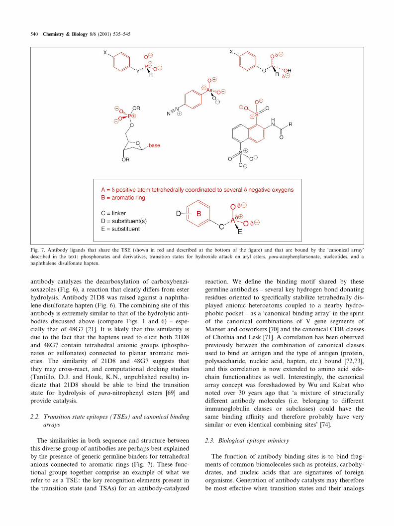
The similarities in both sequence and structure betweenthis diverse group of antibodies are perhaps best explainedby the presence of generic germline binders for tetrahedralanions connected to aromatic rings (Fig. 7). These func-tional groups together comprise an example of what werefer to as a TSE: the key recognition elements present inthe transition state (and TSAs) for an antibody-catalyzed
reaction. We de¢ne the binding motif shared by thesegermline antibodies ^ several key hydrogen bond donatingresidues oriented to speci¢cally stabilize tetrahedrally dis-played anionic heteroatoms coupled to a nearby hydro-phobic pocket ^ as a `canonical binding array' in the spiritof the canonical combinations of V gene segments ofManser and coworkers [70] and the canonical CDR classesof Chothia and Lesk [71]. A correlation has been observedpreviously between the combination of canonical classesused to bind an antigen and the type of antigen (protein,polysaccharide, nucleic acid, hapten, etc.) bound [72,73],and this correlation is now extended to amino acid side-chain functionalities as well. Interestingly, the canonicalarray concept was foreshadowed by Wu and Kabat whonoted over 30 years ago that `a mixture of structurallydi¡erent antibody molecules (i.e. belonging to di¡erentimmunoglobulin classes or subclasses) could have thesame binding a¤nity and therefore probably have verysimilar or even identical combining sites' [74].
2.3. Biological epitope mimicry
The function of antibody binding sites is to bind frag-ments of common biomolecules such as proteins, carbohy-drates, and nucleic acids that are signatures of foreignorganisms. Generation of antibody catalysts may thereforebe most e¡ective when transition states and their analogs
Fig. 7. Antibody ligands that share the TSE (shown in red and described at the bottom of the ¢gure) and that are bound by the `canonical array'described in the text: phosphonates and derivatives, transition states for hydroxide attack on aryl esters, para-azophenylarsonate, nucleotides, and anaphthalene disulfonate hapten.
CHBIOL 100 5-6-01 Cyaan Magenta Geel Zwart
540 Chemistry & Biology 8/6 (2001) 535^545

resemble naturally occurring biological molecules. In thisscenario, mature binding sites that are complementary totransition states for non-biological reactions are derivedfrom germline binding sites that are complementary tobiological epitopes. Phosphate and phosphodiester groupsof nucleic acids are biological epitopes which are mim-icked [75,76] by phosphonate, sulfonate, and arsonatehaptens. Interestingly, some of the most e¤cient antibodycatalysts known were raised against phosphonate TSA 1(Scheme 1) and promote the transesteri¢cation reactionshown in Scheme 1 to produce the corresponding ester 2with (kcat/Km)/kuncat values of approximately 108 and ef-fective molarities (kcat/kuncat) of approximately 105 [77^79].The similarity of hapten 1 to thymine dinucleotide is ob-vious.
Evidence exists for other cases of biological epitopemimicry as well. The three-dimensional structures of twoantibodies, 39-A11 and 1E9, that catalyze Diels^Alder re-actions have recently been determined to high resolution[14,80]. Stevens and Schultz [80] have noted the similarityin structure between 39-A11 and two other structurallyrelated antibodies, DB3 and TE33, and Houk, Hilvert,and Wilson [14] have discussed the relationships between39-A11, DB3, and 1E9 in detail. Antibody TE33 binds the15-residue cholera toxin peptide [81,82], which contains atype II L-turn when bound, and DB3 binds steroids suchas progesterone [80,83^85]. The L-turn structure of cholera
toxin has been shown to bind in a pocket similar to thosethat bind progesterone and the bicyclic ring systems ofTSA haptens 3 and 4 used to elicit 39-A11 and 1E9, re-spectively (Chart 1). The valine^proline^glycine sequenceinvolved in the L-turn and the hydrocarbon rings of thehaptens and steroids have all led to antibody hosts whichcontain deep hydrophobic pockets. Since type II L-turnsoften contain hydrophobic proline and glycine residues forgeometric reasons [86], a deep hydrophobic pocket wouldseem ideal as a generic binding site for these structures. Itis therefore plausible that generic L-turn binders in thenaive repertoire have led to TE33, DB3, 39-A11, and1E9, and the connections between the germline precursorsto these antibodies have been noted [14,80]. This is anelegantly simple case of biological epitope mimicry inwhich a host for a biological hydrophobic array has ledto catalysts for reactions that proceed through non-polartransition states.
2.4. A¤nity maturation and the development of speci¢city
The fact that the binding sites of antibodies 39-A11,1E9, TE33, and DB3 are similar does not necessarilymean that they will cross-react. While DB3 successfullybinds many di¡erent steroids [83^85], it does not acceler-ate the Diels^Alder reaction promoted by 1E9 to any ap-preciable extent [14]. Moreover, when screened against a
Chart 1.
Scheme 1.
CHBIOL 100 5-6-01 Cyaan Magenta Geel Zwart
Review Canonical binding arrays D.J. Tantillo, K.N. Houk 541
`panel of 72 structurally diverse hapten^BSA conjugates',the germline precursor to 39-A11 displayed broad cross-reactivity, while no cross-reactivity was observed for 39-A11, suggesting that germline `polyspeci¢city is temperedupon a¤nity maturation' [80]. These results emphasizethat the alterations to antibody combining sites that occurupon maturation, while often structurally subtle, can haveconsiderable consequences for cross-reactivity.
A similar loss of polyspeci¢city upon maturation hasbeen reported for anti-arsonate antibodies [70]. It was ob-served that antibodies isolated from early in the primaryimmune response to para-azophenylarsonate conjugatesbind single stranded DNA (the similarities between anti-arsonate, anti-DNA, and hydrolytic antibodies were dis-cussed above). Antibodies isolated during the secondaryimmune response, however, lacked this polyspeci¢city.
An elegant study of hydrolytic antibody 48G7 bySchultz and Stevens [25^29], in which this antibody andits germline precursor were characterized both biochemi-cally and structurally, provides great insights into thechemical details of a¤nity maturation. Mature 48G7 andits germline precursor di¡er by only nine mutations; theseincrease the a¤nity for hapten (a phosphonate TSA, Table1) by a factor of 104 and the rate of hydrolysis by a factorof 102 [26,27]. None of the nine mutated residues actuallycontacts the hapten, but these mutations in£uence detailsof the combining site structure by causing subtle reorien-tations of combining site functionalities (Fig. 1).
In light of experiments such as these which show thata¤nity maturation proceeds with relatively few mutations,much of a mature antibody's ability to bind a substratemust be encoded by the germline. The a¤nity maturationprocess tends to tighten binding through relatively few anddistant mutations [25^29,87], suggesting that the initialrecognition events in the immune response need only toinvolve relatively loose binding. The ability of the immuneresponse to produce antibody catalysts is most likely aresult of the fact that TSAs (and transition states them-selves) happen to ¢t into generic germline sites reasonablywell.
If antibodies adopt multiple conformations [88^90], thediversity of the naive repertoire is greatly increased [91]. Infact, it has been observed in several cases that conforma-tional changes upon binding in di¡erent antibodies lead tosimilar combining sites. This is an unusual and unexpectedtype of structural convergence.
In the case of hydrolytic antibodies D2.3, D2.4, andD2.5, crystal structure analysis did not reveal any signi¢-cant conformational changes upon hapten binding [52^54].However, subsequent kinetic experiments revealed that apre-equilibrium exists for each of these antibodies thatconverts between active and inactive conformers. Thisequilibrium is shifted towards the active conformer inthe presence of hapten [55], and it is the active conformerof each that displays the canonical binding array. It wasestimated that this conformational change leads to a 30^
170-fold increase in a¤nity for hapten. This situation sug-gests that the pressure of hapten binding can select for£exible antibodies capable of obtaining productive confor-mations in addition to those that are rigidly preorganizedfor binding.
The convergence of hydrolytic catalyst structures fromdi¡erent immunizations with di¡erent haptens was dis-cussed above [61,62]. In the case of antibody CNJ206, adrastically di¡erent antibody conformation was observedin the X-ray structures of free and hapten-bound antibody[30^33], an observation that is consistent with a conforma-tional change upon binding [32]. Again the canonical ar-ray was not necessarily preorganized in solution, but wasselected for in the presence of hapten.
A similar situation has been reported for antibodies tofoot-and-mouth disease virus [92]. Crystal structures oftwo di¡erent antibodies ^ complexed to their peptide anti-gens and antigen-free ^ showed that conformationalchanges upon antigen binding lead to a situation in which`the two Fab fragments are closer in structure in the com-plexes than in the unbound state' [92].
The extra diversity a¡orded the immune system by con-formational £exibility is likely to be most signi¢cant in thenaive repertoire. Based on their crystallographic studies,Schultz and Stevens suggested that the conformational£exibility of hydrolytic antibody 48G7 is considerablyless than that of its germline precursor [25^29], and recentmolecular dynamics calculations by Kollman and cowork-ers support this hypothesis [93].
The blind versatility of the immune system also oftenleads to combining site functionalities that can interactfavorably with elements of transition states not capturedby TSA haptens. Automated docking of quantum me-chanical transition states into antibody structures has pro-vided insights into catalysis by pinpointing speci¢c non-covalent interactions between transition states and anti-body combining sites [14^24]. In the case of hydrolyticantibodies 48G7, CNJ206, 17E8 and 29G11, this type ofanalysis (Tantillo, D.J. and Houk, K.N., unpublished re-sults) revealed that one of two possible enantiomeric path-ways for reaction (si and re attack of hydroxide ion onaryl esters) is often heavily favored in antibody combiningsites despite their selection for tight binding to symmetri-cal haptens. This selectivity arises from the inherent asym-metry of binding sites constructed from enantiopure ami-no acid residues, rather than elements of asymmetry in thehaptens.
In¢delity between haptens and the transition states thatthey are intended to mimic [69] might also be expected tolead to mature antibody combining sites that lack certainfunctional groups that could enhance catalysis, and this isexactly what is observed in many cases. However, func-tionality not engineered through hapten design has alsobeen found to interact with elements of transition states.For example, the increase in catalysis upon a¤nity matu-ration of antibody 48G7 can be largely explained by the
CHBIOL 100 5-6-01 Cyaan Magenta Geel Zwart
542 Chemistry & Biology 8/6 (2001) 535^545

interaction between the transition state and an additionalhydrogen bond acceptor (Tyr33H) upon maturation (seeFig. 1) [25^29,69]. The presence of a hydrogen bond ac-ceptor in a combining site elicited against a phosphonatehapten with no hydrogen bond donor functionality is sur-prising ^ in fact, this residue actually behaves as a hydro-gen bond donor when 48G7 is bound to its phosphonatehapten. These observations suggest that by displaying res-idues that may switch between donor and acceptor roles,the immune system again acquires increased diversity atno genetic cost. One might also consider the presence ofactive site nucleophilic residues, as found for antibodies43C9 [51] and 7C8 [60], to be similarly serendipitous.
3. Summary
The available experimental and theoretical evidence sug-gests that there is a direct connection between TSEs andcanonical binding arrays. Although a connection betweenhapten structure and mature antibody combining sites isnot at all surprising ^ it is the very assumption upon whichthe proposal of antibody catalysis was predicated [3] ^ theconnection between relatively small hapten substructuresand particular combining site residues is more intriguingand of greater utility for catalyst engineering. Accordingto our model, the origins of this correspondence can betraced to one or more of the following:
1. the presence of generic germline binders for particulartypes of functionality
2. biological epitope mimicry3. sampling of multiple antibody conformations
These principles provide new avenues for design bychemists of `input' that can be translated and expressedby nature in architectures compatible with transition statestabilization.
Acknowledgements
We are grateful to the National Science Foundation andthe National Institute of General Medical Sciences, Na-tional Institutes of Health for ¢nancial support of thisresearch.
References
[1] D.R. Burton, Monoclonal antibodies from combinatorial libraries,Acc. Chem. Res. 26 (1993) 405^411.
[2] J. Kuby, Immunology, 3rd edn., W.H. Freeman, New York, 1997.[3] W.P. Jencks, Catalysis in Chemistry and Enzymology, McGraw Hill,
New York, 1969, p. 288.[4] D. Hilvert, in: S.E. Denmark (Ed.) Topics in Stereochemistry, John
Wiley and Sons, New York, 1999, pp. 83^135.
[5] J.-L. Reymond, in: W.-D. Fessner (Ed.), Topics in Current Chemis-try, Springer-Verlag, Heidelberg, 1999, pp. 59^93.
[6] D.R. Liu, P.G. Schultz, Generating new molecular function: A lessonfrom nature, Angew. Chem. Int. Ed. Engl. 38 (1999) 36^54.
[7] D. Hilvert, G. MacBeath, J.A. Shin, in: S.M. Hecht (Ed.), BioorganicChemistry: Peptides and Proteins, Oxford University Press, NewYork, 1998, pp. 335^366.
[8] D.B. Smithrud, S.J. Benkovic, The state of antibody catalysis, Curr.Opin. Chem. Biol. 8 (1997) 459^466.
[9] A.J. Kirby, The potential of catalytic antibodies, Acta Chem. Scand.50 (1996) 203^210.
[10] P.G. Schultz, R.A. Lerner, From molecular diversity to catalysis ^lessons from the immune system, Science 269 (1995) 1835^1842.
[11] R.A. Lerner, S.J. Benkovic, P.G. Schultz, At the crossroads of chem-istry and immunology ^ catalytic antibodies, Science 252 (1991) 659^667.
[12] M.M. Mader, P.A. Bartlett, Binding energy and catalysis : the impli-cations for transition-state analogs and catalytic antibodies, Chem.Rev. 97 (1997) 1281^1301.
[13] D.J. Tantillo, J.G. Chen, K.N. Houk, Theozymes and compuzymes:Theoretical models for biological catalysis, Curr. Opin. Chem. Biol. 2(1998) 743^750.
[14] J. Xu, Q. Deng, J. Chen, K.N. Houk, J. Bartek, D. Hilvert, I.A.Wilson, Evolution of shape complementarity and catalytic e¤ciencyfrom a primordial antibody template, Science 286 (1999) 2345^2348.
[15] A. Heine, E.A. Stora, J.T. Yli-Kauhaluoma, C. Gao, Q. Deng, B.R.Beno, K.N. Houk, K.D. Janda, I.A. Wilson, An antibody exo Diels^Alderase inhibitor complex at 1.95 angstrom resolution, Science 279(1998) 1934^1940.
[16] O. Wiest, K.N. Houk, Stabilization of the transition state of thechorismate^prephenate rearrangement ^ an ab initio study of enzymeand antibody catalysis, J. Am. Chem. Soc. 117 (1995) 11628^11639.
[17] V.E. Gouverneur, K.N. Houk, B. De Pascual-Teresa, B. Beno, K.D.Janda, R.A. Lerner, Control of the exo-pathway and endo-pathwayof the Diels^Alder reaction by antibody catalysis, Science 262 (1993)204^208.
[18] K. Gruber, B. Zhou, K.N. Houk, R.A. Lerner, C.G. Shevlin, I.A.Wilson, Structural basis for antibody catalysis of a disfavored ringclosure reaction, Biochemistry 38 (1999) 7062^7074.
[19] J. Na, K.N. Houk, Predicting antibody catalyst selectivity from op-timum binding of catalytic groups to a hapten, J. Am. Chem. Soc.118 (1996) 9204^9205.
[20] J. Na, K.N. Houk, C.G. Shevlin, K.D. Janda, R.A. Lerner, Theenergetic advantage of 5-exo versus 6-endo epoxide openings ^ apreference overwhelmed by antibody catalysis, J. Am. Chem. Soc.115 (1993) 8453^8454.
[21] K. Hotta, H. Lange, D.J. Tantillo, K.N. Houk, D. Hilvert, I.A.Wilson, Catalysis of decarboxylation by a preorganized heterogenousmicroenvironment: Crystal structures of abzyme 21D8, J. Mol. Biol.302 (2000) 1213^1225.
[22] J.K. Lee, K.N. Houk, Cation-cyclization selectivity: Variable struc-tures of protonated cyclopropanes and selectivity control by catalyticantibodies, Angew. Chem. Int. Ed. Engl. 36 (1997) 1003^1005.
[23] J. Na, K.N. Houk, D. Hilvert, Transition state of the base-promotedring-opening of isoxazoles ^ theoretical prediction of catalytic func-tionalities and design of haptens for antibody production, J. Am.Chem. Soc. 118 (1996) 6462^6471.
[24] H. Zipse, G. Apaydin, K.N. Houk, A quantum mechanical and sta-tistical mechanical exploration of the thermal decarboxylation ofKemp's other acid (benzisoxazole-3-carboxylic acid) ^ the in£uenceof solvation on the transition state geometries and kinetic isotopee¡ects of a reaction with an awesome solvent e¡ect, J. Am. Chem.Soc. 117 (1995) 8608^8617.
[25] S.A. Lesley, P.A. Patten, P.G. Schultz, A genetic approach to thegeneration of antibodies with enhanced catalytic activities, Proc.Natl. Acad. Sci. USA 90 (1993) 1160^1165.
[26] P.A. Patten, N.S. Gray, P.L. Yang, C.B. Marks, G.J. Wedemayer,
CHBIOL 100 5-6-01 Cyaan Magenta Geel Zwart
Review Canonical binding arrays D.J. Tantillo, K.N. Houk 543

J.J. Boniface, R.C. Stevens, P.G. Schultz, The immunological evolu-tion of catalysis, Science 271 (1996) 1086^1091.
[27] G.J. Wedemayer, P.A. Patten, L.H. Wang, P.G. Schultz, R.C. Ste-vens, Structural insights into the evolution of an antibody combiningsite, Science 276 (1997) 1665^1669.
[28] G.J. Wedemayer, L.H. Wang, P.A. Patten, P.G. Schultz, R.C. Ste-vens, Crystal structures of the free and liganded form of an esterolyticcatalytic antibody, J. Mol. Biol. 268 (1997) 390^400.
[29] P.L. Yang, P.G. Schultz, Mutational analysis of the a¤nity matura-tion of antibody 48G7, J. Mol. Biol. 294 (1999) 1191^1201.
[30] R. Zemel, D.G. Schindler, D.S. Taw¢k, Z. Eshhar, B.S. Green, Dif-ferences in the biochemical properties of esterolytic antibodies corre-late with structural diversity, Mol. Immunol. 31 (1994) 127^137.
[31] B. Golinelli-Pimpaneau, B. Gigant, T. Bizebard, J. Navaza, P. Sal-udjian, R. Zemel, D.S. Taw¢k, Z. Eshhar, B.S. Green, M. Knossow,Crystal structure of a catalytic antibody Fab with esterase-like activ-ity, Structure 2 (1994) 175^183.
[32] J.-B. Charbonnier, B. Golinelli-Pimpaneau, B. Gigant, B.S. Green,M. Knossow, pH in£uences on the crystal structures and mechanisticproperties of a hydrolytic antibody, Isr. J. Chem. 36 (1996) 143^149.
[33] B. Gigant, J.-B. Charbonnier, B. Golinelli-Pimpaneau, R.R. Zemel,Z. Eshhar, B.S. Green, M. Knossow, Mechanism of inactivation of acatalytic antibody by p-nitrophenyl esters, Eur. J. Biochem. 246(1997) 471^476.
[34] J. Guo, W. Huang, T.S. Scanlan, Kinetic and mechanistic character-ization of an e¤cient hydrolytic antibody ^ evidence for the forma-tion of an acyl intermediate, J. Am. Chem. Soc. 116 (1994) 6062^6069.
[35] G.W. Zhou, J. Guo, W. Huang, R.J. Fletterick, T.S. Scanlan, Crystalstructure of a catalytic antibody with a serine protease active site,Science 265 (1994) 1059^1064.
[36] J. Guo, W. Huang, G.W. Zhou, R.J. Fletterick, T.S. Scanlan, Mech-anistically di¡erent catalytic antibodies obtained from immunizationwith a single transition-state analog, Proc. Natl. Acad. Sci. USA 92(1995) 1694^1698.
[37] H. Wade, T.S. Scanlan, P1^S1 interactions control the enantioselec-tivity and hydrolytic activity of the norleucine phenylesterase cata-lytic antibody 17E8, J. Am. Chem. Soc. 118 (1996) 6510^6511.
[38] T. Fox, T.S. Scanlan, P.A. Kollman, Ligand binding in the catalyticantibody 17E8. A free energy perturbation calculation study, J. Am.Chem. Soc. 119 (1997) 11571^11577.
[39] M. Baca, T.S. Scanlan, R.C. Stephensen, J.A. Wells, Phage display ofa catalytic antibody to optimize a¤nity for transition-state analogbinding, Proc. Natl. Acad. Sci. USA 94 (1997) 10063^10068.
[40] J.L. Buchbinder, R.C. Stephenson, T.S. Scanlan, R.J. Fletterick, Acomparison of the crystallographic structures of two catalytic anti-bodies with esterase activity, J. Mol. Biol. 282 (1998) 1033^1041.
[41] H. Wade, T.S. Scanlan, Remote binding energy in antibody catalysis :Studies of a catalytically unoptimized speci¢city pocket, J. Am.Chem. Soc. 121 (1999) 1434^1443.
[42] H. Wade, T.S. Scanlan, Expression of binding energy on an antibodyreaction coordinate, J. Am. Chem. Soc. 121 (1999) 11935^11941.
[43] S.J. Benkovic, J.A. Adams, C.L. Borders Jr., K.D. Janda, R.A. Ler-ner, The enzymic nature of antibody catalysis ^ development of mul-tistep kinetic processing, Science 250 (1990) 1135^1139.
[44] R.A. Gibbs, P.A. Benkovic, K.D. Janda, R.A. Lerner, S.J. Benkovic,Substituent e¡ects on an antibody-catalyzed hydrolysis of phenylesters ^ further evidence for an acyl^antibody intermediate, J. Am.Chem. Soc. 114 (1992) 3528^3534.
[45] J.D. Stewart, L.J. Liotta, S.J. Benkovic, Reaction mechanisms dis-played by catalytic antibodies, Acc. Chem. Res. 26 (1993) 396^404.
[46] V.A. Roberts, J. Stewart, S.J. Benkovic, E.D. Getzo¡, Catalytic anti-body model and mutagenesis implicate arginine transition-state sta-bilization, J. Mol. Biol. 235 (1994) 1098^1116.
[47] J.D. Stewart, V.A. Roberts, M.W. Crowder, E.D. Getzo¡, S.J. Ben-kovic, Creation of a novel biosensor for Zn(II), J. Am. Chem. Soc.116 (1994) 415^416.
[48] M.W. Crowder, J.D. Stewart, V.A. Roberts, C.J. Bender, E.Tevelrakh, J. Peisach, E.D. Getzo¡, B.J. Ga¡ney, S.J. Benkovic,Spectroscopic studies on the designed metal-binding sites of the43C9 single chain antibody, J. Am. Chem. Soc. 117 (1995) 5627^5634.
[49] G.P. Miller, B.A. Posner, S.J. Benkovic, Expanding the 43C9 class ofcatalytic antibodies using a chain-shu¥ing approach, Bioorg. Med.Chem. Lett. 5 (1997) 581^590.
[50] K.D. Janda, D. Schloeder, S.J. Benkovic, R.A. Lerner, Induction ofan antibody that catalyzes the hydrolysis of an amide bond, Science241 (1988) 1188^1191.
[51] M.M. Thayer, E.H. Olender, A.S. Arvai, C.K. Koike, I.L. Canestrel-li, J.D. Stewart, S.J. Benkovic, E.D. Getzo¡, V.A. Roberts, Structur-al basis for amide hydrolysis catalyzed by the 43C9 antibody, J. Mol.Biol. 291 (1999) 329^345.
[52] S.-H. Kim, D.G. Schindler, A.B. Lindner, D.S. Taw¢k, Z. Eshhar,Expression and characterization of recombinant single-chain Fv andFv fragments derived from a set of catalytic antibodies, Mol. Immu-nol. 34 (1997) 891^906.
[53] J.-B. Charbonnier, B. Golinelli-Pimpaneau, B. Gigant, D.S. Taw¢k,R. Chap, D.G. Schindler, S.-H. Kim, B.S. Green, Z. Eshhar, M.Knossow, Structural convergence in the active sites of a family ofcatalytic antibodies, Science 275 (1997) 1140^1142.
[54] B. Gigant, J.-B. Charbonnier, Z. Eshhar, B.S. Green, M. Knossow,X-ray structures of a hydrolytic antibody and of complexes elucidatecatalytic pathway from substrate binding and transition state stabili-zation through water attack and product release, Proc. Natl. Acad.Sci. USA 94 (1997) 7857^7861.
[55] A.B. Lindner, Z. Eshhar, D.S. Taw¢k, Conformational changes a¡ectbinding and catalysis by ester-hydrolysing antibodies, J. Mol. Biol.285 (1999) 421^430.
[56] H. Miyashita, T. Hara, R. Tanimura, F. Tanaka, M. Kikuchi, I.Fujii, A common ancestry for multiple catalytic antibodies generatedagainst a single transition-state analog, Proc. Natl. Acad. Sci. USA91 (1994) 6045^6049.
[57] I. Fujii, F. Tanaka, H. Miyashita, R. Tanimura, K. Kinoshita, Cor-relation between antigen-combining-site structures and functions witha panel of catalytic antibodies generated against a single transitionstate analog, J. Am. Chem. Soc. 117 (1995) 6199^6209.
[58] H. Miyashita, T. Hara, R. Tanimura, S. Fukuyama, C. Cagnon, A.Kohara, I. Fujii, Site-directed mutagenesis of active site contact res-idues in a hydrolytic abzyme: Evidence for an essential histidineinvolved in transition state stabilization, J. Mol. Biol. 267 (1997)1247^1257.
[59] O. Kristensen, D.G. Vassylyev, F. Tanaka, K. Morikawa, I. Fujii, Astructural basis for transition-state stabilization in antibody-catalyzedhydrolysis : Crystal structures of an abzyme at 1.8 angstrom resolu-tion, J. Mol. Biol. 281 (1998) 501^511.
[60] B. Gigant, T. Tsumuraya, I. Fujii, M. Knossow, Diverse structuralsolutions to catalysis in a family of antibodies, Structure 7 (1999)1385^1393.
[61] G. MacBeath, D. Hilvert, Hydrolytic antibodies ^ variations on atheme, Chem. Biol. 3 (1996) 433^445.
[62] J.B. Charbonnier, B. Gigant, B. Golinelli-Pimpaneau, M. Knossow,Similarities of hydrolytic antibodies revealed by their x-ray struc-tures: A review, Biochimie 79 (1997) 653^660.
[63] D.R. Rose, R.K. Strong, M.N. Margolies, M.L. Gefter, G.A. Petsko,Crystal structure of the antigen-binding fragment of the murine anti-arsonate monoclonal antibody 36-71 at 2.9-Aî resolution, Proc. Natl.Acad. Sci. USA 87 (1990) 338^342.
[64] R.K. Strong, R. Campbell, D.R. Rose, G.A. Petsko, J. Sharon, M.N.Margolies, 3-Dimensional structure of murine anti-para-azophenyl-arsonate Fab-36-71. 1. X-ray crystallography, site-directed mutagen-esis, and modeling of the complex with hapten, Biochemistry 30(1991) 3739^3748.
[65] R.K. Strong, G.A. Petsko, J. Sharon, M.N. Margolies, 3-Dimension-al structure of murine anti-para-azophenylarsonate Fab-36-71. 2.
CHBIOL 100 5-6-01 Cyaan Magenta Geel Zwart
544 Chemistry & Biology 8/6 (2001) 535^545

Structural basis of hapten binding and idiotypy, Biochemistry 30(1991) 3749^3757.
[66] M. Shlomchik, M. Mascelli, H. Shan, M.Z. Radic, D. Pisetsky, A.Marshakrothstein, M. Weigert, Anti-DNA antibodies from autoim-mune mice arise by clonal expansion and somatic mutation, J. Exp.Med. 171 (1990) 265^297.
[67] J.N. Herron, X.M. He, D.W. Ballard, P.R. Blier, P.E. Pace, A.L.M.Bothwell, E.W. Voss Jr., A.B. Edmundson, An autoantibody to sin-gle-stranded DNA ^ comparison of the 3-dimensional structures ofthe unliganded Fab and a deoxynucleotide Fab complex, ProteinsStruct. Funct. Genet. 11 (1991) 159^175.
[68] A.L. Gibson, J.N. Herron, D.W. Ballard, E.W. Voss Jr., X.M. He,V.A. Patrick, A.B. Edmundson, Crystallographic characterization ofthe Fab fragment of a monoclonal anti-ss-DNA antibody, Mol. Im-munol. 22 (1985) 499^502.
[69] D.J. Tantillo, K.N. Houk, Fidelity in hapten design: How analogousare phosphonate haptens to the transition states for alkaline hydrol-yses of aryl esters?, J. Org. Chem. 64 (1999) 3066^3076.
[70] T. Manser, L.J. Wysocki, M.N. Morgolies, M.L. Gefter, Evolution ofantibody variable region structure during the immune response, Im-munol. Rev. 96 (1987) 141^162.
[71] C. Chothia, A.M. Lesk, Canonical structures for the hypervariableregions of immunoglobulins, J. Mol. Biol. 196 (1987) 901^917.
[72] E. Vargas-Madrazo, F. Lara-Ochoa, J.C. Almagro, Canonical struc-ture repertoire of the antigen-binding site of immunoglobulins sug-gests strong geometrical restrictions associated to the mechanism ofimmune recognition, J. Mol. Biol. 254 (1995) 497^504.
[73] F. Lara-Ochoa, J.C. Almagro, E. Vargas-Madrazo, M. Conrad, Anti-body^antigen recognition: A canonical structure paradigm, J. Mol.Evol. 43 (1996) 678^684.
[74] T.T. Wu, E.A. Kabat, Analysis of the sequences of the variableregions of Bence Jones proteins and myeloma light chains and theirimplications for antibody complementarity, J. Exp. Med. 132 (1970)211^249.
[75] H.J. Ditzel, S.M. Barbas, C.F. Barbas, D.R. Burton, The nature ofthe autoimmune antibody repertoire in human immunode¢ciencyvirus type 1 infection, Proc. Natl. Acad. Sci. USA 91 (1994) 3710^3714.
[76] S.L. Harris, L. Craig, J.S. Mehroke, M. Rashed, M.B. Zwick, K.Kenar, E.J. Toone, N. Greenspan, F.-I. Auzanneau, J.-R. Marino-Albernas, B.M. Pinto, J.K. Scott, Exploring the basis of peptide^carbohydrate crossreactivity : Evidence for discrimination by peptidesbetween closely related anti-carbohydrate antibodies, Proc. Natl.Acad. Sci. USA 94 (1997) 2454^2459.
[77] J.R. Jacobsen, J.R. Prudent, L. Kochersperger, S. Yonkovich, P.G.Schultz, An e¤cient antibody-catalyzed aminoacylation reaction, Sci-ence 256 (1992) 365^367.
[78] R.C. Stevens, L.C. Hsieh-Wilson, B.D. Santarsiero, G.J. Wedemayer,B. Spiller, L.H. Wang, D. Barnes, H.D. Ulrich, P.A. Patten, F.E.
Romesberg, P.G. Schultz, Structural studies of catalytic antibodies,Isr. J. Chem. 36 (1996) 121^132.
[79] E.M. Driggers, C.W. Liu, D.E. Wemmer, P.G. Schultz, Structure ofthe Michaelis complex of an e¤cient antibody acyl transferase deter-mined by transferred nuclear Overhauser enhancement spectroscopy,J. Am. Chem. Soc. 120 (1998) 7395^7396.
[80] F.E. Romesberg, B. Spiller, P.G. Schultz, Immunological origins ofbinding and catalysis in a Diels^Alderase antibody, Science 279(1998) 1923^1929.
[81] M. Shoham, Crystal structure of an anticholera toxin peptide com-plex at 2.3-angstrom, J. Mol. Biol. 232 (1993) 1169^1175.
[82] T. Scherf, R. Hilles, F. Naider, M. Levitt, J. Anglister, Induced pep-tide conformations in di¡erent antibody complexes ^ molecular mod-eling of the 3-dimensional structure of peptide antibody complexesusing NMR-derived distance restraints, Biochemistry 31 (1992) 6884^6897.
[83] J.H. Arevalo, M.J. Taussig, I.A. Wilson, Molecular basis of cross-reactivity and the limits of antibody antigen complementarity, Nature365 (1993) 859^863.
[84] J.H. Arevalo, E.A. Stura, M.J. Taussig, I.A. Wilson, 3-Dimensionalstructure of an anti-steroid Fab' and progesterone Fab' complex,J. Mol. Biol. 231 (1993) 103^118.
[85] J.H. Arevalo, C.A. Hassig, E.A. Stura, M.J. Sims, M.J. Taussig, I.A.Wilson, Structural analysis of antibody speci¢city ^ detailed compar-ison of ¢ve Fab'^steroid complexes, J. Mol. Biol. 241 (1994) 663^690.
[86] D. Voet, J.G. Voet, Biochemistry, 2nd edn., Wiley, New York, 1995,p. 152.
[87] P.S. Daugherty, G. Chen, B.L. Iverson, G. Georgiou, Quantitativeanalysis of the e¡ect of the mutation frequency on the a¤nity mat-uration of single chain Fv antibodies, Proc. Natl. Acad. Sci. USA 97(2000) 2029^2034.
[88] C. Frieden, Kinetic aspects of regulation of metabolic processes.Hysteretic enzyme concept, J. Biol. Chem. 245 (1970) 5788^5799.
[89] I.A. Wilson, R.L. Stan¢eld, Antibody^antigen interactions ^ newstructures and new conformational changes, Curr. Opin. Struct.Biol. 4 (1994) 857^867.
[90] U.-B. Hansson, C. Wingren, U. Alkner, Conformational isomerismof IgG antibodies, Biochim. Biophys. Acta 1340 (1997) 53^62.
[91] E.A. Padlan, Anatomy of the antibody molecule, Mol. Immunol. 31(1994) 169^217.
[92] N. Verdaguer, N. Sevilla, M.L. Valero, D. Stuart, E. Brocchi, D.Andreu, E. Giralt, E. Domingo, M.G. Mateu, I. Fita, A similarpattern of interaction for di¡erent antibodies with a major antigenicsite of foot-and-mouth disease virus: Implications for intratypic anti-genic variation, J. Virol. 72 (1998) 739^748.
[93] L.T. Chong, Y. Duan, L. Wang, I. Massova, P.A. Kollman, Molec-ular dynamics and free-energy calculations applied to a¤nity matu-ration in antibody 48G7, Proc. Natl. Acad. Sci. USA 96 (1999)14330^14335.
CHBIOL 100 5-6-01 Cyaan Magenta Geel Zwart
Review Canonical binding arrays D.J. Tantillo, K.N. Houk 545


![[pgr]-Conjugated Anions: From Carbon-Rich Anions to](https://img.dokumen.tips/doc/110x75/62887182fd628c47fb7ebde3/pgr-conjugated-anions-from-carbon-rich-anions-to-.jpg)


