Embed Size (px)
Citation preview

1
Cannabidiol improves brain and liver function in a fulminant hepatic failure-
induced model of hepatic encephalopathy in mice*
Y Avraham1*, N C Grigoriadis2, T Poutahidis3, L Vorobiev1, I Magen1, Y Ilan4, R
Mechoulam5 and EM Berry1
1Department of Human Nutrition and Metabolism, Braun School of Public Health,
Hadassah-Hebrew University Medical School, Jerusalem, Israel 91120 2Department of Neurology, AHEPA University Hospital, Aristotle University of
Thessaloniki, Greece, 54636 3Deparment of Pathology, Veterinary Medical School, Aristotle University of
Thessaloniki, Greece, 4The Liver Unit, Hadassah University Hospital, Ein Kerem, Jerusalem, Israel 91120
Jerusalem, Israel 91120. 5Department of Medicinal Chemistry and Natural Products, Medical Faculty, Hebrew
University Jerusalem, Israel 91120.
Short title: CBD moderates fulminant hepatic failure
*Corresponding author: Dr. Yosefa Avraham
Fax: +972-2-6431105; Phone: 972-2-6757547
E-mail: [email protected]
Word Count: 6126 Figures: 6 Tables: 0 No conflict of interest
This is an Accepted Article that has been peer-reviewed and approved for publication in the British Journal of Pharmacology, but has yet to undergo copy-editing and proof correction. Please cite this article as an “Accepted Article”; doi: 10.1111/j.1476-5381.2010.01179.x
2
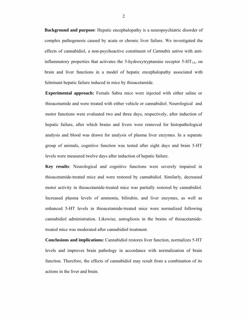
Background and purpose: Hepatic encephalopathy is a neuropsychiatric disorder of
complex pathogenesis caused by acute or chronic liver failure. We investigated the
effects of cannabidiol, a non-psychoactive constituent of Cannabis sativa with anti-
inflammatory properties that activates the 5-hydroxytryptamine receptor 5-HT1A, on
brain and liver functions in a model of hepatic encephalopathy associated with
fulminant hepatic failure induced in mice by thioacetamide.
Experimental approach: Female Sabra mice were injected with either saline or
thioacetamide and were treated with either vehicle or cannabidiol. Neurological and
motor functions were evaluated two and three days, respectively, after induction of
hepatic failure, after which brains and livers were removed for histopathological
analysis and blood was drawn for analysis of plasma liver enzymes. In a separate
group of animals, cognitive function was tested after eight days and brain 5-HT
levels were measured twelve days after induction of hepatic failure.
Key results: Neurological and cognitive functions were severely impaired in
thioacetamide-treated mice and were restored by cannabidiol. Similarly, decreased
motor activity in thioacetamide-treated mice was partially restored by cannabidiol.
Increased plasma levels of ammonia, bilirubin, and liver enzymes, as well as
enhanced 5-HT levels in thioacetamide-treated mice were normalized following
cannabidiol administration. Likewise, astrogliosis in the brains of thioacetamide-
treated mice was moderated after cannabidiol treatment.
Conclusions and implications: Cannabidiol restores liver function, normalizes 5-HT
levels and improves brain pathology in accordance with normalization of brain
function. Therefore, the effects of cannabidiol may result from a combination of its
actions in the liver and brain.
3
Keywords: hepatic encephalopathy; cannabidiol; cognition; liver enzymes;
thioacetamide;
Abbreviations: ALT, alanine transaminase; AST, aspartate transaminase; AUC, area
under the curve; CBD, cannabidiol; FHF, fulminant hepatic failure; GFAP, glial
fibrillary acidic protein; HE, hepatic encephalopathy; 5-HT, 5-hydroxytryptamine;
TAA, thioacetamide; NS, normal saline

4
Introduction
Hepatic encephalopathy (HE) is a syndrome observed in patients with end-stage liver
disease. It is defined as a spectrum of neuropsychiatric abnormalities in patients with
liver dysfunction, after exclusion of other known brain diseases, and is characterized
by personality changes, intellectual impairments, and a depressed level of
consciousness associated with multiple neurotransmitter systems, astrocyte
dysfunction and cerebral perfusion (Riggio et al., 2005; Avraham et al.,2006;
2008a,2009; Magen et al.,2008; Butterworth, 2010). Subtle signs of HE are observed
in nearly 70% of patients with cirrhosis and approximately 30% of patients dying of
end-stage liver disease experience significant encephalopathy (Ferenci,1995). HE,
accompanying the acute onset of severe hepatic dysfunction, is the hallmark of
fulminant hepatic failure (FHF) and patients with HE have been reported to have
elevated levels of ammonia in their blood (Stahl, 1963). In addition, the infiltration of
TNF-α-secreting monocytes into the brain of bile-duct ligated mice, a model of
chronic liver disease, has been found 10 days after the ligation, indicating that
neuroinflammation is involved in the pathogenesis of HE. This infiltration was
shown to be associated with activation of the cerebral endothelium and an increase in
the expression of adhesion molecules ( Κerfoot et al., 2006 ).
Cannabidiol (CBD) is a non-psychoactive ingredient of Cannabis sativa (Izzo et al.,
2009). Many mechanisms have been suggested for its action, such as agonism of 5-
HT1A receptors (Russo et al., 2005). It also has a very strong anti-inflammatory
activity both in vivo, as an anti-arthritic therapeutic (Malfait et al., 2000; Durst et
al.,2007), and in vitro, manifested by inhibition of cytokine production in immune
cells (Ben Shabat et al., 2006). The finding that CBD is devoid of any psychotrophic
effects combined with its anti-inflammatory activity makes it a promising tool for

5
treating HE, which is exacerbated by an inflammatory response (Shawcross et al.,
2004). In the present work, we aimed to explore the effects of CBD in the acute
model of HE induced by the hepatotoxin thioacetamide (TAA), focusing on brain
function, brain pathology and 5-HT levels, liver function and pathology as possible
targets for therapeutic effects of CBD.
Methods
Mice
Female Sabra mice (34-36g), 8 to 10 weeks old, were assigned at random to different
groups of 10 mice per cage and were used in all experiments. All cages contained
wood-chip bedding and were placed in a temperature-controlled room at 22°C, on a
12 h light/dark cycle (lights on at 07h00min). The mice had free access to water 24 h
a day. The food provided was Purina chow and the animals were maintained in the
animal facility (SPF unit) of the Hebrew University Hadassah Medical School,
Jerusalem. Mice were killed after each treatment by decapitation between 10h00min
and 12h00min. Animals were kept at the animal facility in accordance with NIH
guidelines and all experiments were approved by the institutional animal use and care
committee, No. MD -89.52-4.
Induction of hepatic failure
We adapted the rat model of acute liver failure induced by thioacetamide (TAA) to
mice (Zimmermann et al ., 1989). The TAA model in mice has been extensively
validated previously (Honda et al., 2002; Fernandez-Martinez A et al., 2004; Schnur
et al., 2004). TAA was obtained from Sigma–Aldrich (Rehovot, Israel) in powder
form and dissolved in sterile normal saline solution (NS); it was injected i.p. as a
single dose of 200mg kg -1. Vehicle (NS) was also administered in a separate group of
animals that served as controls. Twenty-four hours after injection of TAA all animals

6
(including control) were injected s.c. with 0.5 ml of a solution containing 0.45%
NaCl, 5% dextrose and 0.2% KCl in order to prevent hypovolaemia, hypokalaemia
and hypoglycaemia. The mice were intermittently exposed to infrared light in order to
prevent hypothermia.
Administration of CBD
CBD was extracted from cannabis resin (hashish) and purified as previously reported
(Gaoni et al.,1971) and was dissolved in a vehicle solution consisting of ethanol,
emulphor and saline at a ratio of 1:1:18, respectively, and was injected in a single
dose of 5mg kg-1 i.p, one day after either NS or TAA treatment. Similarly, the CBD
related vehicle (the same mixture without CBD) was administered at the same time
points following either NS or TAA treatment. A dose of 5mg kg-1 cannabidiol was
chosen based on the studies done by Magen et al. (2009, 2010) and on preliminary
experiments done in our laboratory, which demonstrated that this does produced a maximal
effect compared to 1 and 10mg kg-1. Four groups of animals were studied: control
naïve animals treated with either CBD or its vehicle, and corresponding TAA-treated
animals.
Assessment of neurological function
Neurological function was assessed by a 10 point scale based on reflexes and task
performance (Chen et al., 1996): exit from a 1 metre in diameter circle in less than 1
min, seeking, walking a straight line, startle reflex, grasping reflex, righting reflex,
placing reflex, corneal reflex, maintaining balance on a beam 3, 2 and 1 cm in width,
climbing onto a square and a round pole. For each task failed or abnormal reflex
reaction a score of 1 was assigned. Thus, a higher score indicates poorer neurological
function. The neurological score was assessed one day after induction of hepatic
failure by TAA (day 2). The mice were then divided between treatment groups so that
all groups had similar baseline neurological scores after TAA induction. The post-

7
treatment neurological score was assessed one day after administration of CBD or
vehicle (day 3).
Assessment of activity
The activity test was performed two days after the induction of hepatic failure.
Activity of two mice was measured simultaneously for a 5 min period. Two mice
were tested together to lower stress to the minimum, as it has been shown that
separation of mice induces stress (van Leeuwen et al.,1997; Hao et al.,2001). Activity
was assessed in the open field (20x30cm field divided into 12 squares of equal size)
as described previously (Fride and Mechoulam., 1993). Locomotor activity was
recorded by counting the number of crossings by the mice at 1 min intervals.
Results are presented as the mean number of crossings min-1.
Cognitive function
Cognitive function studies were performed eight days after the induction of hepatic
failure. The animals were placed in an eight arm maze, which is a scaled-down
version of that developed for rats (Pick and Yanai, 1983,). Mice were deprived of
water 2 h prior to the test and a reward of 50µl of water was presented at the end of
each arm, in order to motivate them to perform the task. Animals were divided
between treatment groups so that all groups had similar baselines neurological scores
after TAA induction. The mice were tested (no. of entries) until they made entries into
all eight arms or until they completed 24 entries, whichever came first. Hence, the
lower the score the better the cognitive function. Food and water were given at the
completion of the test. Maze performance was calculated on each day for five
consecutive days. Results are presented as area under the curve (AUC) utilizing the
formula: (day 2 + day 3 + day 4 + day 5) - 4*(day 1) (Pick and Yanai, 1983,).
Brain histopathology and immunohistochemistry

8
Two days after the induction of hepatic failure, mice were killed by decapitation and
brains were excised and fixed in 4% neutral- buffered paraformaldehyde.
The brain was cut along the midline and separated into 2 pieces containing brain and
cerebellum hemispheres. Both sections were embedded en block in paraffin and 6μm
saggital sections were adhered to slides. Serial sections were taken in 15 groups of
slides (10 slides each, 3 sections per slide) at 100μm intervals. These slides were used
for glial fibrillary acidic protein (GFAP) immunohistochemistry (a total of 90
sections), according to standard protocol. Briefly, paraffin sections were
deparaffinized and hydrated in xylene and alcohol solutions, rinsed with TBS. Citrate
buffer (pH.6) was used for antigen retrieval. The endogenous peroxidase was blocked
with H2O2 (0,3% in PBS). Sections were then incubated in blocking buffer for 1h. A
series of reselected sections were then treated with primary antibody against GFAP
(1:2500, DakoCytomation, Denmark), overnight at 4°C, and then with goat anti-rabbit
(1:200, Vector Burligame, CA, USA) as secondary antibody. Immunoreactions were
visualized with the avidin – biotin complex (Vectastain) and the peroxidase reaction
was visualized with diaminobenzidine (DAB) (Vector), as chromogen. Sections were
finally counterstained with haematoxylin and examined under light microscope (Zeiss
Axioplan 2). Images were captured with a digital camera (NIKON DS-5Mc-L1)
mounted on microscope. Astrocytes were evaluated at the hippocampal area of both
hemispheres. A total of 5-7 randomly selected visual fields, per hemisphere section,
were evaluated. A square with 100 square subdivisions each of 3721 μm2 as defined
by an occular morphometric grid adjusted at the prefrontal lens, was centered at each
visual field. The number of GFAP-positive astrocytes per mm2 was evaluated. Only
those cells with an identifiable nucleus, were counted. In addition, in an attempt to
evaluate the level of activation of the astrocytes (cell size, extension of cell processes)

9
the number of small square subdivisions with a positive GFAP signal and their % of
the total number of square subdivisions counted, was calculated.
Two independent observers who were blinded to sample identity performed all
quantitative assessments. In cases where significant discrepancies were obvious
between the two observers, the evaluation was repeated by a third one.
Liver histopathology
Two days after the induction of hepatic failure, mice were killed by decapitation and
their livers were excised and fixed in 4% neutral- buffered paraformaldehyde.
Liver histopathological analysis and scoring of necrosis (coagulative, centrilobular)
were performed as described previously (Avraham et al,2008a).
Serum ammonia, liver enzymes and bilirubin levels
Serum for ALT, AST, bilirubin and ammonia measurements was obtained on day 3 in
glass tubes, centrifuged, and analysed on the day of sampling using a Kone Progress
Selective Chemistry Analyzer (Kone Instruments, Espoo, Finland). All serum samples
were processed in the same laboratory using the same methods and the same reference
values.
5-HT synthesis
On day 12, mice killed by decapitation and their brains were dissected out for
determination of 5-HT levels. The assays for 5-HT were performed by standard
alumina extraction, and HPLC with electrochemical detection using
dehydroxybenzylamine (DHBA) as an internal standard (Avraham et al, 2006).
Experimental design
Experiment 1 On the first day of this experiment, 20 mice were administered with
TAA (200mg kg-1) and 20 saline. The following day, neurological evaluation was

10
performed and saline-treated and TAA-treated mice were each assigned to two
different sub-groups with approximately equal neurological score, which were
administered either saline or CBD (5mg kg-1). On the third day, mice were evaluated
for neurological and locomotor function, after which they were killed and their brains
and livers were dissected out and fixed with 4% formaldehyde. Blood was drawn and
separated for plasma, in which liver enzymes were quantified.
Experiment 2 This was identical to experiment 1 on days 1-3, only the mice were not
killed on day 3 but were evaluated for cognitive function using the eight arm maze
test, on days 8-12. On day 12, the mice were killed and their livers and brains were
dissected out for determination of 5-HT levels.
Statistical analysis
All data are expressed as mean ±SEM. Statistical analysis was performed using one-
way ANOVA followed by Bonferroni’s post hoc test.
Results
Neurological score
TAA significantly increased the neurological score of mice compared to the control
group (Figure 1; one way ANOVA: F(3,29)=43.19, P<0.0001; Bonferroni: P<0.01).
Administration of 5mg kg-1 CBD to TAA-treated mice improved the neurological
score compared to TAA alone (1±0.15; P<0.01). CBD did not affect the score of the
control animals.
Activity
TAA decreased the activity level of the mice (Figure 2; ANOVA: F(3,29)=64.18,
P<0.0001; Bonferroni: P<0.01) and CBD administration significantly increased the
activity level in these TAA mice, compared to the untreated TAA mice (P<0.01).
CBD did not affect the activity of control animals.

11
Cognitive function
Cognitive function was significantly impaired following TAA exposure, as reflected
by the higher AUC values (Figure 3; ANOVA: F(3,22)=7.22, P=0.001; Bonferroni:
P<0.05) and this was improved following CBD administration (P<0.01 vs TAA only).
CBD did not affect the cognitive function of control animals.
Brain and liver histopathology
Figure 4 shows images of slices from brains of animals from the control group (A),
control + CBD group (B), TAA group (C) and TAA + CBD group (D),
immunostained for the detection of astrogliosis. Astrogliosis was observed in visual
fields studied in TAA animals, as evident both by the increase in the number of
GFAP-positive cells mm-2 (Figure 4E; ANOVA: F(3,298)=26.8, P<0.001; Bonferroni:
P<0.001) and by the increased % of GFAP-positive surface (Figure 4F; ANOVA:
F(3,298)=19.21, P<0.001; Bonferroni: P<0.001). Both parameters were unaffected in
CBD-treated controls (Figure 4E, F). However, the number of GFAP-positive cells
mm-2 in TAA+5mg kg-1 CBD treated animals was reduced compared to TAA-treated
animals (Figure 4E; Bonferroni: P=0.002). In contrast, CBD had no effect on the % of
GFAP-positive surface in TAA animals (Figure 4F). Overall, it seems that TAA
administration increased the number of activated astrocytes and CBD significantly
reduced this effect. However, astrocytes in both CBD- and vehicle–treated TAA
animals did not differ as regards their cellular size or extension of processes.
TAA-treated animals showed the typical TAA-induced liver necrosis lesions that have
been described in detail previously (Avraham et al,2008a). The statistical analysis of
liver histopathology scores did not reveal significant differences in the extent and
severity of necrotic lesions between CBD-treated and untreated mice (data not
shown).

12
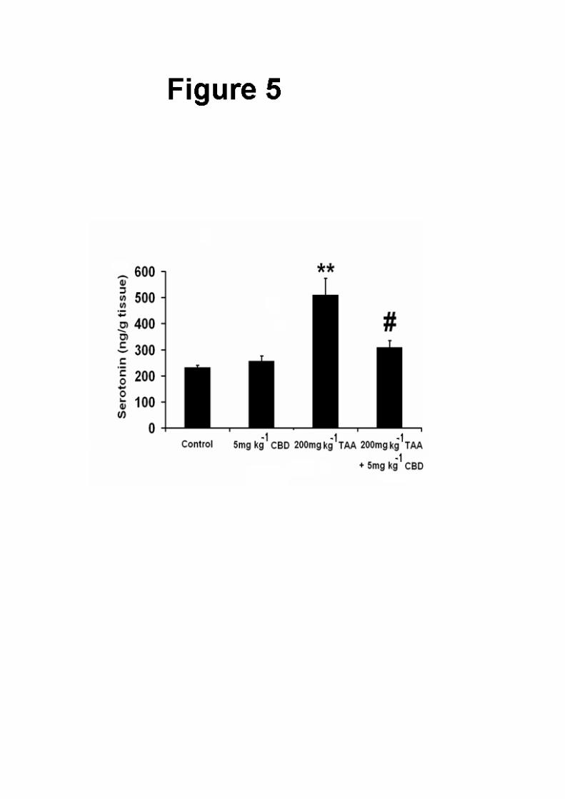
5-HT levels
Whole brain 5-HT levels were increased by TAA administration (Figure 5; ANOVA:
F(3,17)=13.46, P<0.0001; Bonferroni: P<0.01), and CBD partially restored the levels in
TAA-treated animals (P<0.01 vs TAA only). CBD did not affect the levels of 5-HT in
control animals.
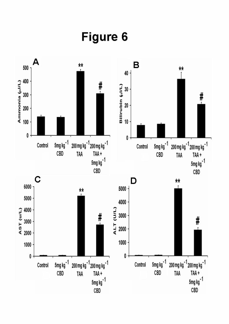
Liver function
The levels of ammonia (Figure 6A), bilirubin (Figure 6B) and the liver enzymes AST
(Figure 6C) and ALT (Figure 6D) were increased after TAA administration
(ammonia: ANOVA: F(3,26)=156.93, P<0.0001; Bonferroni: P<0.01 vs control;
bilirubin: ANOVA: F(3,27)=34.99, P<0.0001; Bonferroni: P<0.01 vs control; AST:
ANOVA: F(3,27)=590.84, P<0.0001; Bonferroni: P<0.01 vs control; ALT: ANOVA:
F(3,27)=314.95, P<0.0001; Bonferroni: P<0.01 vs. control). CBD partially restored all
of these indices in TAA-treated animals (P<0.01 vs TAA only for all parameters).
CBD did not affect the levels of any of these substances in control animals.
Discussion
Thioacetamide administration induces acute liver failure which leads to CNS changes
related to those seen in HE (Zimmerman et al, 1989, Avraham et al, 2006; 2008a;
2009; Magen et al,2008). The hepatotoxicity of TAA is due to the generation of free
radicals and oxidative stress (Zimmerman et al, 1989). However, it is not clear
whether TAA affects the brain directly or the liver (Albrecht et al ., 1996). In
previous studies both a CB1 antagonist and a CB2 or TRPV1 agonist have been
shown to ameliorate the brain and liver damage that occurs in liver disease and HE
(Mallet and Lotersztajn,2008; Avraham et al,2006; 2008a,b; 2009). Also cannabidiol,
an agonist of the 5-HT1A receptor, was found to ameliorate brain damage in a chronic

13
model of HE induced by bile duct ligation. Hence, we investigated the potential of
cannabidiol as a treatment for HE induced by FHF. Our results indicated that it has a
neuroprotective role in HE induced by FHF; cannabidiol was found to restore liver
function, normalize 5-HT levels and improve the brain pathology in accordance with
normalization of brain function. We also showed that cannabidiol affects both
central functions : neurological score, motor and cognitive functions, brain 5-HT
levels as well as astrogliosis and peripheral functions: reduced liver enzymes,
ammonia and bilirubin. Therefore, we conclude that it acts both centrally and
peripherally. In addition it has been shown that cannabidiol can cross the BBB
and act centrally (for review see Pertwee ,2009). Therefore, its effect may
result from a combination of its actions in the liver and the brain. However, to
elucidate its mechanism of action future experiments are needed to determine
the effects of central administration of cannabidiol .
Previous work from our laboratory has demonstrated an impaired neurological and
motor function 3 days, and impaired cognition 12 days after TAA injection to mice
(Avraham et al, 2006; 2008a; 2009). These results were reproduced in the present
study (Figures 1-3). In a more recent study from our laboratory, cognitive and motor
deficits were observed 21 days after bile-duct ligation (BDL), a chronic model of liver
disease (Magen et al, 2009a, b). The different durations of the development of HE
symptoms in the two models apparently result from their different characteristics – an
acute vs a chronic model of HE. In the latter model, cannabidiol was found to
improve cognition and locomotor activity, in accordance with our present data
(Magen et al, 2009). However, in sharp contrast to the findings reported here no
evidence for astrogliosis was found in that study (data not reported); in our acute
14
model induced by TAA we observed astrogliosis after three days (Figure 4C). Those
mice with histopathological alterations displayed an increased neurological score and
decreased activity level, and 5mg kg-1 CBD reversed both the increase in the number
of GFAP(+) cells, an index of neuroinflammation (Figure 4E), and the neurological
and locomotor impairments (Figure 1 and 2), suggesting a link between
neuroinflammation and motor and neurological deficits. Similar results were reported
by Jover et al.(2006) who demonstrated a decrease in motor activity in bile-duct
ligated rats on a high-protein diet, in association with astrogliosis, and by Cauli et
al(2009), who reported that treatment with an anti-inflammatory restored the motor
activity in HE. In the work of Jover et al. (2006), astrogliosis was found only in the
bile-duct ligated rats on a high protein diet, but not in the bile-duct ligated rats on a
regular diet, similar to our previous findings (Avraham et al,2009) This suggests
that TAA causes more severe damage to the brain than bile-duct ligation, and that
hyperammonaemia is required to worsen the damage in BDL rats to an extent that is
equivalent to that observed in TAA mice. The reason for this may be that in chronic
liver disease induced by BDL, compensation mechanisms are activated, which
moderate the brain damage, while in the acute model induced by TAA, no such
mechanisms can come into action because of the severity of the liver insult and the
short interval of time between the induction of liver damage and the histopathological
examination.
Kerfoot et al. (2006) showed the infiltration of peripheral monocytes into the brain of
bile-duct ligated mice ten days after the ligation and suggested that this infiltration
may cause the activation of inflammatory cells in the brain. Therefore, it is
conceivable that such a mechanism was responsible for the astrogliosis observed in
our study, since we found evidence of liver inflammation (data not shown). As

15
evident from the histopathology results, cannabidiol did not appear to
affect the development of TAA-induced necrotic lesions in the liver of
mice. However, the levels of liver transaminases in the serum of
cannabidiol-treated mice were significantly reduced compared to their
untreated counterparts, indicating that this substance contributed to a
partial restoration of liver function. Recent evidence elucidating the
complicated mechanisms involved in the release of hepatocyte cytosolic
enzymes such as ALT and AST in the blood may explain the discrepancy
between histopathology and serum biochemistry data observed in the
present study. Indeed, it is now generally accepted that the release of
cytosolic enzymes during both the reversible and irreversible phases of
hepatocyte injury and, therefore, their appearance in blood does not
necessarily indicate cell death and also that enzyme release during
reversible cell damage occurs with an apparent lack of histological
evidence of necrosis (Solter, 2005). Following this reasoning, it could be
hypothesized that although cannabidiol did not reduce the levels of
histologically detectable necrosis, it may have ameliorated the minute
reversible hepatocyte damage that causes the so-called “leakage” of
cytoplasmic ALT and AST in blood. The interaction between hyperammonaemia
and inflammation as a precipitating factor for HE has been discussed in two recent
reviews (Shawcross and Jalan ,2005 ;Wright and Jalan ,2007). Further work

16
is required to reveal the exact mechanism/s of the manner by which liver damage is
related to dysfunction/damage in the brain, and studies using antagonists of the A2A
adenosine receptors, which are potential targets of cannabidiol that may mediate its
anti-inflammatory effect (Carrier et al, 2006), need to be carried out in order to
elucidate the receptors involved in this effect.
Astrogliosis has also been shown to be involved in learning and memory deficits in a
mouse model of Alzheimer's disease. In this study, astrogliosis was reduced by caloric
restriction, which also reversed the cognitive deficits and increased the expression of
neurogenesis in related genes (Wu et al, 2008). Further studies, such as expression
analysis of such genes using DNA microarray and evaluation of neurogenesis using
BrdU staining, needs to be performed in order to explore the mechanisms through
which TAA-induced astrogliosis impairs cognition, and through which cannabidiol
acts to improve it.
Even though astrogliosis was found a week before cognitive function was observed,
and it is not definite whether it was long-lasting, this mechanism seems, in our eyes,
to account for the cognitive dysfunction, rather than the increase in 5-HT level (Figure
5). The latter mechanism does not seem to be related to the cognitive dysfunction,
even though this increase in 5-HT was reversed by CBD (Figure 5), as 5-HT
depletion, not increase, has been shown to cause memory deficits in the eight arm
maze (Mazer et al., 1997). On the other hand, there is much evidence that
ammonia induces astrocyte swelling which via a number of mechanisms
leads to impaired astrocyte/neuronal communication and synaptic
plasticity, thereby resulting in a disturbance of oscillatory networks. The
latter accounts for the symptoms of hepatic encephalopathy (for review

17
see, Haussinger and Gorg, 2010), among them presumably the cognitive
dysfunction.
An increased level of 5-HT in the brain of rats after TAA administration was reported
by Yurdaydin et al.(1990). In addition, there is indirect evidence that this increase is
related to decreased motor activity, as the nonselective 5-HT receptor antagonist
methysergide increased motor activity in TAA-injected rats, while the selective 5-HT2
receptor antagonist seganserin did not (Yurdaydin et al, 1996). Likewise, we found
that the level of 5-HT was increased following TAA administration and this was
restored after cannabidiol treatment (Figure 5). In parallel, motor activity was
decreased following TAA injection and increased after cannabidiol treatment,
indicating a link between the increase in 5-HT and decrease in motor activity. Hence,
it seems that CBD reversed the increased 5-HT level in the brains of TAA mice and
thus reversed the decrease in their motor activity. A possible mechanism can be
activation of 5-HT1A receptors by CBD (Russo et al, 2005), as these receptors have
been reported to inhibit 5-HT synthesis (Invernizzi et al,1991). We have shown that
the effects of cannabidiol in a chronic model of HE, bile duct ligation, are
mediated via the 5-HT1A receptors (Magen et al., 2010) and in an earlier study
with the same model we demonstrated that the effects of cannabidiol can be
also mediated via A2A adenosine receptors (Magen et al., 2009). Thus, the
effects of cannabidiol, can be mediated by 5-HT1A or/and A2A adenosine
receptors. We think that in the current study the effects of cannabidiol were
mediated by the 5-HT1A receptor since activation of the receptor by cannabidiol
caused depletion of 5-HT (Figure 5). In our previous studies we showed that
18
cognition is multifactorial and not dependent only on 5-HT levels, therefore
there is no direct correlation between cognition and 5-HT levels.
The reversal of astrogliosis was probably related to reduced hepatic toxin
formation. Indeed, there is much evidence that ammonia induces astrocyte
swelling, which via a number of mechanisms leads to impaired astrocyte/neuronal
communication and synaptic plasticity, thereby resulting in a disturbance of
oscillatory networks. The
latter accounts for the symptoms of hepatic encephalopathy (for review see,
Haussinger and Gorg, 2010), among them presumably the cognitive dysfunction.
Neurological and motor functions were improved two and three days,
respectively, after induction of hepatic failure at the same time as a partial
reversal of the astrogliosis and reduced levels of ammonia, bilirubin and liver
enzymes were noticed. It seems that the behavioural effects of cannabidiol are
dramatic and occur within 3 days.
In summary, the present study demonstrates the therapeutic effects of CBD in an
acute model of HE. It appears that this effect of CBD is multi-factorial and involves
cannabinoid (Avraham et al, 2006), vanilloid (Avraham et al, 2008a; 2009) and 5-
HT1A receptors (Magen et al,2009b). CBD improves the symptoms of FHF by
affecting both brain histopathology and liver function, and thus may serve as
therapeutic agent for treating human HE.
Acknowledgement
We would like to thank the Israel Science Foundation for their support
Financial disclosure: none
19
References
Alexander SPH, Mathie A, Peters JA (2008).Guide to Receptors and Channels
(GRAC), 3rd edition (2008 revision). Br J Pharmacol 153(Suppl.2):S1-S209.
Albrecht J, Hilgier W, Januszewski S, Quack G. (1996) “Contrasting effects of
thioacetamide-induced liver damage on the brain uptake indices of ornithine, arginine
and lysine: modulation by treatment with ornithine aspartate”. Metab Brain Dis.
;11:229-237.
Avraham Y, Israeli E, Gabbay E, Okun A, Zolotarev O, Silberman I, et al. (2006)
Endocannabinoids affect neurological and cognitive function in thioacetamide-
induced hepatic encephalopathy in mice. Neurobiol Dis; 21: 237-245.
Avraham Y, Zolotarev O, Grigoriadis NC, Poutahidis T, Magen I, Vorobiav L, et al.
(2008) Cannabinoids and Capsaicin improve liver function following Thioacetamide
–induced acute injury in mice. Am J Gastroenterol (2008a);103: 3047-3056.
Avraham Y, Grigoriadis N, Pautahidis T, Magen I, Vorobiav L, Zolotarev O, et al.
(2009) Capsaicin affects brain function in a model of hepatic encephalopathy
associated with fulminant hepatic failure in mice. Br J Pharmacol 158, 896‐906.
Avraham Y, Magen I, Zolotarev O, Vorobiav L, Pappo O, Ilan Y et al. (2008b). 2-
arachidonoylglycerol, an endogenous cannabinoid receptor agonist, in various rat
tissues during the eveloution of experimental cholestatic liver disease . Prostaglandins
Leukot Essent Fatty Acids. 79:35-40.
Ben-Shabat S, Hanuš LO, Katzavian G, Gallily R. (2006) New cannabidiol
derivatives: synthesis, binding to cannabinoid receptor, and evaluation of their
antiinflammatory activity. J Med Chem; 49: 1113-1117.

20
Butterworth RF. (2010) Altered glial-neuronal crosstalk: Cornerstone in the
pathogenesis of hepatic encephalopathy. Neurochem Int. Mar 27. [Epub ahead of
print]
Carrier EJ, Auchampach JA, Hillard CJ.(2006) Inhibition of an equilibrative
nucleoside transporter by cannabidiol: a mechanism of cannabinoid
immunosuppression. Proc Natl Acad Sci U S A;103: 7895-7900.
Cauli O, Rodrigo R, Piedrafita B, Llansola M, Mansouri MT, Felipo V et al. (2009)
Neuroinflammation contributes to hypokinesia in rats with hepatic encephalopathy:
Ibuprofen restores its motor activity. J Neurosci Res; 87: 1369-1374.
Chen Y, Constantini S, Trembovler V, Weinstock M, Shohami E. (1996) An
experimental model of closed head injury in mice: pathophysiology, histopathology,
and cognitive deficits. J Neurotrauma; 13: 557-568.
Durst R, Danenberg H, Gallily R, Mechoulam R, Meir K, Grad E, et al. (2007)
Cannabidiol, a nonpsychoactive Cannabis constituent, protects against myocardial
ischemic reperfusion injury. Am J Physiol Heart Circ Physiol; 293: H3602-607.
Ferenci P. Hepatic encephalopathy. In: Haubrich WS, Schaffner F, Berk JE,
eds. Bockus Gastroenterology. 5th ed. Philadelphia, Pa: WB Saunders; (1995): 1998-
2003.
Fernández-Martínez A, Callejas NA, Casado M, Boscá L, Martín-Sanz P. (2004)
Thioacetamide-induced liver regeneration involves the expression of cyclooxygenase
2 and nitric oxide synthase 2 in hepatocytes. J Hepatol;40: 963-970.
Fride E, Mechoulam R. (1993) Pharmacological activity of the cannabinoid receptor
agonist, anandamide, a brain constituent. Eur J Pharmacol;231: 313-314.

21
Gaoni Y, Mechoulam R. (1971) The isolation and structure of delta-1-
tetrahydrocannabinol and other neutral cannabinoids from hashish. J Am Chem Soc;
93: 217-224.
Hao S, Avraham Y, Bonne O, Berry EM. (2001) Separation-induced body weight
loss, impairment in alternation behavior, and autonomic tone: effects of tyrosine.
Pharmacol Biochem Behav;68: 273-281.
Häussinger D, Görg B. (2010) Interaction of oxidative stress, astrocyte swelling and
cerebral ammonia toxicity. Curr Opin Clin Nutr Metab Care; 13(1):87-92.
Honda H, Ikejima K, Hirose M, Yoshikawa M, Lang T, Enomoto N, et al. (2002)
Leptin is required for fibrogenic responses induced by thioacetamide in the murine
liver. Hepatology; 36: 12-21.
Invernizzi R, Carli M, Di Clemente A, Samanin R. (1991) Administration of 8-
hydroxy-2-(Di-n-propylamino)tetralin in raphe nuclei dorsalis and medianus reduces
serotonin synthesis in the rat brain: differences in potency and regional sensitivity. J
Neurochem; 56: 243-247.
Izzo AA, Borrelli F, Capasso R, Di Marzo V, Mechoulam R. (2009) Non-
psychotropic plant cannabinoids: new therapeutic opportunities from an ancient herb.
Trends Pharmacol Sci. 30(10):515-527. Review.
Jover R, Rodrigo R, Felipo V, Insausti R, Sáez-Valero J, García-Ayllón MS, et al.
(2006) Brain edema and inflammatory activation in bile duct ligated rats with diet-
induced hyperammonemia: A model of hepatic encephalopathy in cirrhosis.
Hepatology;43: 1257-1266

22
Kerfoot SM, D'Mello C, Nguyen H, Ajuebor MN, Kubes P, Le T, et al. (2006) TNF-α
secreting monocytes are recruited into the brain of cholestatic mice. Hepatology;43:
154-162.
Mallat A, Lotersztajn S. (2008 )Cannabinoid receptors as therapeutic targets in the
management of liver diseases. Drug News Perspect. Sep;21(7):363-8. Review.
Olton DS, Samuelson RJ. (1976) Remembrance of places passed: Spatial memory in
rats. J Exp Psychology: Anim Behav Proc;2: 97–116.
Magen I, Avraham Y, Ackerman Z, Vorobiev L, Mechoulam R, Berry EM.( 2009)
Cannabidiol ameliorates cognitive and motor impairments in mice with bile duct
ligation. J Hepatol 51(3):528-534.
Magen I , Avraham Y, Ackerman Z, Vorobiav L, Mechoulam R, and Berry EM.
(2010)Cannabidiol ameliorates cognitive and motor impairments in bile-duct ligated
mice via 5-HT1A receptor activation. Brit J Pharmacol ;159:950-957.
Magen I , Avraham Y, Berry EM, and Mechoulam R .(2008)Endocannabinoids in liver
disease and hepatic encephalopathy. Curr Pharm Des.; 14(23):2362-2369. Review.
Malfait AM, Gallily R, Sumariwalla PF, Malik AS, Andreakos E, Mechoulam R, et
al. The nonpsychoactive cannabis constituent cannabidiol is an oral anti-arthritic
therapeutic in murine collagen-induced arthritis. Proc Natl Acad Sci U S A. 2000;97:
9561-9566.
Mazer C, Muneyyirci J, Taheny K, Raio N, Borella A, Whitaker-Azmitia P.
(1997)Serotonin depletion during synaptogenesis leads to decreased synaptic density
and learning deficits in the adult rat: a possible model of neuro developmental
disorders with cognitive deficits. Brain Res; 760: 68-73.
23
Osei-Hyiaman D, Liu J, Zhou L, Godlewski G, Harvey-White J, Jeong WI, et
al(2008) Hepatic CB1 receptor is required for development of diet-induced steatosis,
dyslipidemia, and insulin and leptin resistance in mice. J Clin Invest. ;118(9):3160-
3169.
Pertwee RG.,(2009). Emerging strategies for exploiting cannabinoid receptor
agonists as medicines. British J Pharmacol.156,397-411
Pick CG, Yanai J. (1983) Eight arm maze for mice. Int J Neurosci; 21: 63-66.
Riggio O, Efrati C, Catalano C, Pediconi F, Mecarelli O, Accornero N, et al.
(2005) High prevalence of spontaneous portal-systemic shunts in persistent hepatic
encephalopathy: a case-control study. Hepatology ;42: 1158-1165.
Russo EB, Burnett A, Hall B, Parker KK. (2005)Agonistic properties of cannabidiol
at 5-HT1A receptors. Neurochem Res;30: 1037-1043.
Schnur J, Oláh J, Szepesi A, Nagy P, Thorgeirsson SS. (2004) Thioacetamide-
induced hepatic fibrosis in transforming growth factor beta-1 transgenic mice. Eur J
Gastroenterol Hepatol;16: 127-133.
Shawcross DL, Davies NA, Williams R, Jalan R. (2004) Systemic inflammatory
response exacerbates the neuropsychological effects of induced hyperammonemia in
cirrhosis. J Hepatol;40: 247-254.
Shawcross D, Jalan R. (2005) The pathophysiologic basis of hepatic encephalopathy:
central role for ammonia and inflammation. Cell Mol Life Sci;62: 2295-2304. Review.
Stahl J. Studies of the blood ammonia in liver disease. (1963) Its diagnostic,
prognostic, and therapeutic significance. Ann Intern Med. ; 58: 1-24.
Solter PF. Clinical pathology approaches to hepatic injury. Toxicol Pathol.
2005;33(1):9-16.

24
van Leeuwen SD, Bonne OB, Avraham Y, Berry EM. (1997) Separation as a new
animal model for self-induced weight loss. Physiol Behav; 62: 77-81.
Wright G, Jalan R. (2007)Ammonia and inflammation in the pathogenesis of hepatic
encephalopathy: Pandora's box? Hepatology.;46: 291-294.
Wu P, Shen Q, Dong S, Xu Z, Tsien JZ, Hu Y. (2008)Calorie restriction ameliorates
neurodegenerative phenotypes in forebrain-specific presenilin-1 and presenilin-2
double knockout mice. Neurobiol Aging; 29: 1502-1511.
Yurdaydin C, Hörtnagl H, Steindl P, Zimmermann C, Pifl C, Singer EA, et al.(1990)
Increased serotonergic and noradrenergic activity in hepatic encephalopathy in rats
with thioacetamide-induced acute liver failure. Hepatology;12: 695-700.
Yurdaydin C, Herneth AM, Püspök A, Steindl P, Singer E, Ferenci P. (1996)
Modulation of hepatic encephalopathy in rats with thioacetamide-induced acute liver
failure by serotonin antagonists. Eur J Gastroenterol Hepatol;8: 667-671.
Zimmermann C, Ferenci P, Pifl C, Yurdaydin C, Ebner J, Lassmann H, et al. (1989
)Hepatic encephalopathy in thioacetamide-induced acute liver failure in rats:
characterization of an improved model and study of amino acid-ergic
neurotransmission. Hepatology; 9: 594-601.
Figure legends
Figure 1 Neurological function, evaluated two days after induction of hepatic failure,
was impaired in TAA mice and was restored by CBD. **P<0.01 vs control, #P<0.01
vs TAA.

25
Figure 2 Locomotor function, evaluated three days after induction of hepatic failure,
was decreased in TAA mice and was restored by CBD. **P<0.01 vs control, #P<0.01
vs TAA.
Figure 3 Cognitive function, tested eight days after induction of hepatic failure, was
impaired following TAA and was improved by CBD. *P<0.05 vs control, #P<0.01 vs
TAA.
Figure 4 GFAP immunohistochemistry indicating the astrocytic reaction throughout
the parahippocampal area in naïve controls (A, B) and TAA – treated animals (C,D)
following treatment with vehicle (A,C) or CBD (B,D). CBD treatment had no effect
on the astrocytic activation of naïve animals. However, in the case of animals with
HE, CBD treatment induced significant reduction in the total number of activated
astrocytes, although the level of individual cell activation was not impaired. (E)
Quantification of GFAP-positive cells mm-2; the number was reduced in TAA mice
treated with 5mg kg-1 CBD compared to TAA mice treated with vehicle. ***P<0.001
vs control, #P<0.01 vs TAA. (F) Quantification of GFAP-positive surface in μm2;
5mg kg-1 CBD had no effect on the GFAP-positive surface in the brains of TAA-
treated mice. ***P<0.001 vs. control. Scale bars: 100μm.
Figure 5 Brain 5-HT levels, measured twelve days after induction of hepatic failure,
were increased in the brains of TAA mice and were restored by CBD.
Figure 6 Indices of liver function. The levels of ammonia (A), bilirubin (B), AST (C)
and ALT (D) were all increased in the plasma of TAA mice and were all reversed by
CBD. **P<0.01 vs control, #P<0.01 vs TAA.