Embed Size (px)
Citation preview

Cancer Cluster Investigation within the Mission Memorial Hospital Laboratory Final Report
March 31, 2006 Dr. George Astrakianakis Dr. Phil Bigelow Dr. Trevor Corneil Dr. Victor Omelchenko Dr. Malcolm Steinberg Dr. Annalee Yassi Dr. Shicheng Yu
Occupational Health & Safety Agency for Healthcare in BC #301-1195 West Broadway, Vancouver, BC V6H 3X5


Executive Summary This investigation was conducted in response to concerns expressed by employees of the Mission
Memorial Hospital (MMH) Laboratory that they were experiencing a high incidence of cancer.
The investigation resulted in an initial report prepared by the Occupational Health & Safety
Agency for Healthcare in BC (OHSAH) and released for comment in March, 2004 (Attachments
2 & 3). The associated presentation is included as Attachment 4. In addition, Attachments 5, 6
and 7 are supportive documents from the initial investigation. Subsequently a new breast cancer
case was identified and errors in staffing levels were corrected. A re-analysis of the breast cancer
incidence rate was completed in April, 2005 (Attachments 8 & 9) and a revised Draft Report was
released in September, 2005. The revised Draft Report still did not address all of the concerns
raised by the MMH Laboratory employees and resulted in a set of critical questions being posed
which were presented to the Fraser Health Authority (FH) and OHSAH in November of 2005
(Attachment 10). The responses to these questions for which OHSAH was responsible were sent
to FH and the Health Sciences Association (HSA) in January, 2006 (Attachment 11) and were
presented at MMH on February 8th, 2006 to representatives of FH, HSA, the BC Nurses’ Union
(BCNU) and the Hospital Employees’ Union (HEU). Some of the questions posed by the
Laboratory employees were responded to by FH and are included as a separate document. This
Final Report is therefore a compilation of the investigation of cancer incidence at the MMH
Laboratory and the results of an extensive consultation process with the employer, labour
representatives, and the individuals involved.
The results of this investigation are presented in three parts: a comprehensive review of the
literature, an epidemiologic cluster analysis, and an occupational exposure investigation. The
investigation procedures followed the established guidelines by the BC Cancer Agency (BCCA)
for cancer cluster investigations1. These guidelines are in keeping with international
approaches2.
In summary, 64 individuals were identified as having worked in the laboratory between January
1, 1970 and December 7, 2004. Information on health status and diagnosis of cancer were
obtained through personal interviews with employees. One person was diagnosed with cancer 1 Guidelines for the Investigation of Cancer Clusters in BC. BC Cancer Agency, Cancer Control Research, November 1998. 2 Guidelines for Investigating Clusters of Health Events. US CDC, July 27, 1990.
ii

prior to working in the MMH laboratory and was excluded from the analysis. Of the remaining
employees, ten employees reported a cancer diagnosis, of which seven were breast cancer. A
total of 974 person-years of observation were available for the data analysis after excluding one
subject because of diagnosis of cancer prior to start of employment. Based on the age and
calendar-year adjusted rates for the BC population, the expected number of breast cancer cases in
the women was 0.8 and the expected number of all cancers for all employees was 2.3. The
Standard Incidence Ratios (SIR), which is the observed number of cases divided by the expected
number, was found to be 8.4 for breast cancer among women at the MMH Laboratory, and 4.7
for all cancers among both men and women at the lab. In other words, the risk for breast cancer
was over 8 times the expected rate; and the rate of all cancers was over 4 times the expected rate.
The 95 percent confidence intervals indicate that both findings were significant. It can be
concluded that the perception of the laboratory workers that they were experiencing an excess in
cancer was confirmed – i.e. this is truly an observed cancer cluster.
The risk of developing breast cancer was also analyzed by the age at first employment at the
MMH Laboratory, the subjects’ length of time at work prior to diagnosis and by their job title.
The most important result was that no association was found between breast cancer risk and
either the age of first employment or the duration of exposure. However, there was a non-
significant increase in risk by job title with ‘technician’ being at greater risk than the grouping
‘aid, clerk, or ECG technician’.
A walk-through survey of the laboratory in its present state did not identify any potentially
hazardous exposures for which control measures are not in place. Review of indoor air quality
records and chemical assessment of carcinogens in the workplace also did not show any obvious
and extreme exposures in the past (based on current scientific literature), which could be related
to the increase in risk. Assessment of radiation exposure in the laboratory was also found to be at
typical natural background and would not contribute measurably to increased cancer risk. Thus,
while it can not be ruled out that workplace factors played some role in the complicated process
of carcinogenesis that led to this tragic outcome for laboratory workers and their families, the
exact relationship between workplace exposures and the cancers that resulted remains elusive.
The evidence collected to date does not allow us to reach scientific conclusions to support the
association between work-related exposures and breast cancer in this cluster. However, this
iii

report has confirmed that this is indeed a statistical significant cluster. This usually points to the
need to follow up with an etiological study with the required statistical power to investigate for
this association while controlling for other non-work related exposures. Prior to embarking on
such a study however, there is a need to establish an etiological hypothesis based on scientific
evidence that provides proposed mechanism(s) for breast cancer causation. Our review of the
literature was unable to establish the basis for such a hypothesis, as we did not find any scientific
evidence for the plausibility of a laboratory work-related etiological hypothesis regarding breast
cancer. While dioxins from the incinerator stack emissions have been implicated with other
cancers, these did not include breast cancers; despite the potential exposure of MMH Laboratory
workers to these emissions. Moreover, the number of people who worked in the Mission
Hospital Laboratory is not sufficiently large to provide an adequate sample size for an etiological
investigation.
Thus, it is recommended that this specific cancer cluster investigation be closed and the analysis
updated in five years. If new evidence emerges to support a disease causation hypothesis for
laboratory work-related breast cancer, and a larger study with an adequate sample size can be
designed, then this subject could be investigated further at that time.
It is important to understand that human beings are exposed to carcinogens in almost all
environments, at home, at work, and even walking in the sunshine. Every effort should be made,
in this and all workplaces, to ensure that the workplace remains as safe and free of carcinogenic
exposures as possible, and that the workforce is able to pursue safe and healthy choices in all
aspects of their lives.
iv

Forward This report is the culmination of work conducted by numerous individuals, either on staff at
OHSAH, serving as an OHSAH consultant or as a UBC trainee on rotation at OHSAH. Key
personnel include the authors, Phil Bigelow, Shicheng Yu, Trevor Corneil, Victor Omelchenko,
Malcolm Steinberg, and George Astrakianakis. We acknowledge the strong support and
assistance of the BC Cancer Agency (Drs Nhu Le, Greg Hislop, and Malcolm Hayes), the School
of Occupational and Environmental Hygiene at the University of British Columbia (Dr. Paul
Demers), the Fraser Health Authority (Mr. Dave Keen and Ms. Rosemary Nemanishen), and the
Health Sciences Association (Mr. Marty Lovick and Ms. Bev Banfield).
Dr. Annalee Yassi, MD MSc Executive Director OHSAH March 31, 2006
v

Table of Contents Executive Summary ........................................................................................................................ ii Forward ........................................................................................................................................... v Table of Contents........................................................................................................................... vi Introduction..................................................................................................................................... 1 Methods........................................................................................................................................... 1
Review of Literature ................................................................................................................... 2 Analysis of Cancer Incidence Data: Epidemiology and Statistics............................................. 2 Field Investigations: Possible Exposures to Potentially Carcinogenic Substances or Physical Agents ......................................................................................................................................... 4
Literature Review............................................................................................................................ 4 Breast Cancer Epidemiology ...................................................................................................... 4 Breast Cancer Risk Factors......................................................................................................... 8
Sociodemographic Factors...................................................................................................... 8 Inherited Risk Factors ........................................................................................................... 10 Breast Conditions.................................................................................................................. 11 Hormone Factors................................................................................................................... 11 Prevention of Breast Cancer ................................................................................................. 16 Occupational and Environmental Factors in Breast Cancer ................................................. 17 Breast Cancer Risk among Laboratory Workers .................................................................. 21 Incinerator Emissions and Cancer ........................................................................................ 25 Cancer Cluster Investigations ............................................................................................... 28 Investigating Cancer Clusters: Methods and limitations ..................................................... 29
Analysis of Mission Memorial Hospital Laboratory Cancer Incidence Data............................... 33 Events in the Mission Memorial Health laboratory cancer cluster investigation ..................... 33 Cancer Cluster at the Mission Memorial Hospital Laboratory................................................. 33 Field Investigations: Potential Exposures to Potentially Carcinogenic Substances or Physical Agents ....................................................................................................................................... 36
Conclusions................................................................................................................................... 37 Summary ................................................................................................................................... 37 Recommendations..................................................................................................................... 39
References..................................................................................................................................... 41 • Attachment 1: Field Investigations: potential exposures to potentially carcinogenic
substances or physical agents (conducted in August 2003) • Attachment 2 and 3: Initial epidemiological and statistical analysis of the cancer
incidence data (March 2004). • Attachment 4: presentation to the MMH Laboratory employees and staff (March 2004) • Attachment 5: Indoor air quality assessment (November 2004) • Attachment 6: Radiation exposure assessment of laboratory area for 70 days (June-
August 2004) • Attachment 7: Chemical assessment of Carcinogens (June 2004) • Attachment 8: Research update (March 2005) • Attachment 9: Presentation to the MMH Laboratory employees and staff (May 2005) • Attachment 10:Critical questions posed by MMH Laboratory personnel (November 8,
2005)
vi

• Attachment 11A: OHSAH responses to critical questions (January 9th, 2006) • Attachment(Separate Document): FH responses to critical questions

Introduction The Occupational Health and Safety Agency for Healthcare (OHSAH) was invited by the Fraser
Health Authority (FHA) to investigate concerns of a greater than expected number of cancer
cases in the Mission Memorial Hospital (MMH) Laboratory. In addition to the seemingly high
total number of cancer cases, a large proportion were the same type (breast cancer) thus further
highlighting the need for an investigation. Occupational health professionals from FHA had
completed some exploratory work on the project but felt they needed the help of outside experts
to resolve the issues. OHSAH contacted the Health Sciences Association (HSA), the union
representing the lab workers, and ascertained that they were in agreement with OHSAH taking
the task and requested us to proceed. The resultant cancer cluster investigation conducted by
OHSAH is the focus of this report.
A cancer cluster is the observation of a higher than usual occurrence of cases of the same type of
cancer, or all cancers, within a geographic location over a specified period of time. The purpose
of a cancer cluster investigation is to determine if the observed number of cases is statistically
higher than expected. If the observed incidence rate is higher, then the investigation should
review if the subjects involved were exposed to a potential causative agent that can be identified,
and if the investigation should progress to a more systematic review of exposure and incidence.
In BC, these investigations are conducted following guidelines recommended by the BC Cancer
Agency in keeping with protocol used in other jurisdictions across North America 1-4.
Prior to the investigation, an explanatory meeting was held at MMH to discuss the incidence of
cancer at MMH Laboratory and the protocol for the investigation. Details of the protocol used
during the investigation are provided below.
Methods The specific aim of this study was to provide a determination if an excess in the number of
cancer cases had occurred in the MMH Laboratory and to provide information regarding
occupational exposures and the possibility that a work-related factor was involved. More
importantly, the goal was to review current workplace conditions and occupational exposures to
confirm they are not likely to result in an increased risk of cancer for employees.
Final Report 1 March 31, 2006

The BC Cancer Agency has adopted standard protocols for investigating clusters 5. The methods
used in our study followed these standard procedures and included determining if an excess
number of cancers were reported, a literature review on the risk factors for the specific cancer
types, assessing the potential for occupational exposures to potentially carcinogenic physical
agents or substances, and determining the feasibility of further epidemiology studies. Our study
was divided into three components as listed below.
Review of Literature
The investigation of a potential cancer cluster is complex. This report includes a review of the
literature regarding risk factors for breast cancer, exposures in laboratories, and epidemiology of
cancer clusters. The summary of the literature review is presented first. It provides background
for interpreting the findings from both the epidemiologic analysis and the field surveys. The
review of the epidemiologic literature of breast cancer highlights the multi-factorial nature of
disease causation and the difficulties in determining the role of environmental and occupational
exposures as causal factors. Finally, the findings of the statistical analysis are provided and
discussed in relation to findings from other studies.
Analysis of Cancer Incidence Data: Epidemiology and Statistics
Employees who worked in the MMH Laboratory from January 1970 to December 2004 were
identified using records from the Human Resources Department of MMH. A total of 64
individuals were identified and the following information was collected from their records: date
of birth, dates of employment at the lab, job title, full or part time employment status, gender,
and other details pertaining to work at the lab and hospital. A health professional (Registered
Nurse) from Fraser Health attempted to contact all individuals (in person or by telephone) to
gather information on whether or not they had a diagnosis of cancer of any type. For individuals
who reported a cancer diagnosis, information on the diagnosis date, type and site of cancer was
obtained. One employee reported a diagnosis of cancer prior to beginning work at the MMH
Laboratory and was excluded from the analysis. Data for all individuals, without personal
identifiers, were entered into a spreadsheet and provided to OHSAH.
The statistical analysis was conducted two ways. In one, the person-years of observation were
defined as being from the start date of employment at MMH Laboratory to the end date of
Final Report 2 March 31, 2006

employment. In the second analysis, the person-years of observation were defined as the time
between the employee start date and the end of the follow up period (August 2004). In this
report the latter analysis is provided, as it is the most appropriate for the study design that was
used.3
Since a large proportion of the cases were classified as breast cancers, statistical analyses were
conducted using rates of breast and total cancers obtained from the BC Cancer Agency. Rates
for breast and total cancers for each year from 1970 to 2004, grouped by 5-year age intervals,
were used to calculate the expected number of cases in the study group. The expected number of
cases is the number of cases expected in the Laboratory if the rate was the same as the rate in BC
adjusted for age and calendar year. The expected number of cases was computed by multiplying
the population (person-years of observation) within each specific age range and year by the rate
of breast or total cancers for the same age interval and year. The results of these computations
were summed across all the age and year categories to get the total number of expected cases.
Computations and statistical analyses were conducted using Excel and SPSS software.
The observed number of cases was divided by the expected number of breast cancer and total
cancer cases to determine the Standard Incidence Ratios (SIR). A SIR exceeding 1.0 indicates
the observed number is higher than expected. Confidence intervals are used to assess variation
in the SIR and 95% Poisson confidence intervals were calculated using the procedure suggested
by Breslow 6. To investigate the relationship of occupational factors on the rate of developing
breast cancer, a Cox proportional hazard model was developed that included independent
variables for job title, job status (full or part time) and age at start of employment at the
laboratory.
Initially, the epidemiological and statistical analysis of the cancer incidence data was conducted
for 57 individuals and the report that has been produced in March 2004 was based on this
number of employees (Attachments 2 and 3). However, an update for the report was performed
in March 2005 since a new case of breast cancer in the workforce came to light, and along with
it, a request to redo the analysis. This new report was based on the updated number of 63 eligible
employees (57 of whom were female). This change in the number of cases and the employee 3 In many occupational cohort studies, when subjects leave employment, their health status at that time is known and their end date of employment is used in the computation of person-years of observation. In this study, we contacted all study subjects from August to November, 2003 to determine their health status.
Final Report 3 March 31, 2006

numbers affected both numerator and the denominator of the incidence calculations (Attachment
8).
Field Investigations: Possible Exposures to Potentially Carcinogenic Substances or Physical Agents
Prior to this investigation, work had been conducted by occupational health professionals at
Fraser Health to determine the adequacy of procedures to control exposures to chemicals in the
laboratory and to ensure exposures did not exceed government or consensus standards.
Investigations also focused on potential sources of chemical exposures resulting from work tasks
that are typically performed by laboratory personnel. Additionally, studies that involved
reviewing past renovations of the laboratory in hopes of identifying unusual sources of indoor air
contaminants were performed.
In August of 2003, as part of the investigation, a walk-through survey of the laboratory was
completed. Typical work procedures were reviewed to assess the potential for exposures to
hazardous agents. Employees in the laboratory provided information on historical procedures as
well as an indication of the general levels of exposure to air contaminants.
In 2004, an environmental review was conducted and included: chemical agent assessments
(specifically assessment of possible exposure for known carcinogens), physical agent assessment
(radiation assessment) and indoor air quality assessment.
Literature Review
Breast Cancer Epidemiology
Breast cancer is one of the most common female cancers in North America, the second most
common cause of cancer death in women (after lung cancer), and the main cause of death in
women ages 45 to 55. Canada has one of the highest rates of breast cancer. The incidence and
the age standardized rate in 1995 exceeded 225 per 100,000 women aged 40 and over 7,8. In
Canada, breast cancer accounts for over 30% of new cancer cases per year 9. In 2005, an
estimated 21,600 women will be diagnosed with breast cancer and 5,300 will die of it. According
to the Canadian Cancer Society, 415 Canadian women will be diagnosed with breast cancer
every week and, on average, 102 Canadian women will die of breast cancer every week 10. The
Final Report 4 March 31, 2006

province of British Columbia is not an exception. Every year in British Columbia, breast cancer
is diagnosed in approximately 2,500 women and causes more than 500 deaths, second only to
tobacco-induced lung cancer as the cause of cancer deaths amongst women 11. In 2003, among
all cancers, the breast cancer incidence rate for all age groups was 118.1 per 100,000 which is
twice as high as the incidence of lung cancer (second leading cancer) and the mortality rate was
the second highest (29.2 per 100,000). (Figure 1).
Figure 1. Cancer incidence and mortality in 2003 in British Columbia. Source: BCCA Cancer Statistics 2003
The incidence rate, however, in 2003 was found to be the lowest in the last five years; the
mortality rate did not change (Figure 2).
Figure 2.
Final Report 5 March 31, 2006

The majority of breast cancer cases can be explained by known risk factors, such as age at
menarche, first live birth, and menopause, proliferate-breast disease, and correlated factors such
as socioeconomic situation. An additional 10 percent of breast cancers are associated with a
positive family history 9.
Comparative analysis of the incidence and mortality rates of British Columbia versus other
Canadian provinces, conducted in 1998, show that in 1994-1996, British Columbia rates of
breast cancer were high. However, mortality from breast cancer in British Columbia was among
the lowest in Canada. (Figure 3).
Figure 3. Incidence and Mortality rates of Breast Cancer in Canada (1984-1994/1996) Source: Cancer Bureau, Laboratory Centre for Disease Control, Health Canada, based on data supplied by Statistics Canada (April 1999) http://www.phac-aspc.gc.ca/publicat/updates/breast-99_e.html
According to estimations, in the year 2004, over 215,990 American women were diagnosed with
breast cancer, and 40,580 women died because of this disease 12. Recent analysis, performed by
the Surveillance, Epidemiology, and End Results (SEER) program at the National Cancer
Institute (US) shows that the lifetime probability of developing breast cancer is one in six, and
for invasive breast cancer, it is one in nine 13.
Globally, the breast cancer incidence is highest in North America and Northern Europe and
lowest in Asia and Africa (Figure 4). Incidence rates in Japan and urban China have been rising
in recent years. Variation in international differences is believed to be related to societal changes
which occur as part of industrialization (e.g. changes in fat intake, body weight, age at menarche,
and/or lactation).
Final Report 6 March 31, 2006
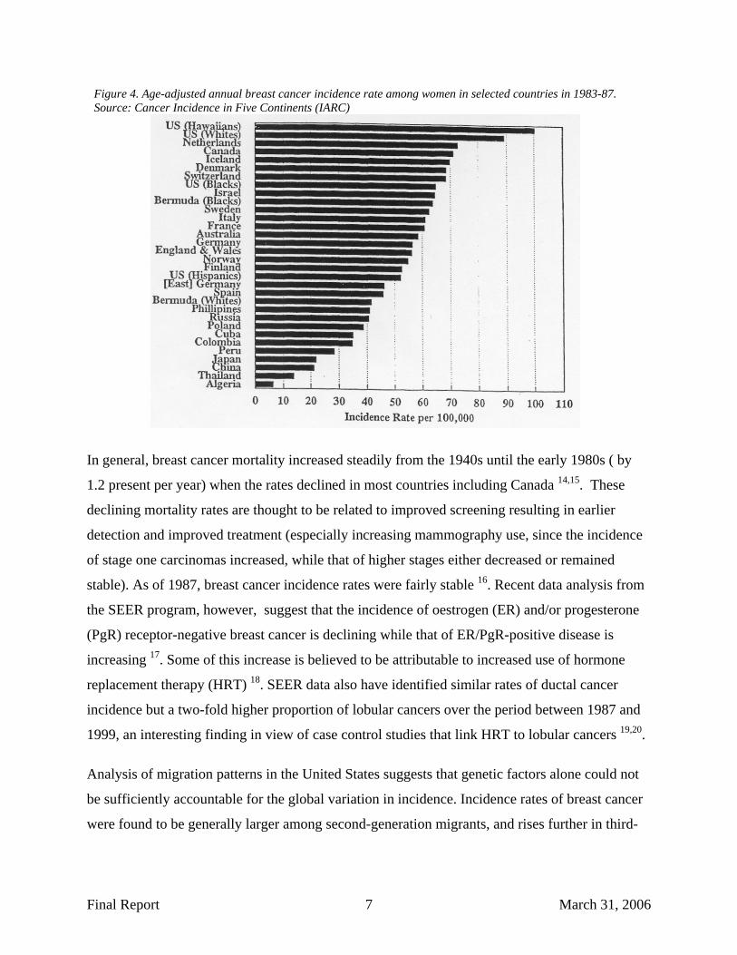
Figure 4. Age-adjusted annual breast cancer incidence rate among women in selected countries in 1983-87. Source: Cancer Incidence in Five Continents (IARC)
In general, breast cancer mortality increased steadily from the 1940s until the early 1980s ( by
1.2 present per year) when the rates declined in most countries including Canada 14,15. These
declining mortality rates are thought to be related to improved screening resulting in earlier
detection and improved treatment (especially increasing mammography use, since the incidence
of stage one carcinomas increased, while that of higher stages either decreased or remained
stable). As of 1987, breast cancer incidence rates were fairly stable 16. Recent data analysis from
the SEER program, however, suggest that the incidence of oestrogen (ER) and/or progesterone
(PgR) receptor-negative breast cancer is declining while that of ER/PgR-positive disease is
increasing 17. Some of this increase is believed to be attributable to increased use of hormone
replacement therapy (HRT) 18. SEER data also have identified similar rates of ductal cancer
incidence but a two-fold higher proportion of lobular cancers over the period between 1987 and
1999, an interesting finding in view of case control studies that link HRT to lobular cancers 19,20.
Analysis of migration patterns in the United States suggests that genetic factors alone could not
be sufficiently accountable for the global variation in incidence. Incidence rates of breast cancer
were found to be generally larger among second-generation migrants, and rises further in third-
Final Report 7 March 31, 2006
and fourth- generation migrants. This information suggests that environmental and lifestyle
determinants are important factors of breast cancer risk 21,22.
Mortality rates from breast cancer have been stable since 1950, although mortality rates of
various subgroups have changed. The mortality rate for white women under age 55 has
decreased, while it has increased for women age 55 and older 23. The reasons for the decrease in
the former are suspected to be related to the increase in mammography use in younger women
and the aggressive use of adjuvant therapies. Mortality rates have increased in African-American
women of all ages. Mortality rates are highest in the very young (less than age 35) and the very
old (greater than age 75).
Breast Cancer Risk Factors
Sociodemographic Factors
A number of sociodemographic factors are associated with breast cancer risk. Most of these are
likely surrogates for lifestyle, hormonal, and/or reproductive factors 24. In terms of gender, beast
cancer occurs one hundred times more frequently in women than in men. Male breast cancer in
2004 in the US is responsible for 0.2 percent of all cancers, and less than 0.1 percent of all
cancer deaths in men annually 12.
Age is major risk factor for breast cancer and (Figure 5) demonstrates the increased likelihood of
a woman developing breast cancer in the next five years at various ages 25. Incidence rates rise
very sharply with age until about the age of 45 to 50 when the rise is less steep. This change in
slope probably reflects the impact of hormonal change (menopause) that occurs about this time,
although alternative hypotheses have been proposed 26. At age 75 to 80, the curve flattens and
decreases slightly thereafter 27. Amazingly, age was the only identifiable risk factor in 76% of
women who developed a breast cancer 28.
Females of higher socioeconomic status are considered to be at greater risk for breast cancer.
There may be as much as a two-fold difference in incidence from the highest to the lowest
classes. The influence of socioeconomic status (educational, occupational, and economic level)
are thought to be mediated by differing reproductive patterns with respect to parity, age at first
birth, and age at menarche 29. In epidemiological studies higher socioeconomic status, as
measured by income and education level, are consistently associated with elevated breast cancer
Final Report 8 March 31, 2006

risk 4,30. Although some of this association may be due to a clustering of reproductive risk factors
in higher socioeconomic status women, the effect is still significant even after controlling for
parity, age at first child and other common reproductive factors 31. Diet has been well studied but
epidemiological investigations have yet to identify foods that significantly increase or decrease
breast cancer risk 32. It is hypothesized that dietary factors may modulate hormone levels so a
number of investigations have focused on foods high in phytoestrogens 33,34 (partial oestrogen
agonists) or containing other endocrine active components 31. Incidence and mortality rates vary
throughout the North America, with the highest reported incidence in Hawaii (128 per 100,000
women) and lowest in Utah (98 per 100,000 women) 35. Urban rates exceed those of rural areas.
These differences are thought to be accounted for by differences in parity and age at first live
birth, at menarche, and at menopause 26.
Figure 5. Probability of developing breast cancer in the next five years (78)
Age
Breast cancer (per 1,000)
30 1,5 35 2,6 40 4,8 45 7,8 50 9,2 55 10,6 60 12,9 65 14,3 70 15,4 80 15,5
In North America, breast cancer is the most common cancer among women of every major
ethnic group, although there are interracial differences. California, as an example, has the highest
rates among Caucasians (110.6 cases per 100,000 women). The rates in African-American
women (96 per 100,000), Latina women (59.2 per 100,000), Asian-Americans (58.2 per
100,000), and others are lower 36. Most of these ethnic difference are attributable to factors
associated with lifestyle and socioeconomic status, which partially could explain differences in
treatment and survival 37.
Final Report 9 March 31, 2006

Inherited Risk Factors
Hereditary risk factors for breast cancer are multifactorial, and should not be simply understood
as just a passage of genetic material. Hereditary factors are traditionally identified through
thorough family histories. Only 10 percent of women diagnosed with breast cancer have a
positive family history. The risk associated with having an affected first or second degree
maternal or paternal relative is modulated by the age of both the case patient and the family
member at diagnosis, and the number of first-degree relatives. In a meta-analysis using data from
over 50,000 women with breast cancer and 100,000 controls, the risk of breast cancer for a
woman with one affected first degree relative was increased 1.80 times. With the two affected
first degree relatives, the risk is increased 2.93 times. The risk ratios (relative risk) were found to
be highest in females with young affected relatives. Therefore, the risk was increased 2.9 times
for a woman whose relative was diagnosed before age 30, but only 1.5 times increased if the
affected relative was diagnosed after age 60 38. The risk of breast cancer before age 40 was
increased 5.7-times if one relative had breast cancer before age 40. The question as to what
extent the influence of a shared environment or a shared lifestyle contributes to the history of
cancer is still open 39,40.
Inherited genes with a low penetrance may account for a familial-specific metabolism of DNA
toxins, which in turn initiates or promote breast cancer. Identification of these genes may be
difficult; however, their expression may be influenced significantly by differences in the
environment. Mutations in BRCA1 and BRCA2 are strong with little respect for environmental
differences. Studies in twins suggest that the majority of familial aggregation of breast cancer
results from inherited susceptibility 41,42. However, specific genetic mutations that predispose to
breast cancer are rare. It is believed that only 5 to 10 percent of all breast cancers are associated
with a specific gene mutation, such as BRCA1, BRCA2, p53, ATM, PTEN, MLH1, or MSH2.
The extent of dense tissue within the breast has recently found to be another risk factor. Breast
density varies within the population, and appears to be largely inherited 43. Besides increasing the
difficulty of detection through mammography, the presence of dense breast tissue increases the
risk of breast cancer by a factor of 1.8 to 6-times compared to women of similar age with less
extensive dense tissue 44.
Final Report 10 March 31, 2006

Breast Conditions
Benign breast conditions include a wide spectrum of pathologic entities. The important
precursors of non-invasive or invasive breast cancer are grouped as proliferative disease with or
without atypia. Non-proliferative lesions are not associated with an increased risk for breast
cancer 24. Poliferative lesions without atypia include fibroadenoma, moderate or florid
hyperplasia, sclerosing adenosis, and intraductal papillomas. Women with these lesions typically
have an increased risk of breast cancer of only 1.3 to 2 times that of the referent group 45,46 .
Proliferative lesions with atypia (both lobular and ductal) possess one or more characteristics of
carcinomas in situ and are associated with a higher relative risk of breast cancer development.
The relative risk of invasive breast cancer (which is in majority of cases is ipsilateral) is
associated with atypical ductal hyperplasia and ranges from four to six-fold 45,46. These lesions
are considered precursors of invasive breast cancer, although invasive disease does not develop
in all cases 47. Although historically regarded as an indicator of the risk of developing invasive
breast cancer in both the ipsilateral and contralateral breast, results from a recent paper suggest
that both atypical lobular hyperplasia (ALH) and lobular carcinoma in situ (LCIS, a lesion that is
qualitatively similar to ALH but more developed quantitatively) are not only indicators of
increased risk, but indeed precursors of invasive cancers. The relative risk of invasive breast
cancer with ALH ranges from three- to fivefold 45-48. The risk for developing breast cancer in
either breast in women with LCIS is 1 percent per year and persists indefinitely.
Malignant breast conditions that increase the risk of a new unrelated breast cancer include ductal
carcinomas in situ and invasive carcinomas. With in situ lesions, the ten-year risk of developing
an invasive breast cancer in the contralateral breast is 5 percent 21; the risk of developing
contralateral breast cancer in women with an invasive cancer is 1 and 0.5 percent per year for
pre- and postmenopausal women, respectively.
Hormone Factors
Epidemiological and animal studies consistently show elevated risk of breast cancer with factors
that increase exposure to estradiol, progesterone, and other hormones 33,49-51. Risk factors such
as alcohol consumption, weight gain after menopause, low pre-menopausal body mass index,
and lack of physical exercise are believed to be associated with exposure to reproductive
hormones 16,52-54. Pharmaceutical hormones appear to have a similar effect and there is evidence
Final Report 11 March 31, 2006

that women exposed to diethylstilbestrol during pregnancy had increased risks for breast cancer 12,13. For oral contraceptives, recent use, not long term exposure, has been associated with an
increased risk 16,17. Similarly, recent use of hormone replacement therapy has been shown to
increase the relative risk of breast cancer, whereas women who stopped over 5 years ago are not
at significantly elevated risk 18.
Prolonged exposure to, and higher concentrations of, endogenous oestrogen will increase the risk
of breast cancer. The production of oestrogen subtypes (estradiol, estriol, estrone) is modulated
by ovarian function: menarche, pregnancy, and menopause. After menopause, the main source of
oestrogen is dehydroepiandrosterone (DHEA), which is produced in the adrenal gland and
metabolized in peripheral fat tissue to estradiol and estrone 55. The roles of progestins, prolactin,
and insulin-like growth factor are less clearly established.
Important factors that influence breast risk are age at menarche, age at first live birth, age at
menopause, and possibly parity and breast-feeding 22,56,57. Younger age at menarche is associated
with a higher risk for developing breast cancer 58 . One study found that for every two year delay
in the onset of menarche there was a 10 percent reduction in breast cancer risk 59. Others have
shown in a case control study of disease-concordant monozygotic twin pairs that the twin with
earlier onset of menses was five times more likely to be diagnosed with breast cancer before the
other 60. In contrast, other hormonal factors (ie, later first pregnancy, lower parity, later
menopause) did not predict an earlier diagnosis when both twins were affected. The explanation
for this protection is that late onset of regular menstrual cycles is associated with later exposure
and thereby less lifetime exposure to hormones.
There is an inverse association between the age at first pregnancy and risk of breast cancer 56.
However, women who give birth to their first child after the age of 30 have a higher risk than
nulliparous women. Women giving birth for the first time at age 35 have a 1.6 times higher risk
of breast cancer than women first giving birth at age 26 to 27 58,61. The explanation for the effect
of early first live birth is that full cellular differentiation, which occurs in the gland during and
after pregnancy, protects it from breast cancer development.
The association between the age of menopause and the risk of breast cancer is straight - the later
a woman undergoes menopause the higher her risk for breast cancer 58. Bilateral oophorectomy
Final Report 12 March 31, 2006

before the age of 40 reduces lifetime risk by 50 percent 60; however, this risk reduction is
eliminated if replacement estrogens are given. The association between late menopausal age and
increased breast cancer risk is thought to reflect longer exposure to the endogenous higher pre-
menopausal levels of hormones 56. Nulliparous women are at increased risk for breast cancer
compared with parous women 58. The risk ranges from 1.2 to 1.7 and appears to affect women
after the age of 40 to 45 56. Whether multiparity confers protection against breast cancer has been
a matter of controversy; the most recent studies suggest a decreased risk with increasing number
of pregnancies 61. Abortions have been hypothesized to increase breast cancer risk. One meta-
analysis of case-control studies supports this theory 62, but the other does not 63. Population based
cohort studies (an epidemiological stronger design) do not support this association 64-66. In March
2003, the National Cancer Institute accepted the findings of a workshop evaluating the link
between early reproductive events and breast cancer, which concluded that induced abortion is
not associated with an increase in breast cancer risk (available at
www.cancer.gov/cancerinfo/ere).
Full term pregnancy is thought to be associated with an increased breast cancer risk. The
protective effects of pregnancy are not seen until after ten years following delivery 67. Other
studies shows that placental factors like maternal floor infarction, smaller size and pre-eclampsia
during pregnancy are associated with a reduced incidence of maternal breast cancer 68.
Breastfeeding was shown to be protective against breast cancer in multiple case-control and
cohort studies, the magnitude of which is dependent on the duration of breastfeeding, and on the
confounding factor of parity 58,61,69-71. In the largest case-control study that included individual
data from 47 epidemiologic studies including 50,302 women with invasive breast cancer and
96,973 controls, the relative risk of breast cancer was reduced by 4.3 percent for every 12
months of breast feeding, in addition to a decrease of 7 percent for each birth 71. Furthermore, it
was estimated that the cumulative incidence of breast cancer in developed countries would be
reduced by more than one-half (from 6.3 to 2.7 per 100 women by age 70) if women had the
average number of births (6.5 versus 2.5) and a lifetime duration of breastfeeding (24 versus 3
months per child) that had been prevalent in the past. Two-thirds of this estimated reduction was
attributable to longer duration of breastfeeding. The mechanism postulated for the protective
effect of breastfeeding is that it may delay the re-establishment of ovulatory cycles. Other
Final Report 13 March 31, 2006

mechanisms may be the increase in prolactin secretion and the concomitant decrease in estrogen
production 24.
Hormone levels were shown to promote breast cancers in animals, and various studies have
manipulated hormones to demonstrate this point 72. Few studies have prospectively examined the
relationship between serum estrogen concentrations and breast cancer risk in humans; much of
the available evidence is primarily based upon observational data. Obese postmenopausal women
have higher oestrogen levels, due to the conversion of adrenal androgens to estrogens in fatty
tissue, and are at increased risk of breast cancer 73. Reducing oestrogen levels (by castration or
use of antiestrogens such as tamoxifen) lowers breast cancer risk. Despite these observations, the
correlation between breast cancer risk and hormone levels from studies examining blood or urine
samples have not been consistent, in part due to the inter-individual and intra-individual
variability of hormone concentrations and difficulties with the assays. Oestrogen levels fluctuate
during menstrual cycles and pregnancy, making reproducible measurements difficult in the pre-
menopausal years. Furthermore, most epidemiologic studies have tended to use a single blood
sample. Thus, it is not surprising that the data on pre-menopausal women are limited and
inconclusive. The data on oestrogen metabolites are similarly limited 24.
A 2002 review of several prospective epidemiologic studies found a positive relationship
between serum estradiol concentration and breast cancer risk 74. Similar results were obtained a
study of 7705 postmenopausal women enrolled on the Multiple Outcomes of Raloxifene
Evaluation (MORE) trial: women with the highest tertile of serum estradiol levels (>12 pmol/L)
had a two-times higher risk of invasive breast cancer than women with lower levels 66. Also,
females in the highest estradiol tertile tended to have a greater reduction in the risk of breast
cancer with raloxifene compared to women in the low estradiol subgroup (79 versus 64 percent).
Results of the Nurses' Health Study suggest that the association is strongest for hormone
receptor-positive breast cancers 75. In this longitudinal study of 121,700 female registered nurses
in the US, endogenous hormone levels were measured in 322 women who developed breast
cancer and in 643 age-matched controls without breast cancer. When the highest and lowest
quartiles of serum hormone concentration were compared, there was a significant direct
association between breast cancer risk and levels of both estrogens and androgens. The strongest
Final Report 14 March 31, 2006

association was found when the analysis was limited to ER and PgR-positive tumors, and in situ
tumours.
Since bone tissue contains oestrogen receptors and is highly sensitive to circulating oestrogen
levels, bone mineral density (BMD) was thought to be a surrogate marker for long-term exposure
to endogenous oestrogen. In the report of elderly women with high BMD it was found that
higher BMD is associated with the greater risk of breast cancer (relative risk 2.7 for women in
the highest compared to the lowest quartile of BMD), compare to women with lower BMD 76.
Postmenopausal females with higher testosterone levels may be at a higher risk of breast cancer 74,75,77-79. Studies of oprogesterone, prolactin, insulin, and insulin growth factor are limited.
However, resent publications suggest a possible increased risk of breast cancer with higher
serum levels of insulin-like growth factor I (IGF 1) and its main binding protein IGFBP-3 in pre-
menopausal but not postmenopausal women 80,81. Some, but not all, studies suggest a slightly
increased risk of breast cancer in postmenopausal but not pre-menopausal women with type 2
diabetes.
There is a rising concern regarding increased risk of breast cancer with usage of oral
contraceptives (OC), infertility treatment and hormone replacement therapy (HRT). Several
epidemiologic studies have not demonstrated any association between OC use and the risk of
breast cancer. However, a large meta-analysis (conducted in 1996) calculated a small but
significant increase in relative risk of breast cancer (RR =1.24) in current oral contraceptive
users 82. Concerns have been raised about this meta-analysis because a low percentage of women
had ‘ever’ used oral contraceptives (40 percent), and it lacked the follow-up necessary to
determine whether there were long-term effects of oral contraceptive use. There are now
reassuring data from two studies that oral contraceptives do not increase breast cancer risk later
in life 83,84.
Causal relationship between the usage of HRT and increased risk of breast cancer has been
supported in the current literature, mostly hormone receptor-positive breast cancer 85. The risk is
small but was consistently demonstrated. Oppositely, a trend towards lower breast cancer risk
was observed in women taking unopposed oestrogen (HR 0.77 for unopposed oestrogen vs.
placebo, 95% CI 0.59-1.01) 86. Long-term use of HRT is associated with the highest risk. In
Final Report 15 March 31, 2006
contrast, short-term HRT appears not to increase the risk of breast cancer significantly, although
it may make detection through mammography more difficult. Concurrent progestin use appears
to further increase risk above that with estrogen alone 87.
Prevention of Breast Cancer
All major North American groups making recommendations about breast cancer screening
recommend routine screening with mammography with or without clinical breast examination
for women aged 50. In the 1996-1997 National Population Health Survey, 79% Canadian women
50-69 years of age report having had a mammogram. The proportion of women in this group
reporting ever having had a mammogram is between 75% and 82% in all provinces except Nova
Scotia (64%) and Newfoundland (54%). Eighty-five percent of those who had a mammogram in
at least 2 years report having had one of the following reasons: routine check-up, family history,
age, or hormone replacement therapy. Low education and income are associated with fewer
mammograms of any type 88.
The Screening Mammography Program is the major preventive health program for British
Columbia women. Currently, between six and eight women out of 10 do not use this program
that costs the system more than $6 million per year. The Minister's Advisory Council on
Women's Health expressed the need to make this program work more effectively or reconsider
its utility in light of other competing priorities. 89. At present, the best preventative strategy for
breast cancer is early detection. Women who detect the breast cancer in situ have a 95 percent
survival rate; if it is not detected in the breast and has metastasized to the rest of the body,
survival rates are much lower 89. Although the Canadian Cancer Society emphasizes a three-
step process involving a monthly breast self-exam, an annual clinical breast exam and
mammography, the Ministry of Health or the Screening Mammography Program do not
monitor the practice of all three aspects of care. Data from the North Shore health unit's 1990
health promotion survey indicated that only 30 per cent of women practice monthly breast self-
exam 90. By health region in British Columbia, the percentage of women aged 50 and older who
received mammogram in 1992-93 ranged from 20 percent (upper Fraser Valley) to 39 present
(South Central). Another way of saying this is, at the present time, between six and eight
women out of 10 in British Columbia do not participate in this publicly-funded preventive
health program 90.
Final Report 16 March 31, 2006

Occupational and Environmental Factors in Breast Cancer
Recent animal studies provided important information in understanding mechanisms of the
development of breast cancer and in the identification of agents that may increase breast cancer
risk. A comprehensive review of chemical carcinogenesis in general is beyond the scope of this
paper. Based on human epidemiological studies, ionizing radiation is one of the few
occupational or environmental exposures that are known to cause breast cancer 19,20, although it
should be noted all cancer causing agents, physical or chemical, will also have the potential to
initiate or promote cancer.
Cells within the breast are not fully differentiated until they are induced by hormonal stimuli at
the woman’s first pregnancy and lactation. Thus, breast cells are more susceptible to the effects
of carcinogens while the breast is not fully developed. Additionally, the breast cells are
vulnerable to genotoxic agents during pregnancy as there is rapid proliferation of cells 27,91. This
explanation of the susceptibility of mammary cells to carcinogens provides a framework for
understanding the increased risk of breast cancer in humans in relation to reproductive events as
well as after exposure to mammary carcinogens. It has been hypothesized that, because the
breast is very susceptible to carcinogen exposures up until the first full-term pregnancy, there
may be an interaction of age (a known risk factor) and the risk associated with exposures to
chemicals 29.
Despite the complex mechanisms and interactions between chemical exposures and hormones,
animal studies have clearly identified numerous mammary carcinogens through standard cancer
bioassays. The US National Toxicology Program (NTP) has tested over 500 chemicals and
identified 42 as causing mammary tumours 35. The human evidence for identifying chemicals
causing breast cancer is more scant and of the 42 chemicals cited above, only four are classified
as human carcinogens: benzene, 1,3-butadiene, ethylene oxide, and C I acid red 114. Also, it
should be noted that epidemiology studies of these compounds have shown exposed employees
at higher risk of cancer, but not specifically breast cancer. Mammary carcinogens that may be
associated with exposures in chemical and medical laboratories are presented in Table 1 below.
Both animal and human studies show that the relationships between hormonal factors and
mammary carcinogens are complex. Treatment of animals with ovarian, placental, pituitary, and
thyroid hormones modulates the tumorgenic responses 26. The situation is further complicated
Final Report 17 March 31, 2006

with exposures to chemicals that are members of a class of hormonally active chemicals,
sometimes referred to as endocrine active, endocrine disruptors, or estrogenic compounds. The
hypothesis is that exposure increases oestrogen-like responses of cell proliferation that increase
cancer risk. There is also a concern that these endocrine active compounds can act in an additive
manner to produce effects 29,36.
Table 1:Chemicals tested by NTP that produce mammary tumors in experimental animals4
Chemical Use Acronycine Pharmaceuticals
Benzene Gasoline, solvent
2,2-bis(bromomethyl)- 1,3-propanediol Flame retardant
1,3-Butadiene Auto exhaust, rubber manufacture, gasoline
C,1 acid red 114 Dye for silk, jute, wool, leather
C,1 basic red 9 monohydrochloride Dye for textiles, leather, paper, biological stain
2-Chloroacetophenone Flame retardant
Chloroprene Used in neoprene manufacture
Clonitralid Molluskicide
Cytembene Pharmaceuticals
2,4-Diaminotoluene Intermediate in dye synthesis
1,2-Dibromo-3-chloropropane Soil fumigant, pesticide
1,2-Dibromoethane Soil fumigant, lead scavenger in gasoline
1,2-Dibromo-1-propanol Flame retardant
1,1-Dichloroethane Solvent
1,2-Dichloroethane Solvent, chemical intermediate in insecticide formulations, gasoline
1,2-Dichloropropane (propylene dichloride) Chemical intermediate, solvent in dry cleaning fluids, fumigant
Dichlorvos Pesticide
1,2-Dimethoxybenzidine dihydrochloride Dye intermediate
3,3-Dimethylbenzidine dihydrochloride Dye intermediate
2,4-Dinitrotoluene Dye intermediate, explosives, propellants
Ethylene oxide Sterilizing gas for medical equipment
Furosemide Pharmaceuticals
Glycidol Stabilizer in vinyl polymers, intermediate in pesticides and fragrances
Hydrazobenzene Dye intermediate, tobacco pesticides, motor oil
Isophosphamide Pharmaceuticals
Indium phosphide Microelectronics, semiconductors, injection lasers, diodes
Isoprene By-product of ethylene production
Methylene chloride Solvent, furniture stripper, adhesives
Methyleugenol Food additive, flavoring, also naturally occurring
Nithiazide Antiprotozoal compound
5-Nitroacenaphthene Research chemical
Nitrofurazone Antibiotic
Nitromethane Rocket and engine fuel, solvent, mining explosive
Ochratoxin A Mycotoxin
Phenesterin Pharmaceuticals
Procarbazine hydrochloride Pharmaceuticals
4 From 35. Bennett LM, Davis BJ. Identification of mammary carcinogens in rodent bioassays. Environ Mol Mutagen 2002;39(2-3):150-7.
Final Report 18 March 31, 2006

Reserpine Pharmaceuticals
Sulfallate Herbicide
2,4- and 2,6-Toluene diisocyanate Used in manufacture of flexible polyurethane foams
o-Toluidine hydrochloride Dye intermediate
1,2,3-Trichloropropane Chemical intermediate, former solvent and paint remover
Chemicals, including some pesticides, also can act as co-carcinogens or tumuor promoters 37. A
good example of a breast cancer promoter in experimental animals is
dichlorodiphenyltrichloroethane (DDT). Experimental animals fed a known mammary
carcinogen, and then given DDT, developed breast tumuors earlier than when the carcinogen was
given alone; however, when DDT was given alone, it did not induce breast tumuors in these
animals 39. The human evidence of DDT’s effects as a promoter is more equivocal, although a
recent study reported significantly elevated mean levels of serum DDT and hexachlorobenzene
(HCB) in breast cancer patients as compared to controls 40. Other organochlorine compounds
have been implicated as being associated with an increased risk of breast cancer. The hypothesis
is that this group of compounds possess estrogenic activity. However, both the hypothesis and
the magnitude of any possible effect on human risk of breast cancer is controversial. Recent
reviews suggest that the estrogenic contribution of organochlorine compounds is small in view of
the presence of natural hormone and anti-hormone mimics in our diet 21,92. Other endocrine
active compounds, such as alkyl phenols and phthalates are still under investigation 41.
Studies of breast cancer risk in working populations have not provided strong evidence of causal
links between specific exposures and increased risk. However, there is evidence for positive
associations of several occupations with increase breast cancer risk 42,93,94. The study by Band et
al. (2000) was conducted in British Columbia and found significantly higher breast cancer risks 42 among pre-menopausal women in electronic data-processing operators; barbers and
hairdressers; in sales and material processing occupations; and in the food, clothing, chemical
and transportation industries. Among post-menopausal women, an elevated risk was found in
school teaching; in medicine, health, and nursing occupations; in laundry and dry-cleaning
occupations; and in the aircraft and automotive, including gasoline service station, industries.
Several significant associations were also seen in the combined group of pre- and post-
menopausal women, particularly in crop farmers and in fruit and vegetable farming, publishing
and printing, and motor vehicle repair industries. The authors suggested that there was excess
Final Report 19 March 31, 2006
breast cancer risk in a number of occupations and industries, notably those that entail exposure to
solvents and pesticides 42.
Shift work causes employees to have exposure to light at night and may increase the risk of
cancer by suppressing the normal nocturnal production of melatonin by the pineal gland.
Melatonin is not only a hormone that has anti-proliferative effects which protect against the
development of cancer 95, but it also modulates oestrogen release from the ovaries. When
nocturnal melatonin production is suppressed, the direct anti-proliferative effects are reduced and
oestrogen release may be increased. There are a few studies that support an association between
exposure to light at night and the risk of breast 95. However, the strength of the association has
been variable (Figure 6).
A nationwide population-based case control study included 7035 Danish women with breast
cancer and individually matched controls 96. Among women aged 30 to 54, the OR for breast
cancer among those who worked at night for at least six months was 1.5, with a trend toward
increased OR for longer durations of night time employment. Similar findings were noted in
another case control study: OR 1.6 for night time shift work, with a trend toward higher risk with
more years and more weekly hours of nocturnal shift work 97
A more modest association between night time shift work and breast cancer risk was noted in a
prospective cohort series from the Nurses' Health Study 95. The relative risk of breast cancer was
significantly increased only among women who worked rotating night shifts (at least three nights
per month) for 30 years or longer (RR = 1.36).
Final Report 20 March 31, 2006

Figure 6. Studies, investigated association between night time shift work and breast cancer risk
It is postulated that exposure to light at night suppresses the normal nocturnal production of
melatonin by the pineal gland, which in turn, could increase the release of oestrogen by the
ovaries. In one of the above studies, the risk of developing breast cancer was significantly higher
in women who did not typically sleep between 1 AM and 2 AM, the night time period when
melatonin levels are at their highest (OR 1.14) (62).
Breast Cancer Risk among Laboratory Workers
Clinical laboratory workers have the potential for exposure to a variety of potentially harmful
chemical, biological, as well as physical agents including solvents, radioisotopes, chemical
carcinogens, viruses, bacteria, human and animal tissue samples and lately also recombinant
organisms. 43,44,46. Despite the fact that chemical and clinical laboratories employ many women
(over 1 million in the US) few studies have examined the possible adverse effects of exposures
on this occupational group.
Wennborg et al. (2005) investigated congenital malformations related to maternal exposure to
specific agents in laboratory employees. The study involved 1951 females and the authors found
that the prevalence of "major malformations" were 2.3% (n = 41; exposed) and 1.9% (n = 23;
unexposed). For the major malformations, solvent exposure before the third trimester gave an
odds ratio (OR) of 1.8 (confidence interval (CI) = 1.0-2.9); "laboratory work in general" of 1.2
(CI = 0.7-2.0). The OR for benzene use around conception/organogenesis was 5.3 (CI = 1.4-
Final Report 21 March 31, 2006

21.1) for non-conditioned medium (NCM). No significant risk for laboratory work in general
was seen, but there was an increased ratio for NCM relative to solvents, especially benzene 98.
Wennborg (2000) compared mortality and cancer incidence in 5,035 full time laboratory
employees with a cohort of 2,923 employees of non laboratory departments at Karolinska
Institute and at the Universities of Lund, Gothenburg and Linköping. Findings shown that the
total mortality as well as the incidence of all cancers together was lower in both the laboratory
and the non-laboratory groups than in the general population, but slightly (non significantly)
increased risks were seen for male brain tumours (SIR=3.11 (95% confidence interval 0.85-7.96)
after 10 years of work), and female breast cancer among laboratory personnel (standardized
incidence ratios (SIR)=1.62 (CI 0.78-2.98). Work with solvents showed an elevated SIR of
malignant melanoma in female laboratory personnel. Concerning reproductive health, no major
risks were noted for most outcomes. However, mother's work in laboratory showed an increase
of large for gestational age (LGA) infants and an association was seen between reduced
fecundity and use of solvents, cell techniques or viruses 99.
Burnett et al. (1999) conducted a study to determine if laboratory workers in the US experienced
higher cancer mortality rates than those in other occupations. They found that clinical laboratory
workers had higher proportionate cancer mortality ratios overall (for all cancers) as well as for
breast cancer. The proportionate mortality ratios for leukaemia were also significantly elevated
for clinical laboratory workers 45. The authors suggest that the elevated risks for lymphatic and
hematopoietic neoplasms may have been associated with occupational exposures.
Brown et al (1996) conducted a cohort study among 12,703 individuals employed by biological
research institutes in the UK. Authors found that mortality due to all causes was significantly
reduced in men (standardised) mortality ratio (SMR) 55 and women (SMR 52). Mortality was
also significantly reduced for circulatory and respiratory diseases, and overall there was low
mortality from malignant neoplasms. SMRs exceeded 100, but were not statistically significant,
for infective and parasitic diseases. There were no statistically significant raised SMRs for any
cancer site. Workers were categorised as ever worked in a laboratory (laboratory workers) and
never worked in a laboratory (non-laboratory workers). The all-cause SMR was significantly
reduced in both groups, as was mortality from circulatory and respiratory diseases. The SMR for
malignant neoplasms was also significantly reduced in laboratory workers 100.
Final Report 22 March 31, 2006

A recent large-scale cohort study conducted by van Barneveld et al, (2004) among 7,307
laboratory workers in the Netherlands, found that laboratory workers have a favourable cancer
mortality pattern as compared to the general population. Authors commented that all-cause
mortality among laboratory workers was significantly lower than that in the general population.
Total cancer mortality and lung cancer mortality were also significantly decreased (SMR=0.8;
95% confidence interval CI=0.7–0.9 and SMR=0.7; 95% CI=0.6–0.9), respectively. However,
when compared to the internal reference population, laboratory workers had a slightly, non
significantly, increased cancer mortality (relative risk (RR)=1.3 95% CI=0.9–1.9). Among men,
a 2.5-fold (95% CI=1.0–6.3) increase of lung cancer mortality was observed which could not be
explained by differences in smoking habits. Lung cancer mortality increased with longer follow-
up. Results with regard to a priori defined fields of research showed significantly increased
cancer mortality (in particular from lung cancer) for men working in genetics (RR=3.8), virology
(RR=4.1) and plant physiology (RR=2.1). Authors concluded that the excess lung cancer
mortality among male laboratory workers was concentrated in certain fields of research, which
warrants further research to identify specific exposures related to the increased risk 101 .
Based on the assumption that laboratory work, especially in the area of biomedical research, is
associated with exposure to a mixture of carcinogens, a group of Israel scientists conducted a two
fold research aiming to analyze the cancer incidence among laboratory workers employed in
biomedical research laboratories versus the employees at the routine laboratories. The first
publication presented the results of the analysis of 4,300 laboratory workers whose cancer
incidence was followed from 1960 to 1997. The authors found that work in research and
biomedical laboratories might involve an increased risk of certain types of cancer. In fact,
significantly elevated SIR was found in breast, ovary, and thyroid cancer among women; and
prostate cancer, leukaemia, and melanoma among men 102. The second publication from the
same author aimed to examine whether the excess cancer morbidity that was found can be
explained by exposure to a particular group of substances, taking into consideration potential
confounders. This study (nested case control study) included 163 cases and two matched control
groups: laboratory workers (311) and general population (448) workers. The authors employed
multiple conditional regression analysis which showed that working in research laboratories
involved an increased risk of cancer generally among women [risk ratio 2.2 (1.2-4.3)], and of
breast cancer particularly [risk ratio 2.3 (1.1-4.7). Seventy-six percent (76%) of breast, 87% of
Final Report 23 March 31, 2006

thyroid, 60% of ovary and prostate, 94% of melanoma, and 50% of leukaemia cases were ever
exposed to at least one known human carcinogen. Authors believe that the results of this study
exclude the possibility that the excess cancer morbidity was related to personal risk factors but
they may be explained by exposure factors 103.
With the exception of a few studies that have identified very high occupational exposures to
carcinogenic compounds as causal factors in breast cancer, most investigations have not been
able to clearly determine occupational risk factors 93. The reasons for the failure to identify
specific chemicals or physical agents include not only the complex nature of the initiation,
promotion, and development of breast cancer (Figure 7), but also the presence of many potential
confounding risk factors. Additionally, there appear to be numerous, but so far unidentified, risk
factors that the issue of confounding becomes even more salient. Little is known about the
interaction of known risk factors on the magnitude of increase in breast cancer risk and even less
is known about the possible synergistic, additive, or antagonistic effects of multiple chemical
exposures.
Figure 7. Stages of carcinogenesis
The strength of already known breast cancer risk factors makes the identification of occupational
risk factors very difficult. When examining the role of these major risk factors, it has been
estimated that 41 percent of breast cancer risk is attributable to later childbearing, nulliparity,
higher income, and family history of breast cancer 47. Studies that have focused on genetic
Final Report 24 March 31, 2006

variation have estimated that less that 10 percent of cases are due to gene mutations in the breast
cancer genes BRCA1 and BRCA2 48. Diet, alcohol consumption, physical activity, body mass
index, other reproductive factors, high chest radiation exposure, and exposure to pharmaceutical
hormones all account for some risk in the development of breast cancer. In occupational studies,
if the likelihood of exposure to these known breast cancer risk factors is increased in an
occupational group, an association between the occupation and increased breast cancer risk will
be observed. Conversely, the presence of powerful risk factors known to cause breast cancer
may mask the effect of an occupational exposure that is truly increasing breast cancer risk, unless
methods are used to “control for” time factors.
Traditional epidemiological methods are typically not able to identify occupational risk factors
for breast cancer at the levels of exposure seen in modern industry in Canada or the US. Newer
methods that include the use of biological markers of exposure and incorporating gene-
environment interactions have shown promise. These methods are better able to uncover subtle
differences in risk and also provide an understanding of the underlying mechanisms. An
example of these cutting edge techniques is the measurement of the aromatic amine, o-toluidine,
a rat mammary carcinogen, in human milk samples from mothers. The presence of this chemical
indicates that the ductal epithelial cells of the breast are exposed to this carcinogen 21. The use of
biomarkers and gene-environment interactions have elucidated the complex associations of
smoking, polymorphisms of drug metabolizing enzymes, and reproductive factors in breast
cancer risk 21. These techniques have not been rigorously applied in studies involving
occupational exposures and breast cancer but their use has been advocated 21,55.
Incinerator Emissions and Cancer
Incineration is widely used in the United States and Canada to reduce the volume of waste.
Whether waste incineration poses a health risk to incinerator employees or to people living and
working nearby has been the subject of much debate. When operated properly by well-trained
employees, modern waste incinerators pose little risk to public health. But older designs, human
error, and equipment failure can result in higher-than-normal, short-term emissions that need to
be studied further. A few studies have tried to establish a link between an incinerator and illness
in the surrounding area, but most studies have been unable to detect any adverse health effects.
The studies that did identify effects on health had shortcomings and failed to provide convincing
Final Report 25 March 31, 2006
evidence. However, it should be noted that ailments may occur infrequently or take years to
appear, pollution from other sources may also be present; these factors make it difficult to
determine if waste incineration can indeed be responsible for local health problems 104.
Medical incineration systems are one of the growing public health concerns. In addition to
infectious waste, hospitals burn disposable plastic medical and food items, office waste,
packaging, and construction debris. Burning this waste could discharge poisonous substances
including mercury, lead, acid gases, dioxins and other chlorinated compounds 105. Many of these
chemicals are known to be persistent, bio-accumulative, carcinogenic or endocrine disruptors.
Most heavy metals have been reported to be associated with kidney disease, respiratory diseases,
cardiovascular damage, blood effects, and neurotoxicity 106. Some are classified as proven or
suspected carcinogens (Figure 8). Some are associated with particular health effects 107.
Polyaromatic hydrocarbons (PAHs), released during the incomplete combustion or pyrolisis of
organic matter, may have estrogenic properties 108 and are reported in association with ischemic
heart disease109 and cancer, in particular lung cancer 110 and bladder cancer 111.Polycyclic
aromatics (PCA) have been reported to be mutagenic and mutagenicity was found to be inversely
proportional to the degree of completeness of refuse combustion (188). Poorly controlled
combustion processes can entail the production of dioxins, another class of compounds that
include two families of chemicals, polychlorinated dibenzo-para-dioxins (PCDDs) and
polychlorinated dibenzofurans (PCDFs). These groups consist respectively of 75 and 135
cogeners that determine toxic effects on human health with different grades of severity 107.
Because of these concerns, hospital systems of disposing biomedical waste are under close
observation and recommended to be updated periodically 112,113.
Final Report 26 March 31, 2006

Figure 8. Carcinogenic effects of chemicals according to the IARC evaluation
Franchini et al. (2004) published a comprehensive review of forty six epidemiological studies
(published from 1987 to 2003) on health effect effects in populations living in the neighbourhood
of waste incinerators 107.
The analysis done by Franchini et al. revealed significant exposure-disease associations. No clear
association between breast cancer and exposure to incinerators or exposure to multiple sources
including incinerators were reported. However, some studies have shown significant association
with lung cancer. Two studies reported significant association between non-Hodgkin’s
lymphoma and environmental exposure to incinerators located in the UK and France and a
significant increase in risk of soft tissue sarcomas was found in France and in Italy in association
with residence close to waste incinerators. A UK study pointed out a small increased risk of liver
cancer associated with living within 1 km of an incinerator even after adjustment for other
known risk factors. A small area analysis of mortality among Italian residents near multiple
sources of combustion products did not indicate any clear association between liver cancer
mortality rates and distance from sources of exposure but highlighted an increase - though not
significant - of cancer of the larynx in males as distance from the plants decreased and a
significant excess of mortality for kidney cancer in females between 3 and 8 km from the
exposure sources. (Cited by: Franchini M. et al) 107.
Final Report 27 March 31, 2006
Cancer Cluster Investigations
Incidence rates of breast cancer, and all cancers, vary over time and geography and a cancer
cluster is generally defined as the occurrence of a greater than expected number of cases of a
particular cancer within a group of people, a geographic area, or a period of time. Studying and
describing these spatial and temporal trends have provided clues for identifying previously
undiscovered causes of cancer. In fact, the first causal relationship between an occupational
exposure and cancer was uncovered as the result of a cluster investigation of scrotal skin cancer
among young chimney sweeps in London 114. Epidemiologists, the scientists most often leading
the investigation of clusters, generally encounter clusters because of reports or through discovery
from organized analyses of large databases 115. Although the methods of analysis differ slightly
depending on how the cluster is first identified, in both cases the results are difficult to interpret
and drawing definitive conclusions is often not possible.
As was discussed in the section on breast cancer risk factors, some variation in breast cancer risk
can be explained by the population distribution of known risk factors such as parity, age at first
child and other reproductive factors 116. In fact, grouping of reproductive risk factors and
socioeconomic status play a major role in the findings of positive associations between white
collar occupations and increased risk of breast cancer 29. However, regional patterns of increased
and decreased breast cancer risk may reflect a complex aggregation of diverse factors which may
include diet, demographics, lifestyle factors, and occupational and environmental exposures.
Gaining an understanding of these individual factors and their relationships is necessary to have
a complete understanding of breast cancer risk in individuals and specific groups of women.
For breast cancer, clusters of relatively high incidence rates have been reported in areas of
southern Alberta and British Columbia 117. This type of variation by region is common and it is
most often unclear whether or not the determinants of these differences are related to
environmental, lifestyle, or other exposures. Even in populations that are well studied, such as in
the Long Island, New York Breast Cancer Study Project 118,119, limitations in study design make
the finding of significant environmental risk factors unlikely. In most investigations, biological
data relating to occupational or environmental exposures is sparse or inadequate and other risk
factors are not well controlled. Thus, even very extensive investigations of breast cancer clusters
have high probabilities of failing to identify occupational or environmental risk factors 120.
Final Report 28 March 31, 2006
Breast cancer cluster investigations are often limited because of the effect of the very strong risk
factors related to endogenous hormones that increase breast cancer risk. The question still
remains: do exposures to hormone-mimicking chemicals or other chemical and physical agents
also exert an effect? A multidisciplinary workshop, titled “Hormones, Hormone Metabolism,
Environment, and Breast Cancer," convened by the National Action Plan on Breast Cancer, the
US National Cancer Institute, Tulane University, and the U.S. Public Health Service's Office of
Women's Health, in September 1995 discussed the complexity of factors, unresolved
controversial issues, and the need for improved methodology to measure hormones and their
metabolites 121. As is the case with occupational studies of breast cancer, molecular as well as
bioinformatic techniques were discussed as useful tools in gaining an understanding of the
complex relationships between genes, individual factors, and the environment.
Investigating Cancer Clusters: Methods and limitations
The first of the modern cancer cluster reports began in the 1960s and the increasing number of
reports spurred the development of investigation protocols. At a US National Cancer Institute
conference on clusters, Dr. Langmuir advocated a simple approach: “The constructive approach
to this situation, in my opinion, is not to develop highly refined statistical techniques to
determine whether or not a certain cluster may have resulted by chance alone. But, rather to
investigate each cluster as it is reported and see if additional associations of possible interest can
be found. If none turn up, this is obviously a cold trail, and any good hunting dog will abandon
it, and look for a better one. If the scent strengthens, then hot pursuit is in order” 122.
Langmuir’s advice for a simple approach did not deter the development of statistical models to
resolve the issue of whether cancer cases were occurring independently or if they appeared to be
related. A number of theoretical statistical methods have been developed and modified to detect
clusters and to assess the statistical associations of interest. A detailed review of the theories
underlying these sophisticated statistical approaches is beyond the scope of this discussion and
interested readers should consult articles describing specific statistical techniques as well as
comprehensive reviews of the subject 2,108,122-127. Most of the models developed are useful when
information is available on the observed cases in many discrete geographic locations and time
intervals; the models then provide the likelihood of any one discrete location/time interval
having a number of cases that is excessive. Thus, these techniques have most utility when there
Final Report 29 March 31, 2006

is routine monitoring of cases across large geographic areas (i.e., province-wide or Canada-wide
surveillance programs).
The detection and analysis of cancer clusters most often is the responsibility of public health
agencies such as local health departments, state or provincial health authorities, cancer registries,
or national health agencies (Health Canada or the US Centers for Disease Control (CDC)). The
Canadian Cancer Incidence Atlas is a recently developed national atlas that assesses the
significance and spatial correlation of the age-standardized rates for 290 census divisions across
the country 128. The Atlas provides information about cancer incidence rates and is able to
determine if cancer rates are significantly elevated in certain areas. As discussed previously, the
Lower Mainland of BC is one area in which breast cancer incidence rates are significantly
elevated as compared to the national average 117.
When a cancer cluster is first reported, usually by concerned employees or citizens, the cluster is
termed a perceived cancer cluster. If an investigation determines that the observed number of
cases significantly exceeds the expected number it is termed an observed cancer cluster. If, after
further investigation, a risk factor can be identified the cluster is called an etiologic cancer
cluster 129. In investigating cancer clusters the goal is to determine if the cluster is real (observed
cancer cluster); and, if it is real, to determine if it is or is not an etiologic cancer cluster. If the
investigation uncovers an etiologic cancer cluster, efforts should be made to reduce/modify the
causal factors (exposures) that are responsible for the increased risk.
Public concern pertaining to environmental exposures and cancer resulted in the reporting of
many perceived cancer clusters over the past 20 years 130,131. Public health authorities responded
to these concerns by conducting investigations that varied in scope and cost. Considerable
resources were allocated to cluster investigations and most did not identify etiologic cancer
clusters. The US CDC, from 1961 to 1982 investigated 108 reported cancer clusters in 29 states
and 5 foreign countries; no clear cause of cancer was determined for any of the reported clusters 4. The Minnesota Department of Health (MDOH) investigated more than one thousand cancer
clusters between 1984 and 1995 without identifying a particular cause in any 132. As a result of
these many investigations, the MDOH developed a widely adopted systematic approach for
cluster investigations 133,134.
Final Report 30 March 31, 2006

Cancer clusters also occur in the workplace and a number of the classic exposure-disease
relationships arose from investigations of clusters. The determinations that polycyclic aromatic
hydrocarbons, asbestos, and vinyl chloride monomer are human carcinogens were made though
analyses of cancer clusters in workers where these products were manufactured or used 114,135-137.
These etiologic clusters occurred before modern industrial hygiene controls were implemented
and resulted from very high exposures to potent carcinogens. These early occupational cancer
cluster investigations were effective in identifying and controlling large cancer risks that workers
faced before the 1970s. Consequently, the role of occupational carcinogens in current clusters is
more subtle than in the past and more difficult to detect.
In the US, the National Institute for Occupational Safety and Health (NIOSH), through its Health
Hazards Evaluation Branch, is often called upon to investigate reported cancer clusters. In a
review of 61 cancer clusters investigations that NIOSH completed between 1978 and 1984, a
numerical excess of cases compared with expected numbers was found in 16 of the reported
clusters 2. In most of the reported clusters, no identified environmental exposure could be
identified. In five of the 16 clusters there were exposures to potential carcinogens and the
exposure-disease relationship was plausible (sufficient induction time and timing of exposure).
Almost all of the investigations were limited by small numbers of cases, absence of complete
personnel records, and other methodological and statistical issues that prevented the
identification of specific causal occupational risk factors 2.
In Canada there have been few published cancer cluster investigations that have identified a
specific cause that was occupationally related. The investigation of a cancer cluster in a steel
mill in Ontario attempted to determine if occupational exposures to polycyclic aromatic
hydrocarbons and silica were responsible for an increased risk of lung cancer. Even with
extensive air monitoring data, no significant findings pertaining to environmental exposures were
observed 138. A more recent cluster investigation of an excess number of cancers within a police
detachment in British Columbia involved the follow-up of 174 police personnel who where
associated with the detachment since 1963 139. Sixteen cases of cancer were identified; however
there was no evidence for an underlying event or exposure that could be attributed to the
observed cancer cases. The authors discussed the possible role of police radar on the rate of
cancer in the detachment.
Final Report 31 March 31, 2006

Health agencies in the US, Canada, and Europe have established protocols for investigating
reported cancer clusters. These protocols may differ in some of the specific steps but they do
follow a basic procedure in which increasingly more specific information is gathered and
analyzed in stages. In the Netherlands, a step wise protocol going from exploratory,
qualification, and quantification stages is used 140. Through each of the three stages, attention is
focused both on exposures and disease, and decisions about possible causality are made at the
end of each stage. Additionally, as with most cluster protocols, at the end of each stage a
decision to progress with the investigation is made 129,141.
The primary objective of a cancer cluster investigation is to identify exposures that may be
associated with excess cases in a workplace or location so that exposures can be controlled.
When conducting a cluster investigation it is useful to consider a number of questions as the
work proceeds through the various stages. The initial questions are: (1) is the incidence of
disease really higher than normal and by how much? (2) is the exposure higher than normal or
above allowable limits? and (3) is the link between exposure and cluster biologically plausible 142? The stages of a cluster investigation allow for the collection of the necessary information to
answer these questions and if these answers are affirmative then the investigation may progress
to a full-scale epidemiological study attempting to determine the association between the
exposure and increased risk.
Very detailed protocols for investigating reported cancer clusters have been published by health
agencies and reviews have appeared in the peer reviewed literature 1,3-5,141,143-145. In British
Columbia, the protocol includes: Stage 1 – Initial contact and response, Stage 2 – Assessment,
case evaluation and incidence evaluation, Stage 3 – Major feasibility study, and Stage 4 –
Etiologic investigation 53. In the State of Washington, their 18 page protocol has similar stages:
(1) collect initial information and provide education and information to the informant, (2) assess
the magnitude of the reported cluster, (3) determine utility and feasibility of further
epidemiologic study, and (4) conduct detailed etiological investigation 3. Other health
departments have developed very similar systematic approaches to cluster investigations and all
provide detailed procedures for data collection, analysis, and guidelines for making decisions at
the end of each stage 141.
Final Report 32 March 31, 2006
Analysis of Mission Memorial Hospital Laboratory Cancer Incidence Data
Events in the Mission Memorial Health Laboratory cancer cluster investigation
This investigation was conducted in response to concerns expressed by employees of the Mission
Memorial Hospital (MMH) Laboratory that they were experiencing a high incidence of cancer.
The investigation resulted in an initial report prepared by the Occupational Health & Safety
Agency for Healthcare in BC (OHSAH) and released for comment in March, 2004 (Attachments
2 & 3). The associated presentation is included as Attachment 4. In addition, Attachments 5, 6
and 7 are supportive documents from the initial investigation. Subsequently a new breast cancer
case was identified and errors in staffing levels were corrected. A re-analysis of the breast cancer
incidence rate was completed in April, 2005 (Attachments 8 & 9) and a revised Draft Report was
released in September, 2005. The revised Draft Report still did not address all of the concerns
raised by the MMH Laboratory employees and resulted in a set of critical questions being posed
which were presented to the Fraser Health Authority (FH) and OHSAH in November of 2005
(Attachment 10). The responses to these questions for which OHSAH was responsible were sent
to FH and the Health Sciences Association (HSA) in January, 2006 (Attachment 11) and were
presented at MMH on February 8th, 2006 to representatives of FH, HSA, the BC Nurses’ Union
(BCNU) and the Hospital Employees’ Union (HEU). Some of the questions posed by the
Laboratory employees were responded to by FH and are included as a separate document. This
Final Report is therefore a compilation of the investigation of cancer incidence at the MMH
Laboratory and the results of an extensive consultation process with the employer, labour
representatives, and the individuals involved.
Cancer Cluster at the Mission Memorial Hospital Laboratory
The analysis of records and interviews of present MMH Laboratory employees initially
identified 57 employees who were employed in the MMHL for periods exceeding one year over
the last 30 years. Since the March 2004 report of the same title prepared by OHSAH, a new case
of breast cancer in the workforce came to light, and along with it, a request to OHSAH to re-
calculate the cancer rates. This final report includes the recalculation and takes into account the
Final Report 33 March 31, 2006
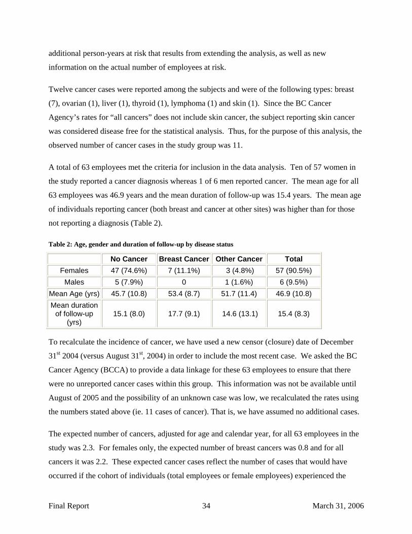
additional person-years at risk that results from extending the analysis, as well as new
information on the actual number of employees at risk.
Twelve cancer cases were reported among the subjects and were of the following types: breast
(7), ovarian (1), liver (1), thyroid (1), lymphoma (1) and skin (1). Since the BC Cancer
Agency’s rates for “all cancers” does not include skin cancer, the subject reporting skin cancer
was considered disease free for the statistical analysis. Thus, for the purpose of this analysis, the
observed number of cancer cases in the study group was 11.
A total of 63 employees met the criteria for inclusion in the data analysis. Ten of 57 women in
the study reported a cancer diagnosis whereas 1 of 6 men reported cancer. The mean age for all
63 employees was 46.9 years and the mean duration of follow-up was 15.4 years. The mean age
of individuals reporting cancer (both breast and cancer at other sites) was higher than for those
not reporting a diagnosis (Table 2).
Table 2: Age, gender and duration of follow-up by disease status
No Cancer Breast Cancer Other Cancer Total Females 47 (74.6%) 7 (11.1%) 3 (4.8%) 57 (90.5%)
Males 5 (7.9%) 0 1 (1.6%) 6 (9.5%) Mean Age (yrs) 45.7 (10.8) 53.4 (8.7) 51.7 (11.4) 46.9 (10.8) Mean duration
of follow-up (yrs)
15.1 (8.0) 17.7 (9.1) 14.6 (13.1) 15.4 (8.3)
To recalculate the incidence of cancer, we have used a new censor (closure) date of December
31st 2004 (versus August 31st, 2004) in order to include the most recent case. We asked the BC
Cancer Agency (BCCA) to provide a data linkage for these 63 employees to ensure that there
were no unreported cancer cases within this group. This information was not be available until
August of 2005 and the possibility of an unknown case was low, we recalculated the rates using
the numbers stated above (ie. 11 cases of cancer). That is, we have assumed no additional cases.
The expected number of cancers, adjusted for age and calendar year, for all 63 employees in the
study was 2.3. For females only, the expected number of breast cancers was 0.8 and for all
cancers it was 2.2. These expected cancer cases reflect the number of cases that would have
occurred if the cohort of individuals (total employees or female employees) experienced the
Final Report 34 March 31, 2006

same rate of cancer as the BC population. The computation of expected numbers of cases is
adjusted for both the age of each individual as well as the calendar years that they were at risk.
The findings from the statistical analyses are presented in Table 3.
Table 3: Observed and expected cases and age/calendar-year adjusted standardized incidence ratios (SIRs) for breast cancer (females only) and all cancers.
Cause Person-years
Number of subjects
Expected cancers
Observed cancers
Standard Incidence
Ratio
95% Confidence
Intervals
Breast Cancer (females)
856.28 57 0.83 7 8.43 3.39 – 17.38
All cancers (females only)
856.28 57 2.18 10 4.59 2.20 – 8.44
All cancers (all subjects) 973.49 63 2.34 11 4.70 2.35 – 8.41
Data Presented as frequency, mean(standard deviation)
A finding of a SIR of 8.4 for breast cancer with 95 percent confidence intervals exceeding 1.0
indicates that the expected number of breast cancers was significantly elevated. The SIR of 8.4
indicates that the women in the MMH Laboratory were experiencing breast cancer incidence at
approximately eight times the rate than women in the BC population. The 95 percent confidence
intervals suggest that, the true SIR (since this is just a statistical approximation) was expected to
be between 3 and 17 with 95 percent certainty. Therefore, statistically speaking, this is a true
cluster of breast cancer cases that exceeds what is expected among women in BC. Similarly, the
standard incidence rates for all cancers in both men and women were significantly elevated as
compared to the rates in BC. However, given the large proportion of cancers that were of the
breast, the excess in the total cancer SIRs was driven by the high number of reported breast
cancers in the employee cohort.
Cox proportional hazard modeling showed that the variables Age at start of work at MMH
Laboratory, Job Position (Technician vs. Aid, Clerk or ECG), and Job Status (Part time vs. Full
Time) were not related to the hazard rate. The hazard rate is defined as the probability per time
unit that a person who has not developed cancer to the beginning of the respective interval will
develop cancer in that interval (Table 4). This is a very important finding. It suggests that there is
Final Report 35 March 31, 2006

no significantly increased risk for women by job position or by job status and it means that
women who started working in the MMH Laboratory at an older age are at a slightly increased
(though non-significant) risk of developing breast cancer than younger women working in the
laboratory.
Table 4: Hazard ratios and 95% confidence intervals (CIs) of breast cancer in relation to age at start of work at MMHL, position and occupational exposure at MMHL
Variables n Hazard Ratio 95% CI p-value *
Age at start work (yrs) 57 1.07 0.95 – 1.21 0.264
Occupational exposure (yrs) 57 1.03 0.92 – 1.15 0.581
Position Aid, clerk or ECG technician Technician
26 31
1.00 4.24 0.36 – 49.38 0.249
* p-value was derived from Cox proportional hazards model with age at start work and year of occupational exposure as a continuous variable and position as categorical variable.
Field Investigations: Potential Exposures to Potentially Carcinogenic Substances or Physical Agents
The walk-through investigation was conducted in August 2003 and included a review of the
current procedures that may result in employee exposures to chemical or physical agents.
Questions were asked about past practices and exposures to gain an understanding about how
exposures may have changed over the years.
Key points from the walkthrough are provided below:
• Current chemical exposures are minimal because liquid volumes are small and handling
is often minimized through the use of “lock and load” systems
• Exposures to physical agents, such as ionizing radiation and electromagnetic fields
appears to not be excessive (heat and noise exposures were also minimal). In September
of 2004, Radiation Protection Services (BCCDC), completed an assessment of radiation
exposure in the laboratory, concluding that “the exposures measured in the Mission
Memorial Laboratory Area are typical natural background and that the X–ray facility is
not contributing to this natural background…this natural background radiation would not
contribute measurably to increased cancer risk.” (Attachment 6)
Final Report 36 March 31, 2006

• Past exposures were likely much higher as a number of procedures have been modified
due to technological advances
o A major change was in the preservation of tissue samples, tissue staining, and
glucose measurement. These procedures, in the past, required open use of
solvents and reagents which included formalin, xylene, and o-toluidine. Most of
these procedures were performed in a separate area of the laboratory, which was
removed when the procedures were modified. It should be noted that o-toluidine,
which was discussed in the literature review, is a rat mammary carcinogen, and
formaldehyde (the major component in formalin) is a known human carcinogen.
o Other areas of the laboratory also were renovated due to changes in laboratory
procedures. Remnants of a local exhaust ventilation system are present in one
area where open chemicals were once mixed and dispensed.
• Poor indoor air quality was a common complaint in the past but appears to be less of a
problem currently. An incinerator at the hospital was a source of very odorous and
potentially hazardous compounds (likely acid gases and possible combustion products of
PVC (monomers of vinyl chloride) and other plastics (halogenated organics)).
Previous air quality studies have been performed at MMH Laboratory; however investigators did
not have access to the historical findings. In discussions with occupational health and safety
professionals at FH, it was mentioned that previous studies were standard IAQ surveys and all
measured concentrations of air contaminants were below regulated limits.
Conclusions
Summary
• The incidence of breast cancer among MMH Laboratory employees (SIR=8.4) statistically
exceeds the expected incidence rate of breast cancer among women. Therefore this is a true
cluster of breast cancer cases.
• We conclude, based on a proportional hazards analysis, that this increase is not statistically
related to age at start of work or duration of exposure. The risk of breast cancer by job
Final Report 37 March 31, 2006

position (technician vs. aid or clerk), is elevated but this increase is not statistically
significant.
• On observation and literature review, no current occupational chemical exposures, or records
of past occupational exposures were found that might relate working in the MMH laboratory
environment to elevated breast cancer risk, or cancer in general. No significant findings were
found during radiation testing in the laboratory, or on basic air quality testing.
• We conclude that this investigation be closed and an update to the analysis conducted in five
years time. Should a larger cohort study be conducted that suggests an increase in breast
cancer in laboratory workers, or if a hypothesis is generated based on new scientific
knowledge, the concerns of the employees of MMH laboratory should be reviewed at that
time.
In our study we did not gather personal information pertaining to known risk factors for breast
cancer. The reason for not gathering this information was that this is a preliminary
epidemiological study and information on risk factors is difficult to interpret without a
comparison population where the prevalence of risk factors is available. For example, in our
study if we had detailed information about reproductive factors, family history of breast cancer,
socioeconomic factors, alcohol consumption, physical exercise, and obesity, we would only be
able to compare the prevalence of those factors with those within the general population. Thus,
such data would provide clues as to the possible reasons for the elevated risk – if the prevalence
of these risk factors were the same as the general population it would suggest that occupational
factor(s) predominate. Only a full-scale etiologic investigation would have the capability of
clearly identifying occupational factors as attributable to the increased breast cancer risk.
A full-scale epidemiologic study is not an appropriate action to take despite the increased rates of
cancer MMH Laboratory employees have experienced. The major goal of cluster investigations
is to identify risk factors so that action can be taken to reduce exposures and risk. Air quality
studies and reviews of procedures indicate that current exposures to carcinogens are minimal.
Past exposures to chemicals like o-toluidine may have resulted in some increased risk for
employees, but these exposures appear to have been eliminated.
Final Report 38 March 31, 2006

Another issue that discourages a major epidemiologic investigation pertains to the statistics of
clusters themselves. Cluster research has shown that elevated rates occur by chance at some
geographic locations and times. In fact, clusters always occur and it is a statistical phenomenon
– even when there is no causal factor that is responsible for the increased incidence (this is why
so few cluster investigations uncover any new risk factors). So, if we look around at many
geographic areas and times we will find some clusters; if a specific cluster is related to statistics
and not an etiologic agent, it is most likely that in the next time period at this location the rate
will not be significantly elevated. Thus, it would be very prudent to continue to evaluate the
incidence of breast cancer in MMH Laboratory employees to see if the rate comes closer to what
is expected.
In summary, this study confirmed that the perceived cluster was an observed cluster and that
MMH Laboratory employees were experiencing an elevated rate of breast cancer. The factors
associated with this increased incidence could not be determined but may have been due to: (1) a
cluster of reproductive and other known, non-occupational, risk factors, (2) past exposures to
chemical carcinogens and less likely to ionizing radiation, and (3) a statistical anomaly.
Recommendations
Our recommendations for action to be considered are:
(1) provide education to all employees about risk factors for breast cancer and the importance of
self exams and mammography, with assistance provided to ensure access to mammography if
needed;
(2) continue to collect information on the incidence of breast and all cancers in the future so that
standardized incidence ratios (SIRs) can be re-calculated in five years time;
(3) a new investigation can be considered at a future time if larger cohort studies suggest a link
between breast cancer and hospital work, or laboratory work in particular, and/or if new
scientific knowledge allows for a hypothesis on work-related causation to be generated for
testing; and .
Final Report 39 March 31, 2006

(4) Every effort should continue to be made, in this and all workplaces, to ensure that
workplaces remain as safe and free of carcinogenic exposures as possible, and that the workforce
is able to pursue safe and healthy choices in all aspects of their lives.
Final Report 40 March 31, 2006
References:
1. Cartwright R. Cluster investigations: are they worth it? Med J Aust 1999;171(4):172.
2. Schulte P, Ehrenberg R, Singal M. Investigation of occupational cancer clusters: theory and practice. Am J Public Health 1987;77(1):52-6.
3. WSDOH. Guidelines for Investigating Clusters of Chronic Disease and Adverse Birth Outcomes: Olympia, Washington, Washington State Department of Health., 2001.
4. Caldwell GG. Twenty-two years of cancer cluster investigations at the Centers for Disease Control. Am J Epidemiol 1990;132(1 Suppl):S43-7.
5. Guidelines for the Investigation of Cancer Clusters in BC. Vancouver: BC Cancer Agency; Cancer Control Research, 1998
6. Breslow N. Statistical Methods in Cancer Research. Lyon, France, 1987.
7. Jacobzone S. Summary of the Results from the Breast Cancer Disease Study. OECD Study on Cross-national Differences of Ageing-related Diseases Workshop. Organization for Economic Cooperation and Development 2002, Paris, France,.
8. Melbye M, Wohlfahrt J, Olsen JH, et al. Induced abortion and the risk of breast cancer. N Engl J Med 1997;336(2):81-5.
9. Erlandsson G, Montgomery SM, Cnattingius S, Ekbom A. Abortions and breast cancer: record-based case-control study. Int J Cancer 2003;103(5):676-9.
10. Breast Cancer Statistics: Canadian Cancer Society, 2006.
11. Provincial Health Officer's Annual Report: The Health of British Columbians
Victoria, BC: Ministry of Health and Ministry Responsible for Seniors, 1994.
12. Schulmeister L. Diethylstilbestrol exposure: how well informed are you about this health risk? Clin J Oncol Nurs 2003;7(4):373-4.
13. Laitman CJ. DES exposure and the aging woman: mothers and daughters. Curr Womens Health Rep 2002;2(5):390-3.
14. Bruzzi P, Negri E, La Vecchia C, et al. Short term increase in risk of breast cancer after full term pregnancy. Bmj 1988;297(6656):1096-8.
15. Cohn BA, Cirillo PM, Christianson RE, van den Berg BJ, Siiteri PK. Placental characteristics and reduced risk of maternal breast cancer. J Natl Cancer Inst 2001;93(15):1133-40.
16. Deligeoroglou E, Michailidis E, Creatsas G. Oral contraceptives and reproductive system cancer. Ann N Y Acad Sci 2003;997:199-208.
Final Report 41 March 31, 2006

17. Burkman RT. Oral contraceptives: an update. Drugs Today (Barc) 1999;35(11):857-66.
18. Vassilopoulou-Sellin R. Breast cancer and hormonal replacement therapy. Ann N Y Acad Sci 2003;997:341-50.
19. Land CE, Tokunaga M, Koyama K, et al. Incidence of female breast cancer among atomic bomb survivors, Hiroshima and Nagasaki, 1950-1990. Radiat Res 2003;160(6):707-17.
20. Ron E. Cancer risks from medical radiation. Health Phys 2003;85(1):47-59.
21. DeBruin LS, Josephy PD. Perspectives on the chemical etiology of breast cancer. Environ Health Perspect 2002;110 Suppl 1:119-28.
22. Yue W, Santen RJ, Wang JP, et al. Genotoxic metabolites of estradiol in breast: potential mechanism of estradiol induced carcinogenesis. J Steroid Biochem Mol Biol 2003;86(3-5):477-86.
23. Wang DY, Allen DS, De Stavola BL, et al. Urinary androgens and breast cancer risk: results from a long-term prospective study based in Guernsey. Br J Cancer 2000;82(9):1577-84.
24. Beral V, Bull D, Doll R, Peto R, Reeves G. Breast cancer and abortion: collaborative reanalysis of data from 53 epidemiological studies, including 83?000 women with breast cancer from 16 countries. Lancet 2004;363(9414):1007-16.
25. Zheng T, Holford TR, Mayne ST, et al. Lactation and breast cancer risk: a case-control study in Connecticut. Br J Cancer 2001;84(11):1472-6.
26. Russo IH, Russo J. Role of hormones in mammary cancer initiation and progression. J Mammary Gland Biol Neoplasia 1998;3(1):49-61.
27. Russo IH, Russo J. Mammary gland neoplasia in long-term rodent studies. Environ Health Perspect 1996;104(9):938-67.
28. Jernstrom H, Lubinski J, Lynch HT, et al. Breast-feeding and the risk of breast cancer in BRCA1 and BRCA2 mutation carriers. J Natl Cancer Inst 2004;96(14):1094-8.
29. Brody JG, Rudel RA. Environmental pollutants and breast cancer. Environ Health Perspect 2003;111(8):1007-19.
30. Parkin DM, Bray F, Ferlay J, Pisani P. Estimating the world cancer burden: Globocan 2000. Int J Cancer 2001;94(2):153-6.
31. Missmer SA, Eliassen AH, Barbieri RL, Hankinson SE. Endogenous estrogen, androgen, and progesterone concentrations and breast cancer risk among postmenopausal women. J Natl Cancer Inst 2004;96(24):1856-65.
32. Halls S. Detailed breast cancer risk calculator, 2004.
Final Report 42 March 31, 2006
33. Kreiger N, Sloan M, Cotterchio M, Kirsh V. The risk of breast cancer following reproductive surgery. Eur J Cancer 1999;35(1):97-101.
34. Hamajima N, Hirose K, Tajima K, et al. Alcohol, tobacco and breast cancer--collaborative reanalysis of individual data from 53 epidemiological studies, including 58,515 women with breast cancer and 95,067 women without the disease. Br J Cancer 2002;87(11):1234-45.
35. Bennett LM, Davis BJ. Identification of mammary carcinogens in rodent bioassays. Environ Mol Mutagen 2002;39(2-3):150-7.
36. Charles GD, Gennings C, Zacharewski TR, Gollapudi BB, Carney EW. Assessment of interactions of diverse ternary mixtures in an estrogen receptor-alpha reporter assay. Toxicol Appl Pharmacol 2002;180(1):11-21.
37. Bounias M. Etiological factors and mechanism involved in relationships between pesticide exposure and cancer. J Environ Biol 2003;24(1):1-8.
38. Safe SH, Zacharewski T. Organochlorine exposure and risk for breast cancer. Prog Clin Biol Res 1997;396:133-45.
39. Snedeker S. Do we need to be concerned about environmental chemicals and breast cancer? An interview with the BCERF Research Project Leader. In: 2. TR, ed, 1997.
40. Charlier C, Foidart JM, Pitance F, et al. Environmental dichlorodiphenyltrichlorethane or hexachlorobenzene exposure and breast cancer: is there a risk? Clin Chem Lab Med 2004;42(2):222-7.
41. Sonnenschein C, Soto AM. An updated review of environmental estrogen and androgen mimics and antagonists. J Steroid Biochem Mol Biol 1998;65(1-6):143-50.
42. Band PR, Le ND, Fang R, Deschamps M, Gallagher RP, Yang P. Identification of occupational cancer risks in British Columbia. A population-based case-control study of 995 incident breast cancer cases by menopausal status, controlling for confounding factors. J Occup Environ Med 2000;42(3):284-310.
43. Weaver VM. Chemical hazards in health care workers. Occup Med 1997;12(4):655-67.
44. Tompa A, Major J, Jakab MG. Is breast cancer cluster influenced by environmental and occupational factors among hospital nurses in Hungary? Pathol Oncol Res 1999;5(2):117-21.
45. Burnett C, Robinson C, Walker J. Cancer mortality in health and science technicians. Am J Ind Med 1999;36(1):155-8.
46. Bigelow P. Application of Threshold Limit Values for Chemical Substances to employees in laboratories Chem Health Safety 2000;8:16-21.
Final Report 43 March 31, 2006

47. Madigan MP, Ziegler RG, Benichou J, Byrne C, Hoover RN. Proportion of breast cancer cases in the United States explained by well-established risk factors. J Natl Cancer Inst 1995;87(22):1681-5.
48. Claus EB. Genetic epidemiology of breast cancer in younger women. J Natl Cancer Inst Monogr 1994(16):49-53.
49. Recchia F, Nuzzo A, Lalli A, De Filippis S, Torchio P. Activity of standard-dose carboplatin, cyclophosphamide, and etoposide in patients with metastatic breast cancer with previous exposure to anthracyclines. Am J Clin Oncol 1997;20(2):166-8.
50. Medina D. Breast cancer: the protective effect of pregnancy. Clin Cancer Res 2004;10(1 Pt 2):380S-4S.
51. Zeleniuch-Jacquotte A, Adlercreutz H, Shore RE, et al. Circulating enterolactone and risk of breast cancer: a prospective study in New York. Br J Cancer 2004;91(1):99-105.
52. PPHB. Breast cancer in Canada: Population and Pulic Health Branch, Health Canada, 2004.
53. CCR. Guidelines for the investigation of cancer clusters investigations in BC. In: Research CC, ed, 1998.
54. Palomares MR, Paz B, Weitzel JN. Genetic cancer risk assessment in the newly diagnosed breast cancer patient is useful and possible in practice. J Clin Oncol 2005;23(13):3165-6; author reply 3166-7.
55. Ward EM, Schulte PA, Bayard S, et al. Priorities for development of research methods in occupational cancer. Environ Health Perspect 2003;111(1):1-12.
56. Jemal A, Tiwari RC, Murray T, et al. Cancer statistics, 2004. CA Cancer J Clin 2004;54(1):8-29.
57. National Cancer Institute:Cancer Statistics, 2004.
58. Tryggvadottir L, Tulinius H, Eyfjord JE, Sigurvinsson T. Breastfeeding and reduced risk of breast cancer in an Icelandic cohort study. Am J Epidemiol 2001;154(1):37-42.
59. Chu KC, Tarone RE, Kessler LG, et al. Recent trends in U.S. breast cancer incidence, survival, and mortality rates. J Natl Cancer Inst 1996;88(21):1571-9.
60. Li CI, Anderson BO, Daling JR, Moe RE. Trends in incidence rates of invasive lobular and ductal breast carcinoma. Jama 2003;289(11):1421-4.
61. Rossouw JE, Anderson GL, Prentice RL, et al. Risks and benefits of estrogen plus progestin in healthy postmenopausal women: principal results From the Women's Health Initiative randomized controlled trial. Jama 2002;288(3):321-33.
62. Verkooijen HM, Fioretta G, Vlastos G, et al. Important increase of invasive lobular breast cancer incidence in Geneva, Switzerland. Int J Cancer 2003;104(6):778-81.
Final Report 44 March 31, 2006
63. Willett W, Rockhill B, Hankinson S. Epidemiology and nongenetic causes of breast cancer, 2000.
64. Henderson BE, Bernstein L. The international variation in breast cancer rates: an epidemiological assessment. Breast Cancer Res Treat 1991;18 Suppl 1:S11-7.
65. Chevarley F, White E. Recent trends in breast cancer mortality among white and black US women. Am J Public Health 1997;87(5):775-81.
66. Peto J, Mack TM. High constant incidence in twins and other relatives of women with breast cancer. Nat Genet 2000;26(4):411-4.
67. Pike C, Spicer D, Dahoush L, Press M. Estrogens, progestogens, normal breast cell proliferation and breast cancer risk. Epidemiol Rev 1993;15(1):17-35.
68. Kelsey JL, Fischer DB, Holford TR, et al. Exogenous estrogens and other factors in the epidemiology of breast cancer. J Natl Cancer Inst 1981;67(2):327-33.
69. Sturgeon SR, Schairer C, Gail M, McAdams M, Brinton LA, Hoover RN. Geographic variation in mortality from breast cancer among white women in the United States. J Natl Cancer Inst 1995;87(24):1846-53.
70. Bradley CJ, Given CW, Roberts C. Race, socioeconomic status, and breast cancer treatment and survival. J Natl Cancer Inst 2002;94(7):490-6.
71. Breast cancer and breastfeeding: collaborative reanalysis of individual data from 47 epidemiological studies in 30 countries, including 50302 women with breast cancer and 96973 women without the disease. Lancet 2002;360(9328):187-95.
72. Chen PL, Sellers TA, Rich SS, Potter JD, Folsom AR. Examination of the effect of nongenetic risk factors on the familial risk of breast cancer among relatives of postmenopausal breast cancer patients. Cancer Epidemiol Biomarkers Prev 1994;3(7):549-55.
73. Magnusson C, Colditz G, Rosner B, Bergstrom R, Persson I. Association of family history and other risk factors with breast cancer risk (Sweden). Cancer Causes Control 1998;9(3):259-67.
74. Lichtenstein P, Holm NV, Verkasalo PK, et al. Environmental and heritable factors in the causation of cancer--analyses of cohorts of twins from Sweden, Denmark, and Finland. N Engl J Med 2000;343(2):78-85.
75. Trock BJ. Breast cancer in African American women: epidemiology and tumor biology. Breast Cancer Res Treat 1996;40(1):11-24.
76. Boyd NF, Dite GS, Stone J, et al. Heritability of mammographic density, a risk factor for breast cancer. N Engl J Med 2002;347(12):886-94.
Final Report 45 March 31, 2006

77. Palmer JR, Wise LA, Horton NJ, Adams-Campbell LL, Rosenberg L. Dual effect of parity on breast cancer risk in African-American women. J Natl Cancer Inst 2003;95(6):478-83.
78. Byrne C, Schairer C, Wolfe J, et al. Mammographic features and breast cancer risk: effects with time, age, and menopause status. J Natl Cancer Inst 1995;87(21):1622-9.
79. Dupont WD, Page DL. Risk factors for breast cancer in women with proliferative breast disease. N Engl J Med 1985;312(3):146-51.
80. Marshall LM, Hunter DJ, Connolly JL, et al. Risk of breast cancer associated with atypical hyperplasia of lobular and ductal types. Cancer Epidemiol Biomarkers Prev 1997;6(5):297-301.
81. Brind J, Chinchilli VM, Severs WB, Summy-Long J. Induced abortion as an independent risk factor for breast cancer: a comprehensive review and meta-analysis. J Epidemiol Community Health 1996;50(5):481-96.
82. Clemons M, Goss P. Estrogen and the risk of breast cancer. N Engl J Med 2001;344(4):276-85.
83. Kelsey JL, Gammon MD, John EM. Reproductive factors and breast cancer. Epidemiol Rev 1993;15(1):36-47.
84. MacMahon B, Trichopoulos D, Brown J, et al. Age at menarche, urine estrogens and breast cancer risk. Int J Cancer 1982;30(4):427-31.
85. Hsieh CC, Trichopoulos D, Katsouyanni K, Yuasa S. Age at menarche, age at menopause, height and obesity as risk factors for breast cancer: associations and interactions in an international case-control study. Int J Cancer 1990;46(5):796-800.
86. Layde PM, Webster LA, Baughman AL, Wingo PA, Rubin GL, Ory HW. The independent associations of parity, age at first full term pregnancy, and duration of breastfeeding with the risk of breast cancer. Cancer and Steroid Hormone Study Group. J Clin Epidemiol 1989;42(10):963-73.
87. Brinton LA, Schairer C, Hoover RN, Fraumeni JF, Jr. Menstrual factors and risk of breast cancer. Cancer Invest 1988;6(3):245-54.
88. Snider J, Beauvais J, Levy I, Villeneuve P, Pennock J. Trends in mammography and Pap smear utilization in Canada. Chronic Dis Can 1996;17(3-4):108-17.
89. Services BMoH. A Report to the Minister of Health from the Minister's Advisory Council on Women's Healt. In: Women BCaBC, ed, 2005.
90. Unit NSH. North Shore Health Promotion Survey. North Vancouver, B.C.: Unpublished report. , 1990.
Final Report 46 March 31, 2006

91. Hu YF, Russo IH, Zalipsky U, Lynch HT, Russo J. Environmental chemical carcinogens induce transformation of breast epithelial cells from women with familial history of breast cancer. In Vitro Cell Dev Biol Anim 1997;33(7):495-8.
92. Safe SH. Is there an association between exposure to environmental estrogens and breast cancer? Environ Health Perspect 1997;105 Suppl 3:675-8.
93. Goldberg MS, Labreche F. Occupational risk factors for female breast cancer: a review. Occup Environ Med 1996;53(3):145-56.
94. Morton WE. Major differences in breast cancer risks among occupations. J Occup Environ Med 1995;37(3):328-35.
95. Fisher B, Dignam J, Wolmark N, et al. Tamoxifen in treatment of intraductal breast cancer: National Surgical Adjuvant Breast and Bowel Project B-24 randomised controlled trial. Lancet 1999;353(9169):1993-2000.
96. Page DL, Dupont WD, Rogers LW, Jensen RA, Schuyler PA. Continued local recurrence of carcinoma 15-25 years after a diagnosis of low grade ductal carcinoma in situ of the breast treated only by biopsy. Cancer 1995;76(7):1197-200.
97. Page DL, Schuyler PA, Dupont WD, Jensen RA, Plummer WD, Jr., Simpson JF. Atypical lobular hyperplasia as a unilateral predictor of breast cancer risk: a retrospective cohort study. Lancet 2003;361(9352):125-9.
98. Wennborg H, Magnusson LL, Bonde JP, Olsen J. Congenital malformations related to maternal exposure to specific agents in biomedical research laboratories. J Occup Environ Med 2005;47(1):11-9.
99. Wennborg H, Bodin L, Vainio H, Axelsson G. Pregnancy outcome of personnel in Swedish biomedical research laboratories. J Occup Environ Med 2000;42(4):438-46.
100. Brown TP, Paulson J, Pannett B, et al. Mortality pattern among biological research laboratory workers. Br J Cancer 1996;73(9):1152-5.
101. van Barneveld TA, Sasco AJ, van Leeuwen FE. A cohort study of cancer mortality among Biology Research Laboratory workers in The Netherlands. Cancer Causes Control 2004;15(1):55-66.
102. Shaham J, Gurvich R, Kneshet Y. Cancer incidence among laboratory workers in biomedical research and routine laboratories in Israel: Part I-the cohort study. Am J Ind Med 2003;44(6):600-10.
103. Shaham J, Gurvich R, Kneshet Y. Cancer incidence among laboratory workers in biomedical research and routine laboratories in Israel: Part II-nested case-control study. Am J Ind Med 2003;44(6):611-26.
104. Mattison D, Austin R, Chrostowski P, et al. New Waste Incinerators Safer, But Some Emissions and Health Concerns Need Further Study.Committee on Health Effects of
Final Report 47 March 31, 2006

Waste Incineration, Board on Environmental Studies and Toxicology, National Research Council., 1999: http://www4.nationalacademies.org/news.nsf/isbn/030906371X?OpenDocument.
105. How to shut down incinerator. Science. technology & health hazards of incineration. Henry Ford Hospital waste incinerator. Sample Fact sheet & Watch dog Follow-up: http://www.noharm.org/library/docs/Toolkit_6_Sample_Fact_Sheet_and_Watchdog_Follo.pdf, 1999.
106. Johnson B. Impact of hazardous waste on human health. US:CRC Press, 1999.
107. Franchini M, Rial M, Buiatti E, Bianchi F. Health effects of exposure to waste incinerator emissions:a review of epidemiological studies. Ann Ist Super Sanita 2004;40(1):101-15.
108. Lloyd OL, Lloyd MM, Williams FL, Lawson A. Twinning in human populations and in cattle exposed to air pollution from incinerators. Br J Ind Med 1988;45(8):556-60.
109. Gustavsson P. Mortality among workers at a municipal waste incinerator. Am J Ind Med 1989;15(3):245-53
110. Doll R, Vessey MP, Beasley RW, et al. Mortality of gasworkers - final report of a prospective study. Br J Ind Med 1972;29(4):394-406.
111. Gustavsson P, Gustavsson A, Hogstedt C. Excess mortality among Swedish chimney sweeps. Br J Ind Med 1987;44(11):738-43.
112. Hospital waste system due in july. Prince George Citizen May 6. 1995.
113. Hawthorne M. Two hospitals made incinerator deals. Chicago Tribune 2005 Aug 15.
114. Brown JR, Thornton JL. Percivall Pott (1714-1788) and chimney sweepers' cancer of the scrotum. Br J Ind Med 1957;14(1):68-70.
115. Kheifets LI. Cluster analysis: a perspective. Stat Med 1993;12(19-20):1755-6.
116. Robbins AS, Brescianini S, Kelsey JL. Regional differences in known risk factors and the higher incidence of breast cancer in San Francisco. J Natl Cancer Inst 1997;89(13):960-5.
117. Breast Cancer Rates, 1986-1995: Natural Resources Canada: Canadian Government Publishing, 2004.
118. Gammon MD, Neugut AI, Santella RM, et al. The Long Island Breast Cancer Study Project: description of a multi-institutional collaboration to identify environmental risk factors for breast cancer. Breast Cancer Res Treat 2002;74(3):235-54.
Final Report 48 March 31, 2006

119. Wittenberg C. Long Island breast cancer studies move forward. J Natl Cancer Inst 1994;86(20):1501-3.
120. Timander LM, McLafferty S. Breast cancer in West Islip, NY: a spatial clustering analysis with covariates. Soc Sci Med 1998;46(12):1623-35.
121. Breast cancer and hormone replacement therapy: collaborative reanalysis of data from 51 epidemiological studies of 52,705 women with breast cancer and 108,411 women without breast cancer. Collaborative Group on Hormonal Factors in Breast Cancer. Lancet 1997;350(9084):1047-59.
122. Langmuir AD. Formal discussion of: epidemiology of cancer: spatial-temporal aggregation. Cancer Res 1965;25(8):1384-6.
123. Kulldorff M, Athas WF, Feurer EJ, Miller BA, Key CR. Evaluating cluster alarms: a space-time scan statistic and brain cancer in Los Alamos, New Mexico. Am J Public Health 1998;88(9):1377-80.
124. Knorr-Held L, Rasser G. Bayesian detection of clusters and discontinuities in disease maps. Biometrics 2000;56(1):13-21.
125. Hanrahan LP, Mirkin I, Olson J, Anderson HA, Fiore BJ. SMRFIT: a Statistical Analysis System (SAS) program for standardized mortality ratio analyses and Poisson regression model fits in community disease cluster investigations. Am J Epidemiol 1990;132(1 Suppl):S116-22.
126. Hall HI, Lee CV, Kaye WE. Cluster: a software system for epidemiologic cluster analysis. Stat Med 1996;15(7-9):943-50.
127. Gangnon RE, Clayton MK. A weighted average likelihood ratio test for spatial clustering of disease. Stat Med 2001;20(19):2977-87.
128. Semenciw RM, Le ND, Marrett LD, Robson DL, Turner D, Walter SD. Methodological issues in the development of the Canadian Cancer Incidence Atlas. Stat Med 2000;19(17-18):2437-49.
129. Aldrich T, Sinks T. Things to know and do about cancer clusters. Cancer Invest 2002;20(5-6):810-6.
130. Siegrist M, Cvetkovich G, Roth C. Shared values, social trust, and the perception of geographic cancer clusters. Risk Analysis 2001;21(6):1047-53.
131. Trumbo CW. Public requests for cancer cluster investigations: a survey of state health departments. Am J Public Health 2000;90(8):1300-2.
132. Garry VF, Jr. Environmental pathology, target environmental cancer. Minn Med 1978;61(5):328-30.
133. Bender AP. On disease clustering. Am J Public Health 1987;77(6):742.
Final Report 49 March 31, 2006

134. Bender AP, Williams AN, Johnson RA, Jagger HG. Appropriate public health responses to clusters: the art of being responsibly responsive. Am J Epidemiol 1990;132(1 Suppl):S48-52.
135. Lewis R, Rempala G. A case-cohort study of angiosarcoma of the liver and brain cancer at a polymer production plant. J Occup Environ Med 2003;45(5):538-45.
136. Lieben J. Malignancies in asbestos workers. Arch Environ Health 1966;13(5):619-21.
137. Lieben J, Pistawka H. Mesothelioma and asbestos exposure. Arch Environ Health 1967;14(4):559-63.
138. Finkelstein MM, Wilk N. Investigation of a lung cancer cluster in the melt shop of an Ontario steel producer. Am J Ind Med 1990;17(4):483-91.
139. van Netten C, Brands RH, Hoption Cann SA, Spinelli JJ, Sheps SB. Cancer cluster among police detachment personnel. Environ Int 2003;28(7):567-72.
140. Drijver M, Woudenberg F. Cluster management and the role of concerned communities and the media. Eur J Epidemiol 1999;15(9):863-9.
141. Fiore BJ, Hanrahan LP, Anderson HA. State health department response to disease cluster reports: a protocol for investigation. Am J Epidemiol 1990;132(1 Suppl):S14-22.
142. Quataert PK, Armstrong B, Berghold A, et al. Methodological problems and the role of statistics in cluster response studies: a framework. Eur J Epidemiol 1999;15(9):821-31.
143. Kipen H, Wartenberg D. Don't close the door: some observations on cancer cluster investigation. J Occup Med 1988;30(8):661-2.
144. Frelick RW, Topham A. The value of cluster and environmentally related cancer studies in Delaware. Del Med J 1991;63(9):559-61.
145. Smith D, Neutra R. Approaches to disease cluster investigations in a state health department. Stat Med 1993;12(19-20):1757-62.
Final Report 50 March 31, 2006
Attachment 1
Potential Exposures to Potentially Carcinogenic Substances or Physical Agents
August 2003


Field Investigations: Potential Exposures to Potentially Carcinogenic Substances or
Physical Agents
The walk-through investigation was conducted in August 2003 and included a review of the
current procedures that may result in employee exposures to chemical or physical agents.
Questions were asked about past practices and exposures to gain an understanding about how
exposures may have changed over the years.
Key points from the walkthrough are provided below:
• Current chemical exposures are minimal because liquid volumes are small and handling is
often minimized through the use of “lock and load” systems
• Exposures to physical agents, such as ionizing radiation and electromagnetic fields appears to
not be excessive (heat and noise exposures were also minimal)
• Past exposures were likely much higher as a number of procedures have been modified due
to technological advances
o A major change was in the preservation of tissue samples, tissue staining, and glucose
measurement. These procedures, in the past, required open use of solvents and
reagents which included formalin, xylene, and o-toluidine. Most of these procedures
were performed in a separate area of the laboratory, which was removed when the
procedures were modified. It should be noted that o-toluidine, which was discussed
in the literature review, is a rat mammary carcinogen, and formaldehyde (the major
component in formalin) is a known human carcinogen.
o Other areas of the laboratory also were renovated due to changes in laboratory
procedures. Remnants of a local exhaust ventilation system are present in one area
where open chemicals were once mixed and dispensed.
• Poor indoor air quality was a common complaint in the past but appears to be less of a
problem currently. An incinerator at the hospital was a source of very odourous and
potentially toxic compounds (likely acid gases and possible combustion products of PVC
(monomers of vinyl chloride) and other plastics (halogenated organics)).
• Previous air quality studies have been performed at MMHL, however investigators did not
have access to the findings. In discussions with occupational health and safety professionals
at FHA, it was mentioned that all measured concentrations of air contaminants were below
regulated limits.


Attachment 2
An Investigation of a Cancer Cluster within the Mission Memorial Hospital Laboratory
March 2004


An Investigation of a Cancer Cluster within the Mission Memorial Hospital Laboratory
March 2004
By: Philip Bigelow,1,4
Shicheng Yu,4 Rosemary Nemanishen,2
Annalee Yassi1,3,4
1 Occupational Health and Safety Agency for Healthcare in BC #301- 1195 West Broadway, Vancouver, BC V6H 3X5 Tel: 604-775-4034 Fax: 604-775-4031 Web: www.ohsah.bc.ca 2 Fraser Health Authority 3 Department of Health Care and Epidemiology, University of British Columbia 5804 Fairview Ave., Vancouver, BC V6T 1Z3 4 Institute of Health Promotion Research, University of British Columbia Library Processing Centre, 2206 East Mall, Rm. 435 Vancouver, B.C. Canada V6T 1Z3

2
Abstract An excess in the number of cases of cancer was suspected by employees of the Mission Memorial Hospital Laboratory and this investigation was conducted in response to the above concern. The study had three parts, an epidemiologic cluster analysis, a comprehensive review of the literature on breast cancer risk factors and the analysis of clusters, and an exposure investigation. A total of 57 individuals were identified as having worked in the laboratory between January 1, 1970 and August 31, 2003. Information on health status and diagnoses of cancer was obtained through personal interviews with employees. Ten employees reported a cancer diagnosis, of which 6 were breast cancer. A total of 704 person-years of observation were available for the data analysis after the exclusion of subjects because of diagnoses of cancer prior to start of employment (n=1) and lost to follow-up (n=10). Based on the age and calendar-year adjusted rates for the BC population, the expected number of breast cancer cases in the women was 0.56, and the expected number of all cancers for all employees was 1.51. The Standard Incidence Ratios (SIR), which are the observed number of cases divided by the expected number, were 10.7 for breast cancer in women, and 6.6 for all cancers in both men and women. The 95 percent confidence intervals indicate both findings were significant. A walk-through survey of the laboratory did not identify any potentially hazardous exposures. Recommendations include a comprehensive assessment of all potential exposures to chemicals and physical agents, continuing to collect epidemiological information to determine temporal trends in the SIR, providing information on breast cancer risks to employees, and collecting information on known risk factors for breast cancer and confirming cancer diagnoses through linkage with the BC Cancer Agency’s registry.

3
Introduction The Occupational Health and Safety Agency for Healthcare (OHSAH) was invited by the Fraser Health Authority (FHA) to investigate concerns of a greater than expected number of cancer cases in the Mission Memorial Hospital Laboratory (MMHL). In addition to the seemingly high total number of cancer cases, a large proportion were the same type (breast cancer) thus further highlighting the need for an investigation. Occupational health professionals from FHA had completed some exploratory work on the project but felt they needed the help of outside experts to resolve the issues. OHSAH conducted a preliminary cluster investigation which is the focus of this report. Dr. Philip Bigelow, a senior scientist at OHSAH, led the investigation and team members included Dr. Annalee Yassi, Rosemary Nemanishen, Dr. Shicheng Yu and William So Yiu Ting. Assistance was also obtained from Dr. Nhu Lee of the British Columbia (BC) Cancer Agency and Dr. Martha Vela Acosta of the Department of Environmental and Radiological Health Sciences at Colorado State University. Cancer clusters are the occurrences of greater numbers of the same type of cancer, or all cancers, within a geographic location over a specified period of time. The purpose of a cancer cluster investigation is to determine if the observed number of cases is higher than expected. If the observed number is higher, the study should provide information that may help to reduce or eliminate exposures that may be associated with the increased risk. Cancer cluster investigations are often conducted by health agencies and this investigation followed general guidelines recommended by the BC Cancer Agency and public health departments in other jurisdictions across North America (Schulte, Ehrenberg et al. 1987; Caldwell 1990; Cartwright 1999; WSDOH 2001). Prior to the investigation, an explanatory meeting was held at Mission Memorial Hospital to discuss the incidence of cancer at MMHL and the protocol for the investigation. Details of the protocol used during the investigation are provided below. Methods The specific aim of this study was to provide a thorough determination if an excess in the number of cancer cases had occurred in the Mission Memorial Hospital Laboratory (MMHL) and to provide information regarding the possibility that a work-related factor was involved. More importantly, the goal was to ensure that current workplace conditions and exposures are not at all likely to result in an increased risk of cancer for employees. The BC Cancer Agency, as well as other health agencies, have adopted standard protocols for investigating clusters. The methods used in our study followed these standard procedures and included determining if an excess number of cancers was reported, a literature review on the risk factors for the specific cancer types, assessing the potential for occupational exposures to potentially carcinogenic physical agents or substances, and determining the feasibility of further epidemiology studies. Our study was divided into three components as listed below.
4
1) Analysis of Cancer Incidence Data: Epidemiology and Statistics Since the concern pertained to employees in the MMHL, only employees who worked in the Laboratory were included in the study. Using data from the Human Resources Department at Mission Memorial Hospital, all employees who were employed in the Laboratory over the past 33 years (January 1970 to August 2003) were identified and this information was provided to an occupational health professional at Fraser Health. A total of 57 individuals were identified and the following information was entered into a computer spreadsheet: date of birth, dates of employment at the lab, job title, full or part time employment status, gender, and other details pertaining to work at the lab and hospital. A health professional (Registered Nurse) from Fraser Health attempted to contact all 57 individuals (in person or by telephone) to gather information on whether or not they had a diagnosis of cancer of any type. For individuals who reported a cancer diagnosis, information on the diagnosis date, type and site of cancer was obtained. Data for all 57 individuals, without personal identifiers, were entered into a spreadsheet and provided to OHSAH. The statistical analysis was conducted two ways. In one, the person-years of observation was defined as being from the start date of employment at MMHL to the end date of employment. In the second analysis, the person-years of observation was defined as the time between the employee start date and the end of the follow up period (August 2003). In this report the latter analysis is provided, as it is the most appropriate for the study design that was used.1 Since a large proportion of the cases were classified as breast cancers, statistical analyses were conducted using rates of breast and total cancers obtained from the BC Cancer Agency. Rates for breast and total cancers for each year from 1970 to 2002, grouped by 5 year age intervals, were used to calculate the expected number of cases in the study group. The expected number of cases is the number of cases expected in the Laboratory if the rate was the same as the rate in BC adjusted for age and calendar year.2 The expected number of cases was computed by multiplying the population (person-years of observation) within each specific age range and year by the rate of breast or total cancers for the same age interval and year. The results of these computations were summed across all the age and year categories to get the total number of expected cases. Computations and statistical analyses were conducted using Excel and SPSS software. 1 In many occupational cohort studies, when subjects leave employment their health status at that time is known and their end date of employment is used in the computation of person-years of observation. In this study, we contacted all study subjects from August to November, 2003 to determine their health status. 2 The risk of developing breast cancer was different in 1980 as compared to today. Additionally, age is a major risk factor for all cancers (including breast cancer). Therefore, crude incidence rates may be misleading when comparing regions or time periods where the age of the populations differ from one region to another or from one time period to another. In this study, the expected number of breast and total cancers was adjusted for age and calendar year and this provides the most accurate comparison of rates for this study.

5
The observed number of cases was divided by the expected number of breast cancer and total cancer cases to determine the Standard Incidence Ratios (SIR). A SIR exceeding 1.0 indicates the observed number is higher than expected. Confidence intervals are used to assess variation in the SIR and 95% Poisson confidence intervals were calculated using the procedure suggested by Breslow (Breslow 1987). 2) Field Investigations – Potential Exposures to Potentially Carcinogenic Substances or Physical Agents Prior to this investigation, work had been conducted by occupational health professionals at Fraser Health to determine the adequacy of procedures to control exposures to chemicals in the laboratory and to ensure exposures did not exceed government or consensus standards. Investigations also focused on potential sources of chemical exposures resulting from work tasks that are typically performed by laboratory personnel. An additional study included reviewing past renovations of the laboratory in hopes of identifying unusual sources of indoor air contaminants. In August of 2003, as part of OHSAH’s investigation, a walkthrough survey of the laboratory was completed. Typical work procedures were reviewed to assess the potential for exposures to hazardous agents. Employees in the laboratory provided information on historical procedures as well as an indication of the general levels of exposure to air contaminants. 3) Literature Review The investigation and response to cancer cluster reports is exceedingly complex and the report includes brief literature reviews on risk factors for breast cancer, exposures in laboratories, and epidemiology of cancer clusters. This information is provided to help interpret the study findings and provide guidance in deciding whether further study is warranted. Results and Discussion The literature review is presented first as it provides a background for interpreting the findings from both the epidemiologic analysis and the field surveys. The literature review of breast cancer highlights the multifactorial nature of disease causation and the difficulties in determining the role of environmental and occupational exposures as causal factors. There is a substantial body of literature on cancer cluster investigations and a brief review is provided. Finally, the findings of the statistical analysis are provided and discussed in relation to findings from other studies. Literature Review: Breast Cancer Risk Factors and the Role of Occupational and Environmental Exposures Breast cancer. Canada has one of the highest rates of breast cancer incidence and the age standardized rate in 1995 exceeded 225 per 100,000 women aged 40 and over (Jacobzone

6
2002). Breast cancer is the most frequently diagnosed cancer in Canadian women and it accounts for over 30% of new cancer cases per year (NCIC 1999). It is estimated that one in nine women in Canada will develop the disease in their lifetime (DeBruin and Josephy 2002). Breast cancer mortality increased steadily from the 1960s until the early 1980s when the rates declined in most countries including Canada (Parkin, Bray et al. 2001; Parkin, Bray et al. 2001). These declining mortality rates are thought to be related to improved screening resulting in earlier detection and improved treatment. Age is major risk factor for breast cancer and Table 1 shows the increased likelihood of a woman developing breast cancer in the next five years at various ages (PPHB 2004). In addition to age, many correlates of risk for breast cancer have been identified and there are a constellation of hormone-related reproductive factors that predominate. Factors known to confer higher risk include younger age at menarche, older age at menopause, nulliparous, and older at first live birth (Davis, Axelrod et al. 1997). Higher parity, longer lactation, and bilateral ovariectomy have been found to be protective (Davis, Axelrod et al. 1997; Kreiger, Sloan et al. 1999). Using data from large population-based surveys in the United States (US), investigators calculated that 41% of breast cancer risk was explained by nulliparity, later childbearing, higher income, and family history of breast cancer (Madigan, Ziegler et al. 1995). Other risk factors for breast cancer include a history of certain types of benign breast disease as well as high levels of radiation exposure to the chest (medical x-rays) (PPHB 2004). Despite the many studies that have been conducted, additional factors, likely modest in magnitude, remain to be discovered. Interestingly, in 76% of women who develop breast cancer, age is the only identifiable risk factor (Halls 2003).
Table 1.3 Probability of developing breast cancer in the next five years.
Age Breast cancer per 1,000 women
30 1.5
35 2.6
40 4.8
45 7.8
50 9.2
55 10.6
60 12.9
65 14.3
70 15.4
80 15.5
Epidemiological and animal studies consistently show elevated risk of breast cancer with factors that increase exposure to estradiol, progesterone, and other hormones (Kreiger, Sloan et al. 1999; Medina 2004; Recchia, Vivacqua et al. 2004; Zeleniuch-Jacquotte, 3 From PPHB (2004). Breast Cancer in Canada. online at: http://www.hc-sc.gc.ca/pphb-dgspsp/publicat/updates/breast-99_e.html, Population and Public Health Branch, Health Canada, Ottawa, Canada. 2004.
7
Shore et al. 2004). Risk factors such as alcohol consumption, weight gain after menopause, low pre-menopausal body mass index, and lack of physical exercise are believed to be associated with exposure to reproductive hormones (Hamajima, Hirose et al. 2002; McTiernan, Rajan et al. 2003; Patel, Press et al. 2003; Yang, Bernstein et al. 2003). Pharmaceutical hormones appear to have a similar effect and there is evidence that women exposed to diethylstilbestrol during pregnancy had increased risks for breast cancer (Laitman 2002; Schulmeister 2003). For oral contraceptives, recent use, not long term exposure, has been associated with an increased risk (Burkman 1999; Deligeoroglou, Michailidis et al. 2003). Similarly, recent use of hormone replacement therapy has been shown to increase the relative risk of breast cancer, whereas women who stopped over 5 years ago are not at significantly elevated risk (Vassilopoulou-Sellin 2003). In epidemiological studies higher socioeconomic status, as measured by income and education level, are consistently associated with elevated breast cancer risk (Mackillop, Zhang-Salomons et al. 2000; Gordon 2003). Although some of this association may be due to a clustering of reproductive risk factors in higher socioeconomic status women, the effect is still significant even after controlling for parity, age at first child and other common reproductive factors (Brody and Rudel 2003). Diet has been well studied but epidemiological investigations have yet to identify foods that significantly increase or decrease breast cancer risk (Higginbotham, Zhang et al. 2004). It is hypothesized that dietary factors may modulate hormone levels so a number of investigations have focused on foods high in phytoestrogens (Hargreaves, Potten et al. 1999; Sarkar and Li 2003) (partial estrogen agonists) or containing other endocrine active components (Brody and Rudel 2003). Studies of occupational and environmental factors. Animal studies have provided important information in understanding mechanisms of the development of breast cancer and in the identification of agents that may increase breast cancer risk. A comprehensive review of chemical carcinogenesis in general is beyond the scope of this paper, but it should be noted that all cancer causing agents, physical or chemical, will also have the potential to initiate or promote breast cancer. A good example is ionizing radiation which is know to cause cancer at multiple sites; based on human epidemiological studies, it is one of the few occupational or environmental exposures that is a known cause of breast cancer (Land, Tokunaga et al. 2003; Ron 2003). Studies indicate that estrogen receptor (ER) alpha mediates the breast cancer promoting effects of estrogen. Estradiol binds to ER alpha and induces estrogen receptor-mediated transcription, DNA synthesis, cell division, and cell proliferation which is associated with an increase in errors in DNA transcription. Estrogen and progesterone, both essential for mammary gland growth and function, cause cell proliferation and may be procarcinogenic (DeBruin and Josephy 2002). Both estrogen and progesterone are cytotoxic as they interact with specific receptor proteins in the cell nucleus. Recent research provides evidence that estrogens can be metabolically activated to genotoxic compounds that induce oncogenic mutations (Yue, Santen et al. 2003). Thus, the carcinogenicity of estrogens, as well as their action in increasing susceptibility, may be

8
related to a receptor mediated stimulation of cellular proliferation. Human studies have provided some evidence to support this hypothesis (Wang, Allen et al. 2000). Even without a complete understanding of the mechanism, it is clear that lifetime exposure to estrogen and other hormones explains many identified risk factors for breast cancer. Cells within the breast are not fully differentiated until they are induced by hormonal stimuli at the woman’s first pregnancy and lactation. Thus, breast cells are more susceptible to the effects of carcinogens while the breast is not fully developed. Additionally, the breast cells are vulnerable to genotoxic agents during pregnancy as there is rapid proliferation of cells (Russo and Russo 1996; Russo and Russo 1997). This explanation of the susceptibility of mammary cells to carcinogens provides a framework for understanding the increased risk of breast cancer in humans in relation to reproductive events as well as after exposure to mammary carcinogens. It has been hypothesized that, because the breast is very susceptible to carcinogen exposures up until the first full-term pregnancy, there may be an interaction of age (a known risk factor) and the risk associated with exposures to chemicals (Brody and Rudel 2003). Despite the complex mechanisms and interactions between chemical exposures and hormones, animal studies have clearly identified numerous mammary carcinogens through standard cancer bioassays. The US National Toxicology Program (NTP) has tested over 500 chemicals and identified 42 as causing mammary tumors (Bennett and Davis 2002). The human evidence for identifying chemicals causing breast cancer is more scant and of the 42 chemicals cited above, only four are classified as human carcinogens: benzene, 1,3-butadiene, ethylene oxide, and C I acid red 114. Also, it should be noted that epidemiology studies of these compounds have shown exposed employees at higher risk of cancer, but not specifically breast cancer. Mammary carcinogens that may be associated with exposures in chemical and medical laboratories are presented in Table 2 below.

9
Table 2.4 Chemicals tested by NTP that produce mammary tumors in experimental animals
Chemical Use Acronycine Pharmaceuticals
Benzene Gasoline, solvent
2,2-bis(bromomethyl)- 1,3-propanediol Flame retardant
1,3-Butadiene Auto exhaust, rubber manufacture, gasoline
C,1 acid red 114 Dye for silk, jute, wool, leather
C,1 basic red 9 monohydrochloride Dye for textiles, leather, paper, biological stain
2-Chloroacetophenone Flame retardant
Chloroprene Used in neoprene manufacture
Clonitralid Molluskicide
Cytembene Pharmaceuticals
2,4-Diaminotoluene Intermediate in dye synthesis
1,2-Dibromo-3-chloropropane Soil fumigant, pesticide
1,2-Dibromoethane Soil fumigant, lead scavenger in gasoline
1,2-Dibromo-1-propanol Flame retardant
1,1-Dichloroethane Solvent
1,2-Dichloroethane Solvent, chemical intermediate in insecticide formulations, gasoline
1,2-Dichloropropane (propylene dichloride) Chemical intermediate, solvent in dry cleaning fluids, fumigant
Dichlorvos Pesticide
1,2-Dimethoxybenzidine dihydrochloride Dye intermediate
3,3-Dimethylbenzidine dihydrochloride Dye intermediate
2,4-Dinitrotoluene Dye intermediate, explosives, propellants
Ethylene oxide Sterilizing gas for medical equipment
Furosemide Pharmaceuticals
Glycidol Stabilizer in vinyl polymers, intermediate in pesticides and fragrances
Hydrazobenzene Dye intermediate, tobacco pesticides, motor oil
Isophosphamide Pharmaceuticals
Indium phosphide Microelectronics, semiconductors, injection lasers, diodes
Isoprene By-product of ethylene production
Methylene chloride Solvent, furniture stripper, adhesives
Methyleugenol Food additive, flavoring, also naturally occurring
Nithiazide Antiprotozoal compound
5-Nitroacenaphthene Research chemical
Nitrofurazone Antibiotic
Nitromethane Rocket and engine fuel, solvent, mining explosive
Ochratoxin A Mycotoxin
Phenesterin Pharmaceuticals
Procarbazine hydrochloride Pharmaceuticals
Reserpine Pharmaceuticals
Sulfallate Herbicide
2,4- and 2,6-Toluene diisocyanate Used in manufacture of flexible polyurethane foams
o-Toluidine hydrochloride Dye intermediate
1,2,3-Trichloropropane Chemical intermediate, former solvent and paint remover
4 From Bennett, L. M. and B. J. Davis (2002). "Identification of mammary carcinogens in rodent bioassays." Environ Mol Mutagen 39(2-3): 150-7.

10
Both animal and human studies show that the relationships between hormonal factors and mammary carcinogens is complex. Treatment of animals with ovarian, placental, pituitary, and thyroid hormones modulates the tumorgenic responses (Russo and Russo 1998). The situation is further complicated with exposures to chemicals that are members of a class of hormonally active chemicals, sometimes referred to as endocrine active, endocrine disruptors, or estrogenic compounds. The hypothesis is that exposure increases estrogen-like responses of cell proliferation that increase cancer risk. There is also a concern that these endocrine active compounds can act in an additive manner to produce effects (Charles, Gennings et al. 2002; Brody and Rudel 2003). Chemicals, including some pesticides, also can act as co-carcinogens or tumor promoters (Bounias 2003). A good example of a breast cancer promoter in experimental animals is dichlorodiphenyltrichloroethane (DDT). Experimental animals fed a known mammary carcinogen, and then given DDT, developed breast tumors earlier than when the carcinogen was given alone; however, when DDT was given alone, it did not induce breast tumors in these animals (Snedeker 1997). The human evidence of DDT’s effects as a promoter is more equivocal, although a recent study reported significantly elevated mean levels of serum DDT and hexachlorobenzene (HCB) in breast cancer patients as compared to controls (Charlier, Albert et al. 2003). Other organochlorine compounds have been implicated as being associated with an increased risk of breast cancer. The hypothesis is that this group of compounds possess estrogenic activity. However, both the hypothesis and the magnitude of any possible effect on human risk of breast cancer is controversial. Recent reviews suggest that the estrogenic contribution of organochlorine compounds is small in view of the presence of natural hormone and antihormone mimics in our diet (Safe and Zacharewski 1997; DeBruin and Josephy 2002). Other endocrine active compounds, such as alkyl phenols and phthalates are still under investigation (Sonnenschein and Soto 1998). Studies of breast cancer risk in working populations have not provided strong evidence of causal links between specific exposures and increased risk. However, there is evidence for positive associations of several occupations with increase breast cancer risk (Morton 1995; Goldberg and Labreche 1996; Band, Le et al. 2000). The study by Band et al. (2000) was conducted in British Columbia and found significantly higher breast cancer risks (1) among pre-menopausal women in electronic data-processing operators; barbers and hairdressers; in sales and material processing occupations; and in the food, clothing, chemical and transportation industries; (2) among post-menopausal women in school teaching; in medicine, health, and nursing occupations; in laundry and dry-cleaning occupations; and in the aircraft and automotive, including gasoline service station, industries. Several significant associations were also seen in the combined group of pre- and post-menopausal women, particularly in crop farmers and in fruit and vegetable farming, publishing and printing, and motor vehicle repair industries. The authors suggested that there was excess breast cancer risk in a number of occupations and industries, notably those that entail exposure to solvents and pesticides (Band, Le et al. 2000).

11
Shiftwork causes employees to have exposure to light at night and may increase the risk of cancer by suppressing the normal nocturnal production of melatonin by the pineal gland. Melatonin, is not only a hormone that has antiproliferative effects which protect against the development of cancer (Schernhammer, Laden et al. 2003), but it also modulates estrogen release from the ovaries. When nocturnal melatonin production is suppressed, the direct antiproliferative effects are reduced and estrogen release may be increased (Davis, Mirick et al. 2001). In a study of female nurses in a large prospective health investigation, women working a rotating night shift at least three nights per month for 15 or more years were at an increased the risk of colorectal cancer (Schernhammer, Laden et al. 2003). A recent population-based, case-control study found that graveyard shiftwork was associated with increased breast cancer risk (OR = 1.6; 95% CI = 1.0 to 2.5), with a trend of increased risk with increasing years and with more hours per week of graveyard shiftwork (Davis, Mirick et al. 2001). Clinical laboratory workers have the potential for exposure to a variety of chemical, biological, as well as physical agents (Weaver 1997; Tompa, Major et al. 1999; Bigelow 2000). Despite the fact that chemical and clinical laboratories employ many women (over 1 million in the US), few studies have examined the possible adverse effects of exposures. Burnett et al. (1999) conducted a study to determine if laboratory workers in the US experienced higher cancer mortality rates than those in other occupations. They found clinical laboratory workers had higher proportionate cancer mortality ratios overall (for all cancers) as well as for breast cancer. The proportionate mortality ratios for leukemia were also significantly elevated for clinical laboratory workers (Burnett, Robinson et al. 1999). The authors suggest that the elevated risks for lymphatic and hematopoietic neoplasms may have been associated with occupational exposures. With the exception of a few studies that have identified very high occupational exposures to carcinogenic compounds as causal factors in breast cancer, most investigations have not been able to clearly determine occupational risk factors (Goldberg and Labreche 1996). The reasons for the failure to identify specific chemicals or physical agents include not only the complex nature of the initiation, promotion, and development of breast cancer, but also the presence of many potential confounding risk factors. Additionally, there appear to be numerous, but so far unidentified, risk factors that the issue of confounding becomes even more salient. Little is known about the interaction of known risk factors on the magnitude of increase in breast cancer risk and even less is known about the possible synergistic, additive, or antagonistic effects of multiple chemical exposures. The strength of already known breast cancer risk factors makes the identification of occupational risk factors very difficult. When examining the role of these major risk factors, it has been estimated that 41 percent of breast cancer risk is attributable to later childbearing, nulliparity, higher income, and family history of breast cancer (Madigan, Ziegler et al. 1995). Studies that have focused on genetic variation have estimated that less that 10 percent of cases are due to gene mutations in the breast cancer genes BRCA1 and BRCA2 (Claus, Schildkraut et al. 1996). Diet, alcohol consumption, physical activity, body mass index, other reproductive factors, high chest radiation exposure, and

12
exposure to pharmaceutical hormones all account for some risk in the development of breast cancer. In occupational studies, if the likelihood of exposure to these known breast cancer risk factors is increased in an occupational group, an association between the occupation and increased breast cancer risk will be observed. Additionally, the presence of powerful risk factors may mask the effect of an exposure that is truly increasing breast cancer risk. Traditional epidemiological methods are typically not able to identify occupational risk factors for breast cancer at the levels of exposure seen in modern industry in Canada or the US. Newer methods that include the use of biological markers of exposure and incorporating gene-environment interactions have shown promise. These methods are better able to uncover subtle differences in risk and also provide an understanding of the underlying mechanisms. An example of these cutting edge techniques is the measurement of the aromatic amine, o-toluidine, a rat mammary carcinogen, in human milk samples from mothers. The presence of this chemical indicates that the ductal epithelial cells of the breast are exposed to this carcinogen (DeBruin, Pawliszyn et al. 1999). The use of biomarkers and gene-environment interactions have elucidated the complex associations of smoking, polymorphisms of drug metabolizing enzymes, and reproductive factors in breast cancer risk (DeBruin and Josephy 2002). These techniques have not been rigorously applied in studies involving occupational exposures and breast cancer but their use has been advocated (DeBruin and Josephy 2002; Ward, Schulte et al. 2003). Literature Review: Cancer Clusters Incidence rates of breast cancer, and all cancers, vary over time and geography and a cancer cluster is generally defined as the occurrence of a greater than expected number of cases of a particular cancer within a group of people, a geographic area, or a period of time. Studying and describing these spatial and temporal trends have provided clues for identifying previously undiscovered causes of cancer. In fact, the first causal relationship between an occupational exposure and cancer was uncovered as the result of a cluster investigation of scrotal skin cancer among young chimney sweeps in London (Pott 1996). Epidemiologists, the scientists most often leading the investigation of clusters, generally encounter clusters because of reports or through discovery from organized analyses of large databases (Kheifets 1993). Although the methods of analysis differ slightly depending on how the cluster is first identified, in both cases the results are difficult to interpret and drawing definitive conclusions is often not possible. As was discussed in the section on breast cancer risk factors, some variation in breast cancer risk can be explained by the population distribution of known risk factors such as parity, age at first child and other reproductive factors (Robbins, Brescianini et al. 1997). In fact, grouping of reproductive risk factors and socioeconomic status play a major role in the findings of positive associations between white collar occupations and increased risk of breast cancer (Brody and Rudel 2003). However, regional patterns of increased and decreased breast cancer risk may reflect a complex aggregation of diverse factors which may include diet, demographics, lifestyle factors, and occupational and

13
environmental exposures. Gaining an understanding of these individual factors and their relationships is necessary to have a complete understanding of breast cancer risk in individuals and specific groups of women. For breast cancer, clusters of relatively high incidence rates have been reported in areas of southern Alberta and British Columbia (NRC 2004). This type of variation by region is common and it is most often unclear whether or not the determinants of these differences are related to environmental, lifestyle, or other exposures. Even in populations that are well studied, such as in the Long Island, New York Breast Cancer Study Project (Wittenberg 1994; Gammon, Neugut et al. 2002), limitations in study design make the finding of significant environmental risk factors unlikely. In most investigations, biological data relating to occupational or environmental exposures is sparse or inadequate and other risk factors are not well controlled. Thus, even very extensive investigations of breast cancer clusters have high probabilities of failing to identify occupational or environmental risk factors (Timander and McLafferty 1998). Breast cancer cluster investigations are often limited because of the effect of the very strong risk factors related to endogenous hormones that increase breast cancer risk. The question still remains: do exposures to hormone-mimicking chemicals or other chemical and physical agents also exert an effect? A multidisciplinary workshop, titled “Hormones, Hormone Metabolism, Environment, and Breast Cancer," convened by the National Action Plan on Breast Cancer, the US National Cancer Institute, Tulane University, and the U.S. Public Health Service's Office of Women's Health, in September 1995 discussed the complexity of factors, unresolved controversial issues, and the need for improved methodology to measure hormones and their metabolites (NCI 1997). As is the case with occupational studies of breast cancer, molecular as well as bioinformatic techniques were discussed as useful tools in gaining an understanding of the complex relationships between genes, individual factors, and the environment. Investigating cancer clusters: Methods and limitations The first of the modern cancer cluster reports began in the 1960s and the increasing number of reports spurred the development of investigation protocols. At a US National Cancer Institute conference on clusters, Dr. Langmuir advocated a simple approach: “The constructive approach to this situation, in my opinion, is not to develop highly refined statistical techniques to determine whether or not a certain cluster may have resulted by chance alone. But, rather to investigate each cluster as it is reported and see if additional associations of possible interest can be found. If none turn up, this is obviously a cold trail, and any good hunting dog will abandon it, and look for a better one. If the scent strengthens, then hot pursuit is in order” (Langmuir 1965). Langmuir’s advice for a simple approach did not deter the development of statistical models to resolve the issue of whether cancer cases were occurring independently or if they appeared to be related. A number of theoretical statistical methods have been developed and modified to detect clusters and to assess the statistical associations of interest. A detailed review of the theories underlying these sophisticated statistical

14
approaches is beyond the scope of this discussion and interested readers should consult articles describing specific statistical techniques as well as comprehensive reviews of the subject (Langmuir 1965; Schulte, Ehrenberg et al. 1987; Hanrahan, Mirkin et al. 1990; Hall, Lee et al. 1996; Kulldorff, Athas et al. 1998; Knorr-Held and Rasser 2000; Lawson 2000; Gangnon and Clayton 2001). Most of the models developed are useful when information is available on the observed cases in many discrete geographic locations and time intervals; the models then provide the likelihood of any one discrete location/time interval having a number of cases that is excessive. Thus, these techniques have most utility when there is routine monitoring of cases across large geographic areas (i.e., province-wide or Canada-wide surveillance programs). The detection and analysis of cancer clusters most often is the responsibility of public health agencies such as local health departments, state or provincial health authorities, cancer registries, or national health agencies (Health Canada or the US Centers for Disease Control (CDC)). The Canadian Cancer Incidence Atlas is a recently developed national atlas that assesses the significance and spatial correlation of the age-standardized rates for 290 census divisions across the country (Semenciw, Le et al. 2000). The Atlas provides information about cancer incidence rates and is able to determine if cancer rates are significantly elevated in certain areas. As discussed previously, the Lower Mainland of BC is one area in which breast cancer incidence rates are significantly elevated as compared to the national average (NRC 2004). When a cancer cluster is first reported, usually by concerned employees or citizens, the cluster is termed a perceived cancer cluster. If an investigation determines that the observed number of cases significantly exceeds the expected number it is termed an observed cancer cluster. If, after further investigation, a risk factor can be identified the cluster is called an etiologic cancer cluster (Aldrich and Sinks 2002). In investigating cancer clusters the goal is to determine if the cluster is real (observed cancer cluster); and, if it is real, to determine if it is or is not an etiologic cancer cluster. If the investigation uncovers an etiologic cancer cluster, efforts should be made to reduce/modify the causal factors (exposures) that are responsible for the increased risk. Public concern pertaining to environmental exposures and cancer resulted in the reporting of many perceived cancer clusters over the past 20 years (Trumbo 2000; Siegrist, Cvetkovich et al. 2001). Public health authorities responded to these concerns by conducting investigations that varied in scope and cost. Considerable resources were allocated to cluster investigations and most did not identify etiologic cancer clusters. The US CDC, from 1961 to 1982 investigated 108 reported cancer clusters in 29 states and 5 foreign countries; no clear cause of cancer was determined for any of the reported clusters (Caldwell 1990). The Minnesota Department of Health (MDOH) investigated more than one thousand cancer clusters between 1984 and 1995 without identifying a particular cause in any (Garry, Jacobs et al. 1989). As a result of these many investigations, the MDOH developed a widely adopted systematic approach for cluster investigations (Bender 1987; Bender, Williams et al. 1990).

15
Cancer clusters also occur in the workplace and a number of the classic exposure-disease relationships arose from investigations of clusters. The determinations that polycyclic aromatic hydrocarbons, asbestos, and vinyl chloride monomer are human carcinogens were made though analyses of cancer clusters in workers where these products were manufactured or used (Lieben 1966; Lieben and Pistawka 1967; Pott 1996; Lewis and Rempala 2003). These etiologic clusters occurred before modern industrial hygiene controls were implemented and resulted from very high exposures to potent carcinogens. These early occupational cancer cluster investigations were effective in identifying and controlling large cancer risks that workers faced before the 1970s. Consequently, the role of occupational carcinogens in current clusters is more subtle than in the past and more difficult to detect. In the US, the National Institute for Occupational Safety and Health (NIOSH), through its Health Hazards Evaluation Branch, is often called upon to investigate reported cancer clusters. In a review of 61 cancer clusters investigations that NIOSH completed between 1978 and 1984, a numerical excess of cases compared with expected numbers was found in 16 of the reported clusters (Schulte, Ehrenberg et al. 1987). In most of the reported clusters, no identified environmental exposure could be identified. In five of the 16 clusters there were exposures to potential carcinogens and the exposure-disease relationship was plausible (sufficient induction time and timing of exposure). Almost all of the investigations were limited by small numbers of cases, absence of complete personnel records, and other methodological and statistical issues that prevented the identification of specific causal occupational risk factors (Schulte, Ehrenberg et al. 1987). In Canada there have been few published cancer cluster investigations that have identified a specific cause that was occupationally related. The investigation of a cancer cluster in a steel mill in Ontario attempted to determine if occupational exposures to polycyclic aromatic hydrocarbons and silica were responsible for an increased risk of lung cancer. Even with extensive air monitoring data, no significant findings pertaining to environmental exposures were observed (Finkelstein and Wilk 1990). A more recent cluster investigation of an excess number of cancers within a police detachment in British Columbia involved the follow-up of 174 police personnel who where associated with the detachment since 1963 (van Netten, Brands et al. 2003). Sixteen cases of cancer were identified, however there was no evidence for an underlying event or exposure that could be attributed to the observed cancer cases. The authors discussed the possible role of police radar on the rate of cancer in the detachment. Health agencies in the US, Canada, and Europe have established protocols for investigating reported cancer clusters. These protocols may differ in some of the specific steps but they do follow a basic procedure in which increasingly more specific information is gathered and analyzed in stages. In the Netherlands, a step wise protocol going from exploratory, qualification, and quantification stages is used (Drijver and Woudenberg 1999). Through each of the three stages, attention is focused both on exposures and disease, and decisions about possible causality are made at the end of each stage. Additionally, as with most cluster protocols, at the end of each stage a decision to

16
progress with the investigation is made (Fiore, Hanrahan et al. 1990; Aldrich and Sinks 2002). The primary objective of a cancer cluster investigation is to identify exposures that may be associated with excess cases in a workplace or location so that exposures can be controlled. When conducting a cluster investigation it is useful to consider a number of questions as the work proceeds through the various stages. The initial questions are: (1) is the incidence of disease really higher than normal and by how much? (2) is the exposure higher than normal or the allowable limit? and (3) is the link between exposure and cluster biologically plausible (Quataert, Armstrong et al. 1999)? The stages of a cluster investigation allow for the collection of the necessary information to answer these questions and if these answers are affirmative then the investigation may progress to a full-scale epidemiological study attempting to determine the association between the exposure and increased risk. Very detailed protocols for investigating reported cancer clusters have been published by health agencies and reviews have appeared in peer-reviewed literature (Kipen and Wartenberg 1988; Caldwell 1990; Fiore, Hanrahan et al. 1990; Frelick and Topham 1991; Smith and Neutra 1993; CCR 1998; Cartwright 1999; WSDOH 2001). In British Columbia, the protocol includes: Stage 1 – Initial contact and response, Stage 2 – Assessment, case evaluation and incidence evaluation, Stage 3 – Major feasibility study, and Stage 4 – Etiologic investigation (CCR 1998). In the State of Washington, their 18 page protocol has similar stages: (1) collect initial information and provide education and information to the informant, (2) assess the magnitude of the reported cluster, (3) determine utility and feasibility of further epidemiologic study, and (4) conduct detailed etiological investigation (WSDOH 2001). Other health departments have developed very similar systematic approaches to cluster investigations and all provide detailed procedures for data collection, analysis, and guidelines for making decisions at the end of each stage (Fiore, Hanrahan et al. 1990). Analysis of MMHL Cancer Incidence Data: Epidemiology and Statistics Analysis of records and interviews of present MMHL employees identified 57 employees who were employed in the MMHL for periods exceeding one year over the last 30 years. We were unable to contact 10 individuals and they were excluded from the data analysis. The mean duration of employment at the MMHL for these 10 excluded subjects was 4.6 years. One subject was also excluded because she reported having a diagnosis of cancer before beginning employment at MMHL. Eleven total cancers were reported among the subjects and were of the following types: breast (6), ovarian (1), liver (1), thyroid (1), lymphoma (1) and skin (1). Since the BC Cancer Agency’s rates for “all cancers” does not include skin cancer, the subject reporting skin cancer was considered disease free for the statistical analysis. Thus, the observed number of cancer cases in the study group was 10. A total of 46 employees met the criteria for inclusion in the data analysis. Nine of the 42 women in the study reported a cancer diagnosis whereas 1 of 4 men reported cancer. The

17
mean age for all 46 employees was 46.4 years and the mean duration of follow-up was 15.3 years. The mean age of individuals reporting cancer (both breast and cancer at other sites) was higher than for those not reporting a diagnosis. The mean age, gender, and duration of follow-up are shown in Table 3.
Table 3. Age, gender and duration of follow-up by disease status
No Cancer Breast Cancer Other Cancer Total Females 33 6 3 42 Males 3 0 1 4
Mean Age (yrs) 44.3 (10.1) 54.2 (11.4) 53.8 (10.8) 46.4 (10.9) Mean duration of follow-up (yrs) 15.9 (7.2) 14.2 (10.5) 12.1 (8.1) 15.3 (7.7)
Data presented as frequency, mean (standard deviation). A total of 704.01 person-years of observation were available for the data analysis (based on start of employment to end of follow-up for all 46 employees). The distribution of person-years of observation by calendar year is shown in Table 4. Since the majority of employees in the study were women and the fact that breast cancer rates are much higher in women, analyses were conducted in which the four males were excluded. Tables 5 and 6 present the person-years of observation for women in the study, grouped by age and calendar year.

18
Table 4. Person-years by calendar year for all employees
Calendar year Person-years Calendar year Person-years
1964 0.50 1984 16.79 1965 1.00 1985 18.14 1966 1.00 1986 19.12 1967 1.00 1987 21.76 1968 1.00 1988 22.00 1969 1.00 1989 23.32 1970 1.00 1990 25.61 1971 1.00 1991 30.05 1972 1.17 1992 32.66 1973 2.00 1993 33.99 1974 2.33 1994 35.88 1975 4.35 1995 36.82 1976 5.16 1996 37.02 1977 6.30 1997 37.41 1978 8.00 1998 38.00 1979 8.59 1999 38.00 1980 11.27 2000 37.87 1981 13.06 2001 38.96 1982 14.76 2002 39.00 1983 15.42 2003 21.72
Total 704.01
n = 46 male and female employees

19
Table 5. Person-years by age group in women in cancer cluster study
Age group (yrs) Person-years
15 – 19 1.34
20 – 24 41.71
25 – 29 99.54
30 – 34 119.92
35 – 39 105.76
40 – 44 101.23
45 – 49 75.58
50 – 54 50.41
55 – 59 25.16
60 – 64 13.49
65 – 69 5.00
70 – 74 2.58
Total 641.72
n = 42 female employees

20
Table 6. Person-years by calendar year for women in cancer cluster study
Calendar year Person-years Calendar year Person-years
1972 0.17 1988 21.00 1973 1.00 1989 22.32 1974 1.33 1990 24.61 1975 3.35 1991 28.43 1976 4.16 1992 30.66 1977 5.30 1993 31.99 1978 7.00 1994 33.88 1979 7.59 1995 34.82 1980 10.27 1996 34.37 1981 12.06 1997 34.41 1982 13.76 1998 35.00 1983 14.42 1999 35.00 1984 15.00 2000 34.87 1985 16.14 2001 35.96 1986 17.12 2002 36.00 1987 19.76 2003 19.98
Total 641.72
n = 42 female employees The expected number of cancers, adjusted for age and calendar year, for all 46 employees in the study was 1.51. For females only, the expected number of breast cancers was 0.56 and total cancers was 1.47. These expected cancer cases reflect the number of cases that would have occurred if the cohort of individuals (total employees or female employees) experienced the same rate of cancer as the BC population. The computation of expected numbers of cases is adjusted for both the age of each individual as well as the calendar years that they were at risk. The findings from the statistical analyses are presented in Table 7.
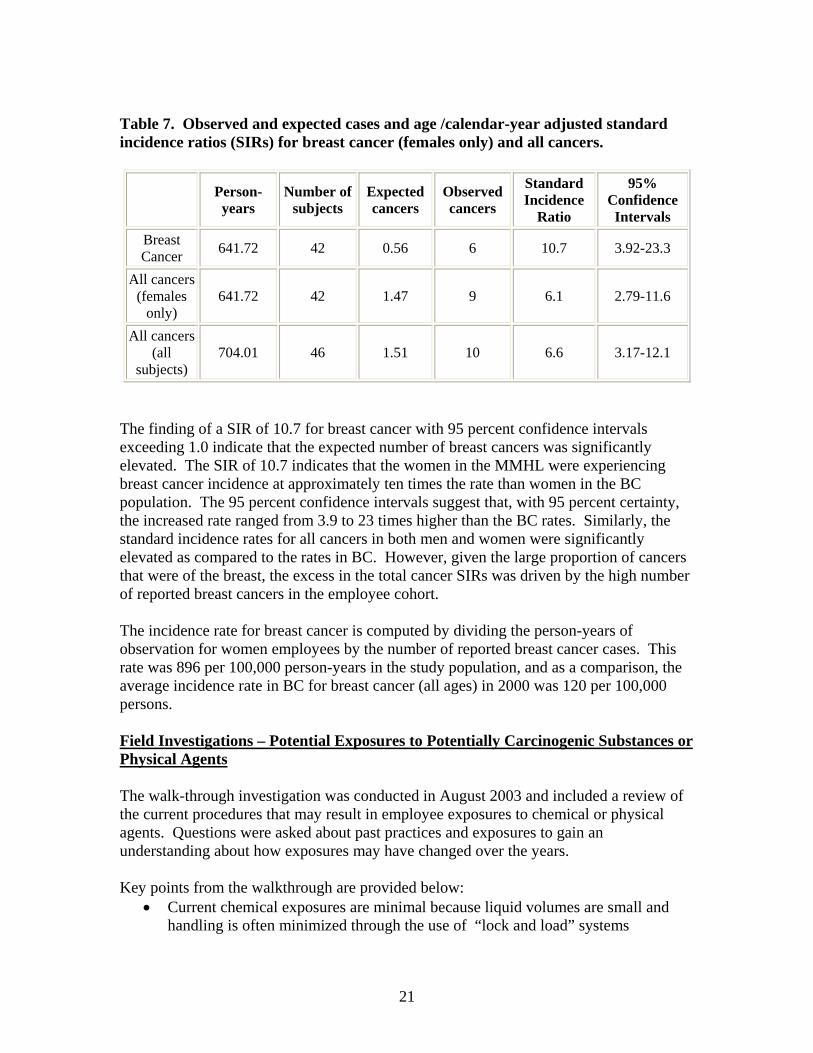
21
Table 7. Observed and expected cases and age /calendar-year adjusted standard incidence ratios (SIRs) for breast cancer (females only) and all cancers.
Person-years
Number of subjects
Expected cancers
Observed cancers
Standard Incidence
Ratio
95% Confidence Intervals
Breast Cancer 641.72 42 0.56 6 10.7 3.92-23.3
All cancers (females
only) 641.72 42 1.47 9 6.1 2.79-11.6
All cancers (all
subjects) 704.01 46 1.51 10 6.6 3.17-12.1
The finding of a SIR of 10.7 for breast cancer with 95 percent confidence intervals exceeding 1.0 indicate that the expected number of breast cancers was significantly elevated. The SIR of 10.7 indicates that the women in the MMHL were experiencing breast cancer incidence at approximately ten times the rate than women in the BC population. The 95 percent confidence intervals suggest that, with 95 percent certainty, the increased rate ranged from 3.9 to 23 times higher than the BC rates. Similarly, the standard incidence rates for all cancers in both men and women were significantly elevated as compared to the rates in BC. However, given the large proportion of cancers that were of the breast, the excess in the total cancer SIRs was driven by the high number of reported breast cancers in the employee cohort. The incidence rate for breast cancer is computed by dividing the person-years of observation for women employees by the number of reported breast cancer cases. This rate was 896 per 100,000 person-years in the study population, and as a comparison, the average incidence rate in BC for breast cancer (all ages) in 2000 was 120 per 100,000 persons. Field Investigations – Potential Exposures to Potentially Carcinogenic Substances or Physical Agents The walk-through investigation was conducted in August 2003 and included a review of the current procedures that may result in employee exposures to chemical or physical agents. Questions were asked about past practices and exposures to gain an understanding about how exposures may have changed over the years. Key points from the walkthrough are provided below:
• Current chemical exposures are minimal because liquid volumes are small and handling is often minimized through the use of “lock and load” systems

22
• Exposures to physical agents, such as ionizing radiation and electromagnetic fields appears to not be excessive (heat and noise exposures were also minimal)
• Past exposures were likely much higher as a number of procedures have been modified due to technological advances
o A major change was in the preservation of tissue samples, tissue staining, and glucose measurement. These procedures, in the past, required open use of solvents and reagents which included formalin, xylene, and o-toluidine. Most of these procedures were performed in a separate area of the laboratory, which was removed when the procedures were modified. It should be noted that o-toluidine, which was discussed in the literature review, is a rat mammary carcinogen, and formaldehyde (the major component in formalin) is a known human carcinogen.
o Other areas of the laboratory also were renovated due to changes in laboratory procedures. Remnants of a local exhaust ventilation system are present in one area where open chemicals were once mixed and dispensed.
• Poor indoor air quality was a common complaint in the past but appears to be less of a problem currently. An incinerator at the hospital was a source of very odourous and potentially toxic compounds (likely acid gases and possible combustion products of PVC (monomers of vinyl chloride) and other plastics (halogenated organics)).
Previous air quality studies have been performed at MMHL, however investigators did not have access to the findings. In discussions with occupational health and safety professionals at FHA, it was mentioned that all measured concentrations of air contaminants were below regulated limits. Summary and Recommendations These findings provide evidence that the female employees within MMHL experienced an elevated rate of breast cancer over the past 30 years. The SIR of 10.2 for breast cancer is statistically significant and the magnitude of the increase rate is of concern. Previous studies have identified nurses and workers in clinical laboratories at higher risk of breast cancer; however, these investigations have not found the magnitude of excess risk found in this study. These previous studies were not designed to determine the causal factors associated with the increased breast cancer risk in laboratory employees or nurses, but it is likely that reproductive factors such as delayed first full-term pregnancy and nulliparity were important in explaining the excess risk. In our study we did not gather personal information pertaining to known risk factors for breast cancer. The reason for not gathering this information was that this is a preliminary epidemiological study and information on risk factors is difficult to interpret without a comparison population where the prevalence of risk factors is available. For example, in our study if we had detailed information about reproductive factors, family history of breast cancer, socioeconomic factors, alcohol consumption, physical exercise, and obesity, we would only be able to compare the prevalence of those factors with those within the general population. Thus, such data would provide clues as to the possible

23
reasons for the elevated risk – if the prevalence of these risk factors were the same as the general population it would suggest that occupational factor(s) predominate. Only a full scale, etiologic investigation would have the capability of clearly identifying occupational factors as attributable to the increased breast cancer risk. A full-scale epidemiologic study may not be the most appropriate action to take despite the increased rates of cancer MMHL employees have experienced. The major goal of cluster investigations is to identify risk factors so that action can be taken to reduce exposures and risk. Air quality studies and reviews of procedures indicate that current exposures to carcinogens are minimal. Past exposures to chemicals like o-toluidine may have resulted in some increased risk for employees, but these exposures appear to have been eliminated. Another issue that discourages a major epidemiologic investigation pertains to the statistics of clusters themselves. Cluster research has shown that elevated rates occur by chance at some geographic locations and times. In fact, clusters always occur and it is a statistical phenomenon – even when there is no causal factor that is responsible for the increased incidence (this is why so few cluster investigations uncover any new risk factors). So, if we look around at many geographic areas and times we will find some clusters; if a specific cluster is related to statistics and not an etiologic agent, it is most likely that in the next time period at this location the rate will not be significantly elevated. Thus, it would be very prudent to continue to evaluate the incidence of breast cancer in MMHL employees to see if the rate comes closer to what is expected. In summary, this study confirmed that the perceived cluster was an observed cluster and that MMHL employees were experiencing an elevated rate of breast cancer. The factors associated with this increased incidence could not be determined but may have been due to: (1) a cluster of reproductive and other known, nonoccupational, risk factors, (2) past exposures to chemical carcinogens and less likely to ionizing radiation, and (3) a chance occurrence (statistical anomaly). Our recommendations for action to be considered are: (1) conduct a thorough inventory of all chemicals currently used in the laboratory and identify any that are listed as animal or known/potential human carcinogens (listing from IARC, NTP, etc.). If any listed compounds are used, conduct a detailed exposure assessment; (2) ensure that exposures to ionizing radiation (one of the few known environmental risk factors for breast cancer) are at background; (3) provide education to all employees about risk factors for breast cancer and the importance of self exams and mammography; (4) continue to collect information on the incidence of breast and all cancers in the future so that SIRs can be computed; and (5) if information as to the possible causes of the high SIR is needed, collect information on known risk factors for breast cancer from all employees and send employee information to the BC Cancer Agency for linkage with the cancer registry.

24
References Aldrich, T. and T. Sinks (2002). "Things to know and do about cancer clusters." Cancer
Invest 20(5-6): 810-6. Band, P. R., N. D. Le, et al. (2000). "Identification of occupational cancer risks in British
Columbia. A population-based case-control study of 995 incident breast cancer cases by menopausal status, controlling for confounding factors." J Occup Environ Med 42(3): 284-310.
Bender, A. P. (1987). "On disease clustering." Am J Public Health 77(6): 742. Bender, A. P., A. N. Williams, et al. (1990). "Appropriate public health responses to
clusters: the art of being responsibly responsive." Am J Epidemiol 132(1 Suppl): S48-52.
Bennett, L. M. and B. J. Davis (2002). "Identification of mammary carcinogens in rodent bioassays." Environ Mol Mutagen 39(2-3): 150-7.
Bigelow, P. (2000). "Application of Threshold Limit Values for Chemical Substances to employees in laboratories." Chem. Health Safety 8: 16-21.
Bounias, M. (2003). "Etiological factors and mechanism involved in relationships between pesticide exposure and cancer." J Environ Biol 24(1): 1-8.
Breslow, N. a. D., N. (1987). Statistical Methods in Cancer Research. Lyon, France, International Agency for Research on Cancer.
Brody, J. G. and R. A. Rudel (2003). "Environmental pollutants and breast cancer." Environ Health Perspect 111(8): 1007-19.
Burkman, R. T. (1999). "Oral contraceptives: an update." Drugs Today (Barc) 35(11): 857-66.
Burnett, C., C. Robinson, et al. (1999). "Cancer mortality in health and science technicians." Am J Ind Med 36(1): 155-8.
Caldwell, G. G. (1990). "Twenty-two years of cancer cluster investigations at the Centers for Disease Control." Am J Epidemiol 132(1 Suppl): S43-7.
Cartwright, R. A. (1999). "Cluster investigations: are they worth it?" Med J Aust 171(4): 172.
CCR (1998). Guidelines for the Investigation of Cancer Clusters in BC. Vancouver, BC, Cancer Control Research, British Columbia Cancer Agency.
Charles, G. D., C. Gennings, et al. (2002). "Assessment of interactions of diverse ternary mixtures in an estrogen receptor-alpha reporter assay." Toxicol Appl Pharmacol 180(1): 11-21.
Charlier, C., A. Albert, et al. (2003). "Breast cancer and serum organochlorine residues." Occup Environ Med 60(5): 348-51.
Claus, E. B., J. M. Schildkraut, et al. (1996). "The genetic attributable risk of breast and ovarian cancer." Cancer 77(11): 2318-24.
Davis, D. L., D. Axelrod, et al. (1997). "Avoidable causes of breast cancer: the known, unknown, and the suspected." Ann N Y Acad Sci 833: 112-28.
Davis, S., D. K. Mirick, et al. (2001). "Night shift work, light at night, and risk of breast cancer." J Natl Cancer Inst 93(20): 1557-62.
DeBruin, L. S. and P. D. Josephy (2002). "Perspectives on the chemical etiology of breast cancer." Environ Health Perspect 110 Suppl 1: 119-28.

25
DeBruin, L. S., J. B. Pawliszyn, et al. (1999). "Detection of monocyclic aromatic amines, possible mammary carcinogens, in human milk." Chem Res Toxicol 12(1): 78-82.
Deligeoroglou, E., E. Michailidis, et al. (2003). "Oral contraceptives and reproductive system cancer." Ann N Y Acad Sci 997: 199-208.
Drijver, M. and F. Woudenberg (1999). "Cluster management and the role of concerned communities and the media." Eur J Epidemiol 15(9): 863-9.
Finkelstein, M. M. and N. Wilk (1990). "Investigation of a lung cancer cluster in the melt shop of an Ontario steel producer." Am J Ind Med 17(4): 483-91.
Fiore, B. J., L. P. Hanrahan, et al. (1990). "State health department response to disease cluster reports: a protocol for investigation." Am J Epidemiol 132(1 Suppl): S14-22.
Frelick, R. W. and A. Topham (1991). "The value of cluster and environmentally related cancer studies in Delaware." Del Med J 63(9): 559-61.
Gammon, M. D., A. I. Neugut, et al. (2002). "The Long Island Breast Cancer Study Project: description of a multi-institutional collaboration to identify environmental risk factors for breast cancer." Breast Cancer Res Treat 74(3): 235-54.
Gangnon, R. E. and M. K. Clayton (2001). "A weighted average likelihood ratio test for spatial clustering of disease." Stat Med 20(19): 2977-87.
Garry, V. F., D. R. Jacobs, Jr., et al. (1989). "An approach to the investigation of cancer in tool and die workers. I. Epidemiologic findings in a suspected cancer cluster among tool and die workers." Mutat Res 225(3): 107-13.
Goldberg, M. S. and F. Labreche (1996). "Occupational risk factors for female breast cancer: a review." Occup Environ Med 53(3): 145-56.
Gordon, N. H. (2003). "Socioeconomic factors and breast cancer in black and white Americans." Cancer Metastasis Rev 22(1): 55-65.
Hall, H. I., C. V. Lee, et al. (1996). "Cluster: a software system for epidemiologic cluster analysis." Stat Med 15(7-9): 943-50.
Halls, S. B. (2003). Detailed Breast Cancer Risk Calculator, Steven B. Halls, MD. 2004. Hamajima, N., K. Hirose, et al. (2002). "Alcohol, tobacco and breast cancer--
collaborative reanalysis of individual data from 53 epidemiological studies, including 58,515 women with breast cancer and 95,067 women without the disease." Br J Cancer 87(11): 1234-45.
Hanrahan, L. P., I. Mirkin, et al. (1990). "SMRFIT: a Statistical Analysis System (SAS) program for standardized mortality ratio analyses and Poisson regression model fits in community disease cluster investigations." Am J Epidemiol 132(1 Suppl): S116-22.
Hargreaves, D. F., C. S. Potten, et al. (1999). "Two-week dietary soy supplementation has an estrogenic effect on normal premenopausal breast." J Clin Endocrinol Metab 84(11): 4017-24.
Higginbotham, S., Z. F. Zhang, et al. (2004). "Dietary glycemic load and breast cancer risk in the Women's Health Study." Cancer Epidemiol Biomarkers Prev 13(1): 65-70.
Jacobzone, S. (2002). Summary of the Results from the Breast Cancer Disease Study. OECD Study on Cross-national Differences of Ageing-related Diseases Workshop, June 20-21, Paris, France, Organization for Economic Cooperation and Development.
Kheifets, L. I. (1993). "Cluster analysis: a perspective." Stat Med 12(19-20): 1755-6. Kipen, H. and D. Wartenberg (1988). "Don't close the door: some observations on cancer
cluster investigation." J Occup Med 30(8): 661-2.

26
Knorr-Held, L. and G. Rasser (2000). "Bayesian detection of clusters and discontinuities in disease maps." Biometrics 56(1): 13-21.
Kreiger, N., M. Sloan, et al. (1999). "The risk of breast cancer following reproductive surgery." Eur J Cancer 35(1): 97-101.
Kulldorff, M., W. F. Athas, et al. (1998). "Evaluating cluster alarms: a space-time scan statistic and brain cancer in Los Alamos, New Mexico." Am J Public Health 88(9): 1377-80.
Laitman, C. J. (2002). "DES exposure and the aging woman: mothers and daughters." Curr Womens Health Rep 2(5): 390-3.
Land, C. E., M. Tokunaga, et al. (2003). "Incidence of female breast cancer among atomic bomb survivors, Hiroshima and Nagasaki, 1950-1990." Radiat Res 160(6): 707-17.
Langmuir, A. D. (1965). "Formal discussion of: epidemiology of cancer: spatial-temporal aggregation." Cancer Res 25(8): 1384-6.
Lawson, A. B. (2000). "Cluster modelling of disease incidence via RJMCMC methods: a comparative evaluation. Reversible jump Markov chain Monte Carlo." Stat Med 19(17-18): 2361-75.
Lewis, R. and G. Rempala (2003). "A case-cohort study of angiosarcoma of the liver and brain cancer at a polymer production plant." J Occup Environ Med 45(5): 538-45.
Lieben, J. (1966). "Malignancies in asbestos workers." Arch Environ Health 13(5): 619-21.
Lieben, J. and H. Pistawka (1967). "Mesothelioma and asbestos exposure." Arch Environ Health 14(4): 559-63.
Mackillop, W. J., J. Zhang-Salomons, et al. (2000). "Associations between community income and cancer incidence in Canada and the United States." Cancer 89(4): 901-12.
Madigan, M. P., R. G. Ziegler, et al. (1995). "Proportion of breast cancer cases in the United States explained by well-established risk factors." J Natl Cancer Inst 87(22): 1681-5.
McTiernan, A., K. B. Rajan, et al. (2003). "Adiposity and sex hormones in postmenopausal breast cancer survivors." J Clin Oncol 21(10): 1961-6.
Medina, D. (2004). "Breast cancer: the protective effect of pregnancy." Clin Cancer Res 10(1 Pt 2): 380S-4S.
Morton, W. E. (1995). "Major differences in breast cancer risks among occupations." J Occup Environ Med 37(3): 328-35.
NCI (1997). "Workshop on Hormones, Hormone Metabolism, Environment, and Breast Cancer,
New Orleans, Louisiana, September 28-29, 1995." Monographs in Environ Hlth Perspect Supp 105(3): 557-688. NCIC (1999). Canadian Cancer Statistics. Toronto, Canada, National Cancer Institute of
Canada. NRC (2004). Breast Cancer Rates, 1986-1995 URL:
http://atlas.gc.ca/site/english/maps/health/status/breastcancerrates1986-1995?map=%2Fhome%2Fmapdata%2Faoc_v3%2Fenglish%2F876.map&imgext=-2032143.858627+341765.863549+-1820995.950293+529233.632629&mode=browse&scale=1398434.17556762&imgxy

27
=&mapext=&img.x=213&img.y=191, Natural Resources Canada, Communication Canada, Canadian Government Publishing, Ottawa, Canada.
Parkin, D. M., F. Bray, et al. (2001). "Estimating the world cancer burden: Globocan 2000." Int J Cancer 94(2): 153-6.
Parkin, D. M., F. I. Bray, et al. (2001). "Cancer burden in the year 2000. The global picture." Eur J Cancer 37 Suppl 8: S4-66.
Patel, A. V., M. F. Press, et al. (2003). "Lifetime recreational exercise activity and risk of breast carcinoma in situ." Cancer 98(10): 2161-9.
Pott, P. (1996). Observation of scrotal tumors among London Chimney Sweeps (republished). Classics in Oncology. A. C. Society. New York, NY.
PPHB (2004). Breast Cancer in Canada. online at: http://www.hc-sc.gc.ca/pphb-dgspsp/publicat/updates/breast-99_e.html, Population and Public Health Branch, Health Canada, Ottawa, Canada. 2004.
Quataert, P. K., B. Armstrong, et al. (1999). "Methodological problems and the role of statistics in cluster response studies: a framework." Eur J Epidemiol 15(9): 821-31.
Recchia, A. G., A. Vivacqua, et al. (2004). "Xenoestrogens and the induction of proliferative effects in breast cancer cells via direct activation of oestrogen receptor alpha." Food Addit Contam 21(2): 134-44.
Robbins, A. S., S. Brescianini, et al. (1997). "Regional differences in known risk factors and the higher incidence of breast cancer in San Francisco." J Natl Cancer Inst 89(13): 960-5.
Ron, E. (2003). "Cancer risks from medical radiation." Health Phys 85(1): 47-59. Russo, I. H. and J. Russo (1996). "Mammary gland neoplasia in long-term rodent
studies." Environ Health Perspect 104(9): 938-67. Russo, I. H. and J. Russo (1998). "Role of hormones in mammary cancer initiation and
progression." J Mammary Gland Biol Neoplasia 3(1): 49-61. Russo, J. and I. H. Russo (1997). "Toward a unified concept of mammary
carcinogenesis." Prog Clin Biol Res 396: 1-16. Safe, S. H. and T. Zacharewski (1997). "Organochlorine exposure and risk for breast
cancer." Prog Clin Biol Res 396: 133-45. Sarkar, F. H. and Y. Li (2003). "Soy isoflavones and cancer prevention." Cancer Invest
21(5): 744-57. Schernhammer, E. S., F. Laden, et al. (2003). "Night-shift work and risk of colorectal
cancer in the nurses' health study." J Natl Cancer Inst 95(11): 825-8. Schulmeister, L. (2003). "Diethylstilbestrol exposure: how well informed are you about
this health risk?" Clin J Oncol Nurs 7(4): 373-4. Schulte, P. A., R. L. Ehrenberg, et al. (1987). "Investigation of occupational cancer
clusters: theory and practice." Am J Public Health 77(1): 52-6. Semenciw, R. M., N. D. Le, et al. (2000). "Methodological issues in the development of
the Canadian Cancer Incidence Atlas." Stat Med 19(17-18): 2437-49. Siegrist, M., G. T. Cvetkovich, et al. (2001). "Shared values, social trust, and the
perception of geographic cancer clusters." Risk Anal 21(6): 1047-53. Smith, D. and R. Neutra (1993). "Approaches to disease cluster investigations in a state
health department." Stat Med 12(19-20): 1757-62.

28
Snedeker, S. (1997). Do we need to be concerned about environmental chemicals and breast cancer? An interview with the BCERF Research Project Leader. The Ribbon. 2.
Sonnenschein, C. and A. M. Soto (1998). "An updated review of environmental estrogen and androgen mimics and antagonists." J Steroid Biochem Mol Biol 65(1-6): 143-50.
Timander, L. M. and S. McLafferty (1998). "Breast cancer in West Islip, NY: a spatial clustering analysis with covariates." Soc Sci Med 46(12): 1623-35.
Tompa, A., J. Major, et al. (1999). "Is breast cancer cluster influenced by environmental and occupational factors among hospital nurses in Hungary?" Pathol Oncol Res 5(2): 117-21.
Trumbo, C. W. (2000). "Public requests for cancer cluster investigations: a survey of state health departments." Am J Public Health 90(8): 1300-2.
van Netten, C., R. H. Brands, et al. (2003). "Cancer cluster among police detachment personnel." Environ Int 28(7): 567-72.
Vassilopoulou-Sellin, R. (2003). "Breast cancer and hormonal replacement therapy." Ann N Y Acad Sci 997: 341-50.
Wang, D. Y., D. S. Allen, et al. (2000). "Urinary androgens and breast cancer risk: results from a long-term prospective study based in Guernsey." Br J Cancer 82(9): 1577-84.
Ward, E. M., P. A. Schulte, et al. (2003). "Priorities for development of research methods in occupational cancer." Environ Health Perspect 111(1): 1-12.
Weaver, V. M. (1997). "Chemical hazards in health care workers." Occup Med 12(4): 655-67.
Wittenberg, C. (1994). "Long Island breast cancer studies move forward." J Natl Cancer Inst 86(20): 1501-3.
WSDOH (2001). Guidelines for Investigating Clusters of Chronic Disease and Adverse Birth Outcomes. online: http://www.doh.wa.gov/EHSPHL/Epidemiology/NICE/publications/ClusterProt.pdf. Olympia, Washington, Washington State Department of Health.
Yang, D., L. Bernstein, et al. (2003). "Physical activity and breast cancer risk among Asian-American women in Los Angeles: a case-control study." Cancer 97(10): 2565-75.
Yue, W., R. J. Santen, et al. (2003). "Genotoxic metabolites of estradiol in breast: potential mechanism of estradiol induced carcinogenesis." J Steroid Biochem Mol Biol 86(3-5): 477-86.
Zeleniuch-Jacquotte, A., R. E. Shore, et al. (2004). "Postmenopausal levels of oestrogen, androgen, and SHBG and breast cancer: long-term results of a prospective study." Br J Cancer 90(1): 153-9.

Attachment 3
An Investigation of a Cancer Cluster within the Mission Memorial Hospital Laboratory
March 2004


An Investigation of a Cancer Cluster within the Mission Memorial Hospital Laboratory
March 2004
By: Philip Bigelow 1,4
Shicheng Yu 4 Rosemary Nemanishen 2
Annalee Yassi 1,3,4
1 Occupational Health and Safety Agency for Healthcare in BC #301- 1195 West Broadway, Vancouver, BC V6H 3X5 Tel: 604-775-4034 Fax: 604-775-4031 Web: www.ohsah.bc.ca 2 Occupational Health Nurse, Fraser East, Fraser Health Authority 3 Department of Health Care and Epidemiology, University of British Columbia 5804 Fairview Ave., Vancouver, BC V6T 1Z3 4 Institute of Health Promotion Research, University of British Columbia Library Processing Centre, 2206 East Mall, Rm. 435 Vancouver, B.C. Canada V6T 1Z3

2
ABSTRACT
An excess in the number of cases of cancer was suspected by employees of the Mission Memorial Hospital Laboratory and this investigation was conducted in response to the above concern. The study had three parts, an epidemiologic cluster analysis, a comprehensive review of the literature on breast cancer risk factors and the analysis of clusters, and an exposure investigation. A total of 57 individuals were identified as having worked in the laboratory between January 1, 1970 and August 31, 2003. Information on health status and diagnoses of cancer was obtained through personal interviews with employees. Ten employees reported a cancer diagnosis, of which 6 were breast cancer. A total of 751 person-years of observation were available for the data analysis after excluding one subject because of diagnoses of cancer prior to start of employment. Based on the age and calendar-year adjusted rates for the BC population, the expected number of breast cancer cases in the women was 0.59 and the expected number of all cancers for all employees was 1.60. The Standard Incidence Ratios (SIR), which are the observed number of cases divided by the expected number, were 10.2 for breast cancer in women, and 6.3 for all cancers in both men and women. The 95 percent confidence intervals indicate both findings were significant. A walk-through survey of the laboratory did not identify any potentially hazardous exposures. Recommendations include a comprehensive assessment of all potential exposures to chemicals and physical agents, continuing to collect epidemiological information to determine temporal trends in the SIR, providing information on breast cancer risks to employees, and collecting information on known risk factors for breast cancer and confirming cancer diagnoses through linkage with the BC Cancer Agency’s registry.

3
Introduction The Occupational Health and Safety Agency for Healthcare (OHSAH) was invited by the Fraser Health Authority (FHA) to investigate concerns of a greater than expected number of cancer cases in the Mission Memorial Hospital Laboratory (MMHL). In addition to the seemingly high total number of cancer cases, a large proportion were the same type (breast cancer) thus further highlighting the need for an investigation. Occupational health professionals from FHA had completed some exploratory work on the project but felt they needed the help of outside experts to resolve the issues. OHSAH conducted a preliminary cluster investigation which is the focus of this report. Dr. Philip Bigelow, a senior scientist at OHSAH, led the investigation and team members included Dr. Annalee Yassi, Rosemary Nemanishen, Dr. Shicheng Yu and William So Yiu Ting. Assistance was also obtained from Dr. Nhu Lee of the British Columbia (BC) Cancer Agency and Dr. Martha Vela Acosta of the Department of Environmental and Radiological Health Sciences at Colorado State University. Cancer clusters are the occurrences of greater numbers of the same type of cancer, or all cancers, within a geographic location over a specified period of time. The purpose of a cancer cluster investigation is to determine if the observed number of cases is higher than expected. If the observed number is higher, the study should provide information that may help to reduce or eliminate exposures that may be associated with the increased risk. Cancer cluster investigations are often conducted by health agencies and this investigation followed general guidelines recommended by the BC Cancer Agency and public health departments in other jurisdictions across North America (Schulte, Ehrenberg et al. 1987; Caldwell 1990; Cartwright 1999; WSDOH 2001). Prior to the investigation, an explanatory meeting was held at Mission Memorial Hospital to discuss the incidence of cancer at MMHL and the protocol for the investigation. Details of the protocol used during the investigation are provided below. Methods The specific aim of this study was to provide a thorough determination if an excess in the number of cancer cases had occurred in the Mission Memorial Hospital Laboratory (MMHL) and to provide information regarding the possibility that a work-related factor was involved. More importantly, the goal was to ensure that current workplace conditions and exposures are not at all likely to result in an increased risk of cancer for employees. The BC Cancer Agency, as well as other health agencies, have adopted standard protocols for investigating clusters. The methods used in our study followed these standard procedures and included determining if an excess number of cancers was reported, a literature review on the risk factors for the specific cancer types, assessing the potential for occupational exposures to potentially carcinogenic physical agents or substances, and determining the feasibility of further epidemiology studies. Our study was divided into three components as listed below.

4
1) Analysis of Cancer Incidence Data: Epidemiology and Statistics Since the concern pertained to employees in the MMHL, only employees who worked in the Laboratory were included in the study. Using data from the Human Resources Department at Mission Memorial Hospital, all employees who were employed in the Laboratory over the past 33 years (January 1970 to August 2003) were identified and this information was provided to an occupational health professional at Fraser Health. A total of 57 individuals were identified and the following information was entered into a computer spreadsheet: date of birth, dates of employment at the lab, job title, full or part time employment status, gender, and other details pertaining to work at the lab and hospital. A health professional (Registered Nurse) from Fraser Health attempted to contact all 57 individuals (in person or by telephone) to gather information on whether or not they had a diagnosis of cancer of any type. For individuals who reported a cancer diagnosis, information on the diagnosis date, type and site of cancer was obtained. Data for all 57 individuals, without personal identifiers, were entered into a spreadsheet and provided to OHSAH. The statistical analysis was conducted two ways. In one, the person-years of observation was defined as being from the start date of employment at MMHL to the end date of employment. In the second analysis, the person-years of observation was defined as the time between the employee start date and the end of the follow up period (August 2003). In this report the latter analysis is provided, as it is the most appropriate for the study design that was used.1 Since a large proportion of the cases were classified as breast cancers, statistical analyses were conducted using rates of breast and total cancers obtained from the BC Cancer Agency. Rates for breast and total cancers for each year from 1970 to 2002, grouped by 5-year age intervals, were used to calculate the expected number of cases in the study group. The expected number of cases is the number of cases expected in the Laboratory if the rate was the same as the rate in BC adjusted for age and calendar year.2 The expected number of cases was computed by multiplying the population (person-years of observation) within each specific age range and year by the rate of breast or total cancers for the same age interval and year. The results of these computations were summed across all the age and year categories to get the total number of expected cases. Computations and statistical analyses were conducted using Excel and SPSS software.
1 In many occupational cohort studies, when subjects leave employment their health status at that time is known and their end date of employment is used in the computation of person-years of observation. In this study, we contacted all study subjects from August to November, 2003 to determine their health status. 2 The risk of developing breast cancer was different in 1980 as compared to today. Additionally, age is a major risk factor for all cancers (including breast cancer). Therefore, crude incidence rates may be misleading when comparing regions or time periods where the age of the populations differ from one region to another or from one time period to another. In this study, the expected number of breast and total cancers was adjusted for age and calendar year and this provides the most accurate comparison of rates for this study.

5
The observed number of cases was divided by the expected number of breast cancer and total cancer cases to determine the Standard Incidence Ratios (SIR). A SIR exceeding 1.0 indicates the observed number is higher than expected. Confidence intervals are used to assess variation in the SIR and 95% Poisson confidence intervals were calculated using the procedure suggested by Breslow (Breslow 1987). To investigate the relationship of occupational factors on the rate of developing breast cancer, a Cox proportional hazard model was developed that included independent variables for job title, job status (full or part time) and age at start of employment at the laboratory. 2) Field Investigations: Possible Exposures to Potentially Carcinogenic Substances or Physical Agents Prior to this investigation, work had been conducted by occupational health professionals at Fraser Health to determine the adequacy of procedures to control exposures to chemicals in the laboratory and to ensure exposures did not exceed government or consensus standards. Investigations also focused on potential sources of chemical exposures resulting from work tasks that are typically performed by laboratory personnel. An additional study included Additionally, studies that involved reviewing past renovations of the laboratory in hopes of identifying unusual sources of indoor air contaminants were performed. In August of 2003, as part of OHSAH’s investigation, a walk-through survey of the laboratory was completed. Typical work procedures were reviewed to assess the potential for exposures to hazardous agents. Employees in the laboratory provided information on historical procedures as well as an indication of the general levels of exposure to air contaminants. 3) Literature Review The investigation and response to cancer cluster reports is exceedingly complex and the report includes brief literature reviews on risk factors for breast cancer, exposures in laboratories, and epidemiology of cancer clusters. This information is provided to help interpret the study findings and provide guidance in deciding whether further study is warranted. Results and Discussion The literature review is presented first as it provides a background for interpreting the findings from both the epidemiologic analysis and the field surveys. The literature review of breast cancer highlights the multifactorial nature of disease causation and the difficulties in determining the role of environmental and occupational exposures as causal factors. There is a substantial body of literature on cancer cluster investigations and a brief review is provided. Finally, the findings of the statistical analysis are provided and discussed in relation to findings from other studies.

6
Literature Review: Breast Cancer Risk Factors and the Role of Occupational and Environmental Exposures Breast cancer. Canada has one of the highest rates of breast cancer incidence and the age standardized rate in 1995 exceeded 225 per 100,000 women aged 40 and over (Jacobzone 2002). Breast cancer is the most frequently diagnosed cancer in Canadian women and it accounts for over 30% of new cancer cases per year (NCIC 1999). It is estimated that one in nine women in Canada will develop the disease in their lifetime (DeBruin and Josephy 2002). Breast cancer mortality increased steadily from the 1960s until the early 1980s when the rates declined in most countries including Canada (Parkin, Bray et al. 2001; Parkin, Bray et al. 2001). These declining mortality rates are thought to be related to improved screening resulting in earlier detection and improved treatment. Age is major risk factor for breast cancer and Table 1 shows the increased likelihood of a woman developing breast cancer in the next five years at various ages (PPHB 2004). In addition to age, many correlates of risk for breast cancer have been identified and there are a constellation of hormone-related reproductive factors that predominate. Factors known to confer higher risk include younger age at menarche, older age at menopause, nulliparous, and older at first live birth (Davis, Axelrod et al. 1997). Higher parity, longer lactation, and bilateral ovariectomy have been found to be protective (Davis, Axelrod et al. 1997; Kreiger, Sloan et al. 1999). Using data from large population-based surveys in the United States (US), investigators calculated that 41% of breast cancer risk was explained by nulliparity, later childbearing, higher income, and family history of breast cancer (Madigan, Ziegler et al. 1995). Other risk factors for breast cancer include a history of certain types of benign breast disease as well as high levels of radiation exposure to the chest (medical x-rays) (PPHB 2004). Despite the many studies that have been conducted, additional factors, likely modest in magnitude, remain to be discovered. Interestingly, in 76% of women who develop breast cancer, age is the only identifiable risk factor (Halls 2003).

7
Table 1.3 Probability of developing breast cancer in the next five years.
Age Breast cancer per 1,000 women
30 1.5 35 2.6 40 4.8 45 7.8 50 9.2 55 10.6 60 12.9 65 14.3 70 15.4 80 15.5
Epidemiological and animal studies consistently show elevated risk of breast cancer with factors that increase exposure to estradiol, progesterone, and other hormones (Kreiger, Sloan et al. 1999; Medina 2004; Recchia, Vivacqua et al. 2004; Zeleniuch-Jacquotte, Shore et al. 2004). Risk factors such as alcohol consumption, weight gain after menopause, low pre-menopausal body mass index, and lack of physical exercise are believed to be associated with exposure to reproductive hormones (Hamajima, Hirose et al. 2002; McTiernan, Rajan et al. 2003; Patel, Press et al. 2003; Yang, Bernstein et al. 2003). Pharmaceutical hormones appear to have a similar effect and there is evidence that women exposed to diethylstilbestrol during pregnancy had increased risks for breast cancer (Laitman 2002; Schulmeister 2003). For oral contraceptives, recent use, not long term exposure, has been associated with an increased risk (Burkman 1999; Deligeoroglou, Michailidis et al. 2003). Similarly, recent use of hormone replacement therapy has been shown to increase the relative risk of breast cancer, whereas women who stopped over 5 years ago are not at significantly elevated risk (Vassilopoulou-Sellin 2003). In epidemiological studies higher socioeconomic status, as measured by income and education level, are consistently associated with elevated breast cancer risk (Mackillop, Zhang-Salomons et al. 2000; Gordon 2003). Although some of this association may be due to a clustering of reproductive risk factors in higher socioeconomic status women, the effect is still significant even after controlling for parity, age at first child and other common reproductive factors (Brody and Rudel 2003). Diet has been well studied but epidemiological investigations have yet to identify foods that significantly increase or decrease breast cancer risk (Higginbotham, Zhang et al. 2004). It is hypothesized that 3 From PPHB (2004). Breast Cancer in Canada. online at: http://www.hc-sc.gc.ca/pphb-dgspsp/publicat/updates/breast-99_e.html, Population and Public Health Branch, Health Canada, Ottawa, Canada. 2004.

8
dietary factors may modulate hormone levels so a number of investigations have focused on foods high in phytoestrogens (Hargreaves, Potten et al. 1999; Sarkar and Li 2003) (partial estrogen agonists) or containing other endocrine active components (Brody and Rudel 2003). Studies of occupational and environmental factors. Animal studies have provided important information in understanding mechanisms of the development of breast cancer and in the identification of agents that may increase breast cancer risk. A comprehensive review of chemical carcinogenesis in general is beyond the scope of this paper, but it should be noted that all cancer causing agents, physical or chemical, will also have the potential to initiate or promote breast cancer. A good example is ionizing radiation which is know to cause cancer at multiple sites; based on human epidemiological studies, it is one of the few occupational or environmental exposures that is a known cause of breast cancer (Land, Tokunaga et al. 2003; Ron 2003). Studies indicate that estrogen receptor (ER) alpha mediates the breast cancer promoting effects of estrogen. Estradiol binds to ER alpha and induces estrogen receptor-mediated transcription, DNA synthesis, cell division, and cell proliferation which is associated with an increase in errors in DNA transcription. Estrogen and progesterone, both essential for mammary gland growth and function, cause cell proliferation and may be procarcinogenic (DeBruin and Josephy 2002). Both estrogen and progesterone are cytotoxic as they interact with specific receptor proteins in the cell nucleus. Recent research provides evidence that estrogens can be metabolically activated to genotoxic compounds that induce oncogenic mutations (Yue, Santen et al. 2003). Thus, the carcinogenicity of estrogens, as well as their action in increasing susceptibility, may be related to a receptor mediated stimulation of cellular proliferation. Human studies have provided some evidence to support this hypothesis (Wang, Allen et al. 2000). Even without a complete understanding of the mechanism, it is clear that lifetime exposure to estrogen and other hormones explains many identified risk factors for breast cancer. Cells within the breast are not fully differentiated until they are induced by hormonal stimuli at the woman’s first pregnancy and lactation. Thus, breast cells are more susceptible to the effects of carcinogens while the breast is not fully developed. Additionally, the breast cells are vulnerable to genotoxic agents during pregnancy as there is rapid proliferation of cells (Russo and Russo 1996; Russo and Russo 1997). This explanation of the susceptibility of mammary cells to carcinogens provides a framework for understanding the increased risk of breast cancer in humans in relation to reproductive events as well as after exposure to mammary carcinogens. It has been hypothesized that, because the breast is very susceptible to carcinogen exposures up until the first full-term pregnancy, there may be an interaction of age (a known risk factor) and the risk associated with exposures to chemicals (Brody and Rudel 2003). Despite the complex mechanisms and interactions between chemical exposures and hormones, animal studies have clearly identified numerous mammary carcinogens through standard cancer bioassays. The US National Toxicology Program (NTP) has tested over 500 chemicals and identified 42 as causing mammary tumors (Bennett and

9
Davis 2002). The human evidence for identifying chemicals causing breast cancer is more scant and of the 42 chemicals cited above, only four are classified as human carcinogens: benzene, 1,3-butadiene, ethylene oxide, and C I acid red 114. Also, it should be noted that epidemiology studies of these compounds have shown exposed employees at higher risk of cancer, but not specifically breast cancer. Mammary carcinogens that may be associated with exposures in chemical and medical laboratories are presented in Table 2 below.

10
Table 2.4 Chemicals tested by NTP that produce mammary tumors in experimental animals
Chemical Use Acronycine Pharmaceuticals
Benzene Gasoline, solvent
2,2-bis(bromomethyl)- 1,3-propanediol Flame retardant
1,3-Butadiene Auto exhaust, rubber manufacture, gasoline
C,1 acid red 114 Dye for silk, jute, wool, leather
C,1 basic red 9 monohydrochloride Dye for textiles, leather, paper, biological stain
2-Chloroacetophenone Flame retardant
Chloroprene Used in neoprene manufacture
Clonitralid Molluskicide
Cytembene Pharmaceuticals
2,4-Diaminotoluene Intermediate in dye synthesis
1,2-Dibromo-3-chloropropane Soil fumigant, pesticide
1,2-Dibromoethane Soil fumigant, lead scavenger in gasoline
1,2-Dibromo-1-propanol Flame retardant
1,1-Dichloroethane Solvent
1,2-Dichloroethane Solvent, chemical intermediate in insecticide formulations, gasoline
1,2-Dichloropropane (propylene dichloride) Chemical intermediate, solvent in dry cleaning fluids, fumigant
Dichlorvos Pesticide
1,2-Dimethoxybenzidine dihydrochloride Dye intermediate
3,3-Dimethylbenzidine dihydrochloride Dye intermediate
2,4-Dinitrotoluene Dye intermediate, explosives, propellants
Ethylene oxide Sterilizing gas for medical equipment
Furosemide Pharmaceuticals
Glycidol Stabilizer in vinyl polymers, intermediate in pesticides and fragrances
Hydrazobenzene Dye intermediate, tobacco pesticides, motor oil
Isophosphamide Pharmaceuticals
Indium phosphide Microelectronics, semiconductors, injection lasers, diodes
Isoprene By-product of ethylene production
Methylene chloride Solvent, furniture stripper, adhesives
Methyleugenol Food additive, flavoring, also naturally occurring
Nithiazide Antiprotozoal compound
5-Nitroacenaphthene Research chemical
Nitrofurazone Antibiotic
Nitromethane Rocket and engine fuel, solvent, mining explosive
Ochratoxin A Mycotoxin
Phenesterin Pharmaceuticals
Procarbazine hydrochloride Pharmaceuticals
Reserpine Pharmaceuticals
Sulfallate Herbicide
2,4- and 2,6-Toluene diisocyanate Used in manufacture of flexible polyurethane foams
o-Toluidine hydrochloride Dye intermediate
1,2,3-Trichloropropane Chemical intermediate, former solvent and paint remover
4 From Bennett, L. M. and B. J. Davis (2002). "Identification of mammary carcinogens in rodent bioassays." Environ Mol Mutagen 39(2-3): 150-7.

11
Both animal and human studies show that the relationships between hormonal factors and mammary carcinogens is complex. Treatment of animals with ovarian, placental, pituitary, and thyroid hormones modulates the tumorgenic responses (Russo and Russo 1998). The situation is further complicated with exposures to chemicals that are members of a class of hormonally active chemicals, sometimes referred to as endocrine active, endocrine disruptors, or estrogenic compounds. The hypothesis is that exposure increases estrogen-like responses of cell proliferation that increase cancer risk. There is also a concern that these endocrine active compounds can act in an additive manner to produce effects (Charles, Gennings et al. 2002; Brody and Rudel 2003). Chemicals, including some pesticides, also can act as co-carcinogens or tumor promoters (Bounias 2003). A good example of a breast cancer promoter in experimental animals is dichlorodiphenyltrichloroethane (DDT). Experimental animals fed a known mammary carcinogen, and then given DDT, developed breast tumors earlier than when the carcinogen was given alone; however, when DDT was given alone, it did not induce breast tumors in these animals (Snedeker 1997). The human evidence of DDT’s effects as a promoter is more equivocal, although a recent study reported significantly elevated mean levels of serum DDT and hexachlorobenzene (HCB) in breast cancer patients as compared to controls (Charlier, Albert et al. 2003). Other organochlorine compounds have been implicated as being associated with an increased risk of breast cancer. The hypothesis is that this group of compounds possess estrogenic activity. However, both the hypothesis and the magnitude of any possible effect on human risk of breast cancer is controversial. Recent reviews suggest that the estrogenic contribution of organochlorine compounds is small in view of the presence of natural hormone and antihormone mimics in our diet (Safe and Zacharewski 1997; DeBruin and Josephy 2002). Other endocrine active compounds, such as alkyl phenols and phthalates are still under investigation (Sonnenschein and Soto 1998). Studies of breast cancer risk in working populations have not provided strong evidence of causal links between specific exposures and increased risk. However, there is evidence for positive associations of several occupations with increase breast cancer risk (Morton 1995; Goldberg and Labreche 1996; Band, Le et al. 2000). The study by Band et al. (2000) was conducted in British Columbia and found significantly higher breast cancer risks (1) among pre-menopausal women in electronic data-processing operators; barbers and hairdressers; in sales and material processing occupations; and in the food, clothing, chemical and transportation industries; (2) among post-menopausal women in school teaching; in medicine, health, and nursing occupations; in laundry and dry-cleaning occupations; and in the aircraft and automotive, including gasoline service station, industries. Several significant associations were also seen in the combined group of pre- and post-menopausal women, particularly in crop farmers and in fruit and vegetable farming, publishing and printing, and motor vehicle repair industries. The authors suggested that there was excess breast cancer risk in a number of occupations and industries, notably those that entail exposure to solvents and pesticides (Band, Le et al. 2000).
12
Shiftwork causes employees to have exposure to light at night and may increase the risk of cancer by suppressing the normal nocturnal production of melatonin by the pineal gland. Melatonin, is not only a hormone that has antiproliferative effects which protect against the development of cancer (Schernhammer, Laden et al. 2003), but it also modulates estrogen release from the ovaries. When nocturnal melatonin production is suppressed, the direct antiproliferative effects are reduced and estrogen release may be increased (Davis, Mirick et al. 2001). In a study of female nurses in a large prospective health investigation, women working a rotating night shift at least three nights per month for 15 or more years were at an increased the risk of colorectal cancer (Schernhammer, Laden et al. 2003). A recent population-based, case-control study found that graveyard shiftwork was associated with increased breast cancer risk (OR = 1.6; 95% CI = 1.0 to 2.5), with a trend of increased risk with increasing years and with more hours per week of graveyard shiftwork (Davis, Mirick et al. 2001). Clinical laboratory workers have the potential for exposure to a variety of chemical, biological, as well as physical agents (Weaver 1997; Tompa, Major et al. 1999; Bigelow 2000). Despite the fact that chemical and clinical laboratories employ many women (over 1 million in the US), few studies have examined the possible adverse effects of exposures. Burnett et al. (1999) conducted a study to determine if laboratory workers in the US experienced higher cancer mortality rates than those in other occupations. They found clinical laboratory workers had higher proportionate cancer mortality ratios overall (for all cancers) as well as for breast cancer. The proportionate mortality ratios for leukemia were also significantly elevated for clinical laboratory workers (Burnett, Robinson et al. 1999). The authors suggest that the elevated risks for lymphatic and hematopoietic neoplasms may have been associated with occupational exposures. With the exception of a few studies that have identified very high occupational exposures to carcinogenic compounds as causal factors in breast cancer, most investigations have not been able to clearly determine occupational risk factors (Goldberg and Labreche 1996). The reasons for the failure to identify specific chemicals or physical agents include not only the complex nature of the initiation, promotion, and development of breast cancer, but also the presence of many potential confounding risk factors. Additionally, there appear to be numerous, but so far unidentified, risk factors that the issue of confounding becomes even more salient. Little is known about the interaction of known risk factors on the magnitude of increase in breast cancer risk and even less is known about the possible synergistic, additive, or antagonistic effects of multiple chemical exposures. The strength of already known breast cancer risk factors makes the identification of occupational risk factors very difficult. When examining the role of these major risk factors, it has been estimated that 41 percent of breast cancer risk is attributable to later childbearing, nulliparity, higher income, and family history of breast cancer (Madigan, Ziegler et al. 1995). Studies that have focused on genetic variation have estimated that less that 10 percent of cases are due to gene mutations in the breast cancer genes BRCA1 and BRCA2 (Claus, Schildkraut et al. 1996). Diet, alcohol consumption, physical activity, body mass index, other reproductive factors, high chest radiation exposure, and

13
exposure to pharmaceutical hormones all account for some risk in the development of breast cancer. In occupational studies, if the likelihood of exposure to these known breast cancer risk factors is increased in an occupational group, an association between the occupation and increased breast cancer risk will be observed. Additionally, the presence of powerful risk factors may mask the effect of an exposure that is truly increasing breast cancer risk. Traditional epidemiological methods are typically not able to identify occupational risk factors for breast cancer at the levels of exposure seen in modern industry in Canada or the US. Newer methods that include the use of biological markers of exposure and incorporating gene-environment interactions have shown promise. These methods are better able to uncover subtle differences in risk and also provide an understanding of the underlying mechanisms. An example of these cutting edge techniques is the measurement of the aromatic amine, o-toluidine, a rat mammary carcinogen, in human milk samples from mothers. The presence of this chemical indicates that the ductal epithelial cells of the breast are exposed to this carcinogen (DeBruin, Pawliszyn et al. 1999). The use of biomarkers and gene-environment interactions have elucidated the complex associations of smoking, polymorphisms of drug metabolizing enzymes, and reproductive factors in breast cancer risk (DeBruin and Josephy 2002). These techniques have not been rigorously applied in studies involving occupational exposures and breast cancer but their use has been advocated (DeBruin and Josephy 2002; Ward, Schulte et al. 2003). Literature Review: Cancer Clusters Incidence rates of breast cancer, and all cancers, vary over time and geography and a cancer cluster is generally defined as the occurrence of a greater than expected number of cases of a particular cancer within a group of people, a geographic area, or a period of time. Studying and describing these spatial and temporal trends have provided clues for identifying previously undiscovered causes of cancer. In fact, the first causal relationship between an occupational exposure and cancer was uncovered as the result of a cluster investigation of scrotal skin cancer among young chimney sweeps in London (Pott 1996). Epidemiologists, the scientists most often leading the investigation of clusters, generally encounter clusters because of reports or through discovery from organized analyses of large databases (Kheifets 1993). Although the methods of analysis differ slightly depending on how the cluster is first identified, in both cases the results are difficult to interpret and drawing definitive conclusions is often not possible. As was discussed in the section on breast cancer risk factors, some variation in breast cancer risk can be explained by the population distribution of known risk factors such as parity, age at first child and other reproductive factors (Robbins, Brescianini et al. 1997). In fact, grouping of reproductive risk factors and socioeconomic status play a major role in the findings of positive associations between white collar occupations and increased risk of breast cancer (Brody and Rudel 2003). However, regional patterns of increased and decreased breast cancer risk may reflect a complex aggregation of diverse factors which may include diet, demographics, lifestyle factors, and occupational and

14
environmental exposures. Gaining an understanding of these individual factors and their relationships is necessary to have a complete understanding of breast cancer risk in individuals and specific groups of women. For breast cancer, clusters of relatively high incidence rates have been reported in areas of southern Alberta and British Columbia (NRC 2004). This type of variation by region is common and it is most often unclear whether or not the determinants of these differences are related to environmental, lifestyle, or other exposures. Even in populations that are well studied, such as in the Long Island, New York Breast Cancer Study Project (Wittenberg 1994; Gammon, Neugut et al. 2002), limitations in study design make the finding of significant environmental risk factors unlikely. In most investigations, biological data relating to occupational or environmental exposures is sparse or inadequate and other risk factors are not well controlled. Thus, even very extensive investigations of breast cancer clusters have high probabilities of failing to identify occupational or environmental risk factors (Timander and McLafferty 1998). Breast cancer cluster investigations are often limited because of the effect of the very strong risk factors related to endogenous hormones that increase breast cancer risk. The question still remains: do exposures to hormone-mimicking chemicals or other chemical and physical agents also exert an effect? A multidisciplinary workshop, titled “Hormones, Hormone Metabolism, Environment, and Breast Cancer," convened by the National Action Plan on Breast Cancer, the US National Cancer Institute, Tulane University, and the U.S. Public Health Service's Office of Women's Health, in September 1995 discussed the complexity of factors, unresolved controversial issues, and the need for improved methodology to measure hormones and their metabolites (NCI 1997). As is the case with occupational studies of breast cancer, molecular as well as bioinformatic techniques were discussed as useful tools in gaining an understanding of the complex relationships between genes, individual factors, and the environment. Investigating cancer clusters: Methods and limitations The first of the modern cancer cluster reports began in the 1960s and the increasing number of reports spurred the development of investigation protocols. At a US National Cancer Institute conference on clusters, Dr. Langmuir advocated a simple approach: “The constructive approach to this situation, in my opinion, is not to develop highly refined statistical techniques to determine whether or not a certain cluster may have resulted by chance alone. But, rather to investigate each cluster as it is reported and see if additional associations of possible interest can be found. If none turn up, this is obviously a cold trail, and any good hunting dog will abandon it, and look for a better one. If the scent strengthens, then hot pursuit is in order” (Langmuir 1965). Langmuir’s advice for a simple approach did not deter the development of statistical models to resolve the issue of whether cancer cases were occurring independently or if they appeared to be related. A number of theoretical statistical methods have been developed and modified to detect clusters and to assess the statistical associations of interest. A detailed review of the theories underlying these sophisticated statistical

15
approaches is beyond the scope of this discussion and interested readers should consult articles describing specific statistical techniques as well as comprehensive reviews of the subject (Langmuir 1965; Schulte, Ehrenberg et al. 1987; Hanrahan, Mirkin et al. 1990; Hall, Lee et al. 1996; Kulldorff, Athas et al. 1998; Knorr-Held and Rasser 2000; Lawson 2000; Gangnon and Clayton 2001). Most of the models developed are useful when information is available on the observed cases in many discrete geographic locations and time intervals; the models then provide the likelihood of any one discrete location/time interval having a number of cases that is excessive. Thus, these techniques have most utility when there is routine monitoring of cases across large geographic areas (i.e., province-wide or Canada-wide surveillance programs). The detection and analysis of cancer clusters most often is the responsibility of public health agencies such as local health departments, state or provincial health authorities, cancer registries, or national health agencies (Health Canada or the US Centers for Disease Control (CDC)). The Canadian Cancer Incidence Atlas is a recently developed national atlas that assesses the significance and spatial correlation of the age-standardized rates for 290 census divisions across the country (Semenciw, Le et al. 2000). The Atlas provides information about cancer incidence rates and is able to determine if cancer rates are significantly elevated in certain areas. As discussed previously, the Lower Mainland of BC is one area in which breast cancer incidence rates are significantly elevated as compared to the national average (NRC 2004). When a cancer cluster is first reported, usually by concerned employees or citizens, the cluster is termed a perceived cancer cluster. If an investigation determines that the observed number of cases significantly exceeds the expected number it is termed an observed cancer cluster. If, after further investigation, a risk factor can be identified the cluster is called an etiologic cancer cluster (Aldrich and Sinks 2002). In investigating cancer clusters the goal is to determine if the cluster is real (observed cancer cluster); and, if it is real, to determine if it is or is not an etiologic cancer cluster. If the investigation uncovers an etiologic cancer cluster, efforts should be made to reduce/modify the causal factors (exposures) that are responsible for the increased risk. Public concern pertaining to environmental exposures and cancer resulted in the reporting of many perceived cancer clusters over the past 20 years (Trumbo 2000; Siegrist, Cvetkovich et al. 2001). Public health authorities responded to these concerns by conducting investigations that varied in scope and cost. Considerable resources were allocated to cluster investigations and most did not identify etiologic cancer clusters. The US CDC, from 1961 to 1982 investigated 108 reported cancer clusters in 29 states and 5 foreign countries; no clear cause of cancer was determined for any of the reported clusters (Caldwell 1990). The Minnesota Department of Health (MDOH) investigated more than one thousand cancer clusters between 1984 and 1995 without identifying a particular cause in any (Garry, Jacobs et al. 1989). As a result of these many investigations, the MDOH developed a widely adopted systematic approach for cluster investigations (Bender 1987; Bender, Williams et al. 1990).

16
Cancer clusters also occur in the workplace and a number of the classic exposure-disease relationships arose from investigations of clusters. The determinations that polycyclic aromatic hydrocarbons, asbestos, and vinyl chloride monomer are human carcinogens were made though analyses of cancer clusters in workers where these products were manufactured or used (Lieben 1966; Lieben and Pistawka 1967; Pott 1996; Lewis and Rempala 2003). These etiologic clusters occurred before modern industrial hygiene controls were implemented and resulted from very high exposures to potent carcinogens. These early occupational cancer cluster investigations were effective in identifying and controlling large cancer risks that workers faced before the 1970s. Consequently, the role of occupational carcinogens in current clusters is more subtle than in the past and more difficult to detect. In the US, the National Institute for Occupational Safety and Health (NIOSH), through its Health Hazards Evaluation Branch, is often called upon to investigate reported cancer clusters. In a review of 61 cancer clusters investigations that NIOSH completed between 1978 and 1984, a numerical excess of cases compared with expected numbers was found in 16 of the reported clusters (Schulte, Ehrenberg et al. 1987). In most of the reported clusters, no identified environmental exposure could be identified. In five of the 16 clusters there were exposures to potential carcinogens and the exposure-disease relationship was plausible (sufficient induction time and timing of exposure). Almost all of the investigations were limited by small numbers of cases, absence of complete personnel records, and other methodological and statistical issues that prevented the identification of specific causal occupational risk factors (Schulte, Ehrenberg et al. 1987). In Canada there have been few published cancer cluster investigations that have identified a specific cause that was occupationally related. The investigation of a cancer cluster in a steel mill in Ontario attempted to determine if occupational exposures to polycyclic aromatic hydrocarbons and silica were responsible for an increased risk of lung cancer. Even with extensive air monitoring data, no significant findings pertaining to environmental exposures were observed (Finkelstein and Wilk 1990). A more recent cluster investigation of an excess number of cancers within a police detachment in British Columbia involved the follow-up of 174 police personnel who where associated with the detachment since 1963 (van Netten, Brands et al. 2003). Sixteen cases of cancer were identified, however there was no evidence for an underlying event or exposure that could be attributed to the observed cancer cases. The authors discussed the possible role of police radar on the rate of cancer in the detachment. Health agencies in the US, Canada, and Europe have established protocols for investigating reported cancer clusters. These protocols may differ in some of the specific steps but they do follow a basic procedure in which increasingly more specific information is gathered and analyzed in stages. In the Netherlands, a step wise protocol going from exploratory, qualification, and quantification stages is used (Drijver and Woudenberg 1999). Through each of the three stages, attention is focused both on exposures and disease, and decisions about possible causality are made at the end of each stage. Additionally, as with most cluster protocols, at the end of each stage a decision to
17
progress with the investigation is made (Fiore, Hanrahan et al. 1990; Aldrich and Sinks 2002). The primary objective of a cancer cluster investigation is to identify exposures that may be associated with excess cases in a workplace or location so that exposures can be controlled. When conducting a cluster investigation it is useful to consider a number of questions as the work proceeds through the various stages. The initial questions are: (1) is the incidence of disease really higher than normal and by how much? (2) is the exposure higher than normal or above allowable limits? and (3) is the link between exposure and cluster biologically plausible (Quataert, Armstrong et al. 1999)? The stages of a cluster investigation allow for the collection of the necessary information to answer these questions and if these answers are affirmative then the investigation may progress to a full-scale epidemiological study attempting to determine the association between the exposure and increased risk. Very detailed protocols for investigating reported cancer clusters have been published by health agencies and reviews have appeared in the peer reviewed literature (Kipen and Wartenberg 1988; Caldwell 1990; Fiore, Hanrahan et al. 1990; Frelick and Topham 1991; Smith and Neutra 1993; CCR 1998; Cartwright 1999; WSDOH 2001). In British Columbia, the protocol includes: Stage 1 – Initial contact and response, Stage 2 – Assessment, case evaluation and incidence evaluation, Stage 3 – Major feasibility study, and Stage 4 – Etiologic investigation (CCR 1998). In the State of Washington, their 18 page protocol has similar stages: (1) collect initial information and provide education and information to the informant, (2) assess the magnitude of the reported cluster, (3) determine utility and feasibility of further epidemiologic study, and (4) conduct detailed etiological investigation (WSDOH 2001). Other health departments have developed very similar systematic approaches to cluster investigations and all provide detailed procedures for data collection, analysis, and guidelines for making decisions at the end of each stage (Fiore, Hanrahan et al. 1990). Analysis of MMHL Cancer Incidence Data: Epidemiology and Statistics Analysis of records and interviews of present MMHL employees identified 57 employees who were employed in the MMHL for periods exceeding one year over the last 30 years. We were unable to contact 10 individuals at the end of the follow-up period (August 2003) and for those individuals their disease status at the time they left employment at the laboratory was determined and used in the analysis. One subject was excluded because she reported having a diagnosis of cancer before beginning employment at MMHL. Eleven total cancers were reported among the subjects and were of the following types: breast (6), ovarian (1), liver (1), thyroid (1), lymphoma (1) and skin (1). Since the BC Cancer Agency’s rates for “all cancers” does not include skin cancer, the subject reporting skin cancer was considered disease free for the statistical analysis. Thus, the observed number of cancer cases in the study group was 10. A total of 56 employees met the criteria for inclusion in the data analysis. Nine of 50 women in the study reported a cancer diagnosis whereas 1 of 6 men reported cancer. The

18
mean age for all 56 employees was 43.1 years and the mean duration of follow-up was 13.4 years. The mean age of individuals reporting cancer (both breast and cancer at other sites) was higher than for those not reporting a diagnosis. The mean age, gender, and duration of follow-up are shown in Table 3.
Table 3. Age, gender and duration of follow-up by disease status
No Cancer Breast Cancer Other Cancer Total Females 41 (73.21%) 6 (10.71%) 3 (5.36%) 50 (89.29%) Males 5 (8.93%) 0 1 (1.79%) 6 (10.71%)
Mean age at start work (yrs) 29.07 (8.68) 32.03 (11.29) 33.78 (7.53) 29.72 (8.86)
Mean age at end work or
end study (yrs) 42.47 (10.67) 46.2 (13.24) 45.90 (7.99) 43.12 (10.70)
Mean duration of follow-up
(yrs) 13.41 (8.34) 14.16 (10.50) 12.13 (8.13) 13.40 (8.41)
Data presented as frequency (percent of total), mean (standard deviation); n=56 A total of 751.27 person-years of observation were available for the data analysis (based on start of employment to end of follow-up for all 56 employees). The distribution of person-years of observation by calendar year is shown in Table 4. Since the majority of employees in the study were women and the fact that breast cancer rates are much higher in women, analyses were conducted in which the males were excluded. Tables 5 and 6 present the person-years of observation for women in the study, grouped by age and calendar year.

19
Table 4. Person-years and distribution of all-causes cancer by calendar year for all employees
Calendar year
Person-years
Number of
incidence cases
Calendar year Person-years
Number of incidence
cases
1964 0.50 0 1984 19.79 1 1965 1.00 0 1985 22.14 0 1966 1.00 0 1986 22.12 0 1967 1.00 0 1987 24.76 0 1968 1.00 0 1988 25.92 1 1969 1.00 0 1989 26.32 0 1970 1.00 0 1990 27.96 0 1971 1.00 0 1991 33.05 1 1972 1.17 0 1992 35.51 0 1973 2.00 0 1993 34.99 1 1974 2.33 0 1994 37.21 0 1975 4.35 0 1995 39.57 0 1976 5.16 0 1996 38.02 1 1977 6.30 0 1997 38.58 1 1978 8.00 0 1998 38.00 0 1979 9.59 0 1999 38.00 0 1980 13.27 0 2000 38.77 1 1981 15.06 0 2001 38.96 0 1982 17.75 0 2002 39.00 0 1983 18.42 0 2003 21.72 3
Total (PYRS and cases) 751.27 10
n = 56 male and female employees.

20
Table 5. Person-years and distribution of breast cancer by age group in women in cancer cluster study
Age group (yrs)
Number of
incident cases
Person-years
Number of incident
cases
15 – 19 0 1.34 0
20 – 24 0 43.95 0
25 – 29 0 100.37 0
30 – 34 0 120.91 0
35 – 39 0 112.67 1
40 – 44 0 113.25 0
45 – 49 0 83.50 1
50 – 54 0 51.58 2
55 – 59 0 25.16 0
60 – 64 0 13.49 1
65 – 69 0 5.00 0
70 – 74 0 2.58 1
Total (PYRS and cases) 673.80 6
n = 50 female employees
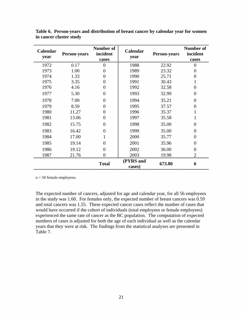
21
Table 6. Person-years and distribution of breast cancer by calendar year for women in cancer cluster study
Calendar year Person-years
Number of incident
cases
Calendar year Person-years
Number of incident
cases 1972 0.17 0 1988 22.92 0 1973 1.00 0 1989 23.32 0 1974 1.33 0 1990 25.71 0 1975 3.35 0 1991 30.43 1 1976 4.16 0 1992 32.58 0 1977 5.30 0 1993 32.99 0 1978 7.00 0 1994 35.21 0 1979 8.59 0 1995 37.57 0 1980 11.27 0 1996 35.37 1 1981 13.06 0 1997 35.58 1 1982 15.75 0 1998 35.00 0 1983 16.42 0 1999 35.00 0 1984 17.00 1 2000 35.77 0 1985 19.14 0 2001 35.96 0 1986 19.12 0 2002 36.00 0 1987 21.76 0 2003 19.98 2
Total (PYRS and cases) 673.80 6
n = 50 female employees. The expected number of cancers, adjusted for age and calendar year, for all 56 employees in the study was 1.60. For females only, the expected number of breast cancers was 0.59 and total cancers was 1.55. These expected cancer cases reflect the number of cases that would have occurred if the cohort of individuals (total employees or female employees) experienced the same rate of cancer as the BC population. The computation of expected numbers of cases is adjusted for both the age of each individual as well as the calendar years that they were at risk. The findings from the statistical analyses are presented in Table 7.

22
Table 7. Observed and expected cases and age/calendar-year adjusted standardized incidence ratios (SIRs) for breast cancer (females only) and all cancers.
Person-years
Number of subjects
Expected cancers
Observed cancers
Standard Incidence
Ratio
95% Confidence Intervals
Breast Cancer 673.80 50 0.59 6 10.2 3.74-22.24
All cancers (females
only)
673.80 50 1.55 9 5.8 2.66-11.02
All cancers
(all subjects)
751.27 56 1.60 10 6.3 3.02-11.59
The finding of a SIR of 10.2 for breast cancer with 95 percent confidence intervals exceeding 1.0 indicate that the expected number of breast cancers was significantly elevated. The SIR of 10.2 indicates that the women in the MMHL were experiencing breast cancer incidence at approximately ten times the rate than women in the BC population. The 95 percent confidence intervals suggest that, with 95 percent certainty, the increased rate was from 3.7 to 22.2 times higher than the BC rates. Similarly, the standard incidence rates for all cancers in both men and women were significantly elevated as compared to the rates in BC. However, given the large proportion of cancers that were of the breast, the excess in the total cancer SIRs was driven by the high number of reported breast cancers in the employee cohort. The incidence rate for breast cancer is computed by dividing the person-years of observation for women employees by the number of reported breast cancer cases. This rate was 890 per 100,000 person-years in the study population, and as a comparison, the average incidence rate in BC for breast cancer (all ages) in 2000 was 120 per 100,000 persons. Cox proportional hazard modeling showed that the variables age at start of work at MMHL, job position, and job status were not related to the hazard rate. The hazard rate is defined as the probability per time unit that a person who has not developed cancer to the beginning of the respective interval will develop cancer in that interval. Findings from the proportional hazard model are presented in Table 8.

23
Table 8. Hazard ratios and 95% confidence intervals (CIs) of breast cancer in relation to age at start of work at MMHL, position, and job status for women
Variables Hazard Ratio 95% CI p-value * Age at start work (yrs) 1.07 0.97, 1.18 0.173 Position Technician Aid, clerk or ECG
1.00 0.67
0.08, 6.04
0.723
Job status Part time or causal Full time
1.00 1.45
0.14, 15.06
0.754
* p-value from Cox proportional hazards model with age at start work as a continuous variable and position and job status as categorical variables.
Field Investigations: Potential Exposures to Potentially Carcinogenic Substances or Physical Agents The walk-through investigation was conducted in August 2003 and included a review of the current procedures that may result in employee exposures to chemical or physical agents. Questions were asked about past practices and exposures to gain an understanding about how exposures may have changed over the years. Key points from the walkthrough are provided below:
• Current chemical exposures are minimal because liquid volumes are small and handling is often minimized through the use of “lock and load” systems
• Exposures to physical agents, such as ionizing radiation and electromagnetic fields appears to not be excessive (heat and noise exposures were also minimal)
• Past exposures were likely much higher as a number of procedures have been modified due to technological advances
o A major change was in the preservation of tissue samples, tissue staining, and glucose measurement. These procedures, in the past, required open use of solvents and reagents which included formalin, xylene, and o-toluidine. Most of these procedures were performed in a separate area of the laboratory, which was removed when the procedures were modified. It should be noted that o-toluidine, which was discussed in the literature review, is a rat mammary carcinogen, and formaldehyde (the major component in formalin) is a known human carcinogen.
o Other areas of the laboratory also were renovated due to changes in laboratory procedures. Remnants of a local exhaust ventilation system are present in one area where open chemicals were once mixed and dispensed.
• Poor indoor air quality was a common complaint in the past but appears to be less of a problem currently. An incinerator at the hospital was a source of very odourous and potentially toxic compounds (likely acid gases and possible

24
combustion products of PVC (monomers of vinyl chloride) and other plastics (halogenated organics)).
Previous air quality studies have been performed at MMHL, however investigators did not have access to the findings. In discussions with occupational health and safety professionals at FHA, it was mentioned that all measured concentrations of air contaminants were below regulated limits. Summary and Recommendations These findings provide evidence that the female employees within MMHL experienced an elevated rate of breast cancer over the past 30 years. The SIR of 10.2 for breast cancer is statistically significant and the magnitude of the increase rate is of concern. Previous studies have identified nurses and workers in clinical laboratories at higher risk of breast cancer; however, these investigations have not found the magnitude of excess risk found in this study. These previous studies were not designed to determine the causal factors associated with the increased breast cancer risk in laboratory employees or nurses, but it is likely that reproductive factors such as delayed first full-term pregnancy and nulliparity were important in explaining the excess risk. In our study we did not gather personal information pertaining to known risk factors for breast cancer. The reason for not gathering this information was that this is a preliminary epidemiological study and information on risk factors is difficult to interpret without a comparison population where the prevalence of risk factors is available. For example, in our study if we had detailed information about reproductive factors, family history of breast cancer, socioeconomic factors, alcohol consumption, physical exercise, and obesity, we would only be able to compare the prevalence of those factors with those within the general population. Thus, such data would provide clues as to the possible reasons for the elevated risk – if the prevalence of these risk factors were the same as the general population it would suggest that occupational factor(s) predominate. Only a full scale, etiologic investigation would have the capability of clearly identifying occupational factors as attributable to the increased breast cancer risk. A full-scale epidemiologic study may not be the most appropriate action to take despite the increased rates of cancer MMHL employees have experienced. The major goal of cluster investigations is to identify risk factors so that action can be taken to reduce exposures and risk. Air quality studies and reviews of procedures indicate that current exposures to carcinogens are minimal. Past exposures to chemicals like o-toluidine may have resulted in some increased risk for employees, but these exposures appear to have been eliminated. Another issue that discourages a major epidemiologic investigation pertains to the statistics of clusters themselves. Cluster research has shown that elevated rates occur by chance at some geographic locations and times. In fact, clusters always occur and it is a statistical phenomenon – even when there is no causal factor that is responsible for the increased incidence (this is why so few cluster investigations uncover any new risk

25
factors). So, if we look around at many geographic areas and times we will find some clusters; if a specific cluster is related to statistics and not an etiologic agent, it is most likely that in the next time period at this location the rate will not be significantly elevated. Thus, it would be very prudent to continue to evaluate the incidence of breast cancer in MMHL employees to see if the rate comes closer to what is expected. In summary, this study confirmed that the perceived cluster was an observed cluster and that MMHL employees were experiencing an elevated rate of breast cancer. The factors associated with this increased incidence could not be determined but may have been due to: (1) a cluster of reproductive and other known, nonoccupational, risk factors, (2) past exposures to chemical carcinogens and less likely to ionizing radiation, and (3) a chance occurrence (statistical anomaly). Our recommendations for action to be considered are: (1) conduct a thorough inventory of all chemicals currently used in the laboratory and identify any that are listed as animal or known/potential human carcinogens (listing from IARC, NTP, etc.). If any listed compounds are used, conduct a detailed exposure assessment; (2) ensure that exposures to ionizing radiation (one of the few known environmental risk factors for breast cancer) are at background; (3) provide education to all employees about risk factors for breast cancer and the importance of self exams and mammography; (4) continue to collect information on the incidence of breast and all cancers in the future so that SIRs can be computed; and (5) if information as to the possible causes of the high SIR is needed, collect information on known risk factors for breast cancer from all employees and send employee information to the BC Cancer Agency for linkage with the cancer registry.

26
References Aldrich, T. and T. Sinks (2002). "Things to know and do about cancer clusters." Cancer
Invest 20(5-6): 810-6. Band, P. R., N. D. Le, et al. (2000). "Identification of occupational cancer risks in British
Columbia. A population-based case-control study of 995 incident breast cancer cases by menopausal status, controlling for confounding factors." J Occup Environ Med 42(3): 284-310.
Bender, A. P. (1987). "On disease clustering." Am J Public Health 77(6): 742. Bender, A. P., A. N. Williams, et al. (1990). "Appropriate public health responses to
clusters: the art of being responsibly responsive." Am J Epidemiol 132(1 Suppl): S48-52.
Bennett, L. M. and B. J. Davis (2002). "Identification of mammary carcinogens in rodent bioassays." Environ Mol Mutagen 39(2-3): 150-7.
Bigelow, P. (2000). "Application of Threshold Limit Values for Chemical Substances to employees in laboratories." Chem. Health Safety 8: 16-21.
Bounias, M. (2003). "Etiological factors and mechanism involved in relationships between pesticide exposure and cancer." J Environ Biol 24(1): 1-8.
Breslow, N. a. D., N. (1987). Statistical Methods in Cancer Research. Lyon, France, International Agency for Research on Cancer.
Brody, J. G. and R. A. Rudel (2003). "Environmental pollutants and breast cancer." Environ Health Perspect 111(8): 1007-19.
Burkman, R. T. (1999). "Oral contraceptives: an update." Drugs Today (Barc) 35(11): 857-66.
Burnett, C., C. Robinson, et al. (1999). "Cancer mortality in health and science technicians." Am J Ind Med 36(1): 155-8.
Caldwell, G. G. (1990). "Twenty-two years of cancer cluster investigations at the Centers for Disease Control." Am J Epidemiol 132(1 Suppl): S43-7.
Cartwright, R. A. (1999). "Cluster investigations: are they worth it?" Med J Aust 171(4): 172.
CCR (1998). Guidelines for the Investigation of Cancer Clusters in BC. Vancouver, BC, Cancer Control Research, British Columbia Cancer Agency.
Charles, G. D., C. Gennings, et al. (2002). "Assessment of interactions of diverse ternary mixtures in an estrogen receptor-alpha reporter assay." Toxicol Appl Pharmacol 180(1): 11-21.
Charlier, C., A. Albert, et al. (2003). "Breast cancer and serum organochlorine residues." Occup Environ Med 60(5): 348-51.
Claus, E. B., J. M. Schildkraut, et al. (1996). "The genetic attributable risk of breast and ovarian cancer." Cancer 77(11): 2318-24.
Davis, D. L., D. Axelrod, et al. (1997). "Avoidable causes of breast cancer: the known, unknown, and the suspected." Ann N Y Acad Sci 833: 112-28.
Davis, S., D. K. Mirick, et al. (2001). "Night shift work, light at night, and risk of breast cancer." J Natl Cancer Inst 93(20): 1557-62.
DeBruin, L. S. and P. D. Josephy (2002). "Perspectives on the chemical etiology of breast cancer." Environ Health Perspect 110 Suppl 1: 119-28.
27
DeBruin, L. S., J. B. Pawliszyn, et al. (1999). "Detection of monocyclic aromatic amines, possible mammary carcinogens, in human milk." Chem Res Toxicol 12(1): 78-82.
Deligeoroglou, E., E. Michailidis, et al. (2003). "Oral contraceptives and reproductive system cancer." Ann N Y Acad Sci 997: 199-208.
Drijver, M. and F. Woudenberg (1999). "Cluster management and the role of concerned communities and the media." Eur J Epidemiol 15(9): 863-9.
Finkelstein, M. M. and N. Wilk (1990). "Investigation of a lung cancer cluster in the melt shop of an Ontario steel producer." Am J Ind Med 17(4): 483-91.
Fiore, B. J., L. P. Hanrahan, et al. (1990). "State health department response to disease cluster reports: a protocol for investigation." Am J Epidemiol 132(1 Suppl): S14-22.
Frelick, R. W. and A. Topham (1991). "The value of cluster and environmentally related cancer studies in Delaware." Del Med J 63(9): 559-61.
Gammon, M. D., A. I. Neugut, et al. (2002). "The Long Island Breast Cancer Study Project: description of a multi-institutional collaboration to identify environmental risk factors for breast cancer." Breast Cancer Res Treat 74(3): 235-54.
Gangnon, R. E. and M. K. Clayton (2001). "A weighted average likelihood ratio test for spatial clustering of disease." Stat Med 20(19): 2977-87.
Garry, V. F., D. R. Jacobs, Jr., et al. (1989). "An approach to the investigation of cancer in tool and die workers. I. Epidemiologic findings in a suspected cancer cluster among tool and die workers." Mutat Res 225(3): 107-13.
Goldberg, M. S. and F. Labreche (1996). "Occupational risk factors for female breast cancer: a review." Occup Environ Med 53(3): 145-56.
Gordon, N. H. (2003). "Socioeconomic factors and breast cancer in black and white Americans." Cancer Metastasis Rev 22(1): 55-65.
Hall, H. I., C. V. Lee, et al. (1996). "Cluster: a software system for epidemiologic cluster analysis." Stat Med 15(7-9): 943-50.
Halls, S. B. (2003). Detailed Breast Cancer Risk Calculator, Steven B. Halls, MD. 2004. Hamajima, N., K. Hirose, et al. (2002). "Alcohol, tobacco and breast cancer--
collaborative reanalysis of individual data from 53 epidemiological studies, including 58,515 women with breast cancer and 95,067 women without the disease." Br J Cancer 87(11): 1234-45.
Hanrahan, L. P., I. Mirkin, et al. (1990). "SMRFIT: a Statistical Analysis System (SAS) program for standardized mortality ratio analyses and Poisson regression model fits in community disease cluster investigations." Am J Epidemiol 132(1 Suppl): S116-22.
Hargreaves, D. F., C. S. Potten, et al. (1999). "Two-week dietary soy supplementation has an estrogenic effect on normal premenopausal breast." J Clin Endocrinol Metab 84(11): 4017-24.
Higginbotham, S., Z. F. Zhang, et al. (2004). "Dietary glycemic load and breast cancer risk in the Women's Health Study." Cancer Epidemiol Biomarkers Prev 13(1): 65-70.
Jacobzone, S. (2002). Summary of the Results from the Breast Cancer Disease Study. OECD Study on Cross-national Differences of Ageing-related Diseases Workshop, June 20-21, Paris, France, Organization for Economic Cooperation and Development.
Kheifets, L. I. (1993). "Cluster analysis: a perspective." Stat Med 12(19-20): 1755-6. Kipen, H. and D. Wartenberg (1988). "Don't close the door: some observations on cancer
cluster investigation." J Occup Med 30(8): 661-2.

28
Knorr-Held, L. and G. Rasser (2000). "Bayesian detection of clusters and discontinuities in disease maps." Biometrics 56(1): 13-21.
Kreiger, N., M. Sloan, et al. (1999). "The risk of breast cancer following reproductive surgery." Eur J Cancer 35(1): 97-101.
Kulldorff, M., W. F. Athas, et al. (1998). "Evaluating cluster alarms: a space-time scan statistic and brain cancer in Los Alamos, New Mexico." Am J Public Health 88(9): 1377-80.
Laitman, C. J. (2002). "DES exposure and the aging woman: mothers and daughters." Curr Womens Health Rep 2(5): 390-3.
Land, C. E., M. Tokunaga, et al. (2003). "Incidence of female breast cancer among atomic bomb survivors, Hiroshima and Nagasaki, 1950-1990." Radiat Res 160(6): 707-17.
Langmuir, A. D. (1965). "Formal discussion of: epidemiology of cancer: spatial-temporal aggregation." Cancer Res 25(8): 1384-6.
Lawson, A. B. (2000). "Cluster modelling of disease incidence via RJMCMC methods: a comparative evaluation. Reversible jump Markov chain Monte Carlo." Stat Med 19(17-18): 2361-75.
Lewis, R. and G. Rempala (2003). "A case-cohort study of angiosarcoma of the liver and brain cancer at a polymer production plant." J Occup Environ Med 45(5): 538-45.
Lieben, J. (1966). "Malignancies in asbestos workers." Arch Environ Health 13(5): 619-21.
Lieben, J. and H. Pistawka (1967). "Mesothelioma and asbestos exposure." Arch Environ Health 14(4): 559-63.
Mackillop, W. J., J. Zhang-Salomons, et al. (2000). "Associations between community income and cancer incidence in Canada and the United States." Cancer 89(4): 901-12.
Madigan, M. P., R. G. Ziegler, et al. (1995). "Proportion of breast cancer cases in the United States explained by well-established risk factors." J Natl Cancer Inst 87(22): 1681-5.
McTiernan, A., K. B. Rajan, et al. (2003). "Adiposity and sex hormones in postmenopausal breast cancer survivors." J Clin Oncol 21(10): 1961-6.
Medina, D. (2004). "Breast cancer: the protective effect of pregnancy." Clin Cancer Res 10(1 Pt 2): 380S-4S.
Morton, W. E. (1995). "Major differences in breast cancer risks among occupations." J Occup Environ Med 37(3): 328-35.
NCI (1997). "Workshop on Hormones, Hormone Metabolism, Environment, and Breast Cancer,
New Orleans, Louisiana, September 28-29, 1995." Monographs in Environ Hlth Perspect Supp 105(3): 557-688. NCIC (1999). Canadian Cancer Statistics. Toronto, Canada, National Cancer Institute of
Canada. NRC (2004). Breast Cancer Rates, 1986-1995 URL:
http://atlas.gc.ca/site/english/maps/health/status/breastcancerrates1986-1995?map=%2Fhome%2Fmapdata%2Faoc_v3%2Fenglish%2F876.map&imgext=-2032143.858627+341765.863549+-1820995.950293+529233.632629&mode=browse&scale=1398434.17556762&imgxy

29
=&mapext=&img.x=213&img.y=191, Natural Resources Canada, Communication Canada, Canadian Government Publishing, Ottawa, Canada.
Parkin, D. M., F. Bray, et al. (2001). "Estimating the world cancer burden: Globocan 2000." Int J Cancer 94(2): 153-6.
Parkin, D. M., F. I. Bray, et al. (2001). "Cancer burden in the year 2000. The global picture." Eur J Cancer 37 Suppl 8: S4-66.
Patel, A. V., M. F. Press, et al. (2003). "Lifetime recreational exercise activity and risk of breast carcinoma in situ." Cancer 98(10): 2161-9.
Pott, P. (1996). Observation of scrotal tumors among London Chimney Sweeps (republished). Classics in Oncology. A. C. Society. New York, NY.
PPHB (2004). Breast Cancer in Canada. online at: http://www.hc-sc.gc.ca/pphb-dgspsp/publicat/updates/breast-99_e.html, Population and Public Health Branch, Health Canada, Ottawa, Canada. 2004.
Quataert, P. K., B. Armstrong, et al. (1999). "Methodological problems and the role of statistics in cluster response studies: a framework." Eur J Epidemiol 15(9): 821-31.
Recchia, A. G., A. Vivacqua, et al. (2004). "Xenoestrogens and the induction of proliferative effects in breast cancer cells via direct activation of oestrogen receptor alpha." Food Addit Contam 21(2): 134-44.
Robbins, A. S., S. Brescianini, et al. (1997). "Regional differences in known risk factors and the higher incidence of breast cancer in San Francisco." J Natl Cancer Inst 89(13): 960-5.
Ron, E. (2003). "Cancer risks from medical radiation." Health Phys 85(1): 47-59. Russo, I. H. and J. Russo (1996). "Mammary gland neoplasia in long-term rodent
studies." Environ Health Perspect 104(9): 938-67. Russo, I. H. and J. Russo (1998). "Role of hormones in mammary cancer initiation and
progression." J Mammary Gland Biol Neoplasia 3(1): 49-61. Russo, J. and I. H. Russo (1997). "Toward a unified concept of mammary
carcinogenesis." Prog Clin Biol Res 396: 1-16. Safe, S. H. and T. Zacharewski (1997). "Organochlorine exposure and risk for breast
cancer." Prog Clin Biol Res 396: 133-45. Sarkar, F. H. and Y. Li (2003). "Soy isoflavones and cancer prevention." Cancer Invest
21(5): 744-57. Schernhammer, E. S., F. Laden, et al. (2003). "Night-shift work and risk of colorectal
cancer in the nurses' health study." J Natl Cancer Inst 95(11): 825-8. Schulmeister, L. (2003). "Diethylstilbestrol exposure: how well informed are you about
this health risk?" Clin J Oncol Nurs 7(4): 373-4. Schulte, P. A., R. L. Ehrenberg, et al. (1987). "Investigation of occupational cancer
clusters: theory and practice." Am J Public Health 77(1): 52-6. Semenciw, R. M., N. D. Le, et al. (2000). "Methodological issues in the development of
the Canadian Cancer Incidence Atlas." Stat Med 19(17-18): 2437-49. Siegrist, M., G. T. Cvetkovich, et al. (2001). "Shared values, social trust, and the
perception of geographic cancer clusters." Risk Anal 21(6): 1047-53. Smith, D. and R. Neutra (1993). "Approaches to disease cluster investigations in a state
health department." Stat Med 12(19-20): 1757-62.
30
Snedeker, S. (1997). Do we need to be concerned about environmental chemicals and breast cancer? An interview with the BCERF Research Project Leader. The Ribbon. 2.
Sonnenschein, C. and A. M. Soto (1998). "An updated review of environmental estrogen and androgen mimics and antagonists." J Steroid Biochem Mol Biol 65(1-6): 143-50.
Timander, L. M. and S. McLafferty (1998). "Breast cancer in West Islip, NY: a spatial clustering analysis with covariates." Soc Sci Med 46(12): 1623-35.
Tompa, A., J. Major, et al. (1999). "Is breast cancer cluster influenced by environmental and occupational factors among hospital nurses in Hungary?" Pathol Oncol Res 5(2): 117-21.
Trumbo, C. W. (2000). "Public requests for cancer cluster investigations: a survey of state health departments." Am J Public Health 90(8): 1300-2.
van Netten, C., R. H. Brands, et al. (2003). "Cancer cluster among police detachment personnel." Environ Int 28(7): 567-72.
Vassilopoulou-Sellin, R. (2003). "Breast cancer and hormonal replacement therapy." Ann N Y Acad Sci 997: 341-50.
Wang, D. Y., D. S. Allen, et al. (2000). "Urinary androgens and breast cancer risk: results from a long-term prospective study based in Guernsey." Br J Cancer 82(9): 1577-84.
Ward, E. M., P. A. Schulte, et al. (2003). "Priorities for development of research methods in occupational cancer." Environ Health Perspect 111(1): 1-12.
Weaver, V. M. (1997). "Chemical hazards in health care workers." Occup Med 12(4): 655-67.
Wittenberg, C. (1994). "Long Island breast cancer studies move forward." J Natl Cancer Inst 86(20): 1501-3.
WSDOH (2001). Guidelines for Investigating Clusters of Chronic Disease and Adverse Birth Outcomes. online: http://www.doh.wa.gov/EHSPHL/Epidemiology/NICE/publications/ClusterProt.pdf. Olympia, Washington, Washington State Department of Health.
Yang, D., L. Bernstein, et al. (2003). "Physical activity and breast cancer risk among Asian-American women in Los Angeles: a case-control study." Cancer 97(10): 2565-75.
Yue, W., R. J. Santen, et al. (2003). "Genotoxic metabolites of estradiol in breast: potential mechanism of estradiol induced carcinogenesis." J Steroid Biochem Mol Biol 86(3-5): 477-86.
Zeleniuch-Jacquotte, A., R. E. Shore, et al. (2004). "Postmenopausal levels of oestrogen, androgen, and SHBG and breast cancer: long-term results of a prospective study." Br J Cancer 90(1): 153-9.
Attachment 4
PowerPoint presentation An Investigation of a Cancer Cluster within the
Mission Memorial Hospital Laboratory


1
An Investigation of a Cancer Cluster within a Hospital Laboratory
P. Bigelow, PhD - Occupational Health and Safety Agency for Healthcare (OHSAH)
S. Yu, PhD - Institute for Health Promotion Research (IHPR), UBC
R. Nemanishen - Fraser Health AuthorityA. Yassi, MD, MSc. - Occupational Health and
Safety Agency for Healthcare and IHPR, UBC
Acknowledgements
Staff at Fraser Health, OHSAH, UBC, Colorado State University, and the BC Cancer Agency, especially
Dr. Nhu Lee (BC Cancer Agency)William SoYiu Ting (OHSAH)Drs. Reif and Burch (Colorado State)Dr. Paul Demers (School of Occupational and Environmental Hygiene, UBC)
Introduction
Report of seemingly high number of cancer cases in laboratory workersNeed for an epidemiological studyPurpose of investigation:
To determine if cluster had occurredTo identify possible occupational factors if a true cluster is identifiedTo ensure current exposures/occupational factors are below recommended standards and current exposures are not increasing the risk of cancer in MMHL employees
Methods - Epidemiology
Enumeration of the occupational cohortEmployee data files for all individuals working from 1970 to 2003 Methods for confidentiality
Analysis of cancer incidence dataCollection of data on health status and cancer incidence from personal interviews Data entered into excel and SPSSRates of cancer and breast cancer from 1970 to 2002 for males and females obtained Person years of observation (PYRS) were computed for study cohort for each age interval (5 yr increments) and calendar yearExpected number of cases (breast and all cancers) computed for each age and calendar year category
FBreastx100,00 0-4 5-9 10-14 15-19 20-24 25-29 30-34 35-39 40-44 45-49 50-54 55-59 60-64 65-69
1970 0 0 0 0 1.153722 9.534447 22.47155 47.04696 98.0408 171.1503 164.6675 184.5657 252.3378 190.6636person yrs 0 0 1 0 0 0 0 0 0 0
1971 0 0 0 0 0 9.872277 27.11824 39.26637 113.3425 205.6203 248.218 219.1901 254.3176 251.9548person yrs 0 0 1 0 0 0 0 0 0 0
1972 0 0 0 0.9374 1.005227 7.84947 12.83239 49.61032 98.31438 161.8765 202.9954 248.5001 241.8511 275.7176person yrs 0.083333 0 1 0 0 0 0 0 0 0
1973 0 0 0 0 0.964376 7.341451 17.30795 51.59797 118.5074 176.5951 204.7583 251.2563 273.3532 235.128person yrs 1 0 0 1 0 0 0 0 0 0
1974 0 0 0 0 1.813615 7.889235 32.11581 39.09774 121.9926 154.3537 209.1224 202.3736 304.1898 255.6846person yrs 1 0 0 1 0.25 0 0 0 0 0
1975 0 0 0 0 0 2.795014 19.79161 62.4102 88.90388 166.5556 203.5073 268.7858 304.6307 256.9967person yrs 1.583333 0 0 1 0.6666667 1 0 0 0 0
1976 0 0 0 0 0.843768 4.469673 20.14054 58.77332 115.0148 153.1928 174.5427 273.2811 267.4778 313.3323person years 0.083333 2 0 1 1 1 0 0 0 0
1977 0 1.044976 0 0 0 7.934969 39.48708 38.77472 97.06382 181.1815 215.1104 192.2752 235.8974 242.17person yrs 1.25 2 0 1 0 2 0 0 0 0
1978 0 0 0 0 0.814545 6.897384 20.5842 64.28923 92.80048 162.9328 183.6711 215.0696 260.0736 312.3669person yrs 2.916667 2 0 0 1 2 0 0 0 0
1979 0 0 0 0 2.383999 10.05151 26.07562 51.17281 95.82438 162.0991 209.9683 221.4242 239.0984 325.1659person yrs 2 3 0 0 1 2 0 0 0 0
1980 0 0 0 0 0.763674 7.224274 17.37363 59.63029 105.4896 180.2289 161.0377 258.5894 261.0714 334.2018person yrs 1 0 1.5 4 0.6666667 0 1 1 1 0 0 0
1981 0 0 0 0 0.735451 7.705585 25.40692 54.50115 98.4756 176.2989 203.9003 225.7557 264.9757 283.29
Breast Cancer Rates and Computation of Expected Number of Cases
Methods - Epidemiology
Data analysesExpected number of cases based on the total number of PYRS of observation was computed Observed number of breast and total cancer cases divided by expected number provided the Standard Incidence Ratio (SIR); 95% confidence intervals computed using Poisson statisticsCox proportional hazard modeling used to determine the association of occupational factors on rate of breast cancer

2
Methods - Exposure Assessment
Review of OHSAH MSDS database for chemicals and products used in hospital laboratoriesWalkthrough survey of MMHLInterviews with current MMHL employees
Changes in processes and controls over timeIssues with regard to air quality in MMHL over time
Methods - Literature Review
Conducted to provide information to assist in interpretation of study findingsFocused on breast cancer risk factors as this cancer type identified in preliminary analyses as likely with a higher incidence in MMHL employees Epidemiology of clusters also provided due to the complexity of the investigations that are performed to identify causal factors underlying clusters
Results - Literature ReviewCarcinogenesis is a multistage process that involves many events, some of which are not well understood.
Age-adjusted Cancer Incidence in 1984-88 (US)
Males Females1. Prostate 93.5 1. Breast 105.62. Lung 84.0 2. Colon/Rectum 42.93. Colon/Rectum 61.1 3. Lung 37.14. Bladder 29.9 4. Uterus 30.35. Lymphoma 19.1 5. Ovary 13.96. Mouth/ Pharynx 16.8 6. Lymphoma 13.17. Leukemia 13.0 7. Melanoma 9.18. Stomach 12.0 8. Pancreas 8.29. Melanoma 11.8 9. Leukemia 7.710. Kidney 11.4 10. Bladder 7.511. Pancreas 11.0 11. Mouth/Pharynx 6.5
Source: Cancer Statistics Review, 1973-1988, SEER
Breast Cancer - Demographics
Canada one of the highest incidence rates for breast cancer (225 per 100.000 women over age 40)Most common malignancy in womenSecond leading cause of cancer mortalityLifetime risk of 1 in 9Most common cause of death in women 35 to 54, age of maximum social responsibilityAverage years of life lost is 19.3
Age-adjusted annual breast cancer incidence rate among women in selected countries in 1983-87.
Source: Cancer Incidence in Five Continents (IARC)

3
Breast Cancer Risk Factors
75% have noneAgeFamily HistoryRadiation exposure in childhoodPrevious malignancy (Hodgkin’s)No children or none before 35Early menarche, late menopauseEstrogen effects in breast cancer
Breast Cancer Genetics
A small proportion of breast cancers appear to be attributable to an autosomal dominant genetic predisposition5-10% of all casesYoung ageStrong family historyBilateral diseaseBRCA-1 and BRCA-2
Breast Cancer Occupational Risk Factors
Animal studies identify mammary carcinogensHuman studies on specific chemicals mostly equivocal Organochlorine compounds implicated due to estrogenic activityMost epidemiology studies not able to establish causal links to breast cancerWorkers exposed to solvents at increased risk
Breast Cancer Occupational Risk Factors
Band et al (2000) higher breast cancer rates in BC for post menopausal women in medicine, health, and nursing occupationsNurses have been studied and shift work is associated with increased cancer risk (suppressed melatonin production)A clustering of risk factors is also suspected as a cause of the observed increased risk in nursing and medical occupations
Cancer Cluster -- Epidemiology
Regional and temporal patterns always existPatterns of increased/decreased risk may be due to aggregation of diverse factors (diet, demographics, lifestyle factors, occupational exposures)The lower mainland of BC has a higher than average incidence of breast cancer
Cancer Cluster -- Epidemiology
Analysis of clusters is responsibility of health agencies (BC Cancer Agency, BC CDC, Health Canada)Modern cancer clusters are widely reported and investigated
Over the last 30 years, thousands have been investigatedFew investigations have identified exposures
etiologically related to the increase risk if an increased risk was found

4
Cancer Cluster -- Epidemiology
Investigations answer questions:Is the cluster real -- first report of a
cluster is defined as a “perceived cluster”If the cluster is real, it is termed an
“observed cluster”Is an agent or exposure the cause of the
observed cluster -- if yes, the cluster is termed an “etiologic cluster”
Cancer Cluster -- Epidemiology
Standard protocols for cluster investigations
Stepwise, going from exploratory to analyticPrimary objective is to identify
exposures that may be increasing the risk of cancer and determining the steps to take to eliminate or reduce such risks
Cancer Cluster Epidemiology MMHL
57 employees working between January 1970 and August 2003
One person excluded from analysisCancer reported: 6 breast, 1 ovarian, 1 thyroid, and 1 lymphoma in cohortAt follow-up mean age of employees was 43.1 yearsTable 3 in report shows demographics of study cohort
Table 3. Age, gender and duration of follow-up by disease status
No Cancer Breast Cancer Other Cancer TotalFemales 41 (73.21%) 6 (10.71%) 3 (5.36%) 50 (89.29%)Males 5 (8.93%) 0 1 (1.79%) 6 (10.71%)
Mean age atstart work (yrs) 29.07 (8.68) 32.03 (11.29) 33.78 (7.53) 29.72 (8.86)
Mean age atend work or
end study (yrs)42.47 (10.67) 46.2 (13.24) 45.90 (7.99) 43.12 (10.70)
Mean durationof follow-up
(yrs)13.41 (8.34) 14.16 (10.50) 12.13 (8.13) 13.40 (8.41)
Data presented as frequency (percent of total), mean (standard deviation); n=56
Table 4. Person-years and distribution of all-causes cancer by calendar year for all employees
Calendaryear
Person-years
Numberof
incidencecases
Calendaryear
Person-years
Number ofincidence
cases
1964 0.50 0 1984 19.79 11965 1.00 0 1985 22.14 01966 1.00 0 1986 22.12 01967 1.00 0 1987 24.76 01968 1.00 0 1988 25.92 11969 1.00 0 1989 26.32 0
1970 1.00 0 1990 27.96 01971 1.00 0 1991 33.05 11972 1.17 0 1992 35.51 01973 2.00 0 1993 34.99 11974 2.33 0 1994 37.21 0
1975 4.35 0 1995 39.57 01976 5.16 0 1996 38.02 11977 6.30 0 1997 38.58 11978 8.00 0 1998 38.00 01979 9.59 0 1999 38.00 01980 13.27 0 2000 38.77 11981 15.06 0 2001 38.96 01982 17.75 0 2002 39.00 01983 18.42 0 2003 21.72 3
Total (PYRS andcases) 751.27 10
n = 56 male and female employees.
Table 5. Person-years and distribution of breast cancer by age group in women incancer cluster study
Age group (yrs)
Numberof
incidentcases
Person-years
Numberof incidentcases
15 – 19 0 1.34 0
20 – 24 0 43.95 0
25 – 29 0 100.37 0
30 – 34 0 120.91 0
35 – 39 0 112.67 1
40 – 44 0 113.25 0
45 – 49 0 83.50 1
50 – 54 0 51.58 2
55 – 59 0 25.16 0
60 – 64 0 13.49 1
65 – 69 0 5.00 0
70 – 74 0 2.58 1
Total (PYRS and cases) 673.80 6
n = 50 female employees

5
Table 6. Person-years and distribution of breast cancer by calendar year for womenin cancer cluster study
Calendaryear
Person-yearsNumber of
incidentcases
Calendar year Person-yearsNumber of
incidentcases
1972 0.17 0 1988 22.92 01973 1.00 0 1989 23.32 01974 1.33 0 1990 25.71 01975 3.35 0 1991 30.43 11976 4.16 0 1992 32.58 01977 5.30 0 1993 32.99 0
1978 7.00 0 1994 35.21 01979 8.59 0 1995 37.57 01980 11.27 0 1996 35.37 11981 13.06 0 1997 35.58 11982 15.75 0 1998 35.00 0
1983 16.42 0 1999 35.00 01984 17.00 1 2000 35.77 01985 19.14 0 2001 35.96 01986 19.12 0 2002 36.00 01987 21.76 0 2003 19.98 2
Total (PYRS andcases) 673.80 6
n = 50 female employees.
Table 7. Observed and expected cases and age/calendar-year adjusted standardizedincidence ratios (SIRs) for breast cancer (females only) and all cancers.
Person-yearsNumber
ofsubjects
Expectedcancers
Observedcancers
StandardIncidence
Ratio
95%Confidence
IntervalsBreastCancer 673.80 50 0.59 6 10.2 3.74-22.24
Allcancers(females
only)
673.80 50 1.55 9 5.8 2.66-11.02
Allcancers
(allsubjects)
751.27 56 1.60 10 6.3 3.02-11.59
Observed Cancer Cluster at MMHL
The SIR if 10.2 with 95% CI of 3.74 to 22.24 indicates the women in the cohort experienced breast cancer at a rate 10 times that which was expectedThe SIR of 6.3 for all cancers indicated that all employees experienced a rate of cancer over 6 times that which was expected; however this was driven by the larger proportion of breast cancer cases
Table 8. Hazard ratios and 95% confidence intervals (CIs) of breast cancer inrelation to age at start of work at MMHL, position, and job status for women
Variables Hazard Ratio 95% CI p-value *Age at start work (yrs) 1.07 0.97, 1.18 0.173Position
Technician Aid, clerk or ECG
1.000.67 0.08, 6.04 0.723
Job status Part time or causal Full time
1.001.45 0.14, 15.06 0.754
* p value from Cox proportional hazards model with age at start work as a continuous variable and position and job status as categorical variables.
Observed Cancer Cluster at MMHL
The hazard ratios from the Cox proportional hazard models were not statistically significantThe occupational factors studied in this investigation were not associated with the increase incidence of cancer in the employees at MMHLThe independent variables were likely too non specific to truly assess the impact of occupational exposures on breast cancer risk
Field Investigations at MMHL
Walkthrough surveyCurrent chemical exposures are minimal due to use of sealed systems which minimizes handlingVolumes of chemicals handled are minimalExposures to physical agents such as heat, noise, RF radiation appear to be minimalNo significant exposures to ionizing radiation were apparent at the time of the survey; radioisotopes are seldom, if ever used, and IR sources are not present in the laboratory

6
Field Investigations at MMHL
Walkthrough surveyPast exposures to carcinogens was likely although the exposures levels are unknownFormaldehyde in formalin is a known carcinogenO-toluidine, a rat mammary carcinogen, was a potential exposure in the past (sample preparation)Poor indoor air quality may have lead to exposures to chemical carcinogens in the past (emissions from a hospital incinerator as well as roofing emissions were reported)
Recommendations for Further Action
Conduct a thorough chemical inventory and identify exposures to hazards compounds and use of any carcinogens and possible mammary carcinogensConduct detailed exposures studies if hazardous chemical exposures are possibleEnsure exposures to all forms of ionizing radiation are at backgroundProvide information about risk factors for cancer to all MMHL employees
Recommendations for Further Action
The finding of this study provide support for an observed cancer cluster but the factor(s) responsible for the cluster are unknown
It is unlikely than a more extensive epidemiology study will identify an occupational/environmental risk factor that is causally related to the increased rate -- this is due to small sample size and the difficulties with confounding, effect modification, and other statistical issues
Recommendations for Further Action
It is recommended that a system be set up to continue the collection and linkage of employment data with cancer reports among employees of MMHL -- if subsequent analyses indicate that the SIR remains very highly elevated then further investigation is warranted
Recommendations for Further Action
To further examine the likelihood of an occupational risk factor being associated with the elevated SIR, a number of actions are possible
Confirm cancer diagnoses with the BC Cancer RegistryCollect information on known breast cancer risk factors to determine if they are more or less prevalent as compared to other populations
Correlation between breast cancer mortality rates and per capita consumption levels of fat (Ecologic comparisons)
0
5
10
15
20
25
0 20 40 60 80 100 120 140 160 180
Total dietary fat intake (g/day)
Age
-adj
uste
d de
ath
rate
per
100
,000

7
Genetic Mutations
Account for only 5-10% of all breast cancer cases
BRCA-1BRCA-2
BRCA Mutations
BRCA-1 positive50-80% chance breast cancer over a lifetime. ovarian cancer colon and prostate cancer.
BRCA-2 positive50-80% chance breast cancer over lifetime Less risk for ovarian cancer than BRCA1Males at increased risk for breast cancer
AJCC Staging System
T (Tumor)N (Nodes)M (Metastases)
TNM System for Breast Cancer
Tis Ductal carcinoma in situ
T1 Tumor 2 cm or lessT1a 0.5 cm or less T1b 1 cm or less and more than 0.5 cmT1c More than 1 cm and not more than 2 cm
T2 Tumor 5 cm or less & more than 2 cmT3 Tumor more than 5 cmT4 Tumor extends to skin or chest wall
T4a Extension to chest wallT4b Edema/ulceration/satellite nodulesT4c Both T4a and T4bT4d Inflammatory breast carcinoma
TNM System for Breast Cancer
Nx Regional nodes cannot be assessedN0 Regional nodes not involvedN1 Metastasis to movable axillarynode (s) /1-3 positiveN2 Metastases to ipsilateralaxillary lymph nodes/4-9 positiveN3 Metastases to internal mammary nodes/10+ , infra or supraclavicular
White = 5th edition; yellow = 6th edition
AJCC Breast Cancer Stage
Stage Criteria0 TisI T1 N0 M0II T2 or N1III T3N1 or T4 or N2-3IV M1
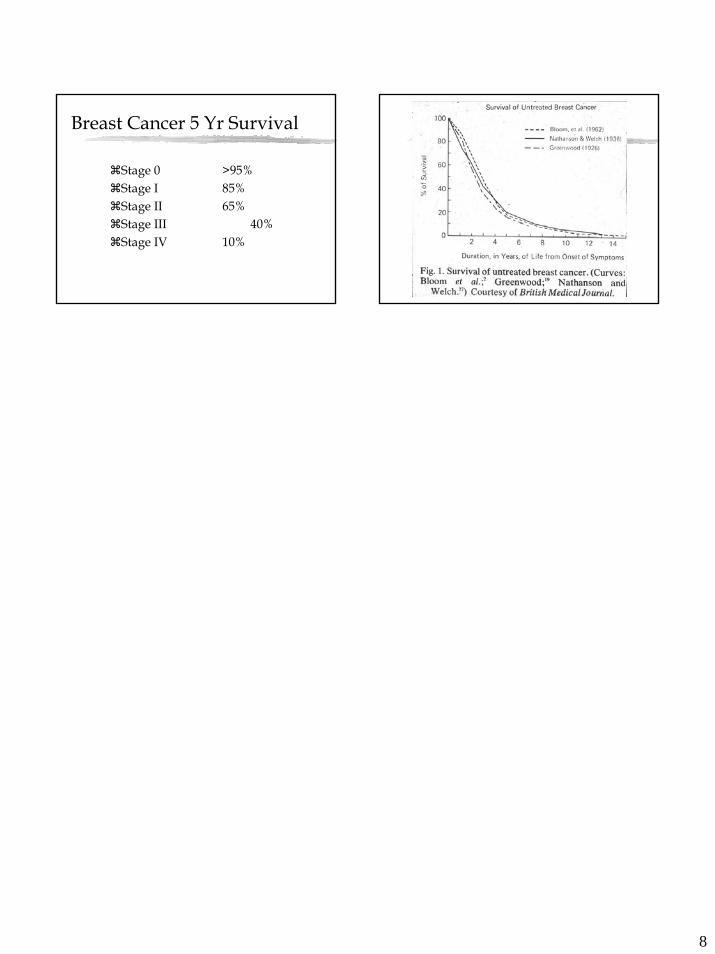
8
Breast Cancer 5 Yr Survival
Stage 0 >95%Stage I 85%Stage II 65%Stage III 40%Stage IV 10%

Attachment 5
Indoor Air Quality Assessment - Mission Memorial Hospital - Laboratory


Indoor Air Quality Assessment - Mission Memorial Hospital - Laboratory
Walk-through survey made November 12, 2004 - 13:34-14:05
Assessment by: Dave Buhr Safety Consultant Fraser Health.
The following indoor air quality parameters were sampled
CO2 - Carbon Dioxide Measured range - 462 PPM to 530 PPMAssessment - Good Fresh Air Exchange WCB max. for acceptable IAQ is 1000 PPM (650 PPM above outside ambient level assumed to be 350 PPM)Health Canada recommendation is 850 PPM Max.
CO - Carbon Monoxide - Measured Range 0 to1 PPM Assessment - Good WCB/ACGIH TLV - 8-hr Time Weighted Average (TWA) 25 PPM
Relative Humidity - Measure Range - 26.1% to 28.3%Assessment - Acceptable - below 25% staff may experience dryness of eyes, nose & throatASHRAE Recommended Range - 25%-60%
Temperature21.2 C to 22.7 CAssessment - NormalHealth Canada Recommendation - 80% staff comfort level
• No exterior air contaminant or exhaust discharges of concern were identified within 30 feet of the West Wingair intake when surveyed in August 2004. See attached photos.
• No interior air contaminant sources of concern were identified within the lab when surveyed August 2004. Ifformalin dispensing begins again in the pathology frozen section room this area should be re-evaluated foracceptable local exhaust ventilation performance.
• Nov 8/04 - Dennis Kruger (MMH - Power Engineer) and John Senetza inspected a typical supply and exhaustduct in the lab. The supply duct was clean with no evidence of dust. The inner exhaust louver intake and ducthad a fine layer of dust on it. Further up the duct, there was little evidence of dust or dirt. The DNT FanSystem supplies the Lab, Emergency, Health Records and two X-ray rooms.
• Last lab air balancing report - 1980 - 9 air changes per hour 4,570 CFM supply/5,068 CFM exhaust• DNT Fan System filtration level - 85% efficiency - filter change out scheduled every 6 months or sooner as
needed.

De-commissionedincinerator stack.
Unused Stat LabVentilationDischarge
West WingIntake Grill
e
Exterior View West Wing Intake
Interior View of West Wing Intak
Attachment 6
Letter from Dave Morley Head, Environmental Radiation Assessment
Programs Radiation Protection Services
At the BC Centre for Disease Control


Radiation Protection Services Tel 604.660.6633Main Floor, 655 12th Ave W Fax 604.660.6628Vancouver, British Columbia www.bccdc.orgCanada V5Z 4R4
September 13, 2004
Quinn Danyluk, MSc.Occupational Hygienist,Workplace Safety and WellnessFraser Health Authority
Dear Mr. Danyluk:
Thanks for your assistance setting up and retrieving the TLD monitors at the Laboratoryworking area in Mission Memorial Hospital in Mission B.C. The following table containsthe results.
RADIATION EXPOSURE IN THE M.M.H. LABORATORYChip
Number Location Exposure inmSv.
2 Std. Dev. InmSv
47 Pathologist Office 0.025 0.04141 Microbiology 0.138 0.05616 Outpatient Area near Exit B 0.143 0.05011 Bloodbank 0.190 0.06250 Chemistry Wall 0.109 0.02642 Exit C 0.098 0.02849 WC near Lab Directors Office 0.063 0.03243 Hematology near Exit D 0.124 0.014
14 Control Administration Area (differentbuilding) 0.103 0.036
45 Field Control 0.109 0.038
The chips were exposed in the laboratory area for 70 days between June 18, 2004 andAugust 26, 2004. A typical natural background exposure for the Fraser Valley areaduring that period would be 0.12 mSv. I would conclude that the exposures measured
. . . /2

-2-
in the Mission Memorial Laboratory Area are typical natural background and that the X–ray facility is not contributing to this natural background. This natural backgroundradiation would not contribute measurably to increased cancer risk.
Yours sincerely,
Dave MorleyHead, Environmental Radiation Assessment ProgramsRadiation Protection Services
k:\inbasket\trs\data\dm\Mission Hospital Radiation Exposure-Lab.doc
Attachment 7
Fraser Health Mission Memorial Hospital Laboratory: Chemical Assessment for Carcinogens


1
Mission Memorial Hospital Laboratory:Chemical Assessment for Carcinogens
SCOPE
This assessment was conducted in response to a cancer cluster within the Laboratory department atMission Memorial Hospital. An assessment conducted by Bigelow et al (2004) recommended anassessment of chemical and radiation hazards within the department that may have carcinogenicpotential. Bigelow et al provided a list of chemical substances known to have the potential of causingmammary tumors.
This report focuses on assessing the potential carcinogenicity of the current chemical products usedwithin this department.
BACKGROUND
Quinn Danyluk (Occupational Hygienist) and Dave Buhr (Safety Consultant) conducted walkthroughsurveys of the laboratory department at Mission Memorial Hospital On June 3rd, June 18th, and July 23rd.
Following these walkthrough surveys, a thorough review of the chemical products currently in useoccurred. The material safety data sheets (MSDSs) were reviewed to determine if any substances withinthe products were carcinogenic. Rita Ciconte (Occupational Hygienist) participated in this review andsummary assessment.
ASSESSMENT OF CARCINOGENICITY
The effect of a substance depends on:
1. Properties and Toxicity of the Substance2. Dose and Concentration3. Duration of Exposure4. Route of Entry into the Body5. Transport, Transformation, Distribution, and Elimination in the Body6. Half-Life7. Time
Carcinogenicity was determined through the use of the International Agency for Research on Cancer(IARC) Monographs. The terms utilized by IARC and throughout this review are provided below with theirassociated definition.

2
Mission Memorial Hospital Laboratory:Chemical Assessment for Carcinogens
DEFINITIONS
Group 1: The agent (mixture) is carcinogenic to humans. The exposure circumstance entails exposuresthat are carcinogenic to humans.
This category is used when there is sufficient evidence of carcinogenicity in humans. Exceptionally, anagent (mixture) may be placed in this category when evidence of carcinogenicity in humans is less thansufficient but there is sufficient evidence of carcinogenicity in experimental animals and strong evidencein exposed humans that the agent (mixture) acts through a relevant mechanism of carcinogenicity.
Group 2A: The agent (mixture) is probably carcinogenic to humans. The exposure circumstance entailsexposures that are probably carcinogenic to humans.
This category is used when there is limited evidence of carcinogenicity in humans and sufficient evidenceof carcinogenicity in experimental animals. In some cases, an agent (mixture) may be classified in thiscategory when there is inadequate evidence of carcinogenicity in humans and sufficient evidence ofcarcinogenicity in experimental animals and strong evidence that the carcinogenesis is mediated by amechanism that also operates in humans. Exceptionally, an agent, mixture or exposure circumstancemay be classified in this category solely on the basis of limited evidence of carcinogenicity in humans.
Group 2B: The agent (mixture) is possibly carcinogenic to humans. The exposure circumstance entailsexposures that are possibly carcinogenic to humans.
This category is used for agents, mixtures and exposure circumstances for which there is limited evidenceof carcinogenicity in humans and less than sufficient evidence of carcinogenicity in experimental animals.It may also be used when there is inadequate evidence of carcinogenicity in humans but there is sufficientevidence of carcinogenicity in experimental animals. In some instances, an agent, mixture or exposurecircumstance for which there is inadequate evidence of carcinogenicity in humans but limited evidence ofcarcinogenicity in experimental animals together with supporting evidence from other relevant data maybe placed in this group.
Group 3: The agent (mixture or exposure circumstance) is not classifiable as to its carcinogenicity tohumans.
This category is used most commonly for agents, mixtures and exposure circumstances for which theevidence of carcinogenicity is inadequate in humans and inadequate or limited in experimental animals.Exceptionally, agents (mixtures) for which the evidence of carcinogenicity is inadequate in humans butsufficient in experimental animals may be placed in this category when there is strong evidence that themechanism of carcinogenicity in experimental animals does not operate in humans. Agents, mixtures and exposure circumstances that do not fall into any other group are also placed in thiscategory.
Group 4: The agent (mixture) is probably not carcinogenic to humans.
This category is used for agents or mixtures for which there is evidence suggesting lack of carcinogenicityin humans and in experimental animals. In some instances, agents or mixtures for which there isinadequate evidence of carcinogenicity in humans but evidence suggesting lack of carcinogenicity inexperimental animals, consistently and strongly supported by a broad range of other relevant data, maybe classified in this group.

3
Mission Memorial Hospital Laboratory:Chemical Assessment for Carcinogens
Antifoam
Manufacturer Ingredients Percent CarcinogenicitySilica (CAS 68855-54-9) <30 Group 3Octamethylcyclotetrasiloxane (CAS 556-67-2) >1.0 Not ApplicableCrystalline Silica (CAS 14464-46-1); >1.0 Group 1Beckman
Methylated silica (CAS 67762-90-7) >20 Not Applicable
Exposure Limits
• Silica/Crystalline Silica: TWA 4 mg/m3 (total); TWA 1.5 mg/m3 (respirable)
Evaluation
Crystalline silica and silica are Group 1 and Group 3 carcinogens, respectively. Their carcinogenicpotential is based upon inhalation of the substance in its particulate form. When contained within anaqueous solution, the risk of inhaling silica particulate is extremely low to impossible. Additionally, verylow quantities of the product are used on an infrequent basis – a few drops may be used occasionally inthe ABL 700 Blood/Gas Machine. Gloves should be worn to prevent skin contact.
� The carcinogenic potential of the product under current usage is LOW.
Anti Seize Lubricant
Manufacturer Ingredients Percent CarcinogenicityMineral Oil (CAS 64742-52-5) >60 Group 1Lithium Soap (CAS 7620-77-1) <3.0 Not ApplicableAluminum (CAS 7429-90-5) <0.1 Not ApplicableCopper (CAS 7440-50-8) <0.1 Not ApplicableGraphite (CAS 7782-42-5); <0.1 Not Applicable
Beckman
Silica gel <0.1 Not Applicable
Exposure Limits
• Mineral Oil: TWA 0.2 mg/m3
Evaluation
Mineral oil (if mildly refined) is a Group 1 carcinogen. Their carcinogenic potential is based uponinhalation of the substance in a mist form. Under current usage, the potential for the generation of mist isextremely low to impossible. Additionally, very low quantities of the product are used as a lubricant for thecentrifuge. Gloves should be worn to prevent skin contact.
� The carcinogenic potential of the product under current usage is LOW.

4
Mission Memorial Hospital Laboratory:Chemical Assessment for Carcinogens
AST (Aspartame Aminotransferase) Reagent
Manufacturer Ingredients Percent CarcinogenicityTris(hydroxymethyl) aminomethane (CAS 77861)
<3.0 Not Applicable
Ethylene Glycol (CAS 107211) >75.0 Not ApplicableSodium Azide (CAS 26628-22-8) <0.1 Not ApplicableSodium hydroxide (CAS 1310-73-2) <0.1 Not ApplicableGlycerol (CAS 56-81-5) >25% Not ApplicableAcetaldehyde (CAS 75-07-0) <0.1 Group 2BEthylene oxide (CAS 75-21-8) <0.1 Group 1
Beckman
1,4-Dioxane (CAS 123-91-1) <0.1 Not Applicable
Exposure Limits
• Acetaldehyde: Ceiling 25 ppm• Ethylene oxide: TWA 0.1 ppm; STEL 1 ppm
Evaluation
Ethylene oxide is a Group 1 carcinogen; acetaldehyde is a Group 2B carcinogen. Ethylene oxide is a gasat room temperature and although used in small quantities in the production of this product, it not presentin the product. Both substances comprise <0.1% of the product and are therefore not reportable as ahazardous product on the MSDS.
The product is used in the Beckman Coulter Synchron CX& Pro Clinical System where threecompartments are mixed together. Acetaldehyde is present in very low concentrations and the risk ofvapour generation is very low. Gloves should be worn to prevent skin contact.
� The carcinogenic potential of the product under current usage is LOW.
B-5 Base
Manufacturer Ingredients Percent CarcinogenicityBDH Mercuric chloride (CAS 7487-94-7) 5-10 Group 3
Exposure Limits
• Mercuric chloride: Not applicable
Evaluation
Mercuric chloride is a Group 3 carcinogen. It is pre-poured and mixed with 40% formaldehyde. Currentlythe product is not in use. If in use it should be located under local exhaust ventilation. Gloves should beworn to prevent skin contact.
� The carcinogenic potential of the product under current usage is LOW.

5
Mission Memorial Hospital Laboratory:Chemical Assessment for Carcinogens
Basic Fuchsin Stain Solution
Manufacturer Ingredients Percent CarcinogenicityBasic fuchsin (CAS 632-99-5) 0.1 Group 2B
PMLEthanol (CAS 64-17-5) 10.0 Not Applicable
Exposure Limits
• Basic fuchsin: Not applicable
Evaluation
Basic fuchsin is a Group 2B carcinogen. It is used as a gram stain. Basic fuchsin comprisesapproximately 0.1% of the stain solution. Very small quantities of the product are utilized at any giventime. The potential for vapour generation is very low. Gloves should be worn to prevent skin contact.
� The carcinogenic potential of the product under current usage is LOW.
BUN (Blood Urea Nitrogen) Reagent
Manufacturer Ingredients Percent CarcinogenicityGlycerol (CAS 56815) >1.0 Not ApplicableEthylene glycol (CAS 107211) >25 Not ApplicableBeckmanGlycerine, N,N-bis(carboxymethyl)- (CAS 139-13-9) <0.1 Group 2B
Exposure Limits
• Glycerine, N,N-bis(carboxymethyl)-: Not applicable
Evaluation
Glycerine, N,N-bis(carboxymethyl)- is a Group 2B carcinogen. The substances carcinogenic potential isbased upon inhalation of the substance in its particulate form (i.e. salt form). The substance is not utilizedin particulate form in the reagent. Additionally, the substance comprises <0.1% of the product and istherefore not reportable as a hazardous product on the MSDS.
The reagent is used in the Beckman Coulter Synchron CX7 Pro Clinical System - two compartments (Aand B) that are mixed together.
� The carcinogenic potential of the product under current usage is LOW.

6
Mission Memorial Hospital Laboratory:Chemical Assessment for Carcinogens
CO2 Alkaline Buffer
Manufacturer Ingredients Percent CarcinogenicityGlycine,N,N-bis(carboxymethyl)- (CAS 139-13-9); <0.1 Group 2B
BeckmanPolyoxyethylated Octyl Phenol (CAS 26027-38-3) <0.1 Not Applicable
Exposure Limits
• Glycerine, N,N-bis(carboxymethyl)-: Not applicable
Evaluation
Glycerine, N,N-bis(carboxymethyl)- is a Group 2B carcinogen. The substances carcinogenic potential isbased upon inhalation of the substance in its particulate form (i.e. salt form). The substance is not utilizedin particulate form in the reagent. Additionally, the substance comprises <0.1% of the product and istherefore not reportable as a hazardous product on the MSDS.
The reagent is used in the Beckman Coulter Synchron CX7 Pro Clinical System – container is openedand placed on the instrument.
� The carcinogenic potential of the product under current usage is LOW.
Dade Behring CTNI Troponin Test Pack
Manufacturer Ingredients Percent CarcinogenicityBovine Serum Albumin (CAS 9048-46-8) 6 Not ApplicableSodium Azide (CAS 26628-22-8) 0.1 Not ApplicableStreptomycin Sulphate (CAS 3810-74-0) 0.025 Not Applicable
Dade Behring
Chloramphenicol (CAS 56-75-7) 0.000016 Group 2A
Exposure Limits
• Chloramphenicol: Not applicable
Evaluation
Chloramphenicol is a Group 2A carcinogen. It is present in extremely low concentrations within the testpack (anything less than 0.1% is not reportable as a hazardous product on the MSDS). It is used in theDade Behring Stratus CS as an in vitro diagnostic test for the measurement of cardiac troponin-I inheparinized whole blood/plasma. The product is used within a sealed container.
� The carcinogenic potential of the product under current usage is LOW.

7
Mission Memorial Hospital Laboratory:Chemical Assessment for Carcinogens
Coluter Clenz Cleaning Agent
Manufacturer Ingredients Percent CarcinogenicityEthylene Oxide (CAS 75-21-8) <0.1 Group 1Ethylene Glycol Monobutyl Ether (CAS 111-76-2) <0.1 Group 3
1,4-Dioxane (CAS 123-91-1) <0.1 Group 2BSodium Hydroxide (CAS 1310-73-2) <0.1 Not ApplicablePhosphoric Acid is (CAS 7664-38-2) <0.1 Not ApplicableMagnesium Nitrate (CAS 10377-60-3) <0.1 Not ApplicableMagnesium Chloride (CAS 7786-30-3) <0.1 Not ApplicablePropylene Glycol (57-55-6) <0.1 Not Applicable
Beckman
Subtilisin (9014-01-1) <0.1 Not Applicable
Exposure Limits
• Ethylene oxide: TWA 0.1 ppm; STEL 1 ppm• Ethylene Glycol Monobutyl Ether: TWA 20 ppm• 1,4-Dioxane: TWA 20 ppm
Evaluation
Ethylene oxide is a Group 1 carcinogen; Ethylene Glycol Monobutyl Ether is a Group 3 carcinogen; 1,4-Dioxane is a Group 2B carcinogen. All of these substances comprise <0.1% of the product and aretherefore not reportable as a hazardous product on the MSDS. Ethylene oxide is a gas at roomtemperature and although used in small quantities in the product production, it not present in the product.
The product is used in Beckman Coulter HmX. Product is in sealed container that is placed in instrument.Product is also placed in a squeeze bottle for cleaning the instrument.
� The carcinogenic potential of the product under current usage is LOW.
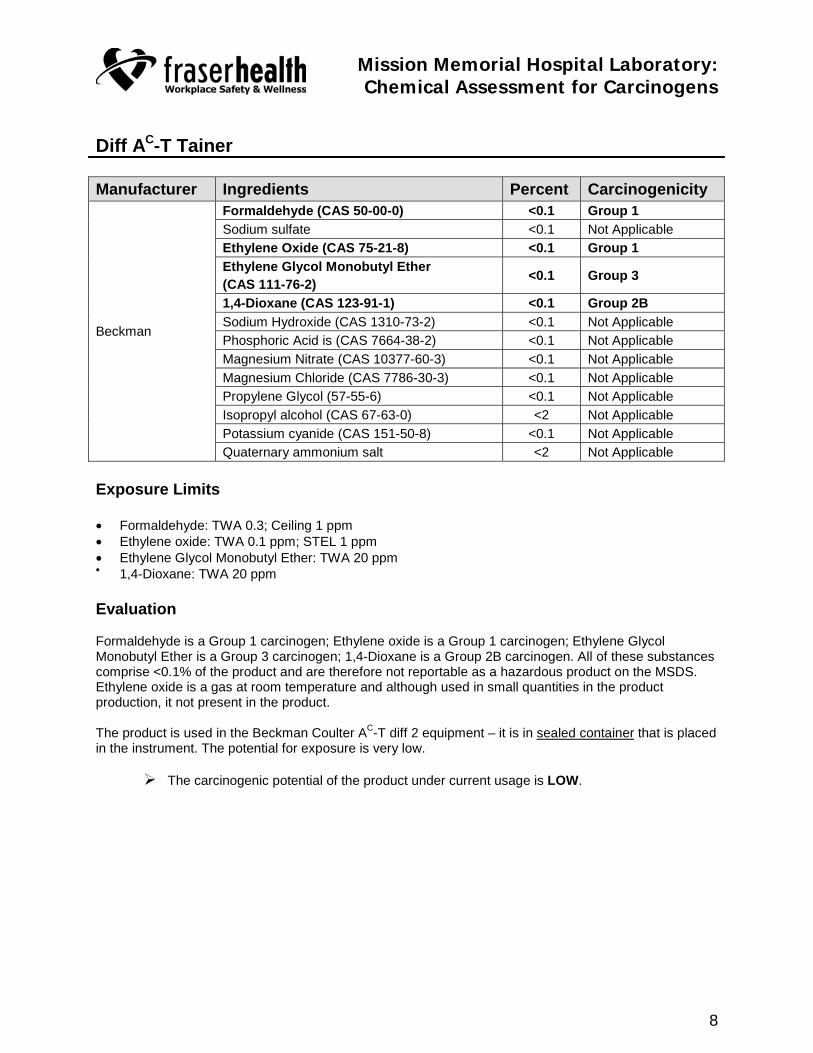
8
Mission Memorial Hospital Laboratory:Chemical Assessment for Carcinogens
Diff AC-T Tainer
Manufacturer Ingredients Percent CarcinogenicityFormaldehyde (CAS 50-00-0) <0.1 Group 1Sodium sulfate <0.1 Not ApplicableEthylene Oxide (CAS 75-21-8) <0.1 Group 1Ethylene Glycol Monobutyl Ether (CAS 111-76-2) <0.1 Group 3
1,4-Dioxane (CAS 123-91-1) <0.1 Group 2BSodium Hydroxide (CAS 1310-73-2) <0.1 Not ApplicablePhosphoric Acid is (CAS 7664-38-2) <0.1 Not ApplicableMagnesium Nitrate (CAS 10377-60-3) <0.1 Not ApplicableMagnesium Chloride (CAS 7786-30-3) <0.1 Not ApplicablePropylene Glycol (57-55-6) <0.1 Not ApplicableIsopropyl alcohol (CAS 67-63-0) <2 Not ApplicablePotassium cyanide (CAS 151-50-8) <0.1 Not Applicable
Beckman
Quaternary ammonium salt <2 Not Applicable
Exposure Limits
• Formaldehyde: TWA 0.3; Ceiling 1 ppm• Ethylene oxide: TWA 0.1 ppm; STEL 1 ppm• Ethylene Glycol Monobutyl Ether: TWA 20 ppm• 1,4-Dioxane: TWA 20 ppm
Evaluation
Formaldehyde is a Group 1 carcinogen; Ethylene oxide is a Group 1 carcinogen; Ethylene GlycolMonobutyl Ether is a Group 3 carcinogen; 1,4-Dioxane is a Group 2B carcinogen. All of these substancescomprise <0.1% of the product and are therefore not reportable as a hazardous product on the MSDS.Ethylene oxide is a gas at room temperature and although used in small quantities in the productproduction, it not present in the product.
The product is used in the Beckman Coulter AC-T diff 2 equipment – it is in sealed container that is placedin the instrument. The potential for exposure is very low.
� The carcinogenic potential of the product under current usage is LOW.
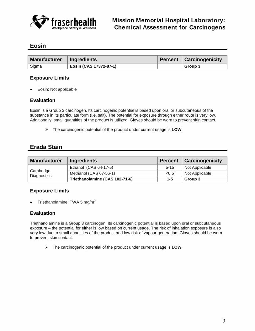
9
Mission Memorial Hospital Laboratory:Chemical Assessment for Carcinogens
Eosin
Manufacturer Ingredients Percent CarcinogenicitySigma Eosin (CAS 17372-87-1) Group 3
Exposure Limits
• Eosin: Not applicable
Evaluation
Eosin is a Group 3 carcinogen. Its carcinogenic potential is based upon oral or subcutaneous of thesubstance in its particulate form (i.e. salt). The potential for exposure through either route is very low.Additionally, small quantities of the product is utilized. Gloves should be worn to prevent skin contact.
� The carcinogenic potential of the product under current usage is LOW.
Erada Stain
Manufacturer Ingredients Percent CarcinogenicityEthanol (CAS 64-17-5) 5-15 Not ApplicableMethanol (CAS 67-56-1) <0.5 Not ApplicableCambridge
DiagnosticsTriethanolamine (CAS 102-71-6) 1-5 Group 3
Exposure Limits
• Triethanolamine: TWA 5 mg/m3
Evaluation
Triethanolamine is a Group 3 carcinogen. Its carcinogenic potential is based upon oral or subcutaneousexposure – the potential for either is low based on current usage. The risk of inhalation exposure is alsovery low due to small quantities of the product and low risk of vapour generation. Gloves should be wornto prevent skin contact.
� The carcinogenic potential of the product under current usage is LOW.

10
Mission Memorial Hospital Laboratory:Chemical Assessment for Carcinogens
Fisher Permount
Manufacturer Ingredients Percent CarcinogenicityToluene (CAS 108-88-3) 55.17 Group 3
FisherPiccolyte (CAS 68240-09-5) 44.85 Not Applicable
Exposure Limits
• Toluene: TWA 50 ppm
Evaluation
Toluene is a Group 3 carcinogen. The risk of exposure through the inhalation route is extremely low dueto small quantities of the product and low risk of vapour generation Risk of exposure through the oral orsubcutaneous route is also low. Additionally, small quantities of the product is utilized. Gloves should beworn to prevent skin contact.
� The carcinogenic potential of the product under current usage is LOW.
Gastroccult Developer
Manufacturer Ingredients Percent CarcinogenicityHydrogen Peroxide (CAS 7722-84-1) <5 Group 3Citric acid (CAS 77-92-9) <5 Not ApplicableOctylphenoxypoly(ethoxyethanol) (CAS 9036-19-5)
<5 Not Applicable
Ethanol-methanol mix (CAS 8013-52-3) <50 Not ApplicableEthylene Oxide (CAS 75-21-8) <0.1 Group 1
Beckman
1,4-Dioxane (CAS 123-91-1) <0.1 Group 2B
Exposure Limits
• Hydrogen peroxide: TWA 1 ppm• Ethylene oxide: TWA 0.1 ppm; STEL 1 ppm• 1,4-Dioxane: TWA 20 ppm
Evaluation
Hydrogen peroxide is a Group 3 carcinogen; Ethylene oxide is a Group 1 carcinogen; 1,4-Dioxane is aGroup 2B carcinogen.
Ethylene oxide is a gas at room temperature and although used in small quantities in the productproduction, it not present in the product. 1,4-Dioxane showed potential risk of carcinogenicity followingoral exposure in rats and mice but not following inhalation exposure – the risk of oral exposure is verylow. Ethylene oxide and 1,4-Dioxane comprise <0.1% of the product and are therefore not reportable as ahazardous product on the MSDS.
This product is rarely used. When it is used a drop is placed on a slide with blood. Very low quantities areused; the risk of exposure through any route is very low. Gloves should be worn to prevent skin contact.
� The carcinogenic potential of the product under current usage is LOW.

11
Mission Memorial Hospital Laboratory:Chemical Assessment for Carcinogens
HCL 0.1%
Manufacturer Ingredients Percent Carcinogenicity
RCH Hydrochloric acid (CAS 7647-01-0) 0.1 Group 3
Exposure Limits
Hydrochloric acid: Ceiling 2 ppm
Evaluation
Hydrochloric acid is a Group 3 carcinogen and thus is not classifiable with respect to humancarcinogenicity. This product is used in urinalysis crystal solubility. The quantity, concentration, methodof use, and exposure potential is sufficiently low to classify as a low risk.
� The carcinogenic potential of the product under current usage is LOW.
HCL 25%
Manufacturer Ingredients Percent CarcinogenicityRCH Hydrochloric acid (CAS 7647-01-0) 25 Group 3
Exposure Limits
Hydrochloric acid: Ceiling 2 ppm
Evaluation
Hydrochloric acid is a Group 3 carcinogen and thus is not classifiable with respect to humancarcinogenicity. This product is used to preserve 24 hour urine. Low quantities of the product are used.The potential for exposure through inhalation is low. Gloves should be worn to prevent skin contact.
� The carcinogenic potential of the product under current usage is LOW.

12
Mission Memorial Hospital Laboratory:Chemical Assessment for Carcinogens
Hemocult SENSA Developer
Manufacturer Ingredients Percent CarcinogenicityEthanol (CAS 64-17-5) 80 Not applicable
Beckman Hydrogen peroxide (CAS 7722-84-1) <4.2 Group 3
Exposure Limits
• Hydrogen peroxide: TWA 1 ppm
Evaluation
Hydrogen peroxide is a Group 3 carcinogen. Very low quantities of the product are used. The solution isdropped onto a card. The potential for exposure through any route is very low.
� The carcinogenic potential of the product under current usage is LOW.
Hydrox 7
Manufacturer Ingredients Percent Carcinogenicity3-Methoxy-3-methyl butanol (CAS 56539-66-3)
3-7 Not applicableVirox
Hydrogen peroxide (CAS 7722-84-1) 3-7 Group 3
Exposure Limits
• Hydrogen peroxide: TWA 1 ppm
Evaluation
Hydrogen peroxide is a Group 3 carcinogen. This product is used for cleaning chairs and benches; theminimum recommended quantity of product should be used whenever performing these tasks so as tominimize exposure. The product contains low quantities of hydrogen peroxide. Hydrogen peroxide has alow vapour pressure (<0.1 kPa); as a result, airborne exposure potential is low. Its carcinogenic potentialis based upon oral or subcutaneous exposure – the potential for either is low based on current usage.Gloves should be worn to prevent skin contact.
� The carcinogenic potential of the product under current usage is LOW.
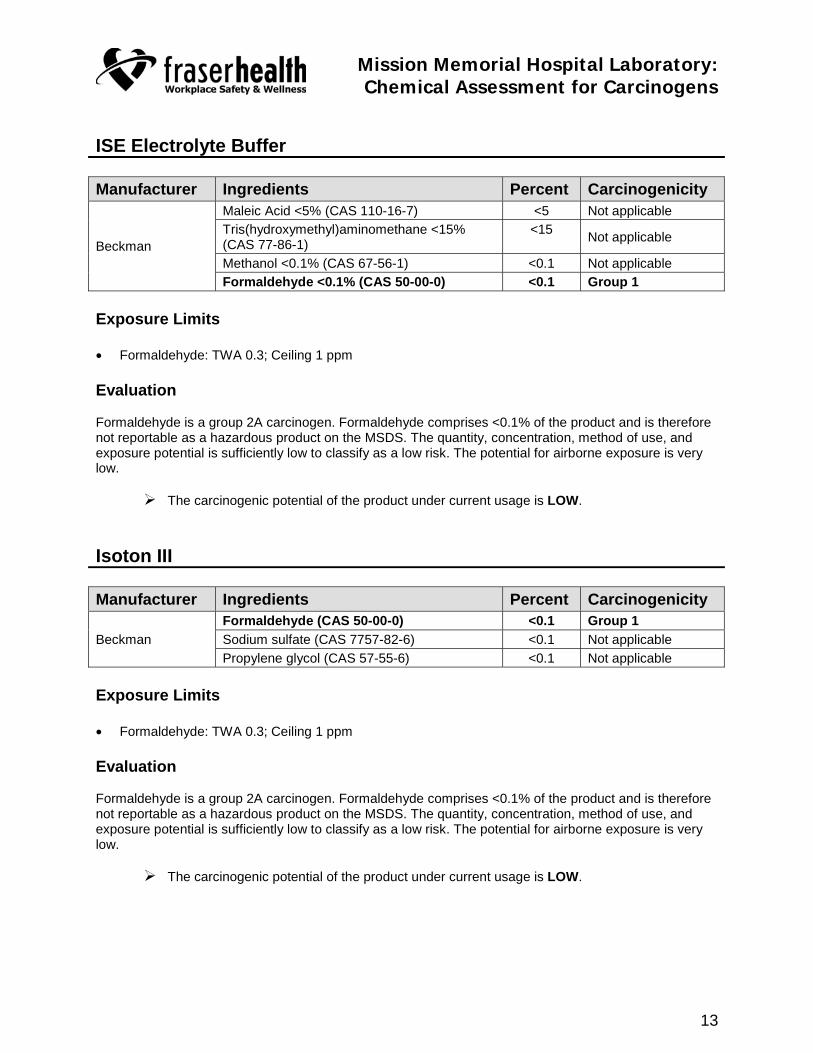
13
Mission Memorial Hospital Laboratory:Chemical Assessment for Carcinogens
ISE Electrolyte Buffer
Manufacturer Ingredients Percent CarcinogenicityMaleic Acid <5% (CAS 110-16-7) <5 Not applicableTris(hydroxymethyl)aminomethane <15%(CAS 77-86-1)
<15 Not applicable
Methanol <0.1% (CAS 67-56-1) <0.1 Not applicableBeckman
Formaldehyde <0.1% (CAS 50-00-0) <0.1 Group 1
Exposure Limits
• Formaldehyde: TWA 0.3; Ceiling 1 ppm
Evaluation
Formaldehyde is a group 2A carcinogen. Formaldehyde comprises <0.1% of the product and is thereforenot reportable as a hazardous product on the MSDS. The quantity, concentration, method of use, andexposure potential is sufficiently low to classify as a low risk. The potential for airborne exposure is verylow.
� The carcinogenic potential of the product under current usage is LOW.
Isoton III
Manufacturer Ingredients Percent CarcinogenicityFormaldehyde (CAS 50-00-0) <0.1 Group 1Sodium sulfate (CAS 7757-82-6) <0.1 Not applicableBeckmanPropylene glycol (CAS 57-55-6) <0.1 Not applicable
Exposure Limits
• Formaldehyde: TWA 0.3; Ceiling 1 ppm
Evaluation
Formaldehyde is a group 2A carcinogen. Formaldehyde comprises <0.1% of the product and is thereforenot reportable as a hazardous product on the MSDS. The quantity, concentration, method of use, andexposure potential is sufficiently low to classify as a low risk. The potential for airborne exposure is verylow.
� The carcinogenic potential of the product under current usage is LOW.

14
Mission Memorial Hospital Laboratory:Chemical Assessment for Carcinogens
Javex
Manufacturer Ingredients Percent CarcinogenicityColgate Palmolive Sodium hypochlorite (CAS 7681-52-9) 5-10 Group 3
Exposure Limits
• Sodium hypochlorite: Not applicable
Evaluation
Sodium hypochlorite is a group 3 carcinogen. Its carcinogenic potential is based upon oral exposure ofthe salt (particulate form); exposure via the skin has not been found to result in an increase risk. The riskof oral exposure to the salt is very low based on current usage. Gloves should be worn to prevent skincontact. This product is used infrequently for equipment maintenance.
� The carcinogenic potential of the product under current usage is LOW.
Potassium dichromate
Manufacturer Ingredients Percent Carcinogenicity
Fisher Scientific Chromic acid, dipotassium salt (CAS 7778-50-9) 100 Group 3
Exposure Limits
• Chromic acid, dipotassium salt: Not applicable
Evaluation
Chromic acid is a Group 3 carcinogen. Its carcinogenic potential is based upon oral exposure of the salt(particulate form) – exposure potential via this route would be low. The product is used during qualitycontrol tests approximately 3 times per week. Used in the Beckman Coulter Synchron CX7 ClinicalSystem for quality control check – done three times per week.
� The carcinogenic potential of the product under current usage is LOW.

15
Mission Memorial Hospital Laboratory:Chemical Assessment for Carcinogens
S-CAL Calibrator Kit
Manufacturer Ingredients Percent CarcinogenicityTreated Human Erythrocytes 45 Not applicable
Beckman 5-Fluorouracil (CAS 51-21-8) <0.1 Group 3
Exposure Limits
• 5-Fluorouracil: Not applicable
Evaluation
5-Fluorouracil is a Group 3 carcinogen. 5-Fluorouracil comprises <0.1% of the product and is thereforenot reportable as a hazardous product on the MSDS. Its carcinogenic potential is based upon oral andintravenous exposure – exposure potential via this route would be very low.
� The carcinogenic potential of the product under current usage is LOW.
Salicylate Reagent
Manufacturer Ingredients Percent CarcinogenicityBeckman Chloroform (CAS 67-66-3) <0.1 Group 2B
Exposure Limits
• Chloroform: TWA 2 ppm
Evaluation
Chloroform is a Group 2B carcinogen. It comprises <0.1% of the product and is therefore not reportableas a hazardous product on the MSDS. Because of the very low concentration, the potential for exposurevia any route is very low. Used in Beckman Coulter Synchron CX7 Pro Clinical System. Gloves should beworn to prevent skin contact.
� The carcinogenic potential of the product under current usage is LOW.

16
Mission Memorial Hospital Laboratory:Chemical Assessment for Carcinogens
TBIL (Total Bilirubin) Reagent
Manufacturer Ingredients Percent CarcinogenicityCaffeine (CAS 58-08-2) <5 Group 3Sodium Acetate (CAS 127-09-3) <10 Not applicableSodium Benzoate (CAS 532-32-1) <10 Not applicableEthoxylated lauryl alcohol (CAS 9002-92-0) <0.1 Not applicableHydrochloric acid (CAS 7647-01-0) <0.5 Group 3
Beckman
Sodium nitrite (CAS 7632-00-0) <10 Not applicable
Exposure Limits
• Caffeine: Not applicable• Hydrochloric acid: Ceiling 2 ppm
Evaluation
Caffeine and hydrochloric acid are Group 3 carcinogens. The carcinogenicity level, potential exposure,quantity within the product, and duration and frequency of use will not result in an increase risk.
� The carcinogenic potential of the product under current usage is LOW.
Wash Concentrate
Manufacturer Ingredients Percent CarcinogenicityMethyl P-Hydroxybenzoate (CAS 99-76-3) 3.0 Not applicable
Beckman Ethylene oxide (CAS 75-21-8) <0.1 Group 1
Exposure Limits
• Ethylene oxide: TWA 0.1 ppm; STEL 1 ppm
Evaluation
Ethylene oxide is a Group 1 carcinogen. Ethylene oxide is a gas at room temperature and although usedin small quantities in the production of this product, it not present in the product. It comprises <0.1% of theproduct and is therefore not reportable as a hazardous product on the MSDS. The quantity,concentration, method of use, and exposure potential is sufficiently low to classify this as low risk.
� The carcinogenic potential of the product under current usage is LOW.
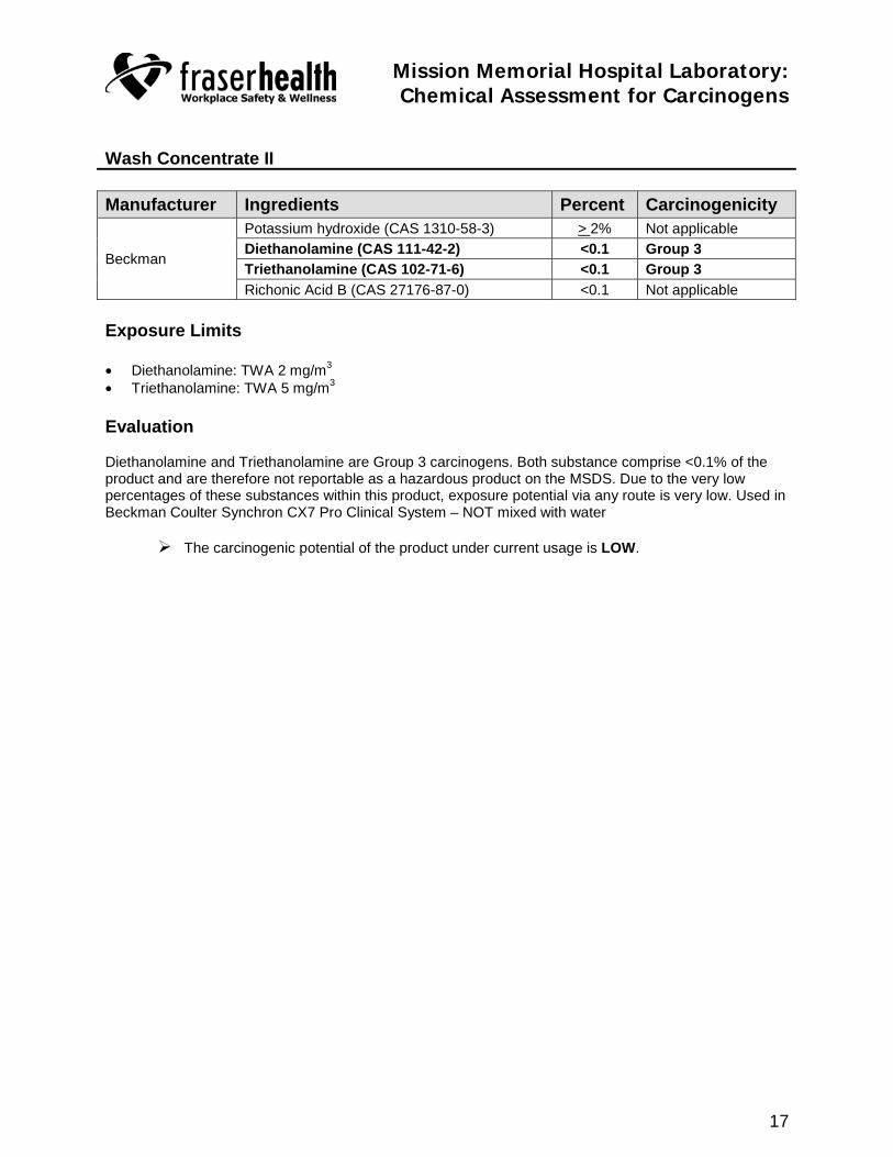
17
Mission Memorial Hospital Laboratory:Chemical Assessment for Carcinogens
Wash Concentrate II
Manufacturer Ingredients Percent CarcinogenicityPotassium hydroxide (CAS 1310-58-3) > 2% Not applicableDiethanolamine (CAS 111-42-2) <0.1 Group 3Triethanolamine (CAS 102-71-6) <0.1 Group 3Beckman
Richonic Acid B (CAS 27176-87-0) <0.1 Not applicable
Exposure Limits
• Diethanolamine: TWA 2 mg/m3
• Triethanolamine: TWA 5 mg/m3
Evaluation
Diethanolamine and Triethanolamine are Group 3 carcinogens. Both substance comprise <0.1% of theproduct and are therefore not reportable as a hazardous product on the MSDS. Due to the very lowpercentages of these substances within this product, exposure potential via any route is very low. Used inBeckman Coulter Synchron CX7 Pro Clinical System – NOT mixed with water
� The carcinogenic potential of the product under current usage is LOW.

18
Mission Memorial Hospital Laboratory:Chemical Assessment for Carcinogens
SUMMARY
Bigelow et al provided a list of chemical substances known to have the potential of causing mammarytumors – see below:
� Acronycine � 1,2-Dibromo-1-propanol� Benzene � 1,1-Dichloroethane� 2,2-bis(bromomethyl)- 1,3-propanediol � 1,2-Dichloroethane� 1,3-Butadiene � 1,2-Dichloropropane (propylene dichloride)� C,1 acid red 114 � Dichlorvos� C,1 basic red 9 monohydrochloride � 1,2-Dimethoxybenzidine dihydrochloride� 2-Chloroacetophenone � 3,3-Dimethylbenzidine dihydrochloride� Chloroprene � 2,4-Dinitrotoluene� Clonitralid � Ethylene oxide� Cytembene � Furosemide� 2,4-Diaminotoluene � Glycidol� 1,2-Dibromo-3-chloropropane � Hydrazobenzene� 1,2-Dibromoethane � Isophosphamide� Indium phosphide � Ochratoxin A� Isoprene � Phenesterin� Methylene chloride � Procarbazine hydrochloride� Methyleugenol � Reserpine� Nithiazide � Sulfallate� 5-Nitroacenaphthene � 2,4- and 2,6-Toluene diisocyanate� Nitrofurazone � o-Toluidine hydrochloride� Nitromethane � 1,2,3-Trichloropropane
Only one of these substances, ethylene oxide, was found to be present in any of the products used in thelab. In every case, the concentration of ethylene oxide was <0.1% of the product and is therefore notreportable as a hazardous product on the MSDS. Additionally, ethylene oxide is a gas at roomtemperature and although used in small quantities in the product production, it not present in the product. Other potential carcinogens are used in the production or are present in some of the products. Theseinclude:
Substance Carcinogenicity Substance CarcinogenicityCrystalline Silica Group 1 Ethylene Glycol Monobutyl Ether Group 3Mineral Oil Group 1 Eosin Group 3Formaldehyde Group 1 Triethanolamine Group 3Ethylene oxide Group 1 Toluene Group 3Chloramphenicol Group 2A Hydrogen Peroxide Group 3Acetaldehyde Group 2B Hydrochloric acid Group 3Basic fuchsin Group 2B Sodium hypochlorite Group 3Glycerine, N,N-bis(carboxymethyl)- Group 2B Chromic acid, dipotassium salt Group 31,4-Dioxane Group 2B 5-fluorouracil Group 3Chloroform Group 2B Caffeine Group 3Silica Group 3 Diethanolamine Group 3Mercuric chloride Group 3 Triethanolamine Group 3
Based on current scientific knowledge none of these substances are present in a quantity or currentlyused in a manner that would result in potential exposure via any occupational exposure route that wouldresult in an increased risk of cancer.

Attachment 8
AN INVESTIGATION OF A CANCER CLUSTER
WITHIN THE MISSION MEMORIAL HOSPITAL
LABORATORY – FINAL UPDATE April 15, 2005

AN INVESTIGATION OF A CANCER CLUSTER WITHIN THE MISSION MEMORIAL HOSPITAL LABORATORY – FINAL UPDATE
April 15, 2005
Occupational Health & Safety Agency for Healthcare in BC
#301-1195 West Broadway, Vancouver, BC V6H 3X5

Page 2 of 9 15 April, 2005
AN INVESTIGATION OF A CANCER CLUSTER WITHIN THE MISSION MEMORIAL HOSPITAL LABORATORY – FINAL UPDATE March 22nd, 2005 Introduction Since the March 2004 report of the same title, prepared by the Occupational Health and Safety Agency for Healthcare (OHSAH), a new case of breast cancer in the workforce came to light, and along with it, a request to OHSAH to re-calculate the cancer rates. This final update of the report includes such a recalculation, taking into account, of course, the additional person-years at risk that comes with extending the analysis, as well as new information on the actual number of employees at risk. This report also includes additional comments on chemical exposure and air quality assessment conducted by Workplace Health as well as comments on radiation exposure conducted by BC Centre for Disease Control (BCCDC). This was done as per the recommendations in the original report. Epidemiology New data 1. Another female employee was diagnosed with breast cancer December 7th of 2004; this brings
the total number of cancer cases to 11: comprising 7 breast (female) and 4 other (3 female and 1 male) which affects the numerator of the incidence calculation
2. There were actually 63 employees (57 of whom were female). The original analysis included only
57 employees. This change in the employee numbers affects the denominator of the incidence calculation.
Methods To recalculate the incidence of cancer, we have used a new censor (closure) date of December 31st 2004 (versus August 31st, 2004) to include the most recent case. We have asked the BC Cancer Agency (BCCA) to provide a data linkage for these 63 employees to ensure that there were no unreported cancer cases within this group. As this information will not be available until August of 2005 and the possibility of an unknown case is very low, we have recalculated the data using the numbers stated above (ie. 11 cases of cancer). That is, we have assumed no additional cases. In the unlikely situation that more new cases are found in the period between August and December 31st, this calculation will be re-done again. Results Table 1 represents disease status of the laboratory employees. Table 2 presents the new calculated incidence of cancer, both breast and other.

Page 3 of 9 15 April, 2005
Interpretation As more time has passed, the denominator (time) has increased faster than the numerator (cancer cases) and the standard incidence ratio (SIR) has in fact decreased despite the new case, for both total cancer cases (SIR = 4.70) and breast cancer cases in females (SIN = 8.43). This is likely due to the additional work hours of the 6 unidentified employees now included in the analysis. Despite this decrease, the SIR is still significantly higher than the expected incidence in BC for both groups (total cancer and female breast cancer), indicating that the perceived cluster is an observed (true) cluster with a high degree of confidence (95% probability that this observation was NOT due to chance alone). However the new information, when used in the Cox regression model to identify cause, continues to indicate that age at start of work at MMHL, job position, and job status do not contribute to this cluster. Environmental review 1. Chemical agents: the field investigation regarding exposure did not show any obvious
carcinogenic exposure in the MMH laboratory, as known from the current scientific literature (see previous report).
2. Physical agents: in September of 2004, Radiation Protection Services (BCCDC), completed an
assessment of radiation exposure in the laboratory, concluding that “the exposures measured in the Mission Memorial Laboratory Area are typical natural background and that the X–ray facility is not contributing to this natural background…this natural background radiation would not contribute measurably to increased cancer risk.”
3. Air Quality: Basic indoor air quality testing was performed and was shown to be within
acceptable standards. Conclusion (summary)
• The increased incidence of breast cancer in laboratory employees is an observed cluster. • Using statistical analysis, we still conclude that this increase is not related to age at start of work,
job position, or job status. • On observation and literature review, no current occupational chemical exposures, or records of
past occupational exposures were found that relate to breast cancer, or cancer in general. No significant findings were found during radiation testing in the laboratory, or on basic air quality testing.
The conclusion thus remains the same. The most likely explanation for the occurrence is a ‘chance cluster’ or statistical anomaly. It is recommended that this cancer cluster investigation be closed. A new cluster investigation may be considered at a future time for comparison (usually 5 years after the original investigation) using new cases and new data. Every effort should continue to be made, in this and all workplaces, to ensure that the workplace remains as safe and free of carcinogenic exposures as possible, and that the workforce is able to pursue safe and healthy choices in all aspects of their lives.

Page 4 of 9 15 April, 2005
Table 1.
Age, gender and duration of follow-up by disease status
No Cancer Breast Cancer Other Cancer Total Females 47 (74.6%) 7 (11.1%) 3 (4.8%) 57 (90.5%)
Males 5 (7.9%) 0 1 (1.6%) 6 (9.5%) Mean Age (yrs) 45.7 (10.8) 53.4 (8.7) 51.7 (11.4) 46.9 (10.8) Mean duration
of follow-up (yrs)
15.1 (8.0) 17.7 (9.1) 14.6 (13.1) 15.4 (8.3)
Data presented as frequency, mean (standard deviation).
Table 2.
Observed and expected cases and age/calendar-year adjusted standard incidence ratios (SIRs) for breast cancer (females only) and all cancers.
Cause Person-years
Number of subjects
Expected cancers
Observed cancers
Standard Incidence
Ratio
95% Confidence
Intervals Breast Cancer
(females) 856.28 57 0.83 7 8.43 3.39 – 17.38
All cancers (females
only) 856.28 57 2.18 10 4.59 2.20 – 8.44
All cancers (all
subjects) 973.49 63 2.34 11 4.70 2.35 – 8.41

Page 5 of 9 15 April, 2005
Table 3a. Age at start of work, age at interview, gender, mean duration of occupational exposure and mean duration of follow-up at interview by disease status for all staff working after 1982 at the Mission Memorial Hospital laboratory
Variable No Cancer Breast Cancer Other Cancer Total Females 47 (74.6%) 7 (11.1%) 3 (4.8%) 57 (90.5%)
Males 5 (7.9%) 0 1 (1.6%) 6 (9.5%) Age at start of
work (yrs) 30.5 (9.1) 30.5 (11.1) 33.8 (7.6) 30.7 (9.1)
Age at interview (yrs) 45.7 (10.8) 54.1 (9.8) 51.8 (11.3) 47.0 (11.0)
Mean duration of follow-up
(yrs)
15.1 (8.0)
Median: 14.0
18.3 (9.6)
Median: 16.0
14.6 (13.1)
Median: 9.6
15.4 (8.3)
Median: 14.1 Mean duration of occupational exposure (yrs)
9.2 (7.6)
Median: 6.0
18.0 (10.6)
Median: 17.8
15.6 (14.0)
Median: 10.7
10.6 (8.8)
Median: 7.2 Data are presented as frequency (percentage) for gender, mean (standard deviation) for age at start of work, age at interview, mean duration of follow-up and mean duration of occupational exposure.
Table 3b. Age at start of work, age at interview, gender, mean duration of occupational exposure and mean duration of follow-up at interview by disease status for female staff working after 1982 at the Mission Memorial Hospital laboratory
Variable No Cancer Breast Cancer Other Cancer Total Females 47 (74.6%) 7 (11.1%) 3 (4.8%) 57 (90.5%)
Age at start of work (yrs) 30.6 (8.9) 30.5 (11.1) 36.7 (6.0) 30.9 (9.0)
Age at interview (yrs) 45.6 (10.8) 54.1 (9.9) 47.1 (7.9) 46.7 (10.8)
Mean duration of follow-up
(yrs)
15.0 (8.2)
Median: 14.0
18.3 (9.6)
Median: 16.0
8.3 (2.9)
Median: 8.0
15.0 (8.3)
Median: 14.0 Mean duration of occupational exposure (yrs)
9.4 (7.8)
Median: 6.4
18.0 (10.6)
Median: 17.8
8.8 (4.3)
Median: 7.8
10.4 (8.4)
Median: 7.2 Data are presented as frequency (percentage) for gender, mean (standard deviation) for age at start of work, age at interview, mean duration of follow-up and mean duration of occupational exposure.

Page 6 of 9 15 April, 2005
Table 4. Person-years and distribution of all-causes cancer by calendar year for all employees
Calendar
year Person-
years Number of
incident cases Calendar
year Person-years
Number of incident cases
1964 0.50 0 1985 23.14 0 1965 1.00 0 1986 24.12 0 1966 1.00 0 1987 26.76 0 1967 1.00 0 1988 28.00 0 1968 1.00 0 1989 29.98 0 1969 1.00 0 1990 32.71 0
1970 1.00 0 1991 38.05 1 1971 1.00 0 1992 40.66 0 1972 1.17 0 1993 42.19 1 1973 2.29 0 1994 44.17 0 1974 3.33 0 1995 46.32 1
1975 5.35 0 1996 46.51 1 1976 6.16 0 1997 47.34 0 1977 7.30 0 1998 48.91 1 1978 8.50 0 1999 49.54 0 1979 9.59 0 2000 53.04 1 1980 12.77 0 2001 55.77 1 1981 15.06 0 2002 54.71 2 1982 17.26 0 2003 53.16 1 1983 18.42 0 2004 52.90 1 1984 20.79 0
Total 973.49
n = 63 male and female employees

Page 7 of 9 15 April, 2005
Table 5. Person-years and distribution of breast cancer by age group in women in cancer cluster study
Age group (yrs) Number of
incident cases Person-years Number of
incident cases
15 – 19 0 1.34 0
20 – 24 0 44.47 0
25 – 29 0 120.18 0
30 – 34 0 150.75 0
35 – 39 0 145.25 2
40 – 44 0 132.74 1
45 – 49 0 106.07 1
50 – 54 0 71.48 1
55 – 59 0 45.49 1
60 – 64 0 22.00 0
65 – 69 0 12.49 1
70 – 74 0 4.00 0
Total 856.28
n = 57 female employees

Page 8 of 9 15 April, 2005
Table 6. Person-years and distribution of breast cancer by calendar year for women in cancer cluster study
Calendar
year Person-
years Number of
incident cases Calendar year
Person-years
Number of incident cases
1972 0.17 0 1989 26.32 0 1973 1.29 0 1990 28.71 0 1974 2.33 0 1991 33.43 1 1975 4.35 0 1992 35.66 0 1976 5.16 0 1993 37.19 0 1977 6.30 0 1994 39.17 0
1978 7.50 0 1995 41.32 1 1979 8.59 0 1996 40.86 1 1980 11.27 0 1997 41.34 0 1981 13.06 0 1998 43.42 0 1982 15.26 0 1999 44.54 0 1983 16.42 0 2000 48.04 0 1984 18.00 0 2001 50.77 1 1985 20.14 0 2002 49.71 1 1986 21.12 0 2003 48.16 1 1987 23.76 0 2004 47.90 1 1988 25.00 0
Total 856.28
n = 57 female employees

Page 9 of 9 15 April, 2005
Table 7. Observed and expected cases and age/calendar-year adjusted standard incidence ratios (SIRs) for breast cancer (females only) and all cancers.
Cause Person-years
Number of subjects
Expected cancers
Observed cancers
Standard Incidence
Ratio
95% Confidence
Intervals Breast Cancer
(females) 856.28 57 0.83 7 8.43 3.39 – 17.38
All cancers (females
only) 856.28 57 2.18 10 4.59 2.20 – 8.44
All cancers (all
subjects) 973.49 63 2.34 11 4.70 2.35 – 8.41
Table 8. Hazard ratios (HR) and 95% confidence intervals (CIs) of breast cancer for women in relation to age at start of work, position and occupational exposure at MMHL
Variable Hazard Ratio (HR) 95% CI p-value *
Age at start of work (yrs) 1.07 0.95 – 1.21 0.264 Occupational exposure (yrs) 1.03 0.92 – 1.15 0.581 Position Aid, clerk or ECG technician Technician
1.00 4.24
0.36 – 49.38
0.249 MMHL = the Mission Memorial Hospital Laboratory. *, p-value was derived from Cox proportional hazards model with age at start of work and year of occupational exposure as continuous variables and position as a categorical variable.


Attachment 9
An Investigation of a Cancer Cluster within a Hospital Laboratory - report regarding the cancer
cluster at Mission Hospital, Fraser Health, BC. Presentation: March 2005


1
An Investigation of a Cancer Cluster
within a Hospital Laboratory
final report regarding the cancer cluster at Mission Hospital,
Fraser Health, BC.
Introduction
Report of seemingly high number of cancer cases in laboratory workersNeed for an epidemiological studyPurpose of investigation:
To determine if cluster had occurredTo identify possible occupational factors if a true cluster is identifiedTo ensure current exposures/occupational factors are below recommended standards and current exposures are not increasing the risk of cancer in MMHL employees
Methods - Epidemiology
Defined occupational cohortEmployee data files for all individuals working from 1970 to 2003
Analysis of cancer incidence dataCollection of data on health status and cancer incidence from personal interviews (57)Data entered into excel and SPSSRates of cancer and breast cancer from 1970 to 2002 for males and females obtained Person years of observation (PYRS) were computed for study cohort for each age interval (5 yr increments) and calendar yearExpected number of cases (breast and all cancers) computed for each age and calendar year category
Methods - EpidemiologyData analyses
Expected number of cases based on the total number of PYRS of observation was computed Observed number of breast and total cancer cases divided by expected number provided the Standard Incidence Ratio (SIR); 95% confidence intervals computed using Poisson statistics (to see if an apparent high incidence is a chance findings)Cox proportional hazard modeling used to determine the association of occupational factors on rate of breast cancer (for example whether there is a relationship between duration of time at work and cancer rate)
Methods - Exposure Assessment
Review of OHSAH MSDS database for chemicals and products used in hospital laboratoriesWalkthrough survey of MMHLInterviews with current MMHL employees
Changes in processes and controls over timeIssues with regard to air quality in MMHL over time
Methods - Literature Review
Conducted to provide information to assist in interpretation of study findings
Focused on breast cancer risk factors as this cancer type identified in preliminary analyses as likely with a higher incidence in MMHL employees

2
Results - Literature ReviewCarcinogenesis is a multistage process that involves many events, some of
which are not well understood.
Cancer Incidence in British Columbia (2003)
Source: BCCA Cancer Statistics 2003
Cancer Mortality in 2003 in British Columbia
Source: BCCA Cancer Statistics 2003
Age-adjusted Cancer Incidence and Mortality in 2003 in British Columbia
Source: BCCA Cancer Statistics 2003
Breast Cancer - Demographics
Canada one of the highest incidence rates for breast cancer
Most common malignancy in womenSecond leading cause of cancer mortalityLifetime risk of 1 in 9Most common cause of death in women 35 to 54, age of maximum social responsibilityAverage years of life lost is 19.3
Age-adjusted annual breast cancer incidence rate among women in selected countries in 1983-87.
Source: Cancer Incidence in Five Continents (IARC)

3
Incidence and Mortality rates of Breast Cancer in Canada (1984-1994/1996)
Source: Cancer Bureau, Laboratory Centre for Disease Control, Health Canada, based on data supplied by Statistics Canada (April 1999)
http://www.phac-aspc.gc.ca/publicat/updates/breast-99_e.html
Breast Cancer Risk Factors
75% have no known risk factorsAgeFamily HistoryRadiation exposure in childhoodPrevious malignancy (Hodgkin’s)No children or none before 35Early menarche, late menopauseEstrogen effects in breast cancer
Breast Cancer Genetics
A small proportion of breast cancers appear to be attributable to an autosomal dominant genetic predisposition 5-10% of all casesYoung ageStrong family historyBilateral diseaseBRCA-1 and BRCA-2
Breast Cancer Occupational Risk Factors
Animal studies identify mammary carcinogensHuman studies on specific chemicals mostly equivocal Organochlorine compounds implicated due to estrogenic activityMost epidemiology studies not able to establish causal links to breast cancer
Breast Cancer Occupational Risk Factors
Band et al (2000) higher breast cancer rates in BC for post menopausal women in medicine, health (OR=1.50; CI: 1.13-1.98), and nursing (OR=1.37; CI: 1.01-1.85) occupations (90% CI)Nurses have been studied and shift work is associated with increased cancer risk (30 or more years on the night shift: RR 1.36; 95%CI=1.04-1.78) (suppressed melatonin production) (E. Schernhammer et al. J. National Cancer Institute 2001; Stevens R. Epidemiology 2005)A clustering of risk factors is also suspected as a cause of the observed increased risk in nursing and medical occupations
Cancer Cluster -- Epidemiology
Regional and temporal patterns always existPatterns of increased/decreased risk may be due to aggregation of diverse factors (diet, demographics, lifestyle factors, occupational exposures)The lower mainland of BC has a higher than average incidence of breast cancer

4
Cancer Cluster -- Epidemiology
Analysis of clusters is responsibility of health agencies (BC Cancer Agency, BC CDC, Health Canada, OHSAH)Modern cancer clusters are widely reported and investigated
Over the last 30 years, thousands have been investigatedFew investigations have identified exposures
etiologically related to the increase risk if an increased risk was found
Cancer Cluster -- Epidemiology
Investigations answer questions:Is the cluster real -- first report of a
cluster is defined as a “perceived cluster”If the cluster is real, it is termed an
“observed cluster”Is an agent or exposure the cause of the
observed cluster -- if yes, the cluster is termed an “etiologic cluster”
Cancer Cluster -- Epidemiology
Standard protocols for cluster investigations
Stepwise, going from exploratory to analyticPrimary objective is to identify
exposures that may be increasing the risk of cancer and determining the steps to take to eliminate or reduce such risks
Cancer Cluster Epidemiology MMHL
57 employees working between January 1970 and August 2003
One person excluded from analysis (Breast Ca identified before employment)
Cancer reported: 6 breast, 1 ovarian, 1 thyroid, 1 skin, and 1 lymphoma in cohortAt follow-up mean age of employees was 43.1 years
Age, gender and duration of follow-up by disease status
No Cancer Breast Cancer
Other Cancer
Total
Females 41(73.21%)
6(10.71%)
3 (5.36%)
50 (89.29%)
Males 5(8.93%)
0 1(1.79%)
6(10.71%)
Mean age at start work (yrs)
29.07 (8.68)
32.03(11.29)
33.78(7.53)
29.72(8.86)
Mean age at end work or end study (yrs)
42.47(10.67)
46.2 (13.24)
45.90 (7.99)
43.12 (10.70)
Mean duration of follow-up (yrs)
13.41 (8.34)
14.16(10.50)
12.13(8.13)
13.40 (8.41)
Data presented as frequency (percent of total), mean (standard deviation); n=56
Age, gender and duration of follow-up by disease status
Person years
Number of
Subjects673.80 50
50
56
673.80
751.27
Expected Cancers Observed
Cancers
Standard Incidence
Ratio
95% Confidence
IntervalBreast Cancer
0.59 610.2
3.74-22.24
All Cancers (Females only)
1.55 95.8
2.66-11.02
All Cancers (All Subjects) 1.60 10 6.3 3.02-11.59
Analysis is based on the March 2004 data
Table. Observed and expected cases and age/calendar-year adjusted standardized incidence ratios (SIRs) for breast cancer (females only) and all cancers
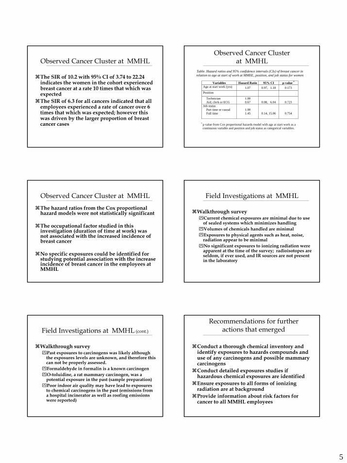
5
Observed Cancer Cluster at MMHL
The SIR of 10.2 with 95% CI of 3.74 to 22.24 indicates the women in the cohort experienced breast cancer at a rate 10 times that which was expectedThe SIR of 6.3 for all cancers indicated that all employees experienced a rate of cancer over 6 times that which was expected; however this was driven by the larger proportion of breast cancer cases
Table. Hazard ratios and 95% confidence intervals (CIs) of breast cancer in relation to age at start of work at MMHL, position, and job status for women
Variables Hazard Ratio 95% CI p-value *
Age at start work (yrs) 1.07 0.97, 1.18 0.173 Position Technician Aid, clerk or ECG
1.00 0.67
0.08, 6.04
0.723
Job status Part time or causal Full time
1.00 1.45
0.14, 15.06
0.754
* p value from Cox proportional hazards model with age at start work as a continuous variable and position and job status as categorical variables.
Observed Cancer Cluster at MMHL
Observed Cancer Cluster at MMHL
The hazard ratios from the Cox proportional hazard models were not statistically significant
The occupational factor studied in this investigation (duration of time at work) was not associated with the increased incidence of breast cancer
No specific exposures could be identified for studying potential association with the increase incidence of breast cancer in the employees at MMHL
Field Investigations at MMHL
Walkthrough surveyCurrent chemical exposures are minimal due to use of sealed systems which minimizes handlingVolumes of chemicals handled are minimalExposures to physical agents such as heat, noise, radiation appear to be minimalNo significant exposures to ionizing radiation were apparent at the time of the survey; radioisotopes are seldom, if ever used, and IR sources are not present in the laboratory
Field Investigations at MMHL (cont.)
Walkthrough surveyPast exposures to carcinogens was likely although the exposures levels are unknown, and therefore this can not be properly assessed.Formaldehyde in formalin is a known carcinogenO-toluidine, a rat mammary carcinogen, was a potential exposure in the past (sample preparation)Poor indoor air quality may have lead to exposures to chemical carcinogens in the past (emissions from a hospital incinerator as well as roofing emissions were reported)
Recommendations for further actions that emerged
Conduct a thorough chemical inventory and identify exposures to hazards compounds and use of any carcinogens and possible mammary carcinogensConduct detailed exposures studies if hazardous chemical exposures are identifiedEnsure exposures to all forms of ionizing radiation are at backgroundProvide information about risk factors for cancer to all MMHL employees

6
Age, gender and duration of follow-up by disease status
Person years
Number of
Subjects856.28 57
57
63
856.28
973.49
Expected Cancers Observed
Cancers
Standard Incidence
Ratio
95% Confidence
IntervalBreast Cancer
0.83 78.43
3.39-17.38
All Cancers (Females only)
2.18 104.59
2.20-8.44
All Cancers (All Subjects) 2.34 11 4.70 2.35-8.41
Since the March 2004 report, a new case of breast Ca was reported ; this brings the total number of cases to 11.With time, the denominator (time) has increased faster than numerator (Ca cases) and the SIR has decreased despite a new case.
Table. Observed and expected cases and age/calendar-year adjusted standardized incidence ratios (SIRs) for breast cancer (females only) and all cancers.
Assessment of indoor air quality
No exterior air contaminant or exhaust discharges within 30 feet of the air intake.No interior air contaminant sources.Fan system filtration level – 85%
Survey (Aug 2004)
CO2 – Assessment: good fresh air exchangeCO – Assessment: goodRelative Humidity – acceptableTemperature – Normal Supply duct clean; Exhaust duct – fine layer of duct
Walk-through survey (Nov 2004)
Assessment of radiation exposure
Thermoluminescent dosimeters (TLD monitors) were exposed in the laboratory area for 70 days (June 18 – Aug 26).Typical natural background for the Fraser Valley during this period 0.12 mSv(milisieverts).Conclusion: Exposures measured at the MMHL area are typical natural background and the X-ray facility is not contributing to this natural backgroundThis natural background would not contribute measurably to increased cancer risk.
Assessment of CarcinogenicityUS National Toxicology Program (NTP) has tested over 500 chemicals for risk of causing mammary tumors42 out of 500 were identifies as mammary carcinogens Four out of 42 are classified as human carcinogens:
Benzene Gasoline, solvent
1,3-Butadiene Auto exhaust, rubber manufacture, gasoline
C,1 acid red 114 Dye for silk, jute, wool, leather
Ethylene oxide Sterilizing gas for medical equipmentL.Bennett, B.Davis Environmental and Molecular Mutagenesis 2002
Assessment of Carcinogenicity (cont.)
Definitions:
1. Group 1: Agent (mixture) is carcinogenic to humans.
2. Group 2a: Agent (mixture) is probablycarcinogenic to humans.
3. Group 2b: Agent (mixture) is possibly carcinogenic to humans.
4. Group 3: Agent (mixture) is not classifiable as to its carcinogenicity to humans.
5. Group 4: Agent (mixture) is probably not carcinogenic to humans.
International Agency for Research on Cancer (IARC)
Assessment of Carcinogenicity (cont.)
Only one among 42 chemical substances known to have potential of causing mammary tumor was found: ETHYLENE OXIDE (Eth Ox) [Group 1].Concentration of Eth Ox was <0.1% => not reportable as hazardous product.27 substances that are used in MMHL were analyzedBased on the scientific knowledge none of the analyzed substances are present in a quantity or currently used in a manner that would result in an increased risk of cancer
Walkthrough surveys on June 3; June 18 and July 23 2004.

7
Conclusions
The increased incidence of breast cancer in laboratory employees is an observed cluster.Using statistical analysis, we conclude that this increase is not related to age at start of work, job position, or job statusOn observation and literature review, no current occupational chemical exposures, or records of past occupational exposures were found that relate to breast cancer, or cancer in generalNo significant findings were found during radiation and carcinogenicity testing or on air quality testing
Conclusions
The most likely explanation for the occurrence is a “chance cluster” or statistical anomalyIt is recommended that this Cancer cluster investigation be closed. A new cluster investigation may be considered at future time for comparison (usually 5 years after the original investigation) using new cases and new data.Every effort should continue to be made, to ensure that the workplace remains as safe and free of carcinogens as possible.
Questions welcome!
Breast Cancer Occupational Risk FactorsBand et al (2000) – population based Case-control study of 995 incident Breast CA (BCA) cases by menopausal status, controlling for Confounding factors
All F < 75 y.o with BCA diagnosed 1988 – 1989 (identified through BC cancer registry)Controls randomly selected from 1989 BC Voter list1489 cases identified, 995 included into analysis⌧318 (31.2%) premenopausal; 700 (68.8%) postmenopausal⌧Histol. confirmation obtained in all cases⌧Matched case-control analysis⌧Controlled for confounders (table)⌧90% CI => 5% sign. (one sided)
Back
Breast Cancer Occupational Risk Factors
Pre-menopausal: electronic data-processing, barbers, sales and material processing, food, clothing, chemical, transportation industries.Post-menopausal: health and nursing, school teaching, laundry/ dry-cleaning, aircraft/automobile incl. gasoline stationsindustries.
Back
Band et al (2000)
Breast Cancer and rotating night shifts
Back

8
Pineal Gland
Back
The pineal body is variable in size and is calcified in 40% of subjects over 20 years of age, but rarely in individuals under this age
Pineal body
Back
Melatonin methabolismMelatonin is a hormone (N-acetyl-5 methoxytryptamine) produced especially at night in the pineal gland. Decreases with age, stimulates anti-oxidant action Its secretion is stimulated by the dark and inhibited by light. The suprachiasmatic nuclei (SCN) of the hypothalamus have melatonin receptors and melatonin may have a direct action on SCN to influence "circadian" rhythms. Melatonin is metabolised to 6-hydroxy-mel in the liver and the main metabolite excreted is 6-sulphatoxy-mel. Isolated measurements of mel are difficult to interpret given its circadian secretion, however urinary excretion of 6-sulphatoxy-mel may be helpful in studying pineal function especially in children.Back
Melatonin as oncostatic in Breast Cancer
Sanchez-Barcelo E, et al. Melatonin-estrogen interactions in breast cancer. J Pineal Res. 2005 38(4):217-22.
Melatonin plays oncostatic role on hormone-dependent mammary tumors
Three antiestrogenic mechanisms:⌧acts through the estrogen receptor interfering
with the effects of endogenous estrogens⌧Interfere with the synthesis of estrogens by
inhibiting the enzymes controlling the interconversion from their androgenic precursors⌧decreases circulating levels of estradiol
Back
Melatonin and night shifts2004 Study by Eva S. Schernhammer et al:
Urinary measurements of 6-sulfatoxymelatonin over a 3-year period in 80 premenopausal women assessed correlations between average urinary melatonin and plasma steroid hormone levels and evaluated potential associations between night work and hormone levels significant inverse association between increasing number of nights worked within the 2 weeks preceding urine collection and urinary melatonin levels (P = 0.008)significantly increased levels of estradiol after longer durations of night work (geometric mean levels of estradiol, 8.8 pg/mL for women who never worked night shifts versus 10.1 pg/mL for women who worked 15 or more years of night shifts; P for trend = 0.03).
Schernhammer E S. et.al. Epidemiology of Urinary Melatonin in Women and Its Relation to Other Hormones and Night Work Cancer Epidemiology Biomarkers & Prevention Vol. 13, 936-943, 2004
Back
Attachment 10
Laboratory Employees’ Questions Arising from MMH Cancer Cluster Draft Report
November 2005


November 9, 2005 QUESTIONS ARISING FROM MMH CANCER CLUSTER DRAFT REPORT 1] Clarification is required as to the comment that findings from previous Air quality studies were not available to investigators in the current process. Does this mean the findings are not archived or were they not provided by the employer? 2] As the report indicates exposures to smoke and odour were likely much higher in the past: a] what is the scientific opinion on the levels required [ e.g. from dioxins or other byproducts of medical incineration] in order to make a causative link to the cancers identified? b] Is it likely that the MMH smokestack produced these kinds of levels? 3] As recently as 2004 there are literature references as to the effects of burning medical waste [e.g. Chicago study]. Has such literature been canvassed in arriving at OHSAH’s conclusions? The members have asked for clarification as to the following: 4] What are the specific types of cancers classified at diagnosis? Do they suggest a common origin/agent? 5] How do these cancers compare to the 7 cancers now listed as occupational diseases for firefighters? Records indicate that MMH was exposed to toxic incinerator smoke twice a day for many years. 6] Is there any technical information about the fire detector system [ particulate matter] which could explain the many “false” alarms over the years? Did other organizations with similar systems have a similar experience with false alarms? 7] Would it be helpful to start up and run the incinerator and CSD ethylene oxide equipment, to monitor air intake of contaminants and get sample evidence for study? 8] Can hospital neighbours, as well as staff, provide anecdotal statements about the air quality issues at the time of the incineration? 9] What substance likely caused the corrosion of the Lab air intake vents? 10] Will FHA provide the sub-committee with the duct cleaning report? 11] Can OHSAH comment on the relationship between cancer causing agents and their probable impact on other systemic health issues, such as pregnancy? 12] What samples and tests are possible to detect toxic build-up in our bodies? [hair, nails, fat tissue etc.]? What type of Lab would do such work, and is FHA willing to participate in funding for this testing?
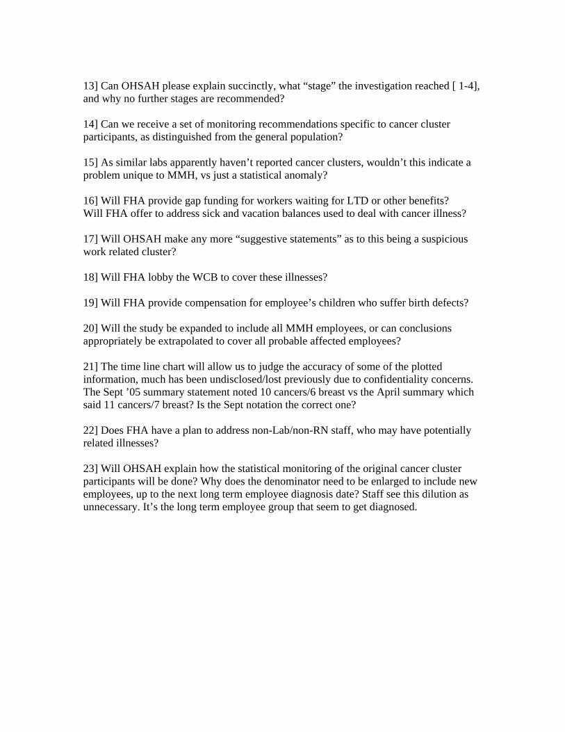
13] Can OHSAH please explain succinctly, what “stage” the investigation reached [ 1-4], and why no further stages are recommended? 14] Can we receive a set of monitoring recommendations specific to cancer cluster participants, as distinguished from the general population? 15] As similar labs apparently haven’t reported cancer clusters, wouldn’t this indicate a problem unique to MMH, vs just a statistical anomaly? 16] Will FHA provide gap funding for workers waiting for LTD or other benefits? Will FHA offer to address sick and vacation balances used to deal with cancer illness? 17] Will OHSAH make any more “suggestive statements” as to this being a suspicious work related cluster? 18] Will FHA lobby the WCB to cover these illnesses? 19] Will FHA provide compensation for employee’s children who suffer birth defects? 20] Will the study be expanded to include all MMH employees, or can conclusions appropriately be extrapolated to cover all probable affected employees? 21] The time line chart will allow us to judge the accuracy of some of the plotted information, much has been undisclosed/lost previously due to confidentiality concerns. The Sept ’05 summary statement noted 10 cancers/6 breast vs the April summary which said 11 cancers/7 breast? Is the Sept notation the correct one? 22] Does FHA have a plan to address non-Lab/non-RN staff, who may have potentially related illnesses? 23] Will OHSAH explain how the statistical monitoring of the original cancer cluster participants will be done? Why does the denominator need to be enlarged to include new employees, up to the next long term employee diagnosis date? Staff see this dilution as unnecessary. It’s the long term employee group that seem to get diagnosed.

Attachment 11
OHSAH Response to Questions raised by Laboratory Employees
January 2006


March 31, 2006 Mr. David Keen Director Workplace Health Fraser Health Authority Mr. Marty Lovick Senior Labour Relations Officer HSABC Dear David and Marty; Enclosed please find a final version of the responses to the questions raised by the laboratory workers at MMH regarding the September 2005 Cancer Cluster Draft Report. This response document was drafted by Dr. Malcolm Steinberg, Ms. Tanya Tang, and me, and was reviewed by Dr. Annalee Yassi. It is included as a separate appendix to the Cancer Cluster Report (Attachment 11a). In order to ensure that this is not separated from the report, it is highlighted in the executive summary as a critical addendum. We have responded to most of the questions conveyed to us in the November 9, 2005 letter, “Questions arising from MMH cancer cluster draft report”. Responses to Questions 6, 8, 9, 10, 16, 18, 21(part), and 22 will be provided by FHA and included in this report as a separate doucument. We have edited the Executive Summary to include details of the steps taken to enable feedback (directly or indirectly), to ensure the clarity and completeness of the report, and to emphasize that the approach taken in this work has followed international guidelines and practice. Finally, we hope that we have given a firm, but sensitively stated conclusion that this investigation is closed, at least with respect to OHSAH’s current involvement. Obviously this has been a difficult and emotional process for all people involved, not the least of which are the women and their families who were directly affected by this disease. We hope that this report will offer an opportunity for all persons involved to find closure. Best Regards; George Astrakianakis, PhD Senior Occupational Hygienist
1Occupational Health & Safety Agency for Healthcare in BC #301-1195 West Broadway, Vancouver, BC V6H 3X5 Tel: 778-328-8000 Fax: 778-328-8001 Web: www.ohsah.bc.ca

Cancer cluster study at Mission Memorial Hospital: A Response to questions by laboratory workers. March 31st, 2006 Questions and responses (to be included as a separate appendix): 1. Clarification is required as to the comment that findings from previous air quality
studies were not available to investigators in the current process. Does this mean the findings are not archived or were they not provided by the employer?[Q.1]1
Response: Based on a telephone conversation with the Mr. Quinn Danyluk, Occupational Hygienist with Fraser Health, the previous air quality surveys were simply standard surveys of ‘indoor air quality’ or IAQ. This included sampling for carbon monoxide, carbon dioxide, temperature and humidity, as well as verification of adequate ventilation. All results were unremarkable. The results of a recent IAQ survey (November 2004) are included as Attachment 5 in the report. More detailed environmental surveys were conducted including an assessment of radiation exposure (See Attachment 6) and a chemical assessment for carcinogens were conducted (See Attachment 7). Again, all results were unremarkable and did not identify any exposure source that might have contributed to increased cancer risk. 2. As the report indicates exposures to smoke and odour were likely much higher in the
past: A) what is the scientific opinion on then levels requires in order to make a causative link to the cancers identified? B) Is it likely that the MMH smokestack produced these kinds of levels? [Q.2]
Response: A) Any comments regarding levels required to make a causative link would be purely speculative without additional information. Among data we would need regarding the source emissions would be historical data on wind direction and velocity; atmospheric conditions (humidity, temperature, etc); as well as characteristics of the emissions, such as what was incinerated. However, decisions to pursue collecting these data would only be warranted if a conclusion were reached to proceed with further investigation. As discussed, this report recommends that this is not required. Moreover, it is not the levels of exposure that are of primary concern but which exposures occurred. B) At this stage we cannot comment on the nature of the emissions nor their levels without knowledge of what was incinerated and their quantity. 3. As recently as 2004 there are literature references as to the effects of burning medical
waste. Has such literature been canvassed in arriving at OHSAH’s conclusions? [Q.3]
Response: The draft report (September, 2005) included a review of the literature from 1972 to 2005 with regards to the health effects of medical waste incineration. In addition to that, another two articles published in 2004 examined the reproductive health effects associated with solid waste incinerator operation. 1 The bolded reference refers to the question number of the November, 2005 Questions from MMH Laboratory Employees document (See Attachment 10)
2

Tango et al (2004) investigated the association of adverse reproductive outcomes with maternal residential proximity to municipal solid waste incinerator in Japan. The study showed a peak-decline in risk with distance from the municipal solid waste incinerators for infant deaths and infant deaths with all congenital malformations combined. However, further investigation was suggested to accumulate good evidence for the reproductive health effects of waste incinerator exposure.2 Cordier et al (2004) assessed the impact of the solid waste incineration emission on birth defect rates at a region level of communities with fewer than 50,000 inhabitants surrounding 70 incinerators that operated at least one year in southeast France. The assessment found that the rate of congenital anomalies was not significantly higher in exposed compared with unexposed communities.3 4. What are the specific types of cancer classified at diagnosis? Do they suggest a
common origin/agent? [Q.4] Response: Based on information available from the pathology reports, at their diagnoses of breast cancer, these women ranged in age from 38 to 67. Of these, 2 women had two tumours diagnosed each. One of these women was diagnosed with two primary tumours in the same breast at the same time; and one woman was diagnosed with one tumour in each breast with the diagnoses being two years apart. Taken as a group, there were 8 ductal-carcinomas and 1 lobular carcinoma diagnosed among 7 different women. In addition, I received the report of one woman who was diagnosed with ovarian cancer and one man who was diagnosed with skin cancer. I do not have the pathology reports for the remaining two women. Both of the breast tumour types diagnosed, lobular and ductal carcinoma, are major sub-types of adenocarcinoma. At the time of diagnosis the pathologist may note adenocarcinoma if it is not possible to clearly identify which sub-type is present. Approximately 80% of breast cancer diagnoses are ductal carcinomas and 8% are lobular carcinomas. In the past, comedo-carcinoma was diagnosed separately; however, it is now recognized that this is a ductal carcinoma at an advanced stage. Comedo refers to the appearance of the tumour cell under the microscope. Tumour cells from two women were found to be oestrogen receptor positive. Four of the tumours were found to be oestrogen receptor negative and the status for three of the tumours was not available from the pathology reports. Oestrogen receptor status is one indicator of potential treatment course. Tumours that are oestrogen receptor positive generally have a better prognosis and are likely to respond to hormonal manipulation.
2 Tango T, Fujita T, et al. Risk of adverse reproductive outcomes associated with proximity to municipal solid waste incinerators with high doxin emission levels in Japan. J.Epidemiol. 2004;14 (3):83-93 3 Cordier S, Chevrier C, et al. Risk of congenital anomalies in the vicinity of municipal solid waste incinerators. Occup Environ Med.2004;61(1):8-15
3

5. How do these cancers compare to the 7 cancers now listed as occupational diseases for firefighters? [Q.5]
Response: There is no overlap with the cancer sites listed as occupational diseases for firefighters, which are: bladder, ureter, kidney, brain, colorectal, leukemia, non-Hodgkin’s lymphoma. 6. Can OHSAH comment on the relationship between cancer causing agents and their
probable impact on other systemic health issues such as pregnancy? [Q.11] Response: There are a number of cancer causing agents that are also mutagens such as radiation and diethyl stilbesterol (DES). In the case of radiation at sufficiently high doses exposure may cause cancer directly (Thyroid). At lower doses and during pregnancy itself, exposure may cause genetic defects among the offspring of exposed individuals. In the case of DES exposure is associated with increased risk of cervical cancer among the female children of exposed women who used this drug to decrease symptoms of morning sickness. 7. What samples and tests are possible to detect toxic build-up in our bodies? What type
of lab would do such work and is FH willing to participate in funding for this testing? [Q.12]
Response: There are many possible analyses for testing the presence of toxic exposures or their metabolites in the body. The choice of what sampling could be done will depend on whether one is searching for a marker of exposure, effect, or simply screening a population thought to be at risk. However, without a suspected target (eg. blood lead among battery manufacturing workers), individuals would be subjected to a number of invasive tests that likely would be of no benefit. 8. Can OHSAH please explain succinctly what stage the investigation reached (1-4),
and why no further stages are recommended? [Q.13] Response: Based on the BC Cancer Agency and US-CDC guidelines, this study completed stage 2B. Since no potential cause has been identified, further evaluation is not suggested. It is important to emphasize that, even if a potential etiologic cause had been identified, a large study sample would be required to demonstrate a statistically significant association between exposure and outcome if this indeed existed. The results of sample size calculations indicate a study that included at least 430 women (4:1 controls:cases or 344 controls and 86 cases) would have an 80% chance of identifying a two-fold risk (OR= 2.0) among a population where 50% of the women were exposed to the agent of interest. If the risk were lower (< 2) or the desired power greater (>80%), then the sample size would increase considerably.
4
Note: Stage 1: involves the gathering of basic information which include geographic, occupational, demographic, and some details regarding the individual cancer cases. Details regarding cancer cases are the most important facts. The outcome of this stage is either to move forward with a more detailed review, if the cancer cases prove to be similar diagnoses, or to conclude the investigation if a variety of different cancers are being suggested as the cluster. Stage 2 is in two parts, a) case evaluation and b) incidence evaluation. Stage 2a: please see the response to Question 4. Stage 2b: involves the evaluation of the incidence of cancer, in this case breast cancer, among the group of subjects as compared to the provincial rates. The rates of incidence were indeed higher than expected with a calculated standardized incidence ratio (SIR) of 8.4; 95%CI (3.4-17.4) as was described in the Draft Report. What this number means is that the incidence of breast cancer among women identified as working in the lab at MMH is 8 times higher than the rate of breast cancer among women in the province of BC. Furthermore, we would expect that 19 times out of 20 (i.e. 95% CI) that the true ratio (since the above number is a statistical calculation) is between 3 and 17. The important point here is that the confidence limits do not include 1, meaning that the elevated ratio is ‘statistically significant’. The outcome from this stage is to move forward if the calculated incidence rate is elevated AND there is evidence to point to a common cause, or to stop if the calculated rate is not elevated. However, even if the rate is elevated, the decision to stop may be made if there is no evidence that would suggest a biologically plausible reason behind this cluster. 9. Can we receive a set of monitoring recommendations specific to cancer cluster
participants as distinguished from the general population? [Q.14] Response: A fully-staffed occupational health unit, with occupational medical expertise as well as occupational health nursing staff, could organize mammograms for the workforce involved, so that results are provided confidentially not only to individuals, but pooled anonymous results are available to inform whether an occupational pattern of risk may exist. This is the recommended best practice in occupational health. However, it is more the custom in BC workplaces, where occupational health professionals (especially occupational medical physicians) are scarce, for individual workers to seek such screening on their own. Our publicly-funded health care system allows for this screening, and it provides individuals with privacy as well as confidentiality; also guaranteed in occupational health practice. However, when individuals seek screening related to workplace concerns on their own, it deprives the workplace from recognizing patterns of risk at early stages. In light on common practice in BC, it is probably more reasonable to suggest that women involved in this cluster consider consulting their family physician in order to seek guidance regarding mammograms and breast-self examination. Interpretation of test results in consideration of each woman’s inherent risk factors (as highlighted in this report) can be provided by their family physician or through services offered by the BC Cancer Agency.
5

10. As similar labs haven’t reported cancer cluster, wouldn’t this indicate a problem unique to MMH versus a statistical anomaly? [Q.15]
Response: Workforces generally do not know whether they are experiencing a cancer cluster unless there is considerable discussion amongst the workforce to alert themselves to this possibility and the investigation then undertaken. A cohort study has been initiated among health care workers affiliated with Vancouver Coastal Health in which the Health Sciences Association of BC has recently agreed to participate. This study my offer further clues regarding whether laboratory workers are at increased risk of breast cancer in BC as a result of what they are exposed to at work. However, at present there is no evidence in the scientific literature to suggest that any of the exposures encountered while working at the MMH are associated with increased risk of breast cancer, thus no comment can be made other than noting that it is indeed a statistically verified cluster of women who experienced a higher rate of cancer than would have been expected based on their age. 11. Will OHSAH make any more suggestive statements as to this being a suspicious
work-related cluster? [Q.17] Response: The cluster is work-related only in that the women afflicted with this disease possessed a common job and a common place of employment. There is not sufficient evidence to support statements suggestive of work-related causation in the absence of evidence linking specific workplace exposures and breast cancer. Had the sample size been larger, a case-control analysis could have been conducted in which individual risk factors are reviewed in depth. The lack of an adequate sample size was cited by BCCA in their decision to not provide OHSAH with the individual level data we requested. It would not have been ethical or scientifically appropriate to approach any of the women diagnosed with breast cancer. If it was determined that there was sufficient justification to progress with the investigation beyond Stage 2B; only then, and in the context of an approved cohort or case-control study, would such detailed personal interviews be warranted. 12. Will the study be expanded to include all MMH employees or can conclusions
appropriately be extrapolated to cover all probably affected employees? [Q.20] Response: Assuming the reference to affected employees refers to the Laboratory employees of MMH, the results of this cluster investigation are conclusive. We recommend that if there is reason to conduct a study of a larger population, such a study should be designed as a cohort study or case-control study, not an expansion of this cancer cluster investigation. 13. The September 2005 summary statement noted 10-cancers/6 breast versus the April
summary statement which said 11 cancers/7 breast? Is the Sept. notation the correct one? [Q.21(partial)]
6

Response: The report dated September 2005, includes a new case identified since the initial report (March 2004). The updated total is 11 diagnoses of cancer of which 7 were breast cancer cases among women. 14. Will OHSAH explain how the statistical monitoring of the original cancer cluster
participants will be done? Why does the denominator need to be enlarged to include new employees up to the next long term employee diagnosis date? Staff sees this dilution as unnecessary. It’s the long term employee group that seems to get diagnosed. [Q.23]
Response: Incidence rates are calculated based on “years at risk”. When rates are recalculated, because the study period has been expanded, person time will be added to the denominator and any new cases will also be added to the numerator. Dilution will occur only if this is a statistical cluster of breast cancer incidence and very few new cases are identified (See response to Q8). Further analyses can be incorporated to examine disease latency in order to investigate if timing of exposure plays a significant role in the determination of risk. George Astrakianakis, PhD With help from: Dr. Malcolm Steinberg, MBBCh, DoH, MScDr. Annalee Yassi, MD MSc Ms. Tanya Tang, MSc
7