Embed Size (px)
Citation preview
FROM THE EDITOR IN CHIEF | MESSAGE DE LA RÉDACTRICE EN CHEF
COLUMNS / CHRONIQUES Educators’ Column – From a Tiny Seed | D’une petit graine jaillit une forêt
Leadership Column – Leading with Problem Solving | Affirmer son leadership par la résolutiondes problems
Students Column – The Journey Begins | J’entreprends mon mandat
DIRECTED READING ARTICLENon-invasive Ventilation in ALI/ARDS
ORIGINAL ARTICLESEthylene Glycol Poisoning and Why Respiratory Therapists Should Always “Mind the Gap”
BMI Percentile a Potential Tool for Predicting Pediatric Obstructive Sleep Apnea
Case Presentation of a Professional Liability Case
Say YES to Evidence based-Respiratory Therapy Practice
CASE STUDY Transpulmonary Pressure and Positive End Expiratory Pressure: A Case Report
BOOK REVIEW When I Nod, I Mean No - Behavioral Guidelines and Cultural Tips for Health ProfessionalsWorking with People of Other Cultures
TECHNOLOGY UPDATEEnhancing the Safety of Medical Suction Through Innovate Technology
ABSRTACTS FROM POSTER PRESENTATIONS / RÉSUMÉS DES PRÉSENTATIONS D’AFFICHES Abstracts from poster presentations at CSRT Education Conference, St. John’s, NL / Résumés desprésentations d’affiches au Congrès éducatif et salon professionnel de St. John’s (T.-N.-L.)
The journal for respiratory health professionals in CanadaLe journal des professionnels de la santé respiratoire au Canada
CJRT RCTRCanadian Journal of Respiratory Therapy | Journal canadien de thérapie respiratoire
Summer | Été 2010Volume | Numéro 46.2
Canadian Journal of Respiratory TherapyJournal canadien de thérapie respiratoire
Summer | Été 2010Volume | Numéro 46.2 1
MARKETING AND ADVERTISING SALES
For advertising rates and information contactRita Hansen, 102-1785 Alta Vista Dr., Ottawa ON, K1G 3Y6;800-267-3422, ex 223; Fax 613-521-4314; [email protected]; or visit our website at www.csrt.com under “Publications”
SUBSCRIPTIONS
CJRT is published four times a year (Spring, Summer, Fall and Winter)
Annual subscriptions are included in annual membership to the CSRT.Subscription rate for 2010 for other individuals and institutions withinCanada is $50. International orders are $60 Cdn. All Canadian ordersare subject to 15% HST. Requests for subscriptions and changes ofaddress: Membership, CSRT, 102 - 1785 Alta Vista Dr., Ottawa ONK1G 3Y6.
Once published, an article becomes the permanent property of TheCanadian Journal of Respiratory Therapy and may not be publishedelsewhere, in whole or in part, without written permission from theCanadian Society of Respiratory Therapists. All editorial matter in CJRTrepresents the opinions of the authors and not necessarily those of The Canadian Journal of Respiratory Therapy, the editors, EditorialBoard, the publisher of the journal, or the CSRT. The Canadian Journalof Respiratory Therapy assumes no responsibility or liability for damagesarising from any error or omission of from the use of any information oradvice contained in the CJRT including editorials, articles, reports, bookand video reviews letters and advertisements.
OFFICIAL JOURNAL OF THE CSRT | SUMMER 2010, VOLUME 46 (2)
Publications Mail Agreement | No. 40012961
Registration No. | 09846
ISSN 0831-2478
Return undeliverable Canadian addresses to:102-1785 Alta Vista Drive, Ottawa ON K1G 3Y6
© 2010 Canadian Journal of Respiratory Therapy – all rights reserved
EDITOR IN CHIEF
Amy Reid, RRT, CREHotel Dieu Grace Hospital, Asthma Research Group Inc., Cottam, ON
EDITORIAL BOARD MEMBERS
Wrae Hill, BSc RRT Corporate Director - Quality Improvement & Patient Safety (QIPS), Interior Health, Canadian Patient Safety Officer - CPSI, Kelowna, BC
Jason Nickerson, RRT, MAPhD Candidate, Centre for Global Health, Institute of Population Health,University of Ottawa, Ottawa ON
Peter J. Papadakos, MD, FCCMDirector, Critical Care MedicineProfessor, Departments Anesthesiology, Surgery and Neurosurgery, University of Rochester, Rochester, New York
Norman H. Tiffin, BSc, RRT, MSAPresident, Tiffin Consulting, Chesterfield VA
Andrea White Markham, RRT, CRE,Faculty, Respiratory Therapy, Adjunct Lecturer Dalhousie UniversityCoordinator PLA, The Michener Institute, Toronto ON
CSRT BOARD REPRESENTATIVE
Christina Dolgowicz, RRT, CRE, Smiths Falls ON
CSRT EXECUTIVE DIRECTOR
Christiane Ménard, Ottawa ON
MANAGING EDITOR
Rita Hansen, Ottawa ON
BOARD OF DIRECTORS 2009-2010
President Mike Lemphers, British Columbia
Past-President Daniel McPhee, Ontario
President-Elect James McCormick, Ontario
Director of Professional Advocacy Cynthia Welton, Ontario
Director of National and Jeff Kobe, British ColumbiaProvincial Relations
Director of Education and Dennis Hunter, OntarioClinical Standards
Director of Membership Services Christina Dolgowicz, Ontario
Treasurer Jeff Dmytrowich, Saskatchewan
Director of Human Resources Angela Coxe, Ontario
Director of Student Relations Krystle Hong, Alberta

Canadian Journal of Respiratory TherapyJournal canadien de thérapie respiratoire
Summer | Été 2010Volume | Numéro 46.2
2
REVUE OFFICIELLE DE LA SCTR | PRINTEMPS 2010 NUMÉRO 46 (1)
MARKETING ET PUBLICITÉ / ANNONCES CLASSÉES
Rita Hansen, 102-1785 Alta Vista prom., Ottawa ON, K1G 3Y6;800-267-3422, poste 223; Courriel 613-521-4314; [email protected];ou visitez notre site Web: www.csrt.com sous « Publications »
ABONNEMENTS
La RCTR paraît 4 fois l’an (en printemps, été, automne et hiver).
L’abonnement annuel est compris dans la cotisation des membres de la SCTR. Le tarif annuel d’abonnement pour les non-membres et lesétablissements au Canada est de 50 $. Les commandes internationalessont 60 $ Canadien. La TVH de 15% est ajoutée aux commandes canadiennes. Veuillez faire parvenir les demandes d’abonnement et les changements d’adresse à l’adresse suivante: Centre des services auxmembres, SCTR, 102 - 1785 prom. Alta Vista, Ottawa ON K1G 3Y6.
Dès qu’un article est publié, il devient propriété permanente de TheCanadian Journal of Respiratory Therapy, et ne peut être publié ailleurs,en totalité ou en partie, sans la permission de la Société canadienne desthérapeutes respiratoires. Tous les articles à caractère éditorial dans leRCTR représentent les opinions de leurs auteurs et n’engagent ni leCanadian Journal of Respiratory Therapy, ni les rédacteurs ou l’éditeurde la revue, ni la SCTR. La Revue canadienne de la thérapie respiratoiredécline toute responsabilité civile ou autre quant à toute erreur ou omis-sion, ou à l’usage de tout conseil ou information fi gurant dans le RCTRet les éditoriaux, articles, rapports, recensions de livres et de vidéos, let-tres et publicités y paraissant.
Concernant l’adhésion à la SCTR :102 – 1785 prom. Alta Vista, Ottawa, ON, K1G 3Y6 800-267-3422 poste 223
Courrier de publications | No. 40012961
No d’enregistrement | 09846
ISSN 0831-2478
Retourner toute correspondencene pouvant être livrée au :102-1785 Alta Vista Drive, Ottawa ON K1G 3Y6
© 2010 Revue canadienne de thérapie respiratoire – tous droits réservés
RÉDACTEUR-EN-CHEF
Amy Reid, TRA, CREHôpital Hôtel Dieu Grace, Asthma Research Group Inc., Cottam (Ont.)
COMITÉ DE RÉDACTION
Wrae Hill, BSc TRA Directeur – Amélioration de la qualité et de la sécurité des patients,Interior Health, Agent de la sécurité des patients canadiens Institut canadien pour la sécurité des patients, Kelowna (C.-B.)
Jason Nickerson, TRA, MACandidat au doctorat, Centre for Global Health, Institut de recherche sur la santé des populations Université d’Ottawa, Ottawa (Ont.)
Peter J. Papadakos, MD, FCCMDirecteur, Médecine des soins intensifsProfesseur, Départements d’anesthésiologie, de chirurgie et de neurochirurgie, Université de Rochester, Rochester (New York)
Norman H. Tiffin, BSc, TRA, MSAPrésident, Tiffin Consulting, Chesterfield VA
Andrea White Markham, TRA, CRE,Membre du corps professoral, Thérapie respiratoire, Chargée de cours associée à l’Université de Dalhousie, Coordinatrice de l’ERA, The Michener Institute, Toronto (Ont.)
REPRÉSENTANT DU CONSEIL D’ADMINISTRATION DE LA SCTR
Christina Dolgowicz, RRT, CRE, Smiths Falls (Ont.)
DIRECTRICE GÉNÉRALE DE LA SCTR
Christiane Ménard, Ottawa (Ont.)
DIRECTRICE DE LA RÉDACTION
Rita Hansen, Ottawa (Ont.)
CONSEIL D’ADMINISTRATION – DE MAI 2009 À MAI 2010
Président Mike Lemphers, (C.-B.)
Ancien président Daniel McPhee, (Ont.)
Président désigné James McCormick, (Ont.)
Directrice de la promotion Cynthia Welton, (Ont.)professionnelle
Directeur des relations Jeff Kobe, (C.-B.)nationales et provinciales
Directeur de la formation et Dennis Hunter, (Ont.)des normes cliniques
Directrice des services aux membres Christina Dolgowicz, (Ont.)
Trésorier Jeff Dmytrowich, (Sask.)
Directrice des ressources humaines Angela Coxe, (Ont.)
Directrice des relations étudiantes Krystle Hong, (Alb.)
Canadian Journal of Respiratory TherapyJournal canadien de thérapie respiratoire
Summer | Été 2010Volume | Numéro 46.2 3
TABLE OF CONTENTS
FROM THE EDITOR IN CHIEF
Amy Reid . . . . . . . . . . . . . . . . . . . . . . . . . . . . . . . . . . . . . . . . . . . . . . . . . . . . . . . . . . . . . . . . . . . . . . . . . . . . . . . . . . . . . . . . . . . . . . . . . . . . . . . . . . . . . . . . . . . . . . . . . . . . . . . . . . . . . . . . . 6
COLUMNS
Educators’ ColumnFrom a Tiny Seed . . . . . . . . . . . . . . . . . . . . . . . . . . . . . . . . . . . . . . . . . . . . . . . . . . . . . . . . . . . . . . . . . . . . . . . . . . . . . . . . . . . . . . . . . . . . . . . . . . . . . . . . . . . . . . . . . . . . . . . . . . . . . 8Amy Reid
Leadership ColumnLeading with Problem Solving . . . . . . . . . . . . . . . . . . . . . . . . . . . . . . . . . . . . . . . . . . . . . . . . . . . . . . . . . . . . . . . . . . . . . . . . . . . . . . . . . . . . . . . . . . . . . . . . . . . . . . . . . . . . . . 11Kirby Peterson
Students ColomnThe Journey Begins . . . . . . . . . . . . . . . . . . . . . . . . . . . . . . . . . . . . . . . . . . . . . . . . . . . . . . . . . . . . . . . . . . . . . . . . . . . . . . . . . . . . . . . . . . . . . . . . . . . . . . . . . . . . . . . . . . . . . . . . . . 14Krystle Hong
DIRECTED READING ARTICLE
Non Invasive Ventilation in ALI/ARDS . . . . . . . . . . . . . . . . . . . . . . . . . . . . . . . . . . . . . . . . . . . . . . . . . . . . . . . . . . . . . . . . . . . . . . . . . . . . . . . . . . . . . . . . . . . . . . . . . . . 16Professor Paolo Pelosi
ORIGINAL ARTICLES
Ethylene Glycol Poisoning and Why Respiratory Therapists Should Always “Mind the Gap” . . . . . . . . . . . . . . . . . . . . . . . . . . . . . . . . . . . . . . 29Dr. Peter Brindley
BMI Percentile a Potential Tool for Predicting Pediatric Obstructive Sleep Apnea . . . . . . . . . . . . . . . . . . . . . . . . . . . . . . . . . . . . . . . . . . . . . . . . . . . . 33Dr. Shane Keene
Case Presentation of a Professional Liability Case . . . . . . . . . . . . . . . . . . . . . . . . . . . . . . . . . . . . . . . . . . . . . . . . . . . . . . . . . . . . . . . . . . . . . . . . . . . . . . . . . . . . . . . . . 38Brian Gomes
Say YES to Evidence-Based Respiratory Therapy Practice . . . . . . . . . . . . . . . . . . . . . . . . . . . . . . . . . . . . . . . . . . . . . . . . . . . . . . . . . . . . . . . . . . . . . . . . . . . . . . . 40Mika Nonoyama
CASE STUDY
Transpulmonary Pressure and Positive End Expiratory Pressure: A Case Report . . . . . . . . . . . . . . . . . . . . . . . . . . . . . . . . . . . . . . . . . . . . . . . . . . . . . 42Tom Piraino
BOOK REVIEW
When I Nod, I Mean No - Behavioral Guidelines and Cultural Tips for Health . . . . . . . . . . . . . . . . . . . . . . . . . . . . . . . . . . . . . . . . . . . . . . . . . . . . 46 Professionals Working with People of Other CulturesAna MacPherson
TECHNOLOGY UPDATE
Enhancing the Safety of Medical Suction Through Innovate Technology . . . . . . . . . . . . . . . . . . . . . . . . . . . . . . . . . . . . . . . . . . . . . . . . . . . . . . . . . . . . 47Patricia Carroll
ABSRTACTS FROM POSTER PRESENTATIONS
Abstracts from poster presentations at CSRT Education Conference, St. John’s, NL . . . . . . . . . . . . . . . . . . . . . . . . . . . . . . . . . . . . . . . . . . . . . . . . 50
Canadian Journal of Respiratory TherapyJournal canadien de thérapie respiratoire
Summer | Été 2010Volume | Numéro 46.2
4
TABLE DES MATIÈRES
MESSAGE DE LA RÉDACTRICE EN CHEF
Amy Reid . . . . . . . . . . . . . . . . . . . . . . . . . . . . . . . . . . . . . . . . . . . . . . . . . . . . . . . . . . . . . . . . . . . . . . . . . . . . . . . . . . . . . . . . . . . . . . . . . . . . . . . . . . . . . . . . . . . . . . . . . . . . . . . . . . . . . . . . . 7
CHRONIQUES
Chronique des enseignantsD’une petit graine jaillit une forêt . . . . . . . . . . . . . . . . . . . . . . . . . . . . . . . . . . . . . . . . . . . . . . . . . . . . . . . . . . . . . . . . . . . . . . . . . . . . . . . . . . . . . . . . . . . . . . . . . . . . . . . . . . . 9Amy Reid
Chronique de la directionAffirmer son leadership par la résolution des problèmes . . . . . . . . . . . . . . . . . . . . . . . . . . . . . . . . . . . . . . . . . . . . . . . . . . . . . . . . . . . . . . . . . . . . . . . . . . . . . . . . . 12Kirby Peterson
Chronique étudianteJ’entreprends mon mandat . . . . . . . . . . . . . . . . . . . . . . . . . . . . . . . . . . . . . . . . . . . . . . . . . . . . . . . . . . . . . . . . . . . . . . . . . . . . . . . . . . . . . . . . . . . . . . . . . . . . . . . . . . . . . . . . . . 15Krystle Hong
LECTURE DIRIGÉE (En anglais seulement)
Non Invasive Ventilation in ALI/ARDS . . . . . . . . . . . . . . . . . . . . . . . . . . . . . . . . . . . . . . . . . . . . . . . . . . . . . . . . . . . . . . . . . . . . . . . . . . . . . . . . . . . . . . . . . . . . . . . . . . . . 16Professor Paolo Pelosi
ARTICLES ORIGINAUX (En anglais seulement)
Ethylene Glycol Poisoning and Why Respiratory Therapists Should Always “Mind the Gap” . . . . . . . . . . . . . . . . . . . . . . . . . . . . . . . . . . . . . . 29Dr. Peter Brindley
BMI Percentile a Potential Tool for Predicting Pediatric Obstructive Sleep Apnea . . . . . . . . . . . . . . . . . . . . . . . . . . . . . . . . . . . . . . . . . . . . . . . . . . . . 33Dr. Shane Keene
Case Presentation of a Professional Liability Case . . . . . . . . . . . . . . . . . . . . . . . . . . . . . . . . . . . . . . . . . . . . . . . . . . . . . . . . . . . . . . . . . . . . . . . . . . . . . . . . . . . . . . . . . 38Brian Gomes
Say YES to Evidence-Based Respiratory Therapy Practice . . . . . . . . . . . . . . . . . . . . . . . . . . . . . . . . . . . . . . . . . . . . . . . . . . . . . . . . . . . . . . . . . . . . . . . . . . . . . . . 40Mika Nonoyama
ÉTUDES DE CAS (En anglais seulement)
Transpulmonary Pressure and Positive End Expiratory Pressure: A Case Report . . . . . . . . . . . . . . . . . . . . . . . . . . . . . . . . . . . . . . . . . . . . . . . . . . . . . 42Tom Piraino
CRITIQUES DE LIVRES (En anglais seulement)
When I Nod, I Mean No - Behavioral Guidelines and Cultural Tips for Health . . . . . . . . . . . . . . . . . . . . . . . . . . . . . . . . . . . . . . . . . . . . . . . . . . . . 46 Professionals Working with People of Other CulturesAna MacPherson
MISE À JOUR SUR LA TECHNOLOGIE (En anglais seulement)
Enhancing the Safety of Medical Suction Through Innovate Technology . . . . . . . . . . . . . . . . . . . . . . . . . . . . . . . . . . . . . . . . . . . . . . . . . . . . . . . . . . . . 47Patricia Carroll
RÉSUMÉS DES PRÉSENTATIONS D’AFFICHES
Résumés des présentations d’affiches au Congrès éducatif et salon professionnel de St. John’s (T.-N.-L.), mai 2010 . . . . . . . . . . . . 54

CSRT/CARESTREAM STUDENT EXCELLENCE AWARDS
The CSRT is proud to partner with CAREstream Medical Ltd., to provide recognition to students in the twenty educational programs across Canada. This award recognizes
students in each respiratory therapy program (accredited through the Council onAccreditation for Respiratory Therapy Education) who have successfully completed
the certification exam and have made a substantial achievement as a student.
Congratulations to this year’s winners!
Sherry Hill - College of the North Atlantic, Newfoundland
Chantale Blanchard - New Brunswick Community College (NBCC) - Saint John, New Brunswick
Mireille Dubé - College communautaire du Nouveau-Brunswick (C.C.N.B.), campus Dieppe, Nouveau-Brunswick
Anthony Robichaud – QEII/Dalhousie School of Health Sciences, Nova Scotia
Jenn Tardiff - Vanier College, Québec
Sandra Di Palma - Le College de Rosemont, Québec
Kaven Godbout - Le Cegep de Ste-Foy, Québec
Justine Boilard - Le Cegep de Sherbrooke, Québec
Julie Tremblay - Cegep de Chicoutimi, Québec
Kimberley Fournier - Algonquin College of Applied Arts and Technology, Ontario
Dominic Séguin - La Cite Collegiale-College d'arts appliques et de technologie, Ontario
Angela Kroll - Canadore College of Applied Arts and Technology, Ontario
Laura Van Bommel - Fanshawe College of Applied Arts and Technology, Ontario
Katie Gyenes - Conestoga College Institute of Technology and Advanced Learning, Ontario
Heather McPhail - The Michener Institute for Applied Health Sciences, Ontario
Vladimir Snovida - University of Manitoba-School of Medical Rehabilitation, Manitoba
Tiffany Duthie - Northern Alberta Institute of Technology, Alberta
Kathryn Sleen - Southern Alberta Institute of Technology, Alberta
Deanna Simons - Thompson Rivers University, British Columbia
Canadian Journal of Respiratory TherapyJournal canadien de thérapie respiratoire
Summer | Été 2010Volume | Numéro 46.2
6
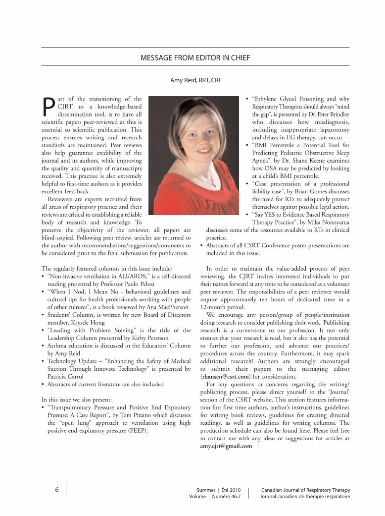
MESSAGE FROM EDITOR IN CHIEF
Part of the transitioning of theCJRT to a knowledge-based dissemination tool, is to have all
scientific papers peer-reviewed as this isessential to scientific publication. Thisprocess ensures writing and researchstandards are maintained. Peer reviewsalso help guarantee credibility of thejournal and its authors, while improvingthe quality and quantity of manuscriptsreceived. This practice is also extremelyhelpful to first-time authors as it providesexcellent feed-back.
Reviewers are experts recruited fromall areas of respiratory practice and theirreviews are critical to establishing a reliablebody of research and knowledge. Topreserve the objectivity of the reviewer, all papers are blind-copied. Following peer review, articles are returned tothe author with recommendations/suggestions/comments tobe considered prior to the final submission for publication.
The regularly featured columns in this issue include:• “Non-invasive ventilation in ALI/ARDS,” is a self-directed
reading presented by Professor Paolo Pelosi• “When I Nod, I Mean No - behavioral guidelines and
cultural tips for health professionals working with peopleof other cultures”, is a book reviewed by Ana MacPherson
• Students’ Column, is written by new Board of Directorsmember, Krystle Hong
• “Leading with Problem Solving” is the title of theLeadership Column presented by Kirby Peterson
• Asthma education is discussed in the Educators’ Columnby Amy Reid
• Technology Update – “Enhancing the Safety of MedicalSuction Through Innovate Technology” is presented byPatricia Carrol
• Abstracts of current literature are also included
In this issue we also present:• “Transpulmonary Pressure and Positive End Expiratory
Pressure: A Case Report”, by Tom Piraino which discussesthe “open lung” approach to ventilation using high positive end-expiratory pressure (PEEP).
• “Ethylene Glycol Poisoning and why Respiratory Therapists should always “mindthe gap”, is presented by Dr. Peter Brindleywho discusses how misdiagnosis, including inappropriate laparotomy and delays in EG therapy, can occur.
• “BMI Percentile a Potential Tool for Predicting Pediatric Obstructive Sleep Apnea”, by Dr. Shane Keene examines how OSA may be predicted by looking at a child’s BMI percentile.
• “Case presentation of a professional liability case”, by Brian Gomes discussesthe need for RTs to adequately protect themselves against possible legal action.
• “Say YES to Evidence Based RespiratoryTherapy Practice”, by Mika Nonoyama
discusses some of the resources available to RTs in clinicalpractice.
• Abstracts of all CSRT Conference poster presentations areincluded in this issue.
In order to maintain the value-added process of peerreviewing, the CJRT invites interested individuals to puttheir names forward at any time to be considered as a volunteerpeer reviewer. The responsibilities of a peer reviewer wouldrequire approximately ten hours of dedicated time in a 12-month period.
We encourage any person/group of people/institutiondoing research to consider publishing their work. Publishingresearch is a cornerstone to our profession. It not onlyensures that your research is read, but it also has the potentialto further our profession, and advance our practices/procedures across the country. Furthermore, it may sparkadditional research! Authors are strongly encouraged to submit their papers to the managing editor([email protected]) for consideration.
For any questions or concerns regarding the writing/publishing process, please direct yourself to the ‘Journal’ section of the CSRT website. This section features informa-tion for: first time authors, author’s instructions, guidelinesfor writing book reviews, guidelines for creating directedreadings, as well as guidelines for writing columns. The production schedule can also be found here. Please feel freeto contact me with any ideas or suggestions for articles [email protected]
Amy Reid, RRT, CRE

Canadian Journal of Respiratory TherapyJournal canadien de thérapie respiratoire
Summer | Été 2010Volume | Numéro 46.2 7
MESSAGE DE LA RÉDACTRICE EN CHEF
Amy Reid, t.r.a., é.r.c.
Une partie de la transition du JCTR à un outil de diffusion du savoirconsiste à faire réviser tous les textes
scientifiques par les pairs, élément essentiel detoute publication scientifique. La méthodegarantit le maintien de hautes normes derecherche et de rédaction. Elle assure aussila crédibilité du Journal et de ses auteurs,tout en permettant d’améliorer la qualité et la quantité des textes. C’est aussi une pratique extrêmement utile aux auteurs depremiers textes puisqu’elle leur permet derecevoir une solide rétroaction sur leur travail.
Les réviseurs sont des experts de différentsdomaines de la thérapie respiratoire et leursrévisions sont essentielles à l’établissementd’un corps de recherche et de savoir fiable. Pour préserver l’objectivité d’un réviseur, les documents qu’on leur soumetsont anonymes. À la suite de la révision, les articles sontretournés à l’auteur accompagnés de recommandations, de suggestions et de commentaires à prendre en compte avant la publication.
Articles courants du présent numéro :• “Non-invasive ventilation in ALI/ARDS,” une lecture
dirigée présentée par le professeur Paolo Pelosi• “When I Nod, I Mean No - behavioral guidelines and
cultural tips for health professionals working with people of other cultures”, une critique de livre par Ana MacPherson
• La chronique étudiante est rédigée par Krystle Hong, une nouvelle venue au conseil d’administration
• “Affirmer son leadership par la résolution des problèmes” est le titre de la chronique de la direction présentée par Kirby Peterson
• La chronique des enseignants nous présente un article sur l’asthme par Amy Reid
• Mise à jour sur la technologie – “Enhancing the Safety of Medical Suction Through Innovate Technology” par Patricia Carrol
• Résumés d’articles récents
Ce numéro présente également :• “Transpulmonary Pressure and Positive End Expiratory
Pressure: A Case Report”, par Tom Piraino; un examen de la méthode de ventilation d’ouverture du poumon au moyen de la pression positive en fin d’expiration.
• “Ethylene Glycol Poisoning and why Respiratory Therapists should always “mind the gap”, une présentation du
Dr Peter Brindley qui explique comment peut se produire une erreur de diagnostic, donnant lieu notammentà une laparatomie inutile et au délai d’une thérapie appropriée.
• “BMI Percentile a Potential Tool for Predicting Pediatric Obstructive Sleep Apnea”, par le Dr Shane Keene qui explique comme prévoir le syndrome des apnées obstructives du sommeil en examinant le percentile de l’IMC d’un enfant.
• “Case presentation of a professional liability case”, par Brian Gomes; un examen de l’importance d’une protection adéquate contre des poursuites en justice pour les TR.
• “Say YES to Evidence Based Respiratory Therapy Practice”,par Mika Nonoyama. Un examen des ressources disponibles aux TR qui travaillent en cliniques.
• Le numéro contient des résumés des présentations d’affichesau congrès de la SCTR.
Dans le but de conserver la valeur ajoutée par les examenspar les pairs, le JCTR invite les personnes intéressées à soumettreleur candidature à titre de bénévoles pour examiner les textesde leurs collègues. Les responsabilités d’un réviseur nécessitentenviron dix heures de travail par période de 12 mois.
Nous encourageons toutes les personnes, les groupes et lesorganismes qui effectuent de la recherche à publier leurstravaux. La publication des recherches est la pierre angulaire denotre profession. La publication nous assure non seulementque notre recherche est lue mais que notre profession progresseet que nos pratiques et nos procédures se répandent à l’ensembledu pays. De plus, c’est un excellent moyen d’encourager lelancement d’autres recherches. Les auteurs sont fortementinvités à soumettre leurs textes à la directrice de la rédaction([email protected]).
Pour de plus amples renseignements concernant les modalitésde rédaction et de publication, veuillez consulter la section duJournal du site Web de la SCTR. La section affiche de l’infor-mation pour les auteurs d’un premier texte, des directives àl’intention des auteurs, sur la rédaction d’une critique de livre,sur la création de lectures dirigées et sur la rédaction d’articles.Vous y trouverez également le calendrier de tombée. N’hésitezpas à communiquer avec moi pour me soumettre vos idéesd’articles ou vos suggestions : [email protected].

Canadian Journal of Respiratory TherapyJournal canadien de thérapie respiratoire
Summer | Été 2010Volume | Numéro 46.2
8
EDUCATORS’ COLUMN
In 2004, Dr. Christopher Licskai (BSc, MD, FRCPC)took an idea, a small seed, and nurtured it. Dr. Licskaiwondered if he could improve the lives of patients with
asthma while also decreasing healthcare costs by placingtrained asthma educators in the community. BetweenOctober 2004 and November 2006 a study was createdusing 523 asthma patients and 33 physicians in 19 sites1.After an initial visit, patients would receive follow-up visitsat 4 weeks, 2 months, 3 months, 6 months, and yearlybased on need. During these visits the objectives for educators were: to complete spirometry, to describe the roleof medication, to provide inhaler device technique teaching,and to discuss environmental control strategies. At the endof the study the results were quite impressive, showing areduction of healthcare utilization by 58.6%.
Previous studies of the same nature had also been conducted (by the Ministry of Health and Long TermCare) with similar results. It is due to those studies and alsodue to the results from the coroner’s inquest, for JoshuaFleuelling, that the Ministry of Health determined thatasthma education was a worthwhile endeavour and developeda fully funded program for asthma. Presently, in EssexCounty (Ontario) we are implementing an asthma educationprogram with 60 physicians, 1350 patients, and 19 asthmaeducators (8 active).
We see patients at 4 weeks, 2 months, 3 months and 6 months as needed. Initial appointments last approximately1 1/2 hours and follow-up appointments are currently sched-uled at 1 hour. Our interviews are all guided by a computerprogram that follows the Asthma Consensus Guidelines2.Questioning includes: family history, personal medical history, symptom assessment, current medications, use ofdevices, environmental assessment (home, work, social), aswell as discussions surrounding smoking and smoking cessation. Spirometry is also completed at each appoint-ment. At the close of each interview a management plan isdeveloped collaboratively between the educator and theprimary care physician.
We are seeing among our patients the dramatic lack ofunderstanding of their illness. It is astounding to learnwhat the definition of ‘asthma control’ is for some of them.There are those who believe that they are under control
despite daily coughing and constant use of their ‘rescue’inhaler! Clearly education is not only necessary, but imper-ative. We need to teach people the importance of asthmacontrol, and how to achieve that control.
Each visit concludes with the patient receiving an indi-vidualized action plan - a tailored strategy which a patientcan employ in order to help manage their asthma when itbecomes out of control. Action plans have been liberatingpatients with asthma, creating an ability to manage theircondition without the need to see a physician for everycold. As a wonderful side benefit, patients have experienceda decreased number of exacerbations! We are witnessing adramatic first-hand improvement in the quality of life ofour patients as well as a decrease in patient reliance onhigher cost medical intervention. Clearly, our investmentin asthma education is benefiting not only patients butreducing the pressure on our health care system.
As this program continues to grow, other programs aredeveloping with the same intention. This past summerARGI (Asthma Research Group INC.), piloted its first daycamp for children with asthma. The program used was based on RAP...aka, ‘Roaring Adventures of Puff3.”RAP is a supported program by the Lung Association andwas created by The University of Alberta – Alberta AsthmaCentre. The ARGI program consists of an 8 hour day filledwith fun and games aimed at helping children to under-stand not only the pathophysiology of asthma, but also the treatments and treatment options available to them. The children also quickly develop a distinct sense of camaraderie as they quickly realize that everyone in theroom has one thing in common...asthma. At the close ofthe day the children provided a wealth of positive feedback,and we are hopeful to continue to offer this program in thefuture.
Another branch from this program is lung health. ARGIhopes to develop a lung health program focused on COPDmanagement. COPD is a leading cause of death and disability in our country. There are many people who areuninformed with regards to their condition. If we couldeducate them and rehabilitate them we could change livesand lessen the demand on valuable health care resources.This is an exciting time for us in Essex County. We as
From a Tiny Seed Grows a ForestOne Respiratory Educators Perspective
Amy Reid, RRT, CREHotel Dieu Grace Hospital,
Asthma Research Group Inc., Cottam ON
Canadian Journal of Respiratory TherapyJournal canadien de thérapie respiratoire
Summer | Été 2010Volume | Numéro 46.2 9
Respiratory Therapists and Certified Respiratory Educatorsare finally being recognized and utilized for the part of ourexpertise which has the potential to have a dramatic impacton the lives of our patients.
In clinics we have seen children who thought that theywould never again play sports, parents who regained theirdreams for their children, adults who thought their breathingwould never get any better. Our patients are controllingtheir asthma and improving their quality of life! We areenabling through education!
And so, as you can see...from one small seed, a beautifulforest is starting to take root!
RÉFÉRENCES1 Licskai, C. (2006). Implementing asthma guidelines: A community-
based, electronically supported, interdisciplinary model for asthma management in Canada.
2 Boulet L-P, Becker A, Bérubé D, et al. (1999). Canadian asthma consensus report 1999. CMAJ, 161 (11 Supplement), S1-S62
3 McGhan S. (2006). Roaring adventures of Puff: Instructor’s guide.Téléchargé le 15 juillet 2009 du site Web de l’Alberta Asthma Centre : http://www.educationforasthma.com
CHRONIQUE DES ENSEIGNANTS
En 2004, le Dr Christopher Licskai (BSc, MD,FRCPC) a pris une idée, une petite graine, et l’anourrie! Le Dr Licskai s’est demandé s’il ne pourrait
pas améliorer la vie des patients asthmatique tout en dimin-uant les coûts de santé en plaçant des éducateurs formés sur l’asthme dans la collectivité. Entre octobre 2004 etnovembre 2006, une enquête a été réalisée auprès de 523patients asthmatiques et de 33 médecins dans 19 sites1.Après une première visite, les patients recevaient des visitesde suivi après quatre semaines, deux mois, trois mois, sixmois ou un an, selon les besoins. Durant ces visites, les éducateurs procédaient à un examen spirométrique,décrivaient le rôle des médicaments, enseignaient l’utilisa-tion de l’inhalateur et discutaient de stratégies de contrôlede l’environnement. À la fin de l’étude, les résultats avaientété fort impressionnants, avec une diminution de 58,6 %de l’utilisation des services de santé.
Des études antérieures de même nature avaient également été menées (par le ministère de la Santé et desSoins de longue durée) avec des résultats similaires. C’est àla suite de ces études et des recommandations du coronerdans le dossier de Joshua Fleuelling que le ministère de laSanté a déterminé que l’éducation des asthmatiques étaitune entreprise utile et entrepris de mettre en place un programme financé pour l’asthme. À l’heure actuelle, dansle comté d’Essex (Ontario) nous mettons en place un
programme d’éducation sur l’asthme auquel participent 60 médecins, 1 350 patients et 19 éducateurs (dont huitsont actifs).
Nous voyons les patients à des intervalles de quatresemaines, deux mois, trois mois ou six mois, selon lesbesoins. La première rencontre est d’une durée d’environ 1 1/2 heure et les visites de suivi sont d’une heure. Nosentrevues sont guidées par un logiciel basé sur les lignesdirectrices sur l’asthme (Asthma Consensus Guidelines2). Lesquestions portent sur les antécédents familiaux, les antécé-dents médicaux personnels, l’utilisation des dispositifs, l’évaluation environnementale (maison, travail, social) etdes discussions sur l’usage du tabac et son abandon. Un testspirométrique est également effectué à chaque visite. À lafin de chaque rencontre, un plan de gestion est préparé encollaboration avec l’éducateur et le médecin traitant.
Nous constatons chez nos patients une absence drama-tique de compréhension de leur maladie. Il est étonnant d’entendre la définition de « contrôle de l’asthme » donnéepar certains d’entre eux. Certains croient qu’ils contrôlentleur maladie même s’ils toussent tous les jours et doiventfaire constamment usage de leur inhalateur de secours! Ilest clair que l’éducation st non seulement nécessaire maisimpérative. Nous devons enseigner aux gens l’importancede contrôler leur asthme et comment y arriver.
Chaque visite se termine par la remise au patient d’un
D’une petit graine jaillit une forêtPoint de vue des éducateurs en thérapie respiratoire
Amy Reid, t.r.a., é.c.r.Hotel Dieu Grace Hospital,
Asthma Research Group Inc., Cottam (Ont.)
Canadian Journal of Respiratory TherapyJournal canadien de thérapie respiratoire
Summer | Été 2010Volume | Numéro 46.2
10
plan d’action individualisé – une stratégie adaptée que lepatient peut utiliser pour l’aider à gérer son asthme lorsquela maladie n’est plus maîtrisée. Les plans d’action on permisde libérer les patients de l’asthme, les rendant capables degérer leur état de santé sans devoir consulter un médecin aumoindre rhume. Dans un autre effet secondaire bénéfique,les patients notent une diminution des exacerbations! Nousconstatons une amélioration remarquable de la qualité devie de nos patients et une diminution de leur dépendanceaux interventions médicales coûteuses. Il est clair que notreinvestissement dans l’éducation des asthmatiques nonseulement bénéficie aux patients mais permet de diminuerla pression exercée sur notre système de santé.
Tandis que notre programme continue de croître,d’autres programmes similaires voient le jour. L’été dernier,ARGI (Asthma Research Group INC.) a tenu sous formede projet pilote un premier camp de vacances à l’intentiondes enfants asthmatiques. Le programme était basé sur RAP(Roaring Adventures of Puff3), un programme soutenu parl’Association pulmonaire et créée par le Alberta AsthmaCentre de l’Université de l’Alberta. Le programme ARGIcomprend une journée de huit heures de plaisir et de jeuxvisant à amener les enfants à comprendre la pathophysiologiede l’asthme ainsi que les traitements et les options de traite-ment disponibles. Les enfants établissent rapidement desliens de camaraderie lorsqu’ils réalisent que toutes les personnes présentes ont un point commun… l’asthme. À la fin de la journée, les enfants ont fait de nombreuxcommentaires positifs, et nous espérons pouvoir poursuivrece programme dans l’avenir.
La santé pulmonaire est un autre élément du programme.ARGI espère pouvoir établir un programme de santé pulmonaire basé sur la gestion de la MPOC. La MPOC estl’une des principales causes de décès et d’invalidité auCanada. De nombreuses personnes sont mal informées surleur état de santé. Si nous pouvions les éduquer et lesréadapter, nous pourrions changer des vies et diminuer lapression sur les ressources de santé importantes.
C’est une période stimulante pour nous dans le comptéd’Essex. Les thérapeutes respiratoires et les éducateurs respiratoires certifiés sont enfin reconnus et utilisés enfonction d’une expertise qui a le potentiel d’avoir une incidence dramatique sur la vie de nos patients.
Dans les cliniques, nous avons vu des enfants qui croyaientne plus jamais pouvoir faire de sport, des parents qui ont retrouvé leurs rêves pour leurs enfants, des adultes quicroyaient que leur respiration ne s’améliorerait jamais. Nospatients contrôlent leur asthme et améliorent leur qualitéde vie. Nous donnons le pouvoir par l’éducation!
Vous pouvez le faire aussi... d’une petite graine, unegrande forêt commence à s’élever!
RÉFÉRENCES1 Licskai, C. (2006). Implementing asthma guidelines: A community-
based, electronically supported, interdisciplinary model for asthma management in Canada.
2 Boulet L-P, Becker A, Bérubé D, et al. (1999). Canadian asthma consensus report 1999. CMAJ, 161 (11 Supplement), S1-S62
3 McGhan S. (2006). Roaring adventures of Puff: Instructor’s guide.Téléchargé le 15 juillet 2009 du site Web de l’Alberta Asthma Centre : http://www.educationforasthma.com
Canadian Journal of Respiratory TherapyJournal canadien de thérapie respiratoire
Summer | Été 2010Volume | Numéro 46.2 11
LEADERSHIP COLUMN
Problem solving can be an intimidating task for anyone in a leadership position. As no institution iswithout its challenges, it is in our best interest to
learn how to deal with them. Effective problem solvinginvolves creating a culture within our institutions: whereproblems are seen to be opportunities for growth; whereproblem solving is focused on processes, and not assigningblame; where people are free to learn; and where our staffand clients are encouraged to make suggestions, point outerrors and identify deficiencies. To be effective in doingthis, we need to use a methodical approach to problems, afairly simple method is the Plan Do Check (Study) Actprocess, also know as the PDCA cycle.
PLANPlanning is the most important step, and should take up amajority of our energy and time. The first task in planningis to identify our problem. We can do this by conductingsurveys, looking at Key Performance Indicators, performingaudits, tracking incident reports, etc. By examining all ofthese you will pick out some trends which suggest that animprovement can be made.
Once you have done this, the problem you are solvingneeds to be defined. Do this by developing a clear andeffective problem statement. This statement is neutral intone and language. It needs to avoid assumptions, blameand has to be concise. Most importantly the stated problemmust be solvable.
Now that you have your problem statement it is time tostart analyzing the problem. Keep an open mind and realizethat all problems have several contributing factors.Problems are usually systemic and the result of a break-down of a process somewhere. Collect data and start breakingthe problem down. A technique used in Root Cause analysisis to keep asking why. Why did this happen? What causedthis? Keep asking until you can no longer ask why. Thereare also many tools to help you analyze your problem: flow charts, Root Cause Analysis matrix, fishbonediagrams, timelines, affinity charts.
The biggest pitfall here is stopping too soon. We come toa point in our analysis where we think that this is obviouslywhat is contributing the most to the problem. Then we gooff with a ready made solution. Solving the wrong problem
can be worse than doing nothing at all. This wastes yourtime and energy, and undermines confidence in your abilityto lead. Remember, your only goal at this point is to under-stand the problem completely. Keep looking at until youcan say, what exactly happened and why.
You now have a good grasp of the many elements thatcontribute to your problem, and now can identify potentialfixes. There are many strategies on how to do this, so don’tbe afraid to get creative. Meet with your staff, talk with others to see how they dealt with similar situations, speakto other managers for their thoughts, seek other’s expertise.
List all the potential solutions, and identify which one isthe best for your organization. Weigh their pros and cons,and evaluate how likely they are to work within your facil-ity. Keep in mind any selection criteria that you may have.It may be necessary to do a cost benefit analysis on some ofthe stronger possibilities. When deciding what your bestoption is, keep in mind that the following categories ofsolutions are more effective: physical changes (human factorsengineering, design and layout), simplifying processes,standardizing equipment and teamwork. Warning labels, newprocedures, training, memos and policies are less efficient.
You have decided on a solution, and now you are readyto develop your action plan. Kevin Taylor’s article on thistopic in the last issue of the CJRT was excellent, and Iencourage you to read it again. Set your goals, and rememberthe acronym SMART. Keep them Specific, Measurable,Achievable, Realistic, and Timely. Do a force field analysisto identify resistance to your plan, and possible promoters.Plan how you are going to overcome the resistance to yourplan. Identify what measures you are taking, and how youare going to monitor the outcomes. Determine your imple-mentation process; would a pilot project be necessary?
DOYou now have your plan, and it is time to start implementingit. Begin by selling your idea. Prepare presentations andannouncements to gain support. Be prepared to adjustyour presentation according to your audience, and showthem how your plan will satisfy their needs. Implement asyou have planned, using your key people to help promote.Track your project as you implement.
Leading with Problem Solving
Kirby Peterson, BA, RRTCovenant Health, St. Mary's Hospital Camrose
Camrose BC

Canadian Journal of Respiratory TherapyJournal canadien de thérapie respiratoire
Summer | Été 2010Volume | Numéro 46.2
12
CHECK (STUDY)Keep tracking your project, and encourage comments.Perform surveys, audits, and talk to the staff involved. Isthis change having the intended effect? Don’t be afraid totweak things to help improve the solution, or make it moreacceptable to staff.
ACTOnce you are satisfied that this is working as intended,implement the change organization wide. Keep tracking,has your goal been met? Go back to how you identified theproblem first. Have your indicators changed any? Have thepractice audits improved?
This brings us to the top of the cycle again. The final stepin act is to go back to the planning stage, what problemsare there now for you to address? This is a continualprocess, integral to improvement.
When we can approach the challenges we face within ourdepartments proactively and methodically, we promoteconfidence in our ability to lead. Staff is more likely tocome to you when they know that you are willing and ableto take action. They are also more likely to cooperate withyou when they realize that you are interested in finding asolution. When we can do this we have gone a long waytowards changing the culture within our departments andinstitutions.
CHRONIQUE DE LA DIRECTION
La résolution de problèmes peut s’avérer une tâche arduepour quiconque se trouve en position d’autorité.Chaque organisme se trouve confronté à des défis et
nous avons tout intérêt à apprendre à y faire face. La réso-lution efficace de problèmes suppose la mise en place, ausein de l’organisation, d’une culture par laquelle les prob-lèmes sont perçus comme une occasion de croissance, oùleur résolution passe par des procédés, où l’on ne se perdpas dans le blâme, où chacun peut apprendre et où le per-sonnel et les clients sont invités à formuler des suggestions,signaler les erreurs et recenser les faiblesses. L’efficacité dansce secteur repose sur une approche méthodique du prob-lème, comme celle du cycle Planifier, Faire, Vérifier(Examiner) et Agir, que l’on appelle aussi le cycle Deming.
PLANIFIERLa planification constitue l’étape la plus importante et vousdevriez y consacrer la plus grande part de votre énergie etde votre temps. La première tâche de la planification vise àdéfinir le problème. Nous pouvons le faire notamment pardes sondages, par l’examen d’indicateurs clés de rendement,par des vérifications ou par le suivi des rapports d’incidents.L’examen de tous ces facteurs fait ressortir des tendancesqui indiquent la nécessité d’apporter des améliorations.
Une fois cette étape franchie, le problème à régler doitêtre défini. On peut le faire en formulant un énoncé clair etefficace du problème. L’énoncé doit être neutre, autant parle ton que par le langage. Il doit éviter les hypothèses et leblâme et il doit être concis. Mais, ce qui est encore plusimportant, le problème énoncé doit être soluble.
Une fois l’énoncé du problème établi, on peut entreprendrel’analyse du problème. Vous devez garder l’esprit ouvert etréaliser que tous les problèmes comportent plusieurs facteursdéclencheurs. Les problèmes sont normalement systémiqueset résultent de l’interruption d’un processus en cours deroute. La collecte de données permettra de fractionner leproblème. Une technique efficace consiste à analyser lescauses profondes et à se poser la question « pourquoi? ».Pourquoi cela s’est-il produit? Qu’est-ce qui a causé cettesituation? Posez-vous la question jusqu’à l’épuisement desréponses. Il existe aussi de nombreux outils pour vous aiderà analyser le problème : les ordinogrammes, l’analyse descauses profondes, les diagrammes cause-effet, les échéancierset les diagrammes d’affinité.
Le plus grand piège consiste à cesser la recherche troprapidement. Il arrive un moment dans l’analyse où nouscroyons avoir trouvé à coup sûr le principal élémentdéclencheur. Puis, nous appliquons une solution toute
Affirmer son leadership par la résolution des problèmes
Kirby Peterson, BA, TRAHôpital St. Mary's
Camrose (C.-B.)
Canadian Journal of Respiratory TherapyJournal canadien de thérapie respiratoire
Summer | Été 2010Volume | Numéro 46.2 13
faite. Résoudre le mauvais problème est souvent plus dom-mageable que ne rien faire du tout. C’est une perte detemps et d’énergie qui mine la confiance en ses proprescapacités de leadership. Il ne faut pas perdre de vue que le seul but à cette étape est de saisir toute la portée du problème. Il faut poursuivre l’examen jusqu’à en venir àpouvoir dire ce qui s’est passé exactement et pourquoi.
Lorsque vous avez bien saisi tous les éléments qui composent le problème, vous pouvez commencer à déter-miner des solutions. Il existe de nombreuses stratégies surla façon de franchir cette étape et il ne faut pas hésiter à semontrer créateur. Réunissez votre personnel, consultezautour de vous et découvrez de quelle façon on a réglé unesituation semblable, parlez à d’autres gestionnaires;n’hésitez surtout pas à vous appuyer sur l’expertise d’autrui.
Faites la liste de toutes les solutions possibles, et retenezcelle qui s’applique le mieux à votre organisme. Pesez lepour et le contre et évaluez de quelle façon une solutionpeut s’appliquer à votre cas. Gardez à l’esprit vos critères desélection. Vous aurez peut-être à effectuer une analysecoûts-avantages pour certaines possibilités s’avérant plusintéressantes. Avant de retenir une option, n’oubliez pasque les solutions suivantes sont plus efficaces : changementsphysiques (ergonomie, conception et aménagement), sim-plification des processus, standardisation de l’équipementet du travail d’équipe. L’affichage de mise en garde, les nouveaux procédés, la formation, les notes de service et lespolitiques sont moins efficaces.
Vous avez opté pour une solution et vous êtes prêt à mettre votre plan d’action en place. L’article de KevinTaylor à ce sujet dans le dernier numéro du JCTR étaitexcellent et je vous invite à le relire. Fixez-vous des objectifset n’oubliez pas l’acronyme SMART. Gardez vos objectifsstratégiques, mesurables, réalisables, réalistes et limités dansle temps. Effectuez une analyse des forces en présence pourrepérer toute résistance à votre plan ainsi que les promo-teurs possibles. Planifiez la façon dont vous vaincrez lesrésistances. Déterminez les mesures que vous mettrez enplace et comment vous surveillerez les résultats. Établissezvotre processus de mise en place. Faut-il un projet-pilote?
FAIREVotre plan est établi et vous vous apprêtez à le mettre enplace. Commencez par faire adopter votre idée. Organisezdes exposés et rédigez des communiqués pour obtenir del’appui. Soyez prêt à modifier vos exposés en fonction del’auditoire et démontrez de quelle façon votre plan répondraà leurs besoins. Mettez votre plan en place comme prévu enayant recours à votre personnel clé pour le promouvoir.Suivez bien toutes les étapes de mise en place.
VÉRIFIER (EXAMINER)Suivez votre projet de près et sollicitez les commentaires.Effectuez des sondages, des vérifications et parlez au personnelqui participe au projet. Les changements produisent-ils leseffets voulus? N’hésitez pas à apporter des mises au pointpour améliorer la situation ou la faire mieux accepter par lepersonnel.
AGIRDès que vous savez que le plan fonctionne comme prévu,étendez les changements à l’ensemble de votre organisme.Suivez toujours les progrès; votre but est-il atteint?Réexaminez les étapes de la détermination du problème.Vos indicateurs ont-ils changé? Constatez-vous de l’amélio-ration?
Vous devez revenir au début du cycle. La dernière étapede l’action consiste à revenir au stade de la planificationpour constater les problèmes qu’il faut encore aborder.C’est un processus sans fin, essentiel à l’amélioration.
En abordant les défis qui se présentent de façon proactiveet méthodique, nous installons la confiance en notre capacitéde gestion. Le personnel ira plus facilement vous consulter,sachant que vous êtes prêt et capable d’agir. Il collaboreraplus facilement avec vous parce que vous avez démontréque vous souhaitez trouver une solution. Une telle façond’agir constitue une étape importante vers le changementde culture au sein de nos services et de nos organismes.
Canadian Journal of Respiratory TherapyJournal canadien de thérapie respiratoire
Summer | Été 2010Volume | Numéro 46.2
14
STUDENTS COLUMN
It is with great pleasure that I introduce myself as the newCSRT Director of Student Relations. I reside inEdmonton, Alberta and have just completed my first year
in Respiratory Therapy at the Northern Alberta Institute ofTechnology (NAIT). I am excited to begin my position toallow me to bring a professional and reliable student voice tothe CSRT Board of Directors during my two-year term.
In January 2010, I applied for my position by submitting aLetter of Intent, in which I stated four goals. It is only reason-able to disclose my platform to my fellow students. My goalsfor the Director of Student Relations would include:I. Improvements on methods of communication between
the CSRT and the studentsII.Increase in awareness of the CSRT and the CSRT mission
and visionIII. Increase awareness of the respiratory therapy profession
to prospective studentsIV. Increase awareness to prospective routes after graduation
Understanding that Canada has numerous RT students andthat my goals are continuous, I hope to diligently improvecommunications and relations between the CSRT and students,satisfying the CSRT’s vision of advocacy, leadership, service,and unity.
My first experience prior to my appointment as Directorwas to attend the CSRT Board of Directors Meeting in May2010 as an observer. I was nervous meeting the board thatappointed me, not knowing of their expectations. However, asan observer, I was able to acquaint myself with the meetingsurroundings and procedures. The two-day meeting was efficient, covering topics that greatly pertain to practicingRRTs and even current RT students. As each director presented, I was able to understand the complexity of eachposition’s responsibilities and how our team works together toachieve targets and goals.
While educators and leaders were in seminars at the 2010CSRT Education Conference and Trade Show (May 13, 2010,St. John’s NL), Chantale Blanchard (Past Director of StudentRelations) and I hosted a Student Focus Group with 12 students from the College of North Atlantic, 2 studentsfrom the College of North Atlantic Qatar, and 2 from theNew Brunswick Community College (NBCC). RepresentingRT students, the hour-long Focus Group meeting allowed usto understand why students choose to enter the RT profession,why students believed they should be a member of the CSRTand what the CSRT does as a professional association. It became obvious that students do not understand the differ-ences between a professional association versus a regulatory
body. This then translates to difficulty in delineating the roleof the CSRT versus a provincial regulatory board, furthertransmitting to confusion about the benefits of a studentmembership in the CSRT. Our goal, then, is to assure that students understand the differences between their provincialboards and the national professional association, to convey to students that a national association is their vehicle for professional advocacy.
After the productive Student Focus Group ended, the student participants were invited to a Student Social Night.The group was able to discuss not only differences in each pro-gram and provincial practicing regulations and norms, but wasalso able to discuss regional cultural differences. The resultingnetworking between students from different areas is beneficialand continual promotion of student-to-student communica-tion across Canada will only build better professionals.
The opening reception for the Education Conference andTrade Show kicked off limitless seminars and detailed discus-sions. Feeling like the least RT-educated person there, I wasunsure if I could understand the material presented, let alonehave the material benefit me. I was naïve, not in knowledge,but in thinking that seminars were beyond my comprehension.Each seminar was intriguing and each speaker was knowledge-able. I began to wish I could split myself up to attend two atonce. It was refreshing to have another person speak to meabout material that my instructors had preached about duringthe year. Presentations “made sense” of the material I waslearning in school and places the material/skills into perspec-tive and practicality. My ultimate favorite – vendor exhibits –I was walking through Santa’s respiratory goodies workshop.
The excitement continues as I accept my position as theDirector of Student Relations at the CSRT 2010 AnnualGeneral Meeting. The challenges begin and the Albertan inme says “take the bull by the horns”. My first ever CSRTEducation Conference and Trade Show allowed me to meetmany new people, learn new things, and see new places.Working with Chantale has also eased me into my position. I know the CSRT Board of Directors will be a great supportas I pursue my goals for increasing student advocacy and communications.
Ultimately, my direction needs to come from the studentmembers, so don’t hesitate. Please feel free to bring any questions, concerns, or comments to my attention. Simplysend me an email through the CSRT website (www.csrt.com).I look forward to receiving your messages.
Join me! My journey begins as the Director of Student Relations.
Krystle Hong, BSc. , Director of Student Relations on the CSRT Board of Directors
Canadian Journal of Respiratory TherapyJournal canadien de thérapie respiratoire
Summer | Été 2010Volume | Numéro 46.2 15
CHRONIQUE DES ÉTUDIANTS
Je suis très fière d’avoir été choisie par le CA de la SCTRpour assurer les relations avec les étudiants. J’habite àEdmonton en Alberta et je viens de terminer ma première
année d’études en thérapie respiratoire au Northern AlbertaInstitute of Technology (NAIT). Je suis très heureuse de commencer mon mandat de deux ans au sein du CA et jecompte représenter les étudiants de façon rigoureuse et fiable.
En janvier 2010, j’ai posé ma candidature au poste enprésentant une lettre d’intention où je définissais quatre objectifs. Je crois qu’il est tout à fait à propos de présenter monprogramme à mes confrères étudiants. Je vise donc à :I. Améliorer les méthodes de communication entre la
SCTR et les étudiants II. Augmenter la sensibilisation à la SCTR, ainsi qu’à sa
vision et sa mission III. Augmenter la sensibilisation à la profession de thérapeute
respiratoire auprès des étudiants IV. Augmenter la sensibilisation aux avenues possibles après
l’obtention du diplôme.
Étant donné que le Canada compte de nombreux étudiantsen TR et que mes objectifs sont à long terme, j’espère sincère-ment pouvoir améliorer les communications et les relationsentre la SCTR et les étudiants, tout en respectant la vision dela SCTR en ce qui a trait à la défense des intérêts, le leader-ship, les services et l’unité.
Avant d’être nommée au sein du CA, j’ai assisté à la réuniondu Conseil de mai 2010 à titre d’observatrice. J’appréhendaislégèrement la rencontre de ceux qui m’avaient élue, ne sachantpas quelles étaient leurs attentes. Cependant, à titre d’observa-trice, j’ai pu me familiariser avec le cadre et les procédures dela réunion. La réunion de deux jours a été teintée d’efficacitéet les thèmes abordés relevaient directement de la pratique desTRA et même des préoccupations des étudiants en TR. Au fur et à mesure des exposés des différents administrateurs duconseil, j’ai pu comprendre la complexité de chaque poste etde ses responsabilités et la façon dont l’équipe fonctionne pouratteindre ses buts et des objectifs.
Pendants que les formateurs et les dirigeants assistaient auxateliers du Congrès éducatif et salon professionnel de mai à St. John’s (T.-N.-L.), Chantale Blanchard (l’ex administratricedes relations avec les étudiants au CA) et moi-même avonstenu un groupe de discussion d’étudiants en compagne de 12 étudiants du College of North Atlantic, 2 étudiants duCollege of North Atlantic Qatar, et 2 autres du Collège com-munautaire du Nouveau-Brunswick.Réuni pendant uneheure, le groupe de discussion des étudiants nous a permis decomprendre pourquoi les étudiants choisissent d’adhérer à laprofession de TR, pourquoi ils croient devoir faire partie de laSCTR et le rôle de la SCTR en tant qu’association profession-
nelle. Il est apparu évident que les étudiants ne comprennentpas la différence entre une association professionnelle et un organisme de réglementation. La méprise se traduit par ladifficulté de saisir le rôle de la SCTR et celui d’un organismede réglementation provincial et de la propagation d’une confusion à propos des avantages d’adhérer à la SCTR pour un étudiant. Notre objectif vise donc à nous assurer que lesétudiants comprennent les différences entre les conseilsprovinciaux et l’association nationale et de faire comprendreaux étudiants que l’association nationale s’occupe de la défensede leurs intérêts.
Après la rencontre du groupe de discussion, les participantsont été conviés à une soirée sociale. Le débat s’est alors pour-suivi sur les différences entre les programmes, les règlements et les normes des provinces, mais aussi sur les différences culturelles régionales. Le réseautage entre les étudiants de différentes régions à cette occasion est bénéfique et il est certain que le fait de favoriser les échanges continus entre étudiants du Canada ne peut qu’enrichir les professionnels dedemain.
La réception d’ouverture du Congrès éducatif et salon professionnel annonçait de nombreux ateliers et débats.Chaque atelier était intéressant et chaque orateur connaissaitsa matière. J’ai vite souhaité pouvoir me diviser pour assister àdeux ateliers en même temps! J’ai beaucoup bénéficié du faitd’entendre parler de connaissances que j’avais apprises durantl’année, sous un angle différent. Les exposés venaient confirmerce que j’avais appris durant l’année scolaire et plaçaient lamatière et les aptitudes en perspective tout en leur conférantune valeur concrète. Et je me suis régalée dans le hall d’expo-sition : un véritable atelier du Père Noël, rempli de jouets derespiration!
Je suis pleine d’enthousiasme à l’idée d’entreprendre mesfonctions dans le cadre de l’assemblée générale annuelle de2010. Le défi commence et l’Albertaine en moi « prend le taureau par les cornes ». Mon premier congrès de la SCTR m’aamenée à faire de nouvelles rencontres, à apprendre de nou-velles choses et à voir de nouvelles places. Ma collaborationavec Chantale m’a également rassurée sur mes nouvelles fonc-tions. Je sais que le conseil d’administration de la SCTR medonnera son appui dans mes objectifs d’augmenter la défensedes intérêts des étudiants et la communication avec eux.
Mais, il est certain que l’orientation que je prendrai me seradonnée par les membres étudiants; alors, n’hésitez pas à mesoumettre des questions, des préoccupations, ou des commen-taires. Vous pouvez me faire parvenir un courriel par le biaisdu site Web de la SCTR (www.csrt.com). Je lirai vos messagesavec grand plaisir.
Vous m’accompagnez? J’entreprends mon mandat au sein du CA
Krystle Hong, BSc. , Administratrice – Relations avec les étudiants – CA de la SCTR
Canadian Journal of Respiratory TherapyJournal canadien de thérapie respiratoire
Summer | Été 2010Volume | Numéro 46.2
16
DIRECTED READING ARTICLE
Noninvasive Respiratory Support (Nrs) In Acute Respiratory Failure
Chidini G MDa, Calderini E MDa and Pelosi P MDb
From the aPediatric Intensive Care Unit, Department of Anesthesia and Critical Care,Fondazione Ospedale Maggiore Policlinico, Mangiagalli Regina Elena, Milan, Italy; b
Department of the Environmental Medicine, Health and Safety, University of Insubria, Varese, Italy
ABSTRACTPurpose of review: The aims of this review are: 1) to discussthe physiological rationale for non invasive respiratory sup-port (NRS) in adults and children with acute respiratoryfailure; 2) to review clinical available data with preventiveand curative NRS and 3) to give some practical recommen-dations to safely apply NRS in adults and children.
Recent findings: NRS refers to techniques allowing respira-tory support without the need of an invasive airway. Twotypes of NRS are commonly used: 1) non invasive contin-uous positive airway pressure (nCPAP) and 2) non invasivepositive pressure ventilation (nPPV). NRS may be animportant tool to prevent (prophylactic treatment) or totreat (curative treatment) Acute Respiratory Failure (ARF)avoiding intubation in adult patients with cardiogenic pulmonary oedema, COPD, postoperative respiratoryinsufficiency, and hematology-oncology patients withALI/ARDS. In general, the evidence to support the use ofNRS in children with acute respiratory failure is scarce.However, two randomized studies have been recently published suggesting that nPPV ameliorates clinical signsand gas exchange while reducing the need for endotrachealintubation. Moreover, nCPAP and heliox may improveclinical scores and CO2 washout in infants with severebronchiolitis, without major complications. Data from noncontrolled studies show that NRS unloads the respiratorymuscles, and that a helmet can be a valid alternative tofacial and/or nasal mask when nCPAP is administered tochildren in the early stage of ARF. The aims of NRS are: 1) to partially compensate for the affected respiratory function by reducing the work of breathing; 2) to improvealveolar recruitment with better gas exchange (oxygenationand ventilation); 3) to reduce left ventricular afterloadincreasing cardiac output and improving hemodynamics.
RÉSUMÉObjet de l’étude : L’étude avait les buts suivants : 1) discuterde la justification physiologique du soutien respiratoire noninvasif (SRNI) chez les adultes et les enfants atteints d’insuffisance respiratoire aiguë; 2) examiner les donnéescliniques disponibles sur le SRNI préventif et curatif; 3) formuler des recommandations pratiques pour l’applica-tion sécuritaire du SRNI chez les adultes et les enfants.
Constats récents : le SRNI renvoie aux techniques permet-tant le soutien respiratoire sans nécessiter une invasion desvoies aériennes. Deux types de SRNI sont communémentutilisés : 1) la ventilation spontanée en pression positivecontinue non invasive (nCPAP) et 2) la ventilation en pres-sion positive non invasive (nPPV). Le SRNI peut être unoutil important pour prévenir (traitement prophylactique)ou traiter l’insuffisance respiratoire aiguë en évitant l’intu-bation (traitement curatif ) chez les patients adultes atteintsd’œdème pulmonaire cardiogène, de MPOC, ou chez lespatients postopératoires et oncohématologiques avecALI/SRAS. En général, les faits soutenant l’utilisation duSRNI chez les enfants atteints d’insuffisance respiratoireaiguë sont rares. Cependant, deux études aléatoires publiéesrécemment laissent croire que la NPPV permet d’amélior-er les signes cliniques et les échanges gazeux tout en dimin-uant le besoin d’intubation endotrachéale. De plus, l’utili-sation de la nCPAP et de l’héliox peut améliorer le scoreclinique et diminuer le lavage du CO2 chez les nouveau-nésprésentant une bronchiolite grave sans complicationsmajeures. Les données d’études non contrôlées indiquentque le SRNI décharge les muscles respiratoires et que lecasque peut être une solution de rechange valable aumasque nasal ou facial lorsque la nCPAP est administrée àdes enfants aux premiers stades de l’insuffisance respiratoireaiguë. Le SRNI vise à 1) compenser partiellement la fonc-tion respiratoire affectée en réduisant le travail de respira-tion; 2) améliorer le recrutement alvéolaire par un meilleuréchange gazeux (oxygénation et ventilation); 3) réduire lapression diastolique du ventricule gauche pour augmenterle débit cardiaque et améliorer l’hémodynamique.
Completion of this directed reading article is equivalent to one hour of professional development

Canadian Journal of Respiratory TherapyJournal canadien de thérapie respiratoire
Summer | Été 2010Volume | Numéro 46.2 17
Summary: The application of NRS by a trained and expe-rienced ICU team, with careful patient selection, can opti-mize patient outcome.
Key words: non invasive ventilation, mechanical ventila-tion, acute respiratory failure, children
Abbreviations: NRS, Noninvasive Respiratory Support;nCPAP Noninvasive Continuous Positive Airway Pressure;nPPV Noninvasive Positive Airway Pressure; ARF AcuteRespiratory Failure; ALI Acute Lung Injury; ARDS AcuteRespiratory Distress Syndrome; ICU Intensive Care Unit;paO2, Partial Arterial Oxygen Tension; paCO2 PartialArterial Carbon Dioxide Tension; FiO2 Inspired OxygenFraction; SpO2, Peripheral Oxygen Saturation;
Sommaire : L’application du SRNI par une équipe d’USIformée et expérimentée, avec un choix prudent despatients, devrait optimiser les résultats pour les patients.Mots clés : non invasive ventilation, mechanical ventila-tion, acute respiratory failure, children
Abréviations : NRS, Noninvasive Respiratory Support;nCPAP Noninvasive Continuous Positive Airway Pressure;nPPV Noninvasive Positive Airway Pressure; ARF AcuteRespiratory Failure; ALI Acute Lung Injury; ARDS AcuteRespiratory Distress Syndrome; ICU Intensive Care Unit;paO2, Partial Arterial Oxygen Tension; paCO2 PartialArterial Carbon Dioxide Tension; FiO2 Inspired OxygenFraction; SpO2, Peripheral Oxygen Saturation;
INTRODUCTION
Noninvasive respiratory support (NRS) has seenincreasing use in ICUs and emergency departmentsin recent years mainly because of several clinical
trials showing improved outcomes in selected patients withacute respiratory failure (ARF).
However the successful application of NRS requirestraining and a good interaction between ICU physicians,respiratory therapists, and nurses with the prompt recognitionof treatment failure and not delaying intubation.
Conventional management of ARF consists of endotra-cheal intubation with its associated risks such as the needfor sedation, infections (ventilator-associated pneumonia,tracheobronchitis), and laryngeal-tracheal damage [1,2].NRS is an alternative form of respiratory treatment whichincludes various techniques directed to improve alveolarventilation, oxygenation, and unloading respiratory muscleswithout the need of an endotracheal tube. NRS includesnoninvasive continuous positive airway pressure (nCPAP)and noninvasive positive pressure ventilation (nPPV) delivered via an interface (nasal/facial mask or helmet) andventilator (ICU or home ventilator) [3]. By virtue of itseffectiveness NRS has become more frequently used in different acute and chronic pathologic conditions in adultand pediatric patients. In adults, high-level evidence supports the use of nCPAP in patients with cardiogenicpulmonary edema [4], postoperative patients with respira-tory insufficiency, and the use of nPPV for exacerbation of
COPD, neuromuscular disorders, or respiratory distress inthe immunocompromised patient [5]. In preterm infantswith respiratory distress syndrome, evidence suggests thatearly or post-extubation application of nCPAP is advanta-geous in reducing the need of mechanical ventilation[6,7,8]. Conversely, the application of NRS in infants and children is less well established, and so far, case seriesconstitute the vast majority of the available knowledge. The aims of this paper are: 1) to examine the physiologicalrationale for NRS in adults and children; 2) to give practi-cal recommendations for its safe application and 3) toreview clinical available data on the use of NRS in acutehypoxemic respiratory failure in both adults and children.
Rationale to deliver NRSNRS can be delivered as non invasive continuous positiveairway pressure (nCPAP) and non invasive positive pressureventilation (nPPV).
nCPAP exerts its effects by increasing intrathoracic pressure. The main physiological effects of CPAP are 1) prevention of atelectasis, thus preventing a loss of functional residual capacity; 2) reduction in work ofbreathing; 3) increase in oxygenation and carbon dioxidewashout, and 4) reduction in left ventricular afterload. In small infants and children nCPAP prevents apnea by a stenting effect on small airways and by stabilizing thehighly compliant chest wall [9].
LEARNING OBJECTIVESAt the completion of this activity, participants will:• Understand the effects of NRS procedures on adults and children with ARF• Know the strategies for implementing NRS in adults and children with ARF• Be able to early identify patients who are responders to NRS vs patients who failed and need intubation.
Canadian Journal of Respiratory TherapyJournal canadien de thérapie respiratoire
Summer | Été 2010Volume | Numéro 46.2
18
During nPPV the patient’s spontaneous inspiratory efforttriggers the ventilator to provide a variable flow of gas thatincreases until airway pressure reaches a selected level.Thus, during each spontaneous inspiration, the patientreceives a pressure supported breath. In contrast to nCPAP,nPPV allows for better respiratory system muscle unloading,alveolar recruitment, oxygenation, and carbon dioxidewashout improvement. However, patient-ventilator asynchrony may become a major issue affecting outcome[10, 11].
NRS applicationNRS has been proposed as a preventive or “prophylactic”application for patients in order to prevent ARF in patientsat risk; and as a “curative” application, once ARF occurs, inorder to avoid endotracheal intubation. In adults NRSshould be initiated according to the following criteria:moderate to severe dyspnea, respiratory rate > 25breaths/min, use of accessory muscles, and gas-exchangeabnormalities (PaCO2 > 45 mmHg, arterial pH < 7.35;paO2/FiO2 <250 mmHg).
In children NRS should be initiated if the following arepresent: dyspnea and/or tachypnea (defined as respiratoryrate > 75 percentile according to age) and gas-exchangederangement: hypoxemia (defined as an oxygen inspiredfraction >0.5 to obtain SpO2 >94%) and/or respiratory acidosis (defined as arterial pH <7.35).
Contraindications for both adults and children are: 1) cardiac or respiratory arrest; 2) non respiratory organfailure (severe encephalopathy (i.e. GCS<10), severe uppergastrointestinal bleeding, hemodynamic instability, unstablecardiac arrhythmia ,etc.) 3) facial surgery, trauma, or defor-mity; 4) upper airway obstruction; 5) inability to protectthe airway (uncooperative, unable to clear secretions,cough), or swallow, and 6) high risk of aspiration.
Setting of NRSAdults: First patients must be prepared and informedbefore and during NRS. In nCPAP, continuous distendingpressure between 7-10 cm H2O is safe and not associatedwith adverse hemodynamic effects. In nPPV, we suggeststarting with nCPAP and positive end expiratory pressure(PEEP) as suggested above, then slowly and progressivelyincreasing the inspiratory pressure level above PEEP at 2cm H2O increments to a maximum pressure of 20 cm H2Oachieving a 6-10 ml/kg expiratory tidal volume, a decreasein the patient’s respiratory rate, and an improvement incomfort (12). The pressure rise time, i.e. the time needed toreach the maximal inspiratory pressure, should be individ-ually set (13-15) in order to optimize tidal volume, andcomfort. These recommendations are based on clinicalexperience without any formal data to support the superi-ority of one technique over another. We recommend
“sequential” use of NRS and total daily use rangingbetween 3 to 12 hrs. In our practice, during the first 24 h,NRS should be applied for approximately 30 to 45 min at2 to 4-h intervals (prophylactic), depending on the patient’sclinical condition. Some patients may be treated during theinitial period with NRS for 60 to 90 min at 2 to 3-h intervals(range, 8 to 12 h/day; curative). Between the periods ofNRS patients may breath through a Venturi mask, toimprove comfort. The length of NRS cycles may be thenprogressively reduced and withdrawn completely as thepatient’s gas-exchange and clinical condition improve.
Children: There are practically no data on how to initiateNRS in children. The present knowledge is based on thedirect experience of clinicians working in the field, and avariety of routines are applied. In nCPAP, continuous distending pressure 4-8 cmH2O is safe and not associatedwith adverse hemodynamic effects. Of note, when nCPAPis delivered by helmet a high flow system should be used toprevent carbon dioxide rebreathing (minimum flow rate:40 L/min). A ventilator should never be connected to an helmet in CPAP mode. nPPV delivered by facial /nasal mask is reported in several papers in children with ARF(see below).
A purposed sequence to starting NRS in adults and children is reported in Table 1.
Equipment to deliver NRSInterfaces Different interfaces may be used for NRS such as nasal (covering the nose but not the mouth), oronasal(covering the nose and mouth) or full face (covering themouth, nose, and eyes) masks, and helmets (covering thewhole head and all or part of the neck; no contact with theface or head). An interface that fits properly is crucial inminimizing air leaks and maximizing NRS efficiency andsuccess mainly in pediatric patients.
The helmets have been recently purposed as possiblealternative to masks with potential advantages: 1) lessresistance to flow; 2) can be applied regardless of the facialcontour, facial trauma, or edentulism; 3) allow coughing;4) less need for patient cooperation; 4) better comfort; 5) less interference with speech; and 6) lower risk of pressure sores. The expectation is that the helmet will bebetter tolerated for a prolonged time of use. The presenceof an antisuffocation valve and a pressure release valve inthe upper hood of the helmet is mandatory to protect thepatient in case of 1) interruption of fresh gas flow with CO2
increase inside the helmet or 2) over pressurization of thecircuit if PEEP valve dysfunction occurs.
In children, the interfaces have included facial masks,moulded masks, modified nasal cannulae, and, in somecases, full-face masks. However, nasal masks seem to be the preferred type, particularly in younger children. Nasal

Canadian Journal of Respiratory TherapyJournal canadien de thérapie respiratoire
Summer | Été 2010Volume | Numéro 46.2 19
cannulas and nasal masks are easy to use and keep in placebut are highly flow resistive and associated with mucosalbleeding, excess of nasal secretion with nares obstruction[16-18]. Nasal masks are associated with large air leaksfrom the mouth leading to airway depressurization andinterruption of respiratory treatment. The facial mask hasthe advantage of limiting oral leak but can increase thenumber of failures due to patient discomfort from tight fitting masks, facial skin breakdown and difficult position-ing [4]. The transparent pediatric helmets, made ofpolyvinyl chloride, have been recently proposed as a possible alternative to masks with better tolerance andreduced need of sedation [19-21]. However, monitoring ofinspired oxygen fraction, pressure and temperature ismandatory even in PICU setting.
nCPAP systems: nCPAP in adults could be administeredvia a high flow system (see below) with an interface suchnasal facial mask, full face mask or helmet. CPAPBoussignac refers to a fluidic logic system were the pressureis generated by an elevated fresh gas flow through a tubewith increased resistance. In children and small infants,nCPAP is delivered via an high flow system which incorpo-rates a blender, a flow-meter and an underwater PEEPvalve (figure 1) or fluidic logic system where the pressure isgenerated by a high gas flow through a tube with increasedresistance. No compelling evidence supports the use of onesystem over the other.
Ventilators: In adults, ineffective inspiratory efforts anddouble-triggering are the most common types of asynchrony leading to patient discomfort, whereas in children auto-triggering has been recently shown to be theprimary cause of difficult patient-ventilator interaction. Inorder to minimize asynchrony the following options can beconsidered: 1) set the inspiratory trigger as sensitive as possible while avoiding auto-triggering; 2) prevent
prolonged inspiratory time by setting a pre-set limitedinspiratory time or an appropriate flow threshold of theexpiratory trigger; or 3) use ventilators with a leak compen-sation software. Both ICU and portable ventilators can beused for nPPV. Studies have shown that leaks play a majorrole in generating patient-ventilator asynchrony and discomfort [22]. The sensitivity of the inspiratory and expiratory triggers is of great importance in children, inparticular in cases of air leaks through the interface.
Humidification: No clear recommendation exists concerninghumidification during NRS and few hygrometric data areavailable. A recent study in nCPAP and nPPV showed thatboth heat and moisture exchangers and heated humidifiersprovided adequate airway conditioning and that absolutehumidity levels above 15 mgH2O/l were well tolerated[24]. Another study showed the efficacy of heated humidifiersduring NRS by helmet [25].
NRS IN SPECIFIC SETTINGSARDS
Several studies have demonstrated that the beneficial effectsof NRS in ARDS patients is reduced compared to othercategories of patients [26]. In a recent randomized trial itwas shown that application of NRS can be helpful toreduce the rate of desaturation at the moment of the intu-bation in hypoxemic patients [27]. One single randomizedstudy investigated the efficacy of nCPAP compared to oxygen therapy delivered by Venturi mask in hypoxemicpatients with ALI/ARDS criteria [28]. They found thatapplication of nCPAP by mask even if was able to improvethe level of oxygenation in the first hours of application,did not reduce the number of intubation or complications.Thus it appears that nCPAP cannot be considered a firstline, non-invasive approach in hypoxemic patients withsevere pneumonia or ALI/ARDS. On the contrary, nPPVhas been found to reduce the intubation rate and compli-cations but not ICU survival compared to standardmechanical ventilation in a group of ARDS patients [29].There is more debate about the use of NRS in hypoxemicpatients with community acquired pneumonia. One randomized study showed beneficial effects of reducing theintubation rate and ICU length stay in COPD patientswith CAP, but not in COPD patients [30]. On the otherhand, another randomized study showed a reduction in thenumber of intubation and ICU mortality in hypoxemicpatients with CAP but not in ARDS [31] .
Another important topic is the use of nPPV in immuno-compromised patients with ARDS. One randomized trialincluded patients undergoing solid organ transplantationwho developed ARF. This study, comparing the use ofnPPV versus standard treatment, showed a reduction inintubation rate and ICU mortality when nPPV was applied
Figure 1
Canadian Journal of Respiratory TherapyJournal canadien de thérapie respiratoire
Summer | Été 2010Volume | Numéro 46.2
20
[32]. The beneficial effects of nPPV were later confirmedin patients with hypoxemic ARF with hematologic malig-nancy or immunosuppression [33]. ln another study, it wasalso found that application of nCPAP was efficient in reducing the number of intubations [34]. A recent meta-analysis [35] suggested beneficial effects of usingnPPV in patients with hypoxemic ARF in reducing intuba-tion rate. The effect on the mortality rate was less clear.
The role of NRS could be relevant in the category ofimmunocompromised patients where the mortality rate isextremely high when they reach criteria for intubation.Thus, NRS has to be considered as a first line treatment inthese patients, whenever possible.
Several factors have to be taken into account to predictNRS failure in this subset of patients: 1) the severity of disease; b) lack of improvement of PaO2/FiO2 higher than150 1 hour after institution of NRS institution. On otherhand, a parameter such as no improvement in arterial pH 1 hour after institution of NRS was not predictive of failure [36].
COPDIn patients with acute exacerbation of COPD, nPPV has tobe preferred over nCPAP. These patients characteristicallydemonstrate accessory muscles recruitment, reduced lungand chest wall recoil, dynamic hyperinflation with intrinsicPEEP, and increased lung volume. A recent meta-analysisshowed that nPPV compared to standard treatment wasable to reduce mortality and morbidity with reduced ICUand hospital stay [37].
Another study showed a reduction in complications anda reduction in ICU and hospital length of stay when nPPVwas compared with invasive mechanical ventilation [38].Furthermore, it has been shown that the use of nPPVimproves survival at 1 year compared to conventionalmechanical ventilation suggesting not only early but alsolate beneficial effects [39]. nPPV has been proposed asalternative way of weaning in COPD patients. In summary,it appears that in this category of patients weaning shouldbe performed according to the following steps: a) earlyextubation then b) nPPV application soon after extubationis performed.
Two randomized studies investigated this topic. The firstone showed that patients who had recovered from a COPDexacerbation within 48 hours after initiating mechanicalventilation, had a reduction in mortality, ICU stay, days onmechanical ventilation, and the rate of ventilator acquiredpneumonia by using this approach [40]. The second studyincluded acute on chronic COPD patients with hypercap-nic respiratory failure and showed a 3 day reduction in themechanical ventilation by endotracheal tube but no signif-icant differences in ICU stay, complications, and survivalwhen nPPV was applied as an alterative to weaning [41].
These favorable results have been also confirmed in arandomized trial including 43 patients (33 with COPD)that failed a spontaneous breathing trial for three consecu-tive days. They found a reduction in endotracheal and totalmechanical ventilation periods, ICU and hospital length of stay, incidence of nosocomial pneumonia, need of tracheostomy, and survival (in ICU and at 6 months) [42].In a recent randomized trial, it was shown that applicationof nPPV reduces the intubation rate and improves survival.This occurred mainly in hypercapnic COPD patients if nPPV was applied as prophylactic assistance after extubation. [43]
Acute Cardiogenic Pulmonary Edema The use of NRS, and in particular the use of nCPAP, has astrong pathophysiological rationale in patients with acutecardiogenic edema. In fact, moderate increases in airwaypressure increase the intrathoracic pressure and end expiratory lung volume. The increase in the intrathoracicpressure is associated with a reduction in venous returnand a reduction in left ventricular afterload. The increase inthe lung volume is associated with better oxygenation,improvement in respiratory compliance, and reduction inwork of breathing. Overall, this improves hemodynamics,in particular the cardiac output, and reduces pulmonarycongestion. A recent meta-analysis showed that nCPAPwas associated with a significant reduction in mortality andintubation rate compared to standard therapy (oxygen byface-mask, diuretics, nitrates, and other supportive care) inpatients with acute cardiogenic pulmonary edema. The useof nPPV was associated with a significant reduction inintubation rate but only a trend toward reduced mortalitywhen compared to standard therapy in this group ofpatients. No significant differences were found betweennCPAP and nPPV in relation to intubation rate and mortality. Weak evidence of an increase of new myocardialinfarction with nPPV versus nCPAP was noted [44]. ThusnCPAP and not nPPV has to be recommended as a firstline treatment of patients with acute cardiogenic pul-monary edema. Several factors should be considered beforeNRS is applied: a) patients with a mean arterial pressure,without vasoactive drugs, <95 mmHg should be consideredfor prompt intubation, b) the presence of myocardialinfarction, without cardiogenic shock is not a contraindica-tion for NRS, and c) patients with hypercapnia (PaCO2 >45 mmHg) are at increased risk to be intubated [45]. nPPVshould be considered as a first line approach if the applica-tion of nCPAP does not reach beneficial effects [46] .
Postoperative NRSMajor changes in respiratory function occur in all patientsfollowing cardiac, thoracic and abdominal surgery becauseof anesthetic and surgical consequences in the first hours
Canadian Journal of Respiratory TherapyJournal canadien de thérapie respiratoire
Summer | Été 2010Volume | Numéro 46.2 21
following surgery. These changes generally regress after oneto two weeks [47]. Anesthetics decrease muscle tone whichincreases lung retractile forces, thus contributing to atelec-tasis development. Surgery disrupts abdominal, thoracicand diaphragmatic muscles forces, reduces phrenic nerveoutput, and induces pain. The expected benefit of NRS inpostoperative period would be: 1) to partially compensatefor the affected respiratory function by reducing the workof breathing; 2) to improve alveolar recruitment with better gas exchange (oxygenation and ventilation); 3) toreduce left ventricular afterload thus increasing cardiac output and improving hemodynamics.
There are a few prospective randomized trials investigat-ed preventative nCPAP with oxygen and physiotherapy inthe postoperative period after abdominal surgery [48,49].All of them reported that nCPAP reduces atelectasis forma-tion in non-cardiac surgery patients. Another studydemonstrated the benefit of nasal CPAP in reducing atelec-tasis formation, pneumonia, and hospital length of stay inpatients with thoracoabdominal aortic aneurysms [50].
Different studies have investigated the role of curativeNRS after abdominal surgery. This included patients suffering from ARF after abdominal surgery. Jaber et al.[51] in an observational study demonstrated the feasibility,good tolerance and safety of nPPV for the treatment ofARF after digestive surgery. More severe initial hypoxemiaand lower improvement of PaO2 after treatment were predictive of nPPV failure. These results were confirmed bya recent study which included 72 patients who developedARF after abdominal surgery. Of these 72 patients, 42 (58%) avoided intubation with nPPV [49]. Conti et al.[52] in a match-controlled study reported nPPV successrate of 80% in the helmet and of 52% in the facial maskgroup. More recently, Michelet et al. showed that nPPVwas associated with a lower intubation rate, decreased frequency of acute respiratory distress syndrome, decreasedincidence of anastomotic leakage, and a reduction in ICUlength of stay in patients with ARF after esophagectomy[53].
Pediatric patientsIn children, hypoxemic respiratory failure mainly occurs indisorders characterized by parenchymal pathologies (suchas bacterial and viral pneumonia) as well as airway obstruc-tion (such as bronchiolitis and status asthmaticus). At thistime, there is a lack of well-designed, controlled experi-ments of nCPAP or nPPV in children with acute hypox-emic respiratory failure [54].
Intubation Rate. The efficacy of nPPV in reducing theintubation rate in children with acute hypoxemic respiratoryfailure was evaluated by only two recent studies. Javouheyet al. [55] conducted a retrospective study comparinginfants with severe bronchiolitis admitted to the PICU
during two different winter epidemics. In the first epidem-ic, invasive ventilation by tracheal intubation was the soleventilatory support strategy available (Invasive Ventilationperiod); during the following winter the attending physi-cian was encouraged to use nPPV by nasal mask as primaryventilation technique (nPPV period). The Authors demon-strated a significantly lower rate of intubation and ventilator-associated pneumonias in the nPPV period as compared tothe invasive ventilation period.
Yanez et al. carried out a prospective, randomized, controlled study on the same topic. Fifty children withARF, mainly from pneumonia and bronchiolitis, were randomly assigned to either nPPV by facial mask or standard medical therapy. nPPV diminished respiratoryrate and heart rate within 1 hour and reduced tracheal intubation by 47% as compared to standard therapy.Moreover, infants with ARF are severely tachy-dyspneicwith respiratory rates as high as 100 bpm. It turns out thatpatient-ventilator asynchronies frequently occur whennPPV is used. nCPAP by helmet could represents a validalternative in non-hypercapnic patients [56-57].
Bronchiolitis, Heliox. nCPAP has been shown to be effective in the early treatment of acute severe bronchiolitis.A preliminary uncontrolled study suggested that the effectiveness of nCPAP could be further increased if a mixture of helium and oxygen (heliox) is added to the gas-mixture [58].
A paper by Martinon-Torres et al. has been recently published on this topic. The Authors carried out a single-center, prospective, randomised, crossover study usingnasal CPAP with and without heliox in 12 infants withsevere bronchiolitis [59]. Nasal CPAP with either gas mixture (air-oxygen or heliox) was safe and effective inameliorating gas exchange and respiratory pattern in thispopulation. Furthermore, when heliox was used in place of an air-oxygen mixture the level of improvement of theclinical scores and transcutaneous CO2 was almost doubled.
Immunosuppressed children. This subset of patients hasbeen regarded as having a poor outcome, especially whentracheal intubation and conventional mechanical ventilationfor respiratory failure is required. Two non randomizedpapers recently explored the feasibility of nPPV in this sub-group: Pancera et al. [60] retrospectively analysed 239 childrenadmitted to the PICU for ARF and treated by either invasive or noninvasive ventilation. In this study, nPPVreduced the need for endotracheal intubation, and hemo-dynamic impairment was identified as an independent factor for intubation. Piastra et al. [61] evaluated nPPV bymask or helmet in 23 consecutive immunocompromisedchildren with ARDS. They showed that it was effective andthat more than 50 percent of children avoided intubation.Moreover, an early improvement in PaO2/FiO2 ratio wasable to predict nPPV success.
Canadian Journal of Respiratory TherapyJournal canadien de thérapie respiratoire
Summer | Été 2010Volume | Numéro 46.2
22
Neuromuscular patients. NRS has been proposed in selected patients with neuro-muscular diseases. In a recent study, in patients with neuromuscular and chest wall diseases with hypercapnicventilator failure, nPPV has been found to be successful inimproving spyrometric parameters and prolonging the timeit takes for further deterioration of respiratory function tooccur [62]. It was also effective in normocapnic patientswith Duchenne disease. Furthermore, a recent randomizedtrial showed that nPPV improved survival and quality oflife in patients with amyotrophic lateral sclerosis withoutsevere bulbar dysfunction.
Do not intubate patients.NRS application has been proposed in selected “do notintubate” patients. A recent study showed better comfort inthese kind of patients when non invasive ventilation wasapplied [63].
Predictors of NRS failureFew studies have evaluated the predictors of NRS failure inARF patients. Antonelli reported a paO2/FiO2 lower than146 at 1 hr after NRS institution as an independent riskfactor for failure. Also associated were severity of illnessscore at admission and age [64]. Rana reported a high NRSfailure rate in a cohort of patients with ALI without cardio-genic edema (70%). In this study, severity of hypoxemiaand metabolic acidosis were significantly correlated to NRSfailure. Moreover a good tolerance seems to be a strong pre-dictor of success [65].
On this basis, good selection of patient candidates toreceive NRS is a key point in obtaining a good outcome:patients with high severity scores at admission and/orpatients who do not correct hypoxemia after 1 hour shouldbe carefully monitored in an ICU by a trained staff ofphysicians, respiratory therapists, and nurses. In suchpatients, intubation should not be delayed if hypoxemiacan not be rapidly corrected.
CONCLUSIONSIn conclusion: 1) NRS plays a relevant role in the treatmentof hypercapnic and hypoxemic critically ill patients, bothadult and pediatric; 2) the right indications and exclusionof poor candidates is essential, as well as the right choice asto when to stop the attempts of non invasive respiratorysupport; 3) NRS has become standard of care in acute exacerbation of COPD patients; 4) Noninvasive positivepressure ventilation has been shown to be effective as analternative to weaning and in post extubation failure inCOPD; 5) CPAP is successful in acute cardiac pulmonaryedema; 6) in immunocompromised hypoxemic patientsnon invasive positive pressure ventilation is first line indication; 7) the application of non invasive positive pres-sure ventilation can be considered but should be stopped ifbeneficial effects have not been achieved within one hour;8) in patients with chronic neuromuscular diseases NRScan be useful to reduce discomfort and improve survival; 9) new developments in the interfaces, such as the helmet,can contribute to the implementation of NRS outside theICU in adults and improve the tolerance and outcome ofNRS in children. NRS in the pediatric population hasbecome an option in the last few years, and is being appliedincreasingly. In general, the evidence supporting its use in infants and children with ARF is still limited and theidentification of the right patient, the right time of application, and the appropriate setting is still lacking.

Canadian Journal of Respiratory TherapyJournal canadien de thérapie respiratoire
Summer | Été 2010Volume | Numéro 46.2 23
Table 1. Protocol for initiation of nPPV in adults and children.PCV, Pressure Volume Control; PSV, Pressure Support Ventilation; BiPAP, Biphasic Positive Airway Pressure; PEEP, Positive EndExpiratory Pressure; FiO2 , Inspired Oxygen Fraction; SpO2, peripheral oxygen saturation
1. Appropriate monitoring: peripheral pulse oximetry, vital signs as clinically indicated
2. Patient sitting at > 30 degree angle in the bedPediatrics: Child sitting at > 30 degree angle in the bed, reassure the child and try to explain what you are going to do
3. Select and fit appropriate interface (mask or helmet or nasal probes in small infants)
4. Check initial ventilator settings before connecting to the mask of the patient:- Type of ventilation: PSV, PCV, BiPAP- Inspiratory trigger: -1 to -2 L/min or -1 to -2 cm H2O
(i.e, the lowest level without induced auto-triggering)- Expiratory trigger: flow set at 40 to 60% of Peak flow or fixed inspiratory time to 0.5 to 1 second.- Pressurization rate: moderate to maximal according to patient’s comfort, expiratory tidal volume and respiratory rate
(the fastest pressurization rate should be used when helmet is applied)- Initial maximal inspiratory pressure level: 10-15 cm H2O- Initial PEEP level: 3 to 5 cm H2O; at least 6 cm H2O when the Helmet is used. - Initial FiO2: minimum at 50-60% to reach SpO2 ³ 95%Pediatrics: Modality: PSV, PCV, BiPAP- Inspiratory trigger: lowest level without induce auto-triggering - Pressure rise time: moderate to maximal according to patient’s comfort (when helmet is used in older children
the fastest pressure rise time must be used)- Initial maximal inspiratory pressure level: 6 to 8 cm H2O- Expiratory trigger (expiratory cycling setting if available): flow: 40 to 60% or time cycled: fixed inspiratory time = 60 / actual RR x 3.- Initial PEEP level: 3 to 5 cm H2O- Initial FiO2: 0.4-0.6
5. After briefly explaining the most important features related to non invasive respiratory support to the patient, the following sequence should be checked: a) apply headgear; b) avoid excessive strap tension (one or two fingers under strap); c) encourage patient to hold mask or helmet; d) propose to the patient to breathe through the interface selected for few seconds without connected to ventilator
6. Connect interface to ventilator tubing and turn on ventilator
7. Start with low pressures (as set previously) and gradually increase maximal inspiratory pressure (up 15 to 20 cm H2O; when helmet is used the maximal inspiratory pressure should be set at least 50% higher than that used during mask ) and PEEP (5 to 10 cm H2O) as tolerated without major leaks to achieve: a) reduction in dyspnea, b) decreased respiratory rate, c) increased expiratory tidal volume, and d) acceptable patient-ventilator synchrony. Never exceed total inspiratory pressure more than 25 cm H2O
8. Set FiO2 to keep SpO2 ³ 90%Pediatrics: - To facilitate tolerance, administer one or two boluses 0.1 mg/kg midazolam iv- Try to adapt the child to the initial settings and then gradually increase maximal inspiratory pressure (8 to 15 cm H2O)
and PEEP (4 to 8 cm H2O) as tolerated without major leaks to achieve a good patient-ventilator interaction. If so, check for HR and RR reduction and SpO2 improvement. When helmet is used, set an inspiratory airway pressure at least 50% higher than that used during ventilation by mask.
- Set FiO2 to keep SpO2 ³ 95%
9. Add humidifier as indicated (heated humidifier or heated and moisture exchanger with low internal volume to avoid excess dead space)
10. Encourage and reassure the patient during the initial application of non invasive respiratory support
11. Monitor blood gases (within 1 to 2 h, then as needed)
Pediatrics: Check for HR,RR and SpO2 after 1 hr of NRS treatment. If no improvement occurs, consider endotracheal intubation without delay.
12. Duration: initial period for 60 to 90 min at 2-to 3-h intervals (range, 8 to 12 h/day).

Canadian Journal of Respiratory TherapyJournal canadien de thérapie respiratoire
Summer | Été 2010Volume | Numéro 46.2
24
REFERENCES1. Orlowski JP, Ellis NG, Amin NP, et al. Complications of airway
intrusion in 100 consecutive cases in a pediatric ICU. Crit Care Med. 1980; 8: 324-331
2. Craven DE, Kunches LM, Kilinsky V, et al. Risk factors for pneumonia and fatality in patients receiving continuous mechanical ventilation. Am Rev Respir Dis.1986; 133: 792-796
3) Essouri S, Nicot F, Clement A, et al. Non invasive positive pressure ventilation in infants with upper airway obstruction: comparison of continuous and bilevel positive pressure. Intensive Care Med.2005; 31: 574-580
4) Mehta S, Al-Hashim AH, Keenan SP. Noninvasive ventilation in patients with acute cardiogenic pulmonary edema. Respir Care. 2009;54: 186-195
5) Nava S, Navalesi P, Carlucci A. Non-invasive ventilation. Minerva Anestesiol. 2009; 75: 31-36
6) Morley CJ, Davis PG, Doyle LW, et al. for the COIN Trial Investigators*. Nasal CPAP or intubation at birth for very preterm infants. N Engl J Med. 2008; 358: 700-708
7) Subramaniam P, Henderson-Smart DJ, Davis PG. Prophylactic nasal continuous positive airway pressure for preventing morbidity and mortality in very preterm infants. Cochrane Database Syst Rev 2005; (3): CD001243
8) Sekar KC, Corff KE. To tube or not to tube babies with respiratory distress syndrome ? J Perinatol. 2009; 29, Suppl 2:S68-72
9) Morley CJ, Davis PG. Continuous positive airway pressure: scientificand clinical rationale. Curr Opin Pediatr. 2008; 20:119-124
10) Vignaux L, Vargas F, Roeseler J, et al. Patient-ventilator asynchronyduring non-invasive ventilation for acute respiratory failure: a multicenter study. Intensive Care Med. 2009; 35:840-846
11) Essouri S, Nicot F, Clement A, et al. Non invasive positive pressure ventilation in infants with upper airway obstruction: comparison of continuous and bilevel positive pressure. Intensive Care Med.2005; 31: 574-580
12) Jaber S, Delay J, Sebbane M, Chanques G, Jacquet E, Souche B, et al. Outcomes of patients with acute respiratory failure after abdominal surgery treated with noninvasive positive-pressure ventilation. Chest. 2005; 128: 2688-2695
13) Chiumello D, Pelosi P, Taccone P, Slutsky A, Gattinoni L. Effect ofdifferent inspiratory rise time and cycling off criteria during pressuresupport ventilation in patients recovering from acute lung injury. Crit Care Med. 2003; 31:2604-2610
14) Chiumello D, Pelosi P, Croci M, Bigatello LM, Gattinoni. The effects of pressurization rate on breathing pattern, work of breathing, gas exchange and patient comfort in pressure support ventilation. Eur Respir J. 2001; 18:107-114
15) Prinianakis G, Delmastro M, Carlucci A, Ceriana P, Nava S. Effect of varying the pressurization rate during noninvasive pressure support ventilation. Eur Respir J. 2004; 23: 314-320
16) Klausner JF, Lee AY, Hutchison AA. Decreased imposed work with a new nasal continuous positive airway pressure device. Pediatr Pulmonol. 1996; 22: 188-194
17) Goldman SL, Brady JP, Dumpit FM. Increased work of breathing associated with nasal prongs. Pediatrics 1979; 64: 160-164
18) Locke RG, Wolfson MR, Shaffer TH, Rubenstein D, Greenspan JS.Inadvertent administration of positive end-distending pressure during increased nasal cannula flow. Pediatrics. 1993; 91: 135-138
19) Codazzi D, Nacoti M, Passoni M, et al. Continuous positive airwaypressure with modified helmet for treatment of hypoxemic acute respiratory failure in infants and a preschool population: A feasibility study. Pediatr Crit Care Med. 2006; 7: 455-460
20) Chidini G, Calderini E, Pelosi P. Treatment of acute hypoxemic respiratory failure with continuous positive airway pressure deliveredby a new pediatric helmet in comparison with a standard full face mask: a prospective pilot study. Pediatr Crit Care Med. DOI: 10.2097/PCC0b013e3181b8063b
21) Piastra M, De Luca D, Pietrini D, et al. Noninvasive pressure-support ventilation in immunocompromised children with ARDS: a feasibility study. Intensive Care Med. 2009; 35: 1420-1427
22) Lofaso F, Brochard L, Hang T, Lorino H, Harf A, Isabey D: Home versus intensive care pressure support devices. Experimental and clinical comparison. Am J Respir Crit Care Med. 1996; 153: 1591-1599
23) Vignaux L, Vargas F, Roeseler J, Tassaux D, Thille AW, Kossowsky MP, et al. Patient-ventilator asynchrony during non-invasive ventilation for acute respiratory failure: a multicenter study. Intensive Care Med. 2009; 35: 840-846
24) Lellouche F, Maggiore SM, Lyazidi A, Deye N, Taillé S, Brochard L.Water content of delivered gases during non-invasive ventilation in healthy subjects. Intensive Care Med. 2009; 35: 987-995
25) Chiumello D, Chierichetti M, Tallarini F, Cozzi P, Cressoni M, Polli F, et al. Effect of a heated humidifier during continuous positive airway pressure delivered by a helmet. Crit Care. 2008;12:R55
26) Antonelli M, Conti G, Moro ML. Predictors of failure of noninvasive positive pressure ventilation in patients with acute hypoxemic respiratory failure: a multi-center study. Intensive Care Med. 2001 Nov; 27: 1718-1728
27) Baillard C, Fosse JP, Sebbane M, Chanques G, Vincent F, Courouble P, et al. Noninvasive ventilation improves preoxygenationbefore intubation of hypoxic patients. Am J Respir Crit Care Med.2006 174: 171-177
28) Delclaux C, L'Her E, Alberti C, Mancebo J, Abroug F, Conti G, et al. Treatment of acute hypoxemic nonhypercapnic respiratory insufficiency with continuous positive airway pressure delivered by aface mask: A randomized controlled trial. JAMA. 2000 Nov 8; 284: 2352-2360
29) Antonelli M, Conti G, Rocco M, Bufi M, De Blasi RA, Vivino G, et al. A comparison of noninvasive positive-pressure ventilation and conventional mechanical ventilation in patients with acute respiratory failure. N Engl J Med. 1998; 339: 429-435
30) Confalonieri M, Potena A, Carbone G, Porta RD, Tolley EA, Umberto Meduri G. Acute respiratory failure in patients with severe community-acquired pneumonia. A prospective randomized evaluation of noninvasive ventilation. Am J Respir Crit Care Med.1999; 160: 1585-1591
31) Ferrer M, Esquinas A, Leon M, Gonzalez G, Alarcon A, Torres A. Noninvasive ventilation in severe hypoxemic respiratory failure: a randomized clinical trial. Am J Respir Crit Care Med. 2003; 168: 1438-1444
32) Antonelli M, Conti G, Bufi M, Costa MG, Lappa A, Rocco M, et al. Noninvasive ventilation for treatment of acute respiratory failure in patients undergoing solid organ transplantation: a randomized trial. JAMA. 2000; 283: 235-241
33) Hilbert G, Gruson D, Vargas F, Valentino R, Gbikpi-Benissan G, Dupon M, et al. Noninvasive ventilation in immunosuppressed patients with pulmonary infiltrates, fever, and acute respiratory failure. N Engl J Med. 2001; 344: 481-487
34) Hilbert G, Gruson D, Vargas F, Valentino R, Chene G, Boiron JM, et al. Noninvasive continuous positive airway pressure in neutropenicpatients with acute respiratory failure requiring intensive care unit admission. Crit Care Med. 2000; 28: 3185-3190
35) Keenan SP, Sinuff T, Cook DJ, Hill NS. Does noninvasive positive pressure ventilation improve outcome in acute hypoxemic respiratoryfailure? A systematic review. Crit Care Med. 2004; 32: 2516-2523
36) Keenan SP. Noninvasive positive pressure ventilation in acute respiratory failure. JAMA. 2000 ; 284: 2376-2378
37) Lightowler JV, Wedzicha JA, Elliott MW, Ram FS. Non-invasive positive pressure ventilation to treat respiratory failure resulting from exacerbations of chronic obstructive pulmonary disease: Cochrane systematic review and meta-analysis. BMJ. 2003; 326: 185-191

Canadian Journal of Respiratory TherapyJournal canadien de thérapie respiratoire
Summer | Été 2010Volume | Numéro 46.2 25
38) Conti G, Antonelli M, Navalesi P, Rocco M, Bufi M, Spadetta G, et al. Noninvasive vs. conventional mechanical ventilation in patients with chronic obstructive pulmonary disease after failure of medical treatment in the ward: a randomized trial. Intensive Care Med. 2002; 28): 1701-1707
39) Plant PK, Elliott MW. Chronic obstructive pulmonary disease * 9: management of ventilatory failure in COPD. Thorax. 2003; 58: 537-542
40) Nava S, Ambrosino N, Clini E, Prato M, Orlando G, Vitacca M, et al. Noninvasive mechanical ventilation in the weaning of patientswith respiratory failure due to chronic obstructive pulmonary disease. A randomized, controlled trial. Ann Intern Med. 1998; 128: 721-728
41) Girault C, Daudenthun I, Chevron V, Tamion F, Leroy J, Bonmarchand G. Noninvasive ventilation as a systematic extubationand weaning technique in acute-on-chronic respiratory failure: a prospective, randomized controlled study. Am J Respir Crit Care Med. 1999; 160: 86-92
42) Ferrer M, Esquinas A, Arancibia F, Bauer TT, Gonzalez G, Carrillo A,et al.Noninvasive ventilation during persistent weaning failure: a randomized controlled trial. Am J Respir Crit Care Med. 2003; 168: 70-76
43) Hilbert G, Gruson D, Portel L, Gbikpi-Benissan G, Cardinaud JP. Noninvasive pressure support ventilation in COPD patients with postextubation hypercapnic respiratory insufficiency. Eur Respir J.1998; 11: 1349-1353
44) Peter JV, Moran JL, Phillips-Hughes J, Graham P, Bersten AD. Effect of non-invasive positive pressure ventilation (NIPPV) on mortality in patients with acute cardiogenic pulmonary oedema: a meta-analysis. Lancet. 2006; 367: 1155-1163
45) Masip J, Páez J, Merino M, Parejo S, Vecilla F, Riera C, et al. Risk factors for intubation as a guide for noninvasive ventilation in patients with severe acute cardiogenic pulmonary edema. Intensive Care Med. 2003; 29: 1921-1928
46) Nava S, Carbone G, DiBattista N, Bellone A, Baiardi P, Cosentini R,et al. Noninvasive ventilation in cardiogenic pulmonary edema: a multicenter randomized trial. Am J Respir Crit Care Med. 2003; 168: 1432-1437
47) Simonneau G, Vivien A, Sartene R, Kunstlinger F, Samii K, Noviant Y, et al. Diaphragm dysfunction induced by upper abdominal surgery. Role of postoperative pain. Am Rev Respir Dis.1983; 128: 899-903
48) Lindner KH, Lotz P, Ahnefeld FW. Continuous positive airway pressure effect on functional residual capacity, vital capacity and its subdivisions. Chest. 1987; 92: 66-70
49) Stock MC, Downs JB, Gauer PK, Alster JM, Imrey PB. Prevention of postoperative pulmonary complications with CPAP, incentivespirometry, and conservative therapy. Chest. 1985; 87: 151-157
50) Squadrone V, Coha M, Cerutti E, Schellino MM, Biolino P, Occella P et al. Continuous positive airway pressure for treatment of postoperative hypoxemia: a randomized controlled trial. JAMA.2005; 293: 589-595
51) Jaber S, Delay J, Sebbane M, Chanques G, Jacquet E, Souche B, et al. Outcomes of patients with acute respiratory failure after abdominal surgery treated with noninvasive positive-pressure ventilation. Chest. 2005; 128: 2688-2695
52) Conti G, Cavaliere F, Costa R, Craba A, Cartaci S, Festa V, et al. Noninvasive positive pressure ventilation with different interfaces in patients with respiratory failure after abdominal surgery. A match-control study. Respir Care. 2007; 52: 1463-1471
53) Michelet P, D'Journo XB, Seinaye F, Forel JM, Papazian L, Thomas P. Non-invasive ventilation for treatment of postoperative respiratory failure after oesophagectomy. Br J Surg. 2009; 96: 54-56
54) Shah PS, Ohlsson A, Shah JP: Continuous negative extrathoracicpressure or continuous positive airway pressure for acute hypoxemic
respiratory failure in children. Cochrane Database Syst Rev. 2008; 1:CD0036990
55) Javouhey E, Barats A, Richard N, et al. Non-invasive ventilation as primary ventilatory support for infants with severe bronchiolitis. Intensive Care Med. 2008; 34: 1608-1614
56) Yanez LJ, Yunge M, Emilfork M, et al. A prospective, randomized, controlled trial of non-invasive ventilation in pediatric acute respiratory failure. Pediatr Crit Care Med. 2008; 9: 484-489
57) Thia LP, McKenzie SA, Blyth TP, et al. Randomised controlled trial of nasal continuous positive airways pressure (CPAP) in bronchiolitis.Arch Dis Child. 2008; 93: 45-47
58) Martinon-Torres F, Rodriguez-Nunez A, Martinon-Sanchez JM. Nasal continuous positive airway pressure with heliox in infants with acute bronchiolitis. Respir Med. 2006; 100: 1458-1462
59) Martinon-Torres F, Rodriguez-Nunez A, Martinon-Sanvhez JM. Nasal continuous positive airway pressure with heliox versus air oxygen in infants with acute bronchiolitis: a crossover study. Respir Med. 2006; 100:1458-1462
60) Pancera CF, Hayashi M, Fregnani JH, et al. Noninvasive ventilationin immunocompromised pediatric patients: eight years of experiencein a pediatric oncology intensive care unit. Pediatr Hematol Oncol2008; 30: 533-538
61) Piastra M, De Luca D, Pietrini D, et al. Noninvasive pressure-support ventilation in immunocompromised children with ARDS: a feasibility study. Intensive Care Med. 2009; 35: 1420-1427
62) Ward S, Chatwin M, Heather S, Simonds AK. Randomised controlled trial of non-invasive ventilation (NIV) for nocturnal hypoventilation in neuromuscular and chest wall disease patients with daytime normocapnia. Thorax. 2005; 60: 1019-1024
63) Schettino G, Altobelli N, Kacmarek RM. Noninvasive positive pressure ventilation reverses acute respiratory failure in select “do-not-intubate” patients. Crit Care Med. 2005; 33: 1976-1982
64) Antonelli M, Conti G, Moro ML, et al. Predictors of failure of non invasive positive pressure ventilation in patients with acute hypoxemic respiratory failure: a multi center study. Intensive Care Med. 2001; 27: 1718- 1728
65) Rana S, Jenad H, Gay PC, et al. Failure of noninvasive ventilation in patients with acute lung injury: observational cohort study. Crit Care. 2006; 10: R79
Correspondence to Giovanna Chidini, Pediatric Intensive Care Unit, Department of Anesthesia and Critical Care, Fondazione IRCCS Ca’ Granda, Ospedale Maggiore Policlinico, via della Commenda 9, 20122 Milan, Italy. Tel: +39 2 5503 2584e-mail: [email protected]
Canadian Journal of Respiratory TherapyJournal canadien de thérapie respiratoire
Summer | Été 2010Volume | Numéro 46.2
26
QUIZ QUESTIONS
1) Which of these parameters represents a clear indication for NRS ?
A. PaO2/FiO2 > 250 mmHg
B. PaO2/FiO2 < 250 mmHg
C. Arterial pH < 7.10
D. None of them
2) Which of these parameters is a clear contraindication to NRS ?
A. Cardiac or respiratory arrest
B. Facial surgery, trauma or deformity
C. Inability to cooperate or protect the airway
D. None of them
E. All of them
3) Which of these parameters is predictor of success or failure of NRS in COPD?
A. No improvement in arterial pH after I hour of NRS institution
B. Poor patients cooperation
C. Arterial pH at admission
D. Lack of improvement in PaO2/FiO2 1 hour after starting NRS
4) Which of these parameters is a good predictor of success or failure of NRS in hypoxemic ARF?
A. The severity of the disease B. Age
C. Lack of improvement of PaO2/FiO2 > 1501 hour after starting NRS
D. Lack of cooperation
E. None of them
F. All of them
5) Which is the evidence that NRS can play a role as an alternative to weaning or after extubation faiure in COPD?
A. High
B. Moderate
C. Poor
D. None
6) Which is the evidence that NRS can play a role as an alternative to weaning or after extubation in hypoxemic respiratory failure?
A. High
B. Moderate
C. Poor
D. None
7) Which of the followings is true in acute cardiogenic pulmonary edema ?
A. n CPAP is equivalent to nPPV
B. NRS cannot be used during acute myocardial infarction
C. NRS can be used when mean arterial pressure is at least 85 mmHg
D. None of them
8) In immunocompromised patients with hypoxemic respiratory failure, the use of NRS :
A. It is clearly contraindicated
B. It should be used only in hypercapnic patients
C. It is a first line treatment when indicated and no contraindication are
D. Present
C. None of them
9) The level of evidence of use of NRS in acute hypoxemic respiratory failure is:
A. High in community acquired pneumonia
B. High in ALI/ARDS
C. Moderate in ALI/ARDS and in community acquired pneumonia
D. None of them
10) nCPAP exerts its physiological effects by:
A. Decreasing mean intrathoracic pressure
B. Increasing left ventricular afterload
C. Dereasing respiratory muscle workload
D. Preventing atelectasis and increasing functional residual capacity

Canadian Journal of Respiratory TherapyJournal canadien de thérapie respiratoire
Summer | Été 2010Volume | Numéro 46.2 27
11) nCPAP exerts its physiological effects by:
A. Increasing mean intrathoracic pressure
B. Reducing left ventricular afterload
C. Reducing work of breathing
D. All of them
12) nCPAP exerts its physiological effects effects in infantsand small children by:
A. Stabilizing the elastic chest wall
B. Stenting small airways
C. Increasing functional residual capacity
D. All of them
13) nPPV allows:
A. To reach a preset pressure level in airways
B. To better wash out CO2 respect to nCPAP
C. To better recruit lung volume respect to nCPAP
D. All of them
14) The use of facial mask to deliver nPPV respect to the helmet in adults leads to:
A. Better comfort
B. Reduced CO2 wash out
D. Better respiratory muscles unloading
E. Increased patient ventilator asynchrony
15) The potential advantages of the pediatric helmet respect to the mask in children:
A. Better CO2 wash out in hypercapnic patients
B. Reduction in patient ventilator asynchrony respectto facial/nasal mask if employed to deliver nPPV via an ICU ventilator
C. In children is associated with less need of sedation in children and prolonged NRS application time respect to the mask
D. None of them
16) High free flow CPAP with helmet incorporates:
A. An ICU ventilator
B. An air oxygen blender, flow meter, PEEP valve, alarm system on pressure and FiO2
C. A capnometer
D. None of them
17) A safe application of NRS by helmet required:
A. Application in ICU/PICU or ED with trained staff and ICU ventilators in case of NRS failure
B. Helmet equipped with antisuffocation and pressure release valve
C. The presence of a monitoring system with acousticpressure and FiO2 alarm in the circuit
D. All of them
18) Interfaces in children:
A. Prolonged use of nasal cannulae leads to nasal bleeding and nares obstruction
B. nPPV could be delivered via nasal cannulae
C. Facial mask is better tolerated respect to helmet
D. Nasal mask is of choice in deliver nPPV in ALI/ARDS
19) Patients ventilator asynchrony:
A. Is frequently due to leaks around the interface
B. Autrotriggering is the main cause in children
C. Ineffective effort is the main cause in adults
D. All of them
20) In order to minimize patients ventilator asynchrony in nPPV :
A. Is mandatory to set the sensitivity of inspiratory and expiratory trigger
B. Is mandatory to set a good level of pressure support
C. Is mandatory to zeroing PEEP level
D. None of them
21) Humidification during NRS:
A. Heat and moisture exchangers and heated humidifiers provided adequate airway conditioning
B. An absolute humidity levels lower 15 mgH2O/l is well tolerated
C. No humidification is required if NRS is delivered by nasal/facial mask
D. Active humidification is contraindicated if NRS is delivered by helmet

Canadian Journal of Respiratory TherapyJournal canadien de thérapie respiratoire
Summer | Été 2010Volume | Numéro 46.2
28
22) Major physiological changes in respiratory function after abdominal/thoracic surgery consist in :
A. Diaphragm function impairment
B. Reduction in FRC
C. Development of atelecatasis and consolidation
D. Are reversible in 2 weeks
E. All of them
23) Predictors of failure in nPPV after abdominal surgery:
A. Low improvement in paO2 level I h after starting nPPV
B. Low improvement in cardiac function I h after starting nPPV
C. Hypotension
D. Metabolic acidosis
24) In small children with pneumonia/bronchiolitis:
A. nPPV could improve oxygenation and reduce intubation rate
B. nPPV is contraindicated
C. nPPV delivered only with nasal facial mask
D. nPPV is indicated if paO2/FiO2 < 100 mmHg
25) In immunosuppressed children with ALI/ARDS:
A. nPPV could reduce intubation rate
B. nPPV is indicated if paO2/FiO2 < 100
C. Moderate hypercapnia is a predictor of nPPV failure
D. nPPV is inficated if paO2/FIO2 > 300
QUIZ ANSWERS ON PAGE 39
SUBMISSIONS TO CANADIAN JOURNAL OF RESPIRATORY THERAPYThe Canadian Journal of Respiratory Therapy publishes articles on recent research, new technology and techniques, professional practices, and respiratory therapists’ viewpoints. As such, it provides information that helps respiratory therapists advance quality and innovation in patient care.
To further promote the dissemination of respiratory information, the journal accepts a variety of original submissions(in English or French) including:
Original manuscripts that describe new research, tech-nologies and practices in the area of respiratory therapy
Commentaries on any number of topics of interest to the respiratory community
Columns on topics of Leadership, Management,Educators and Students that provide perspectives on relevant issues, trends and practices in respiratoryor healthcare.
Case studies including clinical case studies of unusual,unexpected or interesting observations from a variety ofsettings that contribute to respiratory knowledge
Literature reviews including accounts of published material on relevant respiratory topics by accredited scholars and researchers
Directed reading papers which typically summarizerecent research on a respiratory topic of broad scope without making an original contribution to the body of knowledge.
Technology updates covering new services, innovationsand creations of interest to respiratory therapists, oftensubmitted by corporate partners
Papers are peer-reviewed and published in the language they are submitted. Guidelines for writing are available on the CSRT website under “Publications”. For more information, contact [email protected].

Canadian Journal of Respiratory TherapyJournal canadien de thérapie respiratoire
Summer | Été 2010Volume | Numéro 46.2 29
ORIGINAL ARTICLE
Ethylene Glycol Poisoning and why Respiratory Therapists Should Always “Mind the Gap”.
P. G. BrindleyDivision of Critical Care Medicine,
University of Alberta, Edmonton, Alberta, Canada
Modified for CJRT following original publication as:Brindley P.G, et al. Can Med Assn J 2007, 176 (8): 1097-1010.
Please see original publication for additional details and associated tables/figures.
ABSTRACTA 49 year-old female patient was brought to the EmergencyRoom (ER) by Ambulance with severe acidosis (pH < 6.9)and evolving multi-system-organ-failure (MSOF).Decreasing level of consciousness and hypoxic respiratoryfailure necessitated endotracheal intubation. Severehypotension and atrial fibrillation necessitated vasopressorsand cardioversion. She had severe anion-gap metabolic acidosis, and a lactate measured by a point-of-care (POC)arterial-blood-gas (ABG) analyzer of 42 mmol/L.
Limited history was available. The Intensive Care Unit(ICU) team assumed care, and notified Surgery in case thislactate elevation represented severe mesenteric ischemiathus required urgent exploratory laparotomy. Sedation precluded an adequate abdominal examination.Abdominal X-ray showed no abnormal bowel, thumb-printing, or portal venous gas. Although consent wasobtained for immediate laparotomy, we delayed to permitaggressive resuscitation, obtain an abdominal CT, and optimize hemodynamics and acid-base. The patient wasadmitted to the ICU. Admission laboratory work includedplasma lactate. This sample was drawn minutes after theABG sample. Unexpectedly, the plasma lactate was only1.5mmol/L (compared to 42 mmol/L).
Two hours later, laboratory investigations showed anincreased serum osmolarity (353 mOsm/L), and an osmolargap (33). Urine analysis revealed calcium oxalate and hippurate crystals. Therefore, a stat ethylene glycol (EG)level was ordered and found to be 15 mmol/L. As such, itwas now clear that the diagnosis was one of EG poisoning.Resuscitation continued, but now included high-flux dialysis and ethanol-infusion to counter EG. Both POCand laboratory samples were repeated and confirmed thehigh POC lactate and comparatively low plasma-lactate,showing that the discrepancy was real. The EG leveldecreased rapidly with a decrease in the POC lactate.Plasma lactate measured from laboratory testing neverexceeded 3.6 mmol/L. The abdominal CT was grossly normal and no surgery was performed. The patient
RÉSUMÉUne femme de 49 ans a été transportée aux urgences par ambulance souffrant d’acidose grave (pH < 6,9) et d’insuffisance organique multiple évolutive. La pertegraduelle de conscience et la défaillance respiratoire hypoxique ont nécessité une intubation endotrachéale. Unehypotension et une fibrillation auriculaire prononcées ontnécessité l’administration de vasopresseurs et le recours à lacardioversion. La patiente présentait une acidosemétabolique profonde accompagnée d'un trou anioniqueet une mesure lactique de 42 mmol/l, mesurée au point deservice (PDS) par gazométrie du sang artériel (GSA).
Les antécédents disponibles étaient limités. L’unité desoins intensifs (USI) a pris la patiente en charge et prévenule service de chirurgie au cas où l’élévation des lactatesaurait été symptomatique d’une ischémie entérique grave,auquel cas une laparotomie exploratoire d’urgence auraitété nécessaire. La sédation ne permettait par un examenabdominal adéquat. Les radiographies abdominales n’ontrévélé aucune anomalie intestinale, « empreinte de pouce »ou embolie gazeuse d’une veine portale. Bien que la permis-sion ait été obtenue d’effectuer une laparotomie immédiate, l’intervention a été retardée afin de permettreune réanimation agressive, une tomodensitométrie abdominale et l’optimisation hémodynamique et acide-base.La patiente a été admise à l’USI. Les examens de laboratoireà l’admission comprenaient une mesure du taux de lactatedans le plasma. Cet échantillon a été pris quelques minutesaprès l’échantillon de GSA. De façon tout à fait inattendue,le taux n’était plus que de 1,5mmol/L (comparativement à42 mmol/L).
Deux heures plus tard, les examens de laboratoiremontraient une augmentation de l’osmolarité sérique (353mOsm/L), et un écart osmolaire (33). L’analyse d’urine a révélé la présence de cristaux d’oxalate de calcium etd’hippurate. Une mesure du niveau d’éthylèneglycol (ET)a donc été demandée, avec un résultat de 15 mmol/L, cequi indiquait clairement un empoisonnement à l’EG. Lesefforts de réanimation se sont poursuivis, avec l’ajout de
Canadian Journal of Respiratory TherapyJournal canadien de thérapie respiratoire
Summer | Été 2010Volume | Numéro 46.2
30
EXPERIMENTAL CONFIRMATIONTo confirm the hypothesis, venous blood was phlebotomizedinto vacutainersR (Becton Dickinson, Singapore) containingEDTA/F- (for the BeckmanR and VitrosR Analyzers), andheparin (for the i-STATR, Bayer ABGR and ABL 700RadiometerR Analyzers). The two blood collections werepooled separately, and samples quickly pipetted and mixedin equivalent collection tubes. These contained therequired amount of glycolic acid, glyoxylic acid, oxalic acidand formic acid (Sigma-Aldrich, St. Louis, MO) in 1% ofthe blood sample volume to achieve the desired concentra-tions. All blood samples were then rapidly analyzed withthese five machines. The i-STAT analyzer (AbbottLaboratories, East Windsor, NJ) was included due to itscommon usage and dearth of data.
The RadiometerR whole-blood gas POC analyzershowed markedly elevated lactate levels, even at concentra-tions as low as only 5mmol/L, whereas the iSTAT andBayer POC analyzers and the laboratory plasma-analyzersshowed minimal elevations (≤4 mmol/L) even at 40 mmol/Lglycolate. Glyoxylate also causes marked artifactual lactateelevations and to a similar extent when compared to glyco-late. It was also found that 20 mmol/L glyoxlate plus20mmol/L glycolate raised the “apparent” lactate to 31 mmol/L: similar to 40 mmol/L glycolate. Given thatglyoxylate may be the predominant human metabolite ofEG,5 it's effect upon lactate readings is at least as significantas glycolate. We also showed that oxalate (and formate: the
primary metabolite of methanol) do not alter lactate readings.Weeks later, when another patient presented with
discrepant lactate readings: ER POC lactate of 25mmol/L(Radiometer 700) compared to a laboratory plasma-lactateof 2.0mmol/L (Beckman LX 20). The ICU team usedknowledge of the “lac-gap” to make an immediate presumptive diagnosis of EG ingestion. This permitted initiation of ethanol-infusion and high-flux dialysis 2hbefore positive plasma EG was reported. Knowledge of this “lac-gap” reduced the door-to-drug time for a time-dependant therapy. Potential misdiagnosis was changedinto quicker treatment by knowledge of this laboratoryphenomenon.
CLINICAL INSIGHTSDespite multiple causes, lactic acidosis is generally classifiedas either lactate overproduction (Type A) or inadequatebreakdown (Type B). Clinicians are typically most concernedabout overproduction, due to local tissue hypoxia, andpotentially from ischemic-bowel. Historically, elevated lactate have been emphasized in the diagnosis of mesentericischemia due to a sensitivity approaching 100% [1] (anddespite a low specificity of 42-87%).[1,2] Lactate levels >5 mmol/L are associated with poor prognosis in acutemesenteric ischemia.[3] Therefore, elevated lactate is centralin any decision to proceed to laparotomy.
progressed well and was discharged from ICU within twoweeks. She admitted to deliberate EG ingestion, and agreedto this report on condition of anonymity.
This case demonstrates how misdiagnosis, includinginappropriate laparotomy and delays in EG therapy, couldoccur. However, we were eager to determine why discrepantlactates values occurred, and whether this “lac-gap” mightbe useful both for diagnosis and therapy. As such, it wastime to move from the resuscitation bay to the laboratory.
dialyse à flux élevé et d’infusion d’éthanol pour contrer les effets de l’EG. Les échantillons d’examen au point deservice et en laboratoire ont été repris et ont confirmé letaux de lactate élevé au PDS et un taux relativement faiblede lactate sérique, indiquant une divergence réelle. Leniveau d’EG a diminué rapidement avec la baisse du tauxde lactate mesurée au PDS. Les mesures de lactates dans le sang mesurés en laboratoire n’ont jamais dépassé 3,6 mmol/L. La tomodensitométrie abdominale étaitgénéralement normale et aucune chirurgie n’a été effectuée.La patiente a connu un bon rétablissement et a reçu soncongé de l’USI en moins de deux semaines. Elle a admisavoir délibérément ingéré de l'EG, et accepté la productionde ce rapport à condition que son anonymat soit préservé.
Ce dossier montre comment les erreurs de diagnosticpouvaient survenir, y compris le recours inapproprié à lalaparotomie et les retards dans la thérapie EG. Cependant,nous souhaitions vivement déterminer la cause de l’écartdans les valeurs de lactate et déterminer si cet écart pourraitêtre utile à des fins de diagnostic et de traitement. Pour cefaire, il était temps de passer de la salle de réanimation aulaboratoire.
Canadian Journal of Respiratory TherapyJournal canadien de thérapie respiratoire
Summer | Été 2010Volume | Numéro 46.2 31
Following EG ingestion, acidosis and toxicity are primarilyattributed to metabolites. EG is converted to glycolate, glyoxylate and oxalate.[4,5] These metabolites cause systemicacidosis which can contribute to MSOF. This could ultimately cause lactate overproduction and inadequatebreakdown, and thus elevate lactate. However, this wouldnot explain the discrepancy between analyzers. Instead,although not widely appreciated, certain whole-blood analyzers have been reported to misinterpret glycolate forlactate.[4,6] The effect of other metabolites had not beenpreviously studied before this experiment demonstratedthat significant “artifactual” lactate elevation can occurfrom both glycolate and glyoxalate. In contrast to rabbitsand rats, glyoxylate accumulates as the predominant EGmetabolite in humans.[5] As such, our study showed thatglyoxalate’s propensity to cause misdiagnosis is at least assignificant as glycolate.
Most common L-lactate analyzers use L-lactate oxidasewhich accelerates the reaction between L-lactate and oxygen to form hydrogen peroxide (H2O2) and pyruvate.H2O2 is then used in a reaction with peroxidase to producea chromophore that is measured to compute L-lactate concentration. Artifactual L-lactate elevation likely resultsfrom structural similarities between EG metabolites andlactate. These metabolites subsequently cross-react as substrates for L-lactate oxidase.[4] Lactate elevation hasbeen reported in the ABL 700 Radiometer (Radiometer,Copenhagen, Denmark) ABG analyzer used in our hospi-tal for ER POC testing.[4] It has also been described withthe ABL 625 (Radiometer, Copenhagen) and Chiron 865(Chiron Diagnostics, Medfield, MA,).[4] Plasma L-lactatemeasurements in our hospital laboratory also use L-lactateoxidase (Beckman LX 20, Beckman Coulter, Fullerton,CA, and Vitros 250 chemistry analyzer, Johnson andJohnson, NJ), but these are believed to only modestly overestimate L-lactate at very high (>13mmol/L) glycolicacid concentrations. This explains the discrepant lactateresults in our case. This is likely because its chemical structure should not cross-react with L-lactate oxidase.Enzymatic L-lactate assays can also be incorrectly elevatedby some blood gas analyzers following toxic concentrationsof isoniazid, acetaminophen, and thiocyanate.[7]
This case illustrates how a markedly false lactate elevation,measured by a common ER blood gas analyzer, might leadto unnecessary laparotomy in a patient who would havehad significant peri-operative mortality. Delays in the diagnosis of EG ingestion could also postpone treatmentssuch as ethanol-infusion or dialysis. Delays, or misdiagnosis,could have serious sequelae including renal failure, convul-sions or death. Laparotomy was avoided in part becauseICU admission meant routine admission orders thatincluded re-measuring lactate. This fortuitously meant adifferent analyzer, where we showed lactate was likely over-
estimated by <3 mmol/L as compared to >30 mmol/L.Lactate obtained from the Bayer, iSTAT and Vitros wouldnot likely alter clinical decisions, whereas the massive lactate elevation with the Radiometer might well. Of note,the Radiometer is a very common ER analyzer and ERPOC lactate is increasingly used to triage patients. We havealso provided unique data from the i-STAT analyzer,another increasingly common POC analyzer.
This case also illustrates that even with significant lactateelevation, that clinicians should remain alert to other diagnoses. Fixation errors: the tendency to assume a singlediagnosis despite mounting cues suggesting otherwise, arecommon in medicine, and affect even experienced practi-tioners.[9] These risks are likely magnified with patientswho may deny EG ingestion, and where denial may not bedoubted given an apparent predominance of lactate.[10,11]Clues as to the possibility of EG ingestion included severeacidosis with an increased anion and osmol gap, and urinarycalcium oxalate. Lactate elevations to a degree rarely seen(i.e. >30mmol/L) should also raise diagnostic suspicions.The patient’s clinical status, while certainly severe, did notseem consistent with this massive lactate elevation. Wetherefore screened for other diagnoses. This reminds clini-cians to beware of reliance upon any single test to makecritical decisions. In addition to demonstrating limitationsof the Radiometer we have also shown how a “lac-gap” canbe turned into a diagnostic advantage. Other authors havesuggested this phenomenon might be used as a diagnosticclue.[6,8]. In so doing, we are now able to expedite time-dependant treatment by hours.
Patients with late presentations of EG ingestion are oftenparticularly sick due to accumulation of high metaboliteconcentrations. However, they are also often the toughestto confirm diagnostically because both plasma EG and anosmolar-gap can be absent due to metabolism. Diagnosis isdifficult because almost all clinical laboratories measure EGnot its metabolites. The “lac-gap” actually offers a surrogatetest that confirms the presence of major metabolites: glyoxalate and glycolate. “Lac-gap” is therefore a usefuldiagnostic test, particularly in late presentations. In addi-tion, the previous inability to measure metabolites meansclinicians frequently base EG dialysis protocols upon plasmaEG levels, even though metabolites cause toxicity.Alternatively, as long as a “lac-gap” exists, clinicians can beconfident that metabolites still exist. The marked “lac-gap”that we have shown occurs with even low metabolite con-centrations means dialysis can be stopped with confidenceonce it has resolved.
EG is not only found in antifreeze, but in many commonproducts. [12] This case demonstrates that even modestingestion of ethylene glycol-containing agents (resulting incombined glycolate and glyoxylate of 5-10 mmol/L) couldproduce large “apparent” lactates i.e. 10-20 mmol/L. In
Canadian Journal of Respiratory TherapyJournal canadien de thérapie respiratoire
Summer | Été 2010Volume | Numéro 46.2
32
other words it is very sensitive test. A review of 35 cases ofEG ingestion, Porter et al.[8] showed 16 cases had glycolate>13 mmol/L. Booth et al.[5] suggest glyoxalate would be atleast as high. Our experiment shows how both productswould produce additive artifactual lactate elevations. Ourfirst patient shows how a false-positive lactate might causemisdiagnosis. Our second patient has shown how this samephenomenon has been exploited towards faster diagnosisand treatment.
Finally, most clinicians are aware that laboratory errorsoccur, and will have a suspicious value repeated. This workswell for quantitative errors. However, artifactual errors areless commonly considered and require re-testing with a different machine. Our experience illustrates both the dangers of over-reliance on a single analyzer, as well as thepotential benefits from more than one machine. Our casesshowed a “lac-gap” by contrasting a Radiometer POC analyzer and a Beckman laboratory analyzer. However, ourexperiment has shown how this “gap” can equally be diag-nosed by contrasting two POC analyzers: the radiometerversus either the i-STAT or Bayer. This would mean almostimmediate presumptive diagnosis, even further ahead ofplasma EG. It would also maintain other potential benefitsof POC: rapid diagnosis, low cost, and no need to transport samples or occupy laboratory staff. In short, the“lac-gap” is quick, sensitive, inexpensive, and can still beestablished at POC. Without knowledge we might beprone to misdiagnosis with its associated mordibity andmortality. Alternatively, with knowledge, this can be a diagnostic aid. In short, we should always “Mind the Gap”.
ACKNOWLEDGEMENTSTo the RTs of Edmonton Alberta, to Julie Mitchell RRTfor help with this study, and to the other co-investigators.Is paper is a modification of the article written for theCMJA (Brindley et al. CMAJ 2007 - http://www.cmaj.ca/)
REFERENCES1. Murray MJ, Gonze MD, Nowak LR, Cobb CF. Serum D(-)-lactate
levels as an aid to diagnosing acute intestinal ischemia. Am J Surg.1994;167(6):575-8.
2. Lange H, Jackel R. Usefulness of plasma lactate concentration in the diagnosis of acute abdominal disease. Eur J Surg. 1994;160(6-7):381-4.
3. Merle C, Lepouse C, De Garine A, Frayssinet N, Leymarie F, Leon A, Jolly D. Surgery for mesenteric infarction: prognostic factors associated with early death within 72 hours. J Cardiothorac Vasc Anesth. 2004;18(6):734-41.
4. Morgan TJ, Clark C, Clague A. Artifactual elevation of measured plasma L-lactate concentration in the presence of glycolate. Crit Care Med. 1999;27(10):2177-9.
5. Booth ED, Dofferhoff O, Boogaard PJ, Watson WP. Comparison of the metabolism of ethylene glycol and glycolic acid in vitro by precision-cut tissue slices from female rat, rabbit and human liver. Xenobiotica. 2004;34(1):31-48.
6. Woo MY, Greenway DC, Nadler SP, Cardinal P. Artifactual elevation of lactate in ethylene glycol poisoning. J Emerg Med.2003;25(3):289-93.
7. Lacoma FJ, Oud L, Kruse JA. Interference with blood L-lactate determination due to toxic substances associated with lactic acidosis. Crit Care Med. 1997; 25(Suppl):A53.
8. Porter WH, Crellin M, Rutter PW, Oeltgen P. Interference by glycolic acid in the Beckman synchron method for lactate: a useful clue for unsuspected ethylene glycol intoxication. Clin Chem.2000;46(6 Pt 1):874-5.
9. Gaba DM, Fish KJ, Howard SK. Crisis Management in Anesthesia.New York: Churchill Livingstone, 1994.
10. Eder AF, McGrath CM, Dowdy YG, Tomaszewski JE, Rosenberg FM, Wilson RB, Wolf BA, Shaw LM. Ethylene glycol poisoning: toxicokinetic and analytical factors affecting laboratory diagnosis. Clin Chem. 1998;44(1):168-77.
11. Litovitz TL, Smilkstein M, Felberg L, Klein-Schwartz W, Berlin R, Morgan JL. 1996 annual report of the American Association of Poison Control Centers Toxic Exposure Surveillance System. Am J Emerg Med. 1997;15(5):447-500
12. National Institute of Health. National Library of Medicine. Specialized information Services. Available at: http://householdproducts.nlm.nih.gov/cgibin/household/brands?tbl=chem&id=193 .Accessed Sept 5th 2006
Canadian Journal of Respiratory TherapyJournal canadien de thérapie respiratoire
Summer | Été 2010Volume | Numéro 46.2 33
ORIGINAL ARTICLE
BMI Percentile a Potential Tool for Predicting Pediatric Obstructive Sleep Apnea
Shane Keene, D.H.Sc., MBA, RRT-NPS, CPFT, RPSGTRicky Mohon, M.D., FAAP
Donald Samples, Ed.D, RRT, RPSGTDouglas Masini, Ed.D, RRT-NPS, RPFT
Randy Byington, Ed.D, MBA, MT (ASCP)
East Tennessee State University
ABSTRACTMany physicians are reluctant to order overnightpolysomnography studies in children to definitively diag-nose sleeping disorders because of a lack of expertise in thefield, high cost of the examination, or inducing the anxietyof separating the child from its family. The study questioned what if any relationship existed between bodymass index (BMI) percentile and pediatric obstructive sleepapnea, and if so, can BMI percentile be used as an additionalscreening tool (in conjunction with primary snoring) to warrant polysomnography testing. The relevance of discovering such a relationship would be the addition of anoninvasive diagnostic trigger useful to clinicians, specifi-cally pediatricians, as a differential to the diagnosis of OSA.This trigger would add validity to ordering polysomnographytesting to diagnose or rule out OSA in pediatric patients.We performed a retrospective chart review of 158 pediatricpatients who were tested for OSA in a pediatric sleep labo-ratory in Northeast Tennessee. Of those 158 patientsundergoing overnight polysomnography, 129 were foundto be positive for OSA. Of the 129 positive for OSA, 117were at the tails of the distribution for BMI and fell in theless than fifth percentile for body mass index (BMI) orgreater than the ninety-fifth percentile for BMI. This studywas significant using the one way Chi Square method(Alpha=.05, p=.770). The significance of these findings toclinical practice in pediatrics is that in conjunction withprimary snoring, OSA may be predicted by looking at thechild’s BMI percentile. If the child’s BMI percentile is lessthan the 5th percentile or greater than the 95th percentile,overnight polysomnography is indicated when consideringa diagnosis of OSA.
RÉSUMÉDe nombreux médecins sont réticents à demander desétudes de polysomnographie chez les enfants dans le butd’obtenir un diagnostic définitif des troubles du sommeilen raison d’un manque d’expertise dans le domaine, ducoût élevé de l’examen ou de la possibilité de susciter del’anxiété chez l’enfant séparé de sa famille. L’étude sedemande quel est le lien, s’il en est un, entre l’indice demasse corporel (IMC) et le syndrome d’apnées obstructivesdu sommeil (SAOS) pédiatrique; et si c’est le cas, si l’IMCpeut être utilisé comme outil de dépistage additionnel (avecle ronflement primaire) afin de justifier le recours à lapolysomnographie. La pertinence de la découverte d’un tellien réside dans l’ajout d’un déclencheur diagnostique addi-tionnel utile pour les cliniciens, plus précisément les pédia-tres, comme outil différentiel dans le diagnostic du SAOS.Le déclencheur augmenterait la validité de la demande d’untest de polysomnographie pour un diagnostic positif ounégatif de SAOS. Nous avons effectué une étude rétrospec-tive des dossiers de 158 patients pédiatriques ayant faitl’objet de tests visant à détecter la présence du SAOS dansun laboratoire du sommeil dans le nord-est du Tennessee.Parmi les 158 patients ayant été soumis à la polysomnogra-phie, 129 ont obtenu un résultat positif de SAOS. Parmices 129 patients, 117 se trouvaient dans les franges de dis-tribution pour l’IMC (inférieur au cinquième centile ousupérieur au 95e centile pour l’indice de masse corporelle(IMC). Les résultats de l’étude sont significatifs selon laméthode du chi carré unidirectionnel (Alpha=0,05,p=0,770). L’importance de ces conclusions pour la pratiqueclinique en pédiatrie tient au fait qu’en conjonction avec leronflement primaire, l’IMC de l’enfant pourrait permettrede prédire le SAOS. Si l’IMC de l’enfant se situe sous le cinquième centile ou au-dessus du 95e centile, lapolysomnographie est indiquée dans l’évaluation d’undiagnostic de SAOS.
Canadian Journal of Respiratory TherapyJournal canadien de thérapie respiratoire
Summer | Été 2010Volume | Numéro 46.2
34
INTRODUCTIONObstructive sleep apnea is a condition characterized by 5 ormore periods of apnea (cessation of breathing) lasting atleast 10 seconds each occurring per hour of sleep caused byan occlusion of the oropharyngeal airway with continuedefforts to breathe. When most characterize a patient withOSA the stereotypical depiction is an overweight adultpatient, generally male. While the general public’s stereo-typical perception of someone suffering from OSA is thatof someone similar to a Pickwickian patient who is mor-bidly obese with a large neck circumference, the disease isnot just limited to the adult population. Patients of all agesare susceptible to obstructive sleep apnea.
Pediatric OSAPediatric obstructive sleep apnea was first described byGuillenmault in 1976 after he studied a large group of children that snored and had frequent awakeningsthroughout the night. The prevalence of OSA in the pediatric population is estimated to be up to 2% (Ali,Pitson, & Stradling, 1993).
Research has clearly demonstrated the increased risksassociated with pediatric OSA. Children with OSA havebeen shown to have an increased risk for developing systemichypertension as well as right ventricular dysfunction due toelevated pulmonary arterial pressure. Children with OSAalso have a higher risk of failure to thrive and impairmentof growth development. Research suggests that growth hormone secretion is impaired, thus retarding growth(Nieminen, Lopponen, & Tolonen, 2002).
Students that have multiple arousals throughout thenight due to OSA have a hard time reaching Slow WaveSleep (SWS). It is believed that SWS sleep is essential forthe body to repair itself and produce essential hormones(cortisol and human growth hormone). Children that areSWS sleep deprived are at very high risk for diabetes and/orfailure to thrive (Nieminen, Lopponen, & Tolonen, 2002).
The most extensive evidence on the impairment of sleepin children exhibited as neurocognitive and behavioral consequences. Although the hallmark of (EDS) excessivedaytime sleepiness seen in adults does not seem to be themajor symptom in children, behavior problems includinghyperactivity, inattention, aggression, sleepwalking, andnight terrors, as well as impaired learning function anddiminished academic performance, are the common symptoms (Stein, Mendelsohn, Obermyer, Amromin, &Benca, 2001).Studies have demonstrated that the airway is smaller inchildren with OSA (Arens, McDonough, & Corbin,2003). The adenoids and tonsils are larger and the airwayis most restricted where the adenoids and tonsils overlap.The soft palate is also larger in children with OSA, thusadding further restriction. In addition, it is important to
appreciate that many children with OSA have dentofacialabnormalities such as maxillomandibular constriction,maxillomandibular deficiency, and long face syndrome(Zucconi , Caprioglio, & Calori, 1999). The smaller airway found in children with OSA may possibly resultfrom these skeletal deficiencies. Anytime the lumen size ofthe airway is decreased in any patient it increases (RAW)airway resistance as air flow turns from laminar under lowresistance to turbulent in the face of increasing resistance.
Treating Pediatric OSA While medical treatment, such as Continuous Positive
Airway Pressure (CPAP), can be successful in treating pediatric OSA and nasal steroid sprays can be effective inreducing the severity of OSA in children with allergic rhinitis,most investigators would agree that these approaches arenot the ideal long-term treatments for pediatric OSA.Today, surgery remains the first-line treatment, with adenotonsillectomy (T&A) the most commonly performedsurgical procedure for the treatment of pediatric OSA. Therationale for the procedure as a treatment of OSA is simple:the removal of tissues in the airway lumen relieves obstructionand improves airway flow. In addition, it restores airflowfrom turbulent to more laminar thus decreasing thepatient’s work when breathing. Following T&A, the upperairway stability is improved due to reduction of collapsibilityas measured by critical nasal pressure (Li, 2006).
There is extensive evidence validating the improvementof OSA after T&A. This surgery results in significantincrease in patient’s quality of life based on validated ques-tionnaires measuring sleep disturbance, physical symptoms,emotional symptoms, hyperactivity, and daytime functioning(Flanary, 2003). While Flanary’s results showed that forchildren, who had T&A surgery, pulmonary hypertensionwas normalized based upon echocardiography assessment,Miman, Kirazli, & Ozyurek,(2000), study concluded thatschool performance was improved, and health care utilizationwas reduced.
Resolution of abnormal sleep parameters, including respiratory disturbance index, oxygen saturation and arousalindex, has been demonstrated by polysomnogram. Most ofthe complications arising from OSA are highly reversible.Unfortunately thousands of children go undiagnosed andno course of action is implemented.
Research Question Relevance and FeasibilityPediatric sleep apnea is a very serious condition that can
lead to a multitude of problems for the patient if untreated.Most pediatric OSA patients can be cured with adenoton-sillectomy. However, pediatric sleep apnea goes largelyundiagnosed for many reasons. Many pediatricians andfamily practitioners lack the expertise or experience toaccurately diagnose sleep abnormalities in this population.
Canadian Journal of Respiratory TherapyJournal canadien de thérapie respiratoire
Summer | Été 2010Volume | Numéro 46.2 35
While the gold standard and definitive way to diagnoseOSA is the overnight polysomnogram, practitioners havevery few indicators or tools that increase suspicions forOSA outside of the subjective statements of the parents and children.
Many pediatricians who track snoring in their patientsare hesitant to order a sleep study because of the complexity,inconvenience, and cost of the test strictly based on poorreported sleep and snoring. If health professionals had anobjective indicator that would lead them to suspect OSAother than subjective indicators such as snoring or behavioralproblems practitioners would be more likely to orderovernight polysomnography studies. This change in diag-nostic practice would lead to more children being accuratelydiagnosed and treated for OSA.
This following question guided this study: is there a relationship between (BMI) body mass index and pediatricobstructive sleep apnea and if so can BMI be used as anadditional screening tool (in conjunction with snoring) toindicate the need for polysomnography testing. The relevanceof discovering such causality would be the addition of anoninvasive diagnostic trigger that practitioners wouldhave available that would lead them to suspect OSA inpediatric patients. This trigger would add validity to order-ing polysomnography testing to diagnose or rule out OSA.
REVIEW OF LITERATUREOne of the biggest problems associated with pediatric OSAis that it goes undetected in a large number of children.Unfortunately, the problem is hard to address because ofthe complexity of diagnosing the condition and lack ofknowledge regarding pediatric sleep disorders among manypediatricians
While the only definitive diagnostic test for OSA is theovernight polysomnography study, it is often difficult for achild to sleep in a foreign environment with a plethora ofprobes and leads attached to them. The cost of the proce-dure also is a potential barrier preventing some patientsfrom receiving the proper diagnostic testomg. However,the primary reason for lack of focus on OSA in infants andchildren is the low level of awareness in the medical community. Results of a survey by Owens (2001) of 626 pediatricians in Rhode Island, Massachusetts, andConnecticut suggested a lack of basic knowledge aboutpediatric sleep disorders and an inability to incorporatesuch knowledge into clinical practice. The survey also indicated that only a fourth of the respondents routinelyscreened toddlers and school age children for snoring (asign of OSA when a patient is sleeping in any positionbesides flat on their back).
A history of poor weight gain can often be elicited inyoung children with chronic upper airway obstruction
resulting from adenotonsillar hypertrophy. A study of 41 children under the age of 3 years of age who underwenta T&A and were monitored for changes in weight andheight resulted in 37 of the patients being followed long-term. Many of these patients had dramatic improvement ingrowth post T&A. At the time of surgery 19 of the 41 (46%) patients were of the fifth percentile or lower forage corrected weight. After surgery, 28 children (75%)showed a change to a higher percentile to weight. Twenty-four of the children (65%) had an increase of at least 15%or more post T&A. The study concluded that a relationshipexists between improved growth rate and having a T&A.The rapid improvement in growth rate was most prominentin children with primary adenotonsillarhypertrophy. Thestudy further concluded that adenotonsillar hypertrophyshould be suspected as a possible cause during the workupfor children with suboptimum growth (Williams, Woo,Miller, & Kellman, 1991).
The clinical features of two children studied by Everett(1987) in a Philadelphia pediatric clinic who presentedwith failure to thrive and obstructive sleep apnea are note-worthy. Both patients had subnormal growth patterns forheight and weight for months prior to OSA diagnosis. Thepatients both presented with low body weights for theirheight. Corrective surgery in the form of a T&A resulted inthe immediate weight gain and catch up growth spurting.Normal body weight for height was soon achieved andmaintained by both patients. The study concluded thatOSA should be considered in the differential diagnosis for failure to thrive children (Everett, Koch, & Saulsbury,1987).
A study conducted at Churchill Hospital in Oxford, UKamong 61 snoring children selected for T&A due to OSAconcluded that the symptoms were totally reversed after theT&A. The studies were repeated with six months postop-eratively using overnight pulse-oximetry and sleep efficiencyquestionnaires. Preoperatively, 61% of the children haddegrees of sleep hypoxemia above normal and 65% hadabnormally disturbed sleep. Postoperatively the hypoxemiawas resolved in all cases and via a questionnaire completedby the parents sleep disturbances were alleviated in all ofthe children. A growth spurt also occurred in most of thechildren studied that came in with low BMI. Most of thechildren however that required the intervention had a highBMI indicative of moderate to severe obesity (Stradling,Thomas, Warlwy, Willams, & Freeland, 1990).
A study conducted at Ataturk University in Erzurum,Turkey found that poor growth in children and adenoton-sillar hypertrophy may have an association, but the patho-physiological mechanism is unclear. “The study included29 pre-pubertal children with obstructive adenotonsillarhypertrophy, and aimed to investigate the probable difference in energy intake and serum insulin-like growth
Canadian Journal of Respiratory TherapyJournal canadien de thérapie respiratoire
Summer | Été 2010Volume | Numéro 46.2
36
factor 1 (IGF-1) and insulin-like growth factor bindingprotein 3 (IGFBP-3) levels before and 6 months afterT&A”. Weight and height standard deviation scores werefound to be significantly higher after surgery. IGF-1 levelsand energy intake per kilogram were found to be higher aswell. The study suggested that adenotonsillar hypertrophyis associated with poor growth development (Selimoglu,Selimoglu, & Orbak, 2003).
A study conducted ay Boston Children’s HospitalBrietzke, 2004 using an evidence-based technique, system-atically reviewed the literature to evaluate the accuracy ofroutine clinical history and physical examination in thediagnosis of obstructive sleep apnea/hypopnea syndrome(OSAHS) in the pediatric patient. The biomedical literaturewas systematically reviewed. Articles comparing the resultsof clinical evaluation to polysomnography (PSG) wereselected. The level of evidence was assessed using estab-lished evidence-based medicine (EBM) guidelines. Twelvearticles were identified using the search criteria. Eleven of12 articles concluded that clinical evaluation is inaccuratein the diagnosis of OSAHS. Clinical history and physicalexamination are not reliable for diagnosing OSAHS comparedwith overnight PSG. Complicating the interpretation ofthis work is the lack of a validated PSG threshold of clini-cally significant disease. There is an urgent need for thedevelopment of adequate screening tests with validatedclinical outcomes (Brietzke, Katz, & Roberson, 2004).
METHODOLOGYThe design was a case control study of 200 children
between 2 and 13 years of age that had a polysomnographystudy conducted. Data were collected by the researchers asthey performed a retrospective review of the children’smedical records. The children were all Caucasian andresided in Northeast Tennessee. All children were evaluatedby a pediatric pulmonologist as outpatients and ordered toreceive an overnight polysomnography. The results of thepolysomnography, as well as, the patient’s vital signs, history,and chart were readily available for the researchers involvedin the study.
Limitations of the StudyThe study was limited to patients in Northeast Tennessee.Because all of the children positive for OSA usingpolysomnography during the study were included in the sample there was no need to develop procedures forrandomization. While the following categories of OSA arewidely recognized:
- normal (having an apnea/hypopnea index of less than five per hour),
- mild apnea/hypopnea (index of five-ten per hour),
- moderate (eleven-fifteen per hour), and
- severe (over fifteen per hour)
a diagnosis of sleep apnea was defined as some level of OSAvia a polysomnogram. There is limited ethnic diversity inthe general population of Northeast Tennessee therefore nominority children presented for polysomnography and thestudy’s data was limited to Caucasian children.Additionally the study’s participants were between the agesof 2 -17 years. The gender of the study’s participants consisted of 108 boys and 92 girls. Despite the fact that thiswas a case control study with potential biases the study’sfindings could lead to other investigations with strongerempirical evidence such as a cohort study or randomizedclinical trial.
Demographics of the Study’s SampleWhile the population of pediatric patients under study
was 200, only 158 of the children were diagnosed withsleep apnea. These 158 children were the study’s sample.and were between the ages of 2 and 13 years. With nominority patients in the study’s population, the participantsin the sample were 100% Caucasian in ethnicity.
VariablesThe dependent variable in this study is the diagnosis of
pediatric sleep apnea. The independent variable was BMIpercentile. BMI was operationalized in the following manner: BMI percentile taken from the Centers forDisease Control BMI less than the fifth percentile wereconsidered underweight, BMI in the fifth percentile to lessthan the eighty-fifth percentile are considered to be of nor-mal weight, children with a BMI percentile in the eighty-fifth percentile, but less than the ninety-fifth percentile areconsidered at risk for obesity, and those that are greaterthan the ninety-fifth percentile are considered morbidly obese.
FindingsData were entered into SPSS Version 15.0 and a one way
Chi-square was performed. The study found that 117 outof 129 of the patient from the sample were less than thefifth percentile or greater than the ninety-fifth percentilefor BMI. This is statistically significant using the one wayChi Square method. The convenience sample demonstratedthat BMI percentile could be used as an additional tool topredict pediatric OSA.
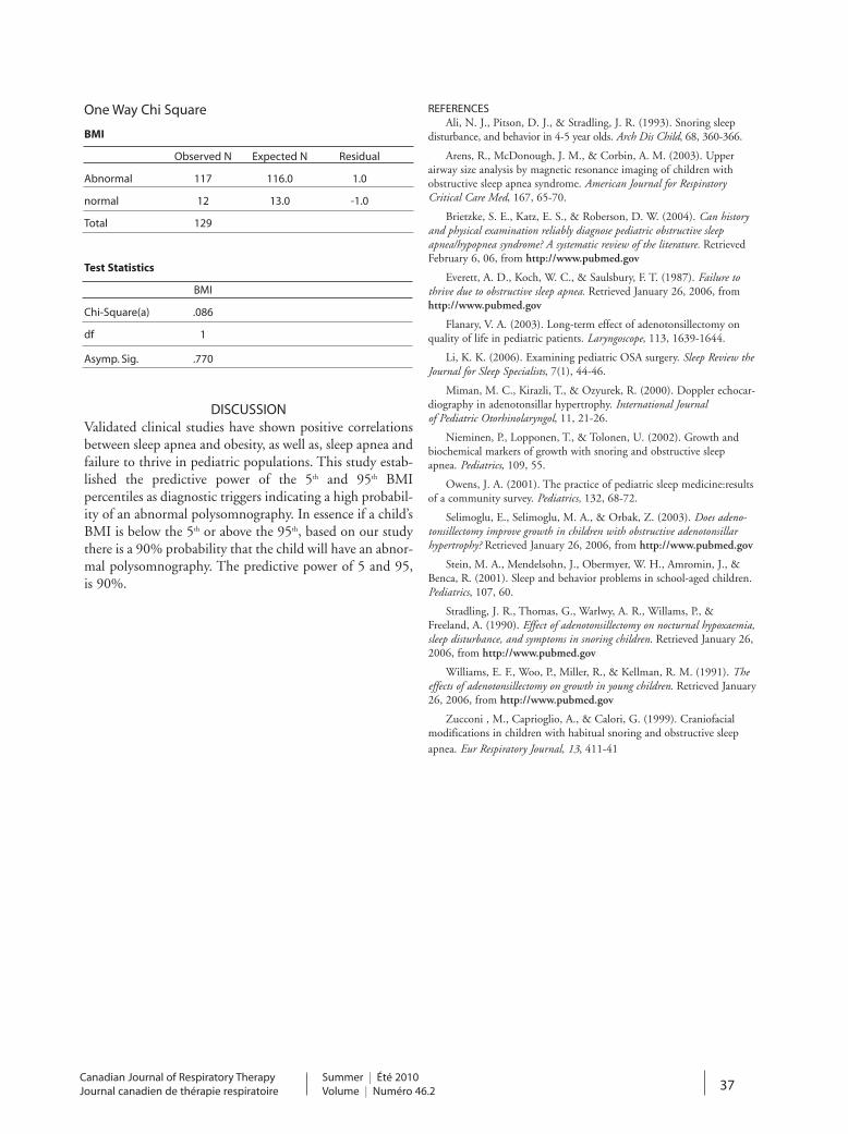
Canadian Journal of Respiratory TherapyJournal canadien de thérapie respiratoire
Summer | Été 2010Volume | Numéro 46.2 37
One Way Chi Square
BMI
Observed N Expected N Residual
Abnormal 117 116.0 1.0
normal 12 13.0 -1.0
Total 129
Test Statistics
BMI
Chi-Square(a) .086
df 1
Asymp. Sig. .770
DISCUSSIONValidated clinical studies have shown positive correlationsbetween sleep apnea and obesity, as well as, sleep apnea andfailure to thrive in pediatric populations. This study estab-lished the predictive power of the 5th and 95th BMI percentiles as diagnostic triggers indicating a high probabil-ity of an abnormal polysomnography. In essence if a child’sBMI is below the 5th or above the 95th, based on our studythere is a 90% probability that the child will have an abnor-mal polysomnography. The predictive power of 5 and 95,is 90%.
REFERENCESAli, N. J., Pitson, D. J., & Stradling, J. R. (1993). Snoring sleep
disturbance, and behavior in 4-5 year olds. Arch Dis Child, 68, 360-366.
Arens, R., McDonough, J. M., & Corbin, A. M. (2003). Upper airway size analysis by magnetic resonance imaging of children withobstructive sleep apnea syndrome. American Journal for RespiratoryCritical Care Med, 167, 65-70.
Brietzke, S. E., Katz, E. S., & Roberson, D. W. (2004). Can historyand physical examination reliably diagnose pediatric obstructive sleepapnea/hypopnea syndrome? A systematic review of the literature. RetrievedFebruary 6, 06, from http://www.pubmed.gov
Everett, A. D., Koch, W. C., & Saulsbury, F. T. (1987). Failure tothrive due to obstructive sleep apnea. Retrieved January 26, 2006, fromhttp://www.pubmed.gov
Flanary, V. A. (2003). Long-term effect of adenotonsillectomy onquality of life in pediatric patients. Laryngoscope, 113, 1639-1644.
Li, K. K. (2006). Examining pediatric OSA surgery. Sleep Review theJournal for Sleep Specialists, 7(1), 44-46.
Miman, M. C., Kirazli, T., & Ozyurek, R. (2000). Doppler echocar-diography in adenotonsillar hypertrophy. International Journal of Pediatric Otorhinolaryngol, 11, 21-26.
Nieminen, P., Lopponen, T., & Tolonen, U. (2002). Growth andbiochemical markers of growth with snoring and obstructive sleepapnea. Pediatrics, 109, 55.
Owens, J. A. (2001). The practice of pediatric sleep medicine:resultsof a community survey. Pediatrics, 132, 68-72.
Selimoglu, E., Selimoglu, M. A., & Orbak, Z. (2003). Does adeno-tonsillectomy improve growth in children with obstructive adenotonsillarhypertrophy? Retrieved January 26, 2006, from http://www.pubmed.gov
Stein, M. A., Mendelsohn, J., Obermyer, W. H., Amromin, J., &Benca, R. (2001). Sleep and behavior problems in school-aged children.Pediatrics, 107, 60.
Stradling, J. R., Thomas, G., Warlwy, A. R., Willams, P., &Freeland, A. (1990). Effect of adenotonsillectomy on nocturnal hypoxaemia,sleep disturbance, and symptoms in snoring children. Retrieved January 26,2006, from http://www.pubmed.gov
Williams, E. F., Woo, P., Miller, R., & Kellman, R. M. (1991). Theeffects of adenotonsillectomy on growth in young children. Retrieved January26, 2006, from http://www.pubmed.gov
Zucconi , M., Caprioglio, A., & Calori, G. (1999). Craniofacialmodifications in children with habitual snoring and obstructive sleepapnea. Eur Respiratory Journal, 13, 411-41
Canadian Journal of Respiratory TherapyJournal canadien de thérapie respiratoire
Summer | Été 2010Volume | Numéro 46.2
38
ORIGINAL ARTICLE
CSRT Members Malpractice Program Risk Advisor
Brian Gomes, Aon Risk Services
An allegation of negligence or wrongful act to a respiratory therapist (RT) who is not adequatelyinsured can be a financially devastating experience.
Even a frivolous accusation can result in crippling defensecosts that no individual should have to bear. The sponsoredinsurance program of the Canadian Society of RespiratoryTherapists was established precisely to protect RT’s in suchcircumstances by providing coverage tailored to membersand that follows them through a variety of practice settings.
The comprehensive group-insurance program is a membership benefit designed to meet the needs of any RTwhile providing professional services to protect individualday-to-day risk exposures. While ensuring adequate risktransfer protection via professional liability insurance is aprudent course of action, it is also important to considerissues surrounding scope of practice, documentation, andrisk management as RT’s deliver health care services.
SCOPE OF PRACTICEAccepting responsibilities beyond the scope of your licenseor can have serious consequences for you, your patients,and your career.
Respiratory therapists should always refuse assignmentsoutside of the legal scope of practice as defined by theapplicable provincial regulatory body. If a RT were toaccept responsibilities that are reserved for other professionals,he or she may be charged with practicing without a license.The professional liability insurance may not cover membersfor penalties resulting from actions beyond ones scope ofpractice that harmed a patient.
In addition, a patient who has been harmed or injured atthe alleged negligence of an RT, may put forth a complaintto a regulatory body or even take formal legal action againsta respiratory therapist if the therapist was practicing withoutthe skills needed to provide safe patient care – even if therespiratory therapist was performing tasks within the scopeof practice.
It is a prudent best practice to always advise the assigningrespiratory therapist when a member is not current on a specific skill needed and if one feels unsafe to provide certain care. Attempting to perform treatments or useequipment without the knowledge and training requiredcan have negative implications to a potential loss where theprofessional is deemed to be held negligent for any damageor injury.
Refusing to perform services in any of the scenario’snoted above can be a difficult response to put forth to colleagues and other medical professionals. A reasonableexplanation that’s assertively explained for saying no will beheard and accepted better than a no that is aggressively ornegatively presented. If saying yes in not in your best inter-est, give your response clearly. Communicate decisively andin a timely manner. Provide suggestions to other ways formeeting the need you’re unable or unwilling to fulfill.
It isn’t always easy to say no to requests being made bypeers and healthcare team members. The manner in whicha refusal is communicated can make a difference in how themessage is received. Good team relations as well as excellentteam communication are important to establish a founda-tion of the work environment that allows you to say no.Regulatory bodies, employee support staff, and associationscan be a tremendous aid when deliberating such scenarios.
DOCUMENTATIONA malpractice judgment can seriously harm your personaland professional life. Your file documentation can be theonly thing between your word and that of a patient whomay be filing a complaint or formal legal action. Oftenyour documentation may be the sole item of evidence in acase. Proper documentation is a key element in adverselegal action. Legally credible documentation involves andaccurate record of the care your client received and yourcompetence in providing appropriate respiratory therapyservices. The file notes should be accurate, honest andappropriate.
Here are some common ensure steps to take to ensure proper documentation.• Document accurately, be sure to note the time and date.• Avoid any exaggeration• Remember that file documentation may be subject to
a subpoena.• Never alter client records.
RISK MANAGEMENTIn addition to traditional transferable risks, such as thosecovered by professional-liability insurance, it is importantfor a respiratory therapist to also to explore any methodsthat minimize other risk exposures.
Canadian Journal of Respiratory TherapyJournal canadien de thérapie respiratoire
Summer | Été 2010Volume | Numéro 46.2 39
To start the risk-management process, RT’s should viewthe daily practice settings from a macro and micro perspec-tive and outline any potential day-to-day operational risksby asking and answering questions such as the following:
• What potential scenarios could have a negative impact on my reputation as a professional?
• What can I do to minimize or control these risks?• Is all my work conducted within my regulatory
guidelines and scope of practice?• Am I following the best possible documentation
standards, such as protecting client or proprietary information, and separating and duplicating valuable records?
Members participating within the CSRT medical malpracticeinsurance program should take a moment to review the specific coverage’s provided through this dedicated compre-hensive insurance policy.
A highlight sheet describing coverage elements alongwith a claims advisory stating necessary actions to take in the event of a potential loss can be found athttp://www.csr t .com/en/profess ional/ l iabi l i ty_insurance.asp
This article is written in conjunction with content fromAon’s HSPO and NSO 2009-2010 Risk Advisor.
NON-INVASIVE VENTILATION IN ALI/ARDS – DIRECTED READING ARTICLE ANSWERS
1) B. PaO2/FiO2 < 250 mmHg
2) E. All of them
3) A. No improvement in arterial pH after I hour of NRS institution
B. Poor patients cooperation
4) F. All of them
5) A. High
6) C. Poor
7) A. n CPAP is equivalent to nPPV
8) D. It is a first line treatment when indicated and no contraindication are present
9) C. Moderate in ALI/ARDS and in community acquired pneumonia
10) D. Preventing atelectasis and increasing functional residual capacity
11) D. All of them
12) D. All of them
13) D. All of them
14) D. Better respiratory muscles unloading
15) C. In children is associated with less need of sedation in children and prolonged NRS application time respect to the mask
16) B. An air oxygen blender, flow meter, PEEP valve, alarm system on pressure and FiO2
17) D. All of them
18) A. Prolonged use of nasal cannulae leads to nasal bleeding and nares obstruction
19) D. All of them
20) A. Is mandatory to set the sensitivity of inspiratoryand expiratory trigger
21) A. Heat and moisture exchangers and heated humidifiers provided adequate airway conditioning
22) E. All of them
23) A. Low improvement in paO2 level I h after starting nPPV
24) A. nPPV could improve oxygenation and reduce intubation rate
25) A. nPPV could reduce intubation rate
Canadian Journal of Respiratory TherapyJournal canadien de thérapie respiratoire
Summer | Été 2010Volume | Numéro 46.2
40
ORIGINAL ARTICLE
Say “YES” to Evidence Based Respiratory Therapy Practice
Mika L. Nonoyama RRT, PhDPost Doctoral Fellow, Toronto Rehabilitation Institute
You are approached by a respirology resident who asksyou what is the most effective puffer to help hispatient with chronic obstructive pulmonary disease
(COPD) relieve her shortness of breath. A certain degree offoreboding sets in. Several answers swirl in your head, all ofwhich come from various places – the pharmacology courseyou took from school, the last journal club you attended, arecent in-service, an abstract posted on the board, yourexperience at your shift last week etc. What do you say tothis resident? Do you say “the blue one” and make a run forit? No doubt that idea pops in your head, but fortunatelyyou are an excellent respiratory therapist (RT) and you goforward to help answer his question. How do you go aboutdoing this?
Evidence based clinical practice (EBCP) has been aroundfor centuries, but its explicit methodologies started takingshape in the 20th century with the help of Dr. ArchieCochrane, Dr. David Sackett and Dr. Gordon Guyatt.(1)There are several definitions of EBCP, the most commonby David Sackett and colleagues: “the integration of bestresearch evidence with clinical expertise and patient valuesand circumstances”.(2) This is a nice definition because itpoints out that EBCP is more than just what is in literatureor within the revered randomized controlled trial (RCT).Our clinical expertise and what patients bring to the tableare equally as important. One challenge of EBCP is the lit-erature “mountain” that accompanies every clinical scenariowe encounter. EBCP provides a systematic approach tofinding the best answers to our clinical questions. I hopethis column will give you a better understanding of EBCP,providing a starting point for its use in your own clinicalpractice.
In an editorial, Haynes and colleagues (3) used Sackett’sdefinition of EBCP to develop a model for evidence basedclinical decision making. Four elements are contained inthis model: 1) clinical state and circumstances; 2) patient preferences and actions; 3) research evidence and; 4) clinical expertise. Clinical expertise must encompass and balance all three elements in order for a successful and satisfying result tooccur.(3)
Clinical state and circumstances can include: the clinicalsetting e.g. remote area vs. tertiary care medical centre; thepatients’ clinical state; the clinical circumstances; etc.(3)For example, a patient with COPD who complains of dyspnea after climbing several flights of stairs would likelybenefit from using a short acting beta-agonist (SABA) onan as needed basis (prn). A patient who becomes dyspneicafter walking 25 metres and suffers from frequent exacerba-tions would benefit from daily use of a combination longacting anti-cholinergic (LAAC) and inhaled corticosteroid(ICS) along with a SABA prn.(4) In both cases the efficacyof the treatment can be modified by adequate follow-upe.g. ensuring these patients are technically and symptomati-cally using the puffers properly.
Patients’ preferences and actions can include: personalvalues and experiences; the degree of aversion to risk;healthcare insurance and resources; family input; willing-ness to take medicines; accurate or misleading informationat hand; etc.(3) For example the preferences of patients forICS medication will depend on the competing side effectsof hoarse voice and/or oral candidiasis (4) and the potentialcost (especially if they do not have monetary coverage). In addition, the preferences of us as RTs may be quite different than our patient. We may be hesitant to suggestICS because of the competing risks but the patient may bewilling to tolerate the adverse effects and costs in order torelieve his/her shortness of breath.
Research evidence can include: systematic observationsfrom the laboratory; preliminary pathophysiological studiesin humans; advanced applied clinical research, e.g. RCTswith outcomes immediately important to patients; etc.(3)The challenge with research evidence is knowing where tolook for the information and then figuring out whether itis the most rigorous and if it is applicable toward the specific clinical scenario.(3) In addition there are oftenalternate therapies each with their own benefit and risk profiles.(3) For example a patient who has dyspnea afterwalking 50 to 75 metres on a level surface with a remotehistory of exacerbations may be prescribed either a long acting beta-agonist (LABA) or a LAAC.(4) A couple ofstudies (RCTs) in patients with COPD have shown theLAAC Tiotropium significantly improved forced expiratory

Canadian Journal of Respiratory TherapyJournal canadien de thérapie respiratoire
Summer | Été 2010Volume | Numéro 46.2 41
volume (FEV1) compared to the LABAs Salmeterol (5) orFormoterol (6), but the magnitude was small (Tiotropiumimproved FEV1 by an average range of 80 to 127mL). Forthis patient the decision may depend on the side effects,convenience and/or cost of the medication.
Finally our RT clinical expertise plays an important rolein EBCP. This includes general basic skills from our school-ing and our experience as an individual practitioner.(3)Clinical expertise must encompass and balance all threeelements in order for us to find the best answer for our
patient. We must be atop research evidence, acquire andhone the skills needed to both interpret the evidence andapply it appropriately to the circumstances and determinewhat the patient wants.(3) All of us as RTs have the skillsto do this.
At this point you may be thinking that EBCP is toocomplicated and time consuming to fit into daily clinicalpractice. It is true that it takes time to become comfortablewith the process. However you don’t have to be expert in allaspects of EBCP at the get-go. A departmental journal tojot down interesting clinical questions and a once-a-monthjournal club to help answer those questions is a good start.We all as RTs must be actively involved in lifelong learning.What better way than incorporating clinical circumstances,patient values, the best research evidence and our clinicalexpertise?
Fortunately there are resources to help. This ranges fromsingle articles to help us search the literature (7), all the wayto detailed textbooks (8). In addition several EBCP groupsaround the world have developed online resources to makethe process less daunting. The Clarity Research Group atMcMaster University provided annual workshops on howto teach EBCP (www.clarityresearch.ca). The Centre forEvidence Based Medicine at Oxford University offers presentations, documents and other handy tools(www.cebm.net). The University of Alberta created a“toolkit” (www.ebm.med.ualberta.ca) for EBCP thatincludes a concise and easy to follow “mini-manual” (9).This mini-manual provides a step-by-step guide – all within 24 pages and open access:
• Analyze the clinical situation.• Ask a focused clinical question• Access the clinical research literature
(i.e., the evidence)• Appraise the best evidence you have found• Apply the evidence to care of your patient• Assess the effectiveness of care based on this evidence
And the list goes on…Based on this brief description of EBCP you may have a
better idea how to answer that respirology resident whoapproached you about his COPD patients. The details onformulating a specific answer go beyond the scope of thiscommentary however I hope it plants the EBCP seed andallows for further RT specific growth in the area. Our peerprofessions such as medicine, nursing, physical therapy,occupational therapy, and speech pathology have alreadycreated a niche in the area of EBCP. Let’s do it too!
REFERENCES1. Evidence-based medicine - history (http://hlwiki.slais.ubc.ca/
index.php/Evidence-based_medicine_-_history). In: Wikipedia, The Free Encyclopedia; 2010.
2. Sackett D, Straus S, Richardson W, Rosenberg W, Haynes R. Evidence-based medicine: how to practice and teach EBM. 2nd ed. Edinburgh: Churchill Livingstone; 2000.
3. Haynes RB, Devereaux PJ, Guyatt GH. Clinical expertise in the era of evidence-based medicine and patient choice. ACP Journal Club 2002;136(2):Mar-Apr.
4. O'Donnell DE, Aaron S, Bourbeau J, Hernandez P, Marciniuk DD, Balter M, et al. Canadian Thoracic Society recommendations for management of chronic obstructive pulmonary disease - 2007 update. Can Respir J 2007;14(Suppl. B):5B-32B.
5. Donohue JF, van Noord JA, Bateman ED, Langley SJ, Lee A, Witek TJ, Jr., et al. A 6-month, placebo-controlled study comparing lung function and health status changes in COPD patients treated with tiotropium or salmeterol. Chest 2002;122(1):47-55.
6. van Noord JA, Aumann JL, Janssens E, Smeets JJ, Verhaert J, Disse B, et al. Comparison of tiotropium once daily, formoterol twice daily and both combined once daily in patients with COPD. European Respiratory Journal 2005;26(2):214-22.
7. Haynes B. Of studies, syntheses, synopses, summaries, and systems: the “5S” evolution of information services for evidence-based healthcare decisions. Evidence Based Nursing 2007;10(1):6-7.
8. Guyatt G, Rennie D, Meade M, Cook D. Users' Guides to the Medical Literature: A Manual for Evidence-Based Clinical Practice. New York: McGraw-Hill Medical; 2008.
9. Buckingham J, Fisher B, Saunders D. Evidence based medicine - mini manual. In: Evidence based medicine tool kit(http://www.library.ualberta.ca/uploads/HealthSciences/eb_mini_manual_2008.pdf ), University of Alberta; 2008.
Canadian Journal of Respiratory TherapyJournal canadien de thérapie respiratoire
Summer | Été 2010Volume | Numéro 46.2
42
CASE STUDY
The Use of Transpulmonary Pressure to set Optimal Positive End-expiratory Pressure: A Case Report
Tom Piraino RRT,Best Practice Clinical Educator, Respiratory Therapy Services, St. Joseph's Healthcare, Hamilton Ontario
ABSTRACTThe airway pressure generated by the mechanical ventilatordoes not distinguish the pressure generated within the lungfrom that which may be present outside of the lung. Truelung pressure is transpulmonary pressure, which representsthe actual movement of the lung (airway pressure – pleuralpressure = transpulmonary pressure). Theoretically, if thepressure outside of the lung at end-exhalation was higherthan the Positive End-expiratory Pressure (PEEP) set onthe ventilator, the result would be atelectasis, and with sub-sequent breaths would place the patient at high risk ofatelectrauma. This case is an example of how estimatingtranspulmonay pressure can assist the clinician to optimal-ly set PEEP in a mechanically ventilated patient.
RÉSUMÉLa pression des voies aériennes générée par un ventilateurmécanique crée une confusion entre la pression produite à l’intérieur du poumon et celle qui peut être présente à l’extérieur du poumon. La véritable pression du poumon estune pression transpulmonaire qui représente le mouvementréel du poumon (pression des voies aériennes – pressionpleurale = pression transpulmonaire). En théorie, si la pres-sion à l’extérieur du poumon à la fin de l’expiration est plusélevée que la pression positive en fin d’expiration (PEEP)insufflée par le ventilateur, il en résultera une atélectasie et lesrespirations subséquentes du patient le mettent en dangerélevé de traumatisme. Le document constitue un exemple dela façon dont l’estimation de la pression pulmonaire peutaider le clinicien à mieux établir la PEEP chez un patientventilé mécaniquement.
INTRODUCTIONStrategies to prevent atelectrauma, volutrauma, and barotrauma during mechanical ventilation are important inlimiting ventilator induced lung injury (VILI). PositiveEnd-expiratory Pressure (PEEP) is a setting of mechanicalventilation used to keep the lungs open and prevent atelec-trauma. A current method for managing patients with pooroxygenation is to use high levels of PEEP. However, twolarge randomized controlled trials compared a low and highPEEP/FiO2 protocol to guide changes in PEEP and/orFiO2 in response to oxygenation. These studies failed toshow an improvement in clinical outcomes such as mortal-ity2,4. The limitation of setting PEEP using a chart or pro-tocol is that it ignores the individual lung mechanics of thepatient and therefore is not optimal. The generalizedapproach to setting PEEP in these studies may be the rea-son why the results were not significant in supporting theuse of high PEEP. The level (low or high) of PEEP may notbe as important as the appropriateness of PEEP, and a lim-itation of mechanical ventilators is they fail to represent theeffect of pleural pressure on the measured airway pressure(Figure 1). The difference between airway pressure andpleural pressure is called transpulmonary pressure (Ptp),and it is normally positive in healthy patients14. This can
drastically change when a patient in respiratory failurerequires sedation and positive pressure ventilation. PEEPthat is set optimally should maintain a neutral Ptp (0-3 cmH2O). A current method used to estimate pleural pressure,calculate Ptp, and set optimal PEEP, is the esophageal balloon catheter.
TRANSPULMONARY PRESSURE AND ESOPHAGEALBALLOON CATHETERS IN CURRENT LITERATURE
Although the ARDSNet recommendation is to limitplateau pressures to ² 30 cm H2O, an interesting findingwith the Lung Open Ventilation Study (LOVS) was thatallowing higher PEEP levels and subsequent higher plateaupressures did not increase mortality, or the incidence ofbarotrauma1,4. The reason for this may be that lung recruit-ment, lung stretch and strain, and atelectasis are not easilyassessed by airway pressure measurements on a mechanicalventilator. The most important pressure to understandwhat is happening to a lung is the Ptp (alveolar pressure -pleural pressure = Ptp). The least invasive method of meas-uring pleural pressure is to estimate it with an esophagealballoon. Chiumello et al. studied lung stress and strainusing esophageal pressure (Pes) measurements at different
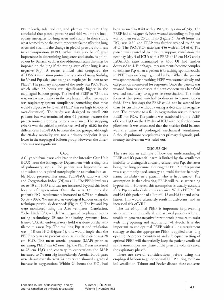
Canadian Journal of Respiratory TherapyJournal canadien de thérapie respiratoire
Summer | Été 2010Volume | Numéro 46.2 43
PEEP levels, tidal volume, and plateau pressures6. Theyconcluded that plateau pressures and tidal volume are inad-equate surrogates for lung stress and strain. In their study,what seemed to be the most important factor affecting lungstress and strain is the change in pleural pressure from restto end-inspiration (Æ PL). What may also be of greatimportance in determining lung stress and strain, as point-ed out by Behazin et al., is the additional strain that may beimposed on the lung if the resting state of the lung is at anegative Ptp7. A recent clinical trial compared theARDSNet ventilation protocol to a protocol using 6mls/kgfor Vt and Ptp calculated using an esophageal balloon to setPEEP8. The primary endpoint of the study was PaO2/FiO2,which after 72 hours was significantly higher in theesophageal balloon group. The level of PEEP at 72 hourswas, on average, higher in the esophageal balloon group, aswas respiratory system compliance, something that mostwould suspect to be lower if PEEP was set high (theory ofover-distention). The study was designed to enroll 200patients but was terminated after 61 patients because thepredetermined stopping criteria were met. The stoppingcriteria was the critical significance level of p <0.02 for thedifference in PaO2/FiO2 between the two groups. Althoughthe 28-day mortality was not a primary endpoint it waslower in the esophageal balloon group. However, the differ-ence was not significant.
CASEA 61 yr old female was admitted to the Intensive Care Unit(ICU) from the Emergency Department with a diagnosisof pulmonary sepsis. The patient was hypotensive onadmission and required norepinephrine to maintain a sta-ble blood pressure. Her initial PaO2/FiO2 ratio was 143and Oxygenation Index (OI) was 11. The PEEP level wasset to 10 cm H2O and was not increased beyond this levelbecause of hypotension. Over the next 13 hours thepatient’s FiO2 requirements increased to 0.75 to maintainSpO2 > 90%. We inserted an esophageal balloon using thetechnique previously described8 (Figure 2). The Pes and Ptpwere monitored using the Avea ventilator (Carefusion,Yorba Linda CA), which has integrated esophageal moni-toring technology (Bicore Monitoring Systems, Inc.,Irvine, CA). An end-expiratory hold was done on the ven-tilator to assess Ptp. The resulting Ptp at end-exhalationwas – 18 cm H2O (Figure 1), this would imply that thePEEP necessary to prevent atelectasis in the patient was 28cm H2O. The mean arterial pressure (MAP) prior toincreasing PEEP was 62 mm Hg, the PEEP was increasedto 28 cm H2O and contrary to expectations the MAPincreased to 74 mm Hg immediately. Arterial blood gaseswere drawn over the next 24 hours and showed a gradualincrease in oxygenation. Within 24 hours the FiO2 had
been weaned to 0.40 with a PaO2/FiO2 ratio of 345. ThePEEP had subsequently been weaned according to Ptp andwas by then set at 25 cm H2O (Figure 3). At 48 hours theFiO2 was 0.30 and PEEP was further weaned to 22 cmH2O. The PaO2/FiO2 ratio was 456 with an OI of 6. Thepatient was switched to pressure support ventilation thenext day (day 3 of ICU) with a PEEP of 16 cm H2O and aPaO2/FiO2 ratio maintained at 453, OI had furtherdecreased to 4. Esophageal measurements become complexto estimate Ptp when a patient is breathing spontaneously,so PEEP was no longer guided by Ptp. When the patientwas spontaneously breathing PEEP was weaned slowly andoxygenation monitored for response. Once the patient wasweaned from vasopressors the next concern was her fluidoverload secondary to aggressive resuscitation. The mainfocus at that point switched to diuresis to remove excessfluid. For a few days the PEEP could not be weaned lessthan 14 cm H2O without causing a decrease in oxygena-tion. The response to a fall in oxygenation was to increasePEEP, not FiOv. The patient was extubated from a PEEPof 6 cm H2O on the 12th day of ICU with no further com-plications. It was speculated that the positive fluid balancewas the cause of prolonged mechanical ventilation.Although pulmonary sepsis was her primary diagnosis, pul-monary involvement was ruled out.
DISCUSSIONThe case was an example of how our understanding ofPEEP and it’s potential harm is limited by the ventilatorsinability to distinguish airway pressure from Ptp, the latterbeing true lung pressure. Limiting the PEEP in this patientwas a commonly used strategy to avoid further hemody-namic instability in a patient who is hypotensive. Theassumption is that elevating PEEP will cause worseninghypotension. However, this assumption is usually accurateif the Ptp at end-exhalation is excessive. With a PEEP of 10cmH2O this patient had a Ptp of - 18 cmH2O at end-exha-lation. This would ultimately result in atelectasis, and anincreased risk of VILI.
The use of optimal PEEP is important in preventingatelectrauma in critically ill and sedated patients who areunable to generate negative intrathoracic pressure to assistwith lung opening and stabilization of alveoli. It is alsoimportant to use optimal PEEP with a lung recruitmentstrategy so that the appropriate PEEP is applied after lungopening. A proper recruitment and subsequent setting ofoptimal PEEP will theoretically keep the patient ventilatedin the most important phase of the pressure volume curve;the expiratory phase11.
There are several considerations before using theesophageal balloon to guide optimal PEEP during mechan-ical ventilation. Talmor and Fessler discuss these concerns
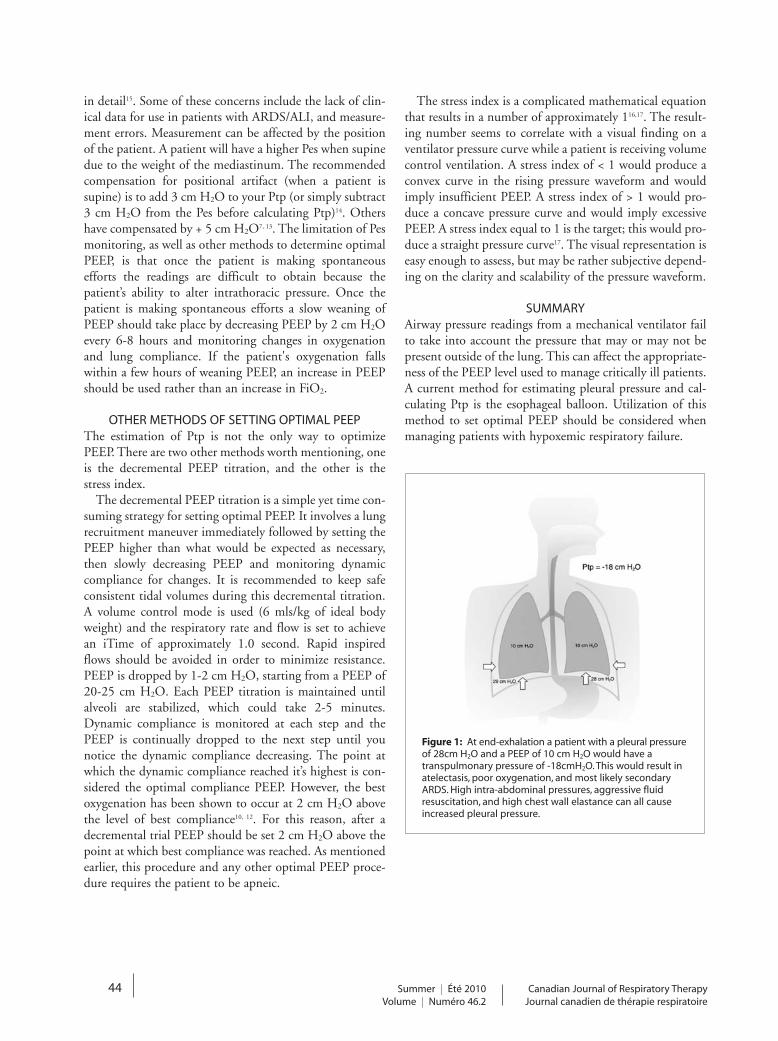
Canadian Journal of Respiratory TherapyJournal canadien de thérapie respiratoire
Summer | Été 2010Volume | Numéro 46.2
44
in detail15. Some of these concerns include the lack of clin-ical data for use in patients with ARDS/ALI, and measure-ment errors. Measurement can be affected by the positionof the patient. A patient will have a higher Pes when supinedue to the weight of the mediastinum. The recommendedcompensation for positional artifact (when a patient issupine) is to add 3 cm H2O to your Ptp (or simply subtract3 cm H2O from the Pes before calculating Ptp)14. Othershave compensated by + 5 cm H2O7, 13. The limitation of Pesmonitoring, as well as other methods to determine optimalPEEP, is that once the patient is making spontaneousefforts the readings are difficult to obtain because thepatient’s ability to alter intrathoracic pressure. Once thepatient is making spontaneous efforts a slow weaning ofPEEP should take place by decreasing PEEP by 2 cm H2Oevery 6-8 hours and monitoring changes in oxygenationand lung compliance. If the patient's oxygenation fallswithin a few hours of weaning PEEP, an increase in PEEPshould be used rather than an increase in FiO2.
OTHER METHODS OF SETTING OPTIMAL PEEPThe estimation of Ptp is not the only way to optimizePEEP. There are two other methods worth mentioning, oneis the decremental PEEP titration, and the other is thestress index.
The decremental PEEP titration is a simple yet time con-suming strategy for setting optimal PEEP. It involves a lungrecruitment maneuver immediately followed by setting thePEEP higher than what would be expected as necessary,then slowly decreasing PEEP and monitoring dynamiccompliance for changes. It is recommended to keep safeconsistent tidal volumes during this decremental titration.A volume control mode is used (6 mls/kg of ideal bodyweight) and the respiratory rate and flow is set to achievean iTime of approximately 1.0 second. Rapid inspiredflows should be avoided in order to minimize resistance.PEEP is dropped by 1-2 cm H2O, starting from a PEEP of20-25 cm H2O. Each PEEP titration is maintained untilalveoli are stabilized, which could take 2-5 minutes.Dynamic compliance is monitored at each step and thePEEP is continually dropped to the next step until younotice the dynamic compliance decreasing. The point atwhich the dynamic compliance reached it’s highest is con-sidered the optimal compliance PEEP. However, the bestoxygenation has been shown to occur at 2 cm H2O abovethe level of best compliance10, 12. For this reason, after adecremental trial PEEP should be set 2 cm H2O above thepoint at which best compliance was reached. As mentionedearlier, this procedure and any other optimal PEEP proce-dure requires the patient to be apneic.
The stress index is a complicated mathematical equationthat results in a number of approximately 116,17. The result-ing number seems to correlate with a visual finding on aventilator pressure curve while a patient is receiving volumecontrol ventilation. A stress index of < 1 would produce aconvex curve in the rising pressure waveform and wouldimply insufficient PEEP. A stress index of > 1 would pro-duce a concave pressure curve and would imply excessivePEEP. A stress index equal to 1 is the target; this would pro-duce a straight pressure curve17. The visual representation iseasy enough to assess, but may be rather subjective depend-ing on the clarity and scalability of the pressure waveform.
SUMMARYAirway pressure readings from a mechanical ventilator failto take into account the pressure that may or may not bepresent outside of the lung. This can affect the appropriate-ness of the PEEP level used to manage critically ill patients.A current method for estimating pleural pressure and cal-culating Ptp is the esophageal balloon. Utilization of thismethod to set optimal PEEP should be considered whenmanaging patients with hypoxemic respiratory failure.
Figure 1: At end-exhalation a patient with a pleural pressureof 28cm H2O and a PEEP of 10 cm H2O would have atranspulmonary pressure of -18cmH2O. This would result inatelectasis, poor oxygenation, and most likely secondaryARDS. High intra-abdominal pressures, aggressive fluid resuscitation, and high chest wall elastance can all causeincreased pleural pressure.

Canadian Journal of Respiratory TherapyJournal canadien de thérapie respiratoire
Summer | Été 2010Volume | Numéro 46.2 45
REFERENCES1. The Acute Respiratory Distress Syndrome Network. Ventilation
with lower tidal volumes as compared with traditional tidal volumesfor acute lung injury and the acute respiratory distress syndrome. N. Engl J Med 2000;342(18): 1301-1308.
2. Brower R, Lanken P, MacIntyre N, Matthay M, Morris A, Ancukiewicz M, et al. Higher versus lower positive end-expiratory pressures in patients with the acute respiratory distress syndrome. N Engl J Med 2004;351(4):327-336.
3. Mercat A, Richard J, Vielle B, Jaber S, Osman D, Diehl J, et al. Positive end-expiratory pressure setting in adults with acute lung injury and acute respiratory distress syndrome: a randomized controlled trial. JAMA 2008;299(6):646-655.
4. Meade M, Cook D, Guyatt G, Slutsky A, Arabi Y, Cooper D, et al. Ventilation strategy using low tidal volumes, recruitment maneuvers,and high positive end-expiratory pressure for acute lung injury and acute respiratory distress syndrome: a randomized con- trolled trial. JAMA 2008;299(6):637-645.
5. Phoenix S, Paravastu S, Columb M, Vincent J, Nirmalan M. Does aHigher Positive End Expiratory Pressure Decrease Mortality in Acute Respiratory Distress Syndrome? A Systematic Review and Meta-analysis. Anesthesiology 2009;110(5):1098-1105
6. Chiumello D, Carlesso E, Cadringher P, Caironi P, Valenza F, Polli F, et al. Lung stress and strain during mechanical ventilation for acute respiratory distress syndrome. Am J Respir Crit Care Med 2008;178(4): 346-355
7. Behazin N, Jones S, Cohen R, Loring S. Esophageal pressures in acute lung injury-do they represent artifact or useful information about transpulmonary pressure, chest wall mechanics, and lung stress?. Journal of Applied Physiology 2010;108(1): 212-218
8. Talmor D, Sarge T, Malhotra A, O’Donnell C, Ritz R, Lisbon A, et al. Mechanical ventilation guided by esophageal pressure in acute lung injury. N Engl J Med 2008;359(20):2095-2104.
9. Borges J, Okamoto V, Matos G, Caramez M, Arantes P, Barros F, et al. Reversibility of lung collapse and hypoxemia in early acute respiratory distress syndrome. Am J Respir Crit Care Med 2006;174(3):268-278.
10. Suarez-Sipmann et al. Use of dynamic compliance for open lung positive end-expiratory pressure titration in an experimental study.Crit Care Med 2007;35(1):214-21
11. Gattinoni L, Carless E, Brazzi L, Caironi P. Positive end-expiratory pressure. Curr Opin Crit Care 2010;16(1):45-52
12. Caramez et al. A comparison of methods to identify open-lung PEEP. Intensive Care Med 2009;35(4): 740-747
13. Talmor D, Sarge T, O’Donnell C, Ritz R, Malhotra A, Lisbon A, Loring S. Esophageal and transpulmonary pressures in acute respiratory failure. Crit Care Med 2006;34(5):1389-1394.
14. Washko G, O’Donnell C, Loring S. Volume-related and volume-independent effects of posture on esophageal and transpulmonary pressures in healthy subjects. J Appl Physiol Mar 2006;100(3): 753-758.
15. Talmor D, Fessler H. Are esophageal pressure measurements important in clinical decision-making in mechanically ventilated patients?. Respir Care 2010;55(2):162-174
16. Grasso S, Terragni P, Mascia L, Fanelli V, Quintel M, Herrmann P, et al. Airway pressure-time curve profile (stress index) detects tidal recruitment/hyperinflation in experimental acute lung injury. Crit Care Med 2004;vol;32(4): 1018-1027
17. Terragni P, Rosboch G, Lisi A, Viale A, Ranieri V. How respiratory system mechanics may help in minimising ventilator-induced lung injury in ARDS patients. Eur Respir J Suppl 2003;42:15-21
Gentle compression of theabdomen to confirm placement Look for cardiac oscillations
Figure 2: The catheter is placed orally to 60 cm. Gentle compression of the abdomen should cause a rise in the Pesmeasurement. The catheter is then pulled back to 40 cmwhile looking for cardiac oscillations to ensure proper placement in the lower third of the esophagus.
Figure 3: Chest radiograph with a PEEP of 10 cm H20 (left)and 24 hours later with a PEEP of 25 cm H2O (right).

Canadian Journal of Respiratory TherapyJournal canadien de thérapie respiratoire
Summer | Été 2010Volume | Numéro 46.2
46
BOOK REVIEW
“When I Nod I Mean “NO!”by Gaynor D. Govias”
Ana MacPherson, RRT, CRE, MAScProvincial Coordinator, PCAP, Ontario
Asthma Plan of Action
This book focuses on behavioural guidelines and cultural tips for health professionals working withpeople from other cultures. The readership is written
for healthcare professionals as well as any public serviceworker. The author hopes to provide these professionalswith basic knowledge and understanding of their patients’or clients’ culture and religion, and to encourage them tothink about the way they use the English language. Theauthor openly states that this guide is intended to be usedas a general, quick-reference source of information. It is areminder that each person we meet should be treated asunique because there is no such thing as a typical person!
As a healthcare professional for more than 20 years, I have lived and worked in Toronto and Mississauga havepopulations of which about 47% and 49% (respectively)are visible minorities. As one of those visible minorities, itwas very helpful for me to have the same cultural back-ground as some of the patients that I came in contact withfrom other countries. I was very fortunate to have grownup in a country that is culturally sensitive to its residents.Equally interesting was that as a young child immigratingto a land of opportunities, I had to learn about the culture,religion and government of the country I was immigratingto. Assimilation would have brought on an easy fix with thecultural diversity, but fortunately, Canada’s Charter ofHuman Rights recognizes people with diverse backgroundsand respects the culture and religion you practise.
I highly recommend this book as a general guide to beused to complement previous or other resources in learningabout other cultures. It is a great little book of “quick-reference” for healthcare professionals who work with adiverse patient population in Canada.
I appreciate the way the guide is organized. The book islogically mapped out, which makes it very user friendly. Section 1 General guidelines of dealing with people from
other culturesSection 2 Culture, beliefs and practices in different
countriesSection 3 Major world religions
Section 4 Major religious, national and cultural holidaysSection 5 Brief descriptions of some complementary
medicines and medical therapiesSection 6 Glossary and Index
The general recommendations are just that. We know fora fact that each person patient is unique. The cultural back-ground will be the predisposing factor that will influencehealth beliefs and understanding. Use the guide as a gener-al quick- reference. For further in-depth answers, furtherresearch is necessary as suggested by the author. I find thisbook helpful, but would make no prejudgement until Ihave spent time with the patient to comprehend theirhealth beliefs. Time spent with the patient will provide uswith the basic knowledge of cultural and religious beliefsthat will guide us in motivational intervention using theprocesses of change and teaching our patients self-manage-ment of their disease.
The book is written in easy simple language which givesit a flavour right at the outset of how we should be convers-ing with our patient: in lay and simple terms that shouldnot be interpreted in any other way. One of the recommen-dations under Section 1 is the cautious use of the Englishlanguage. I am sure we all have thought of this, but theguide, specifically, identifies that the English languageneeds to be used with great thought. As an example thephrase: To “Knock up” could mean: - To impregnate or to knock at the doorThis book shows the care the author has taken in puttingthis “quick-reference” guide. As healthcare professionalswanting to make a difference with our patients’ behaviourand assisting them with self-management, we need tounderstand their health beliefs stemming from their culturaland religious background. This book has provided us withthat basic knowledge. Everyone working as a healthcareprofessional or public service worker should have this reference. Thank you to the author for the time and effort.Two thumbs up!
Canadian Journal of Respiratory TherapyJournal canadien de thérapie respiratoire
Summer | Été 2010Volume | Numéro 46.2 47
TECHNOLOGY UPDATE
Enhancing the Safety of Medical Suction Through Innovative Technology
Patricia Carroll, RN,BC, CEN, RRT, MSSubmitted by Ohio Medical Corporation (Amvex)
ABSTRACTMedical suctioning is essential for patient care. However,few clinicians receive training on the principles of physicsthat govern the safe use of medical suction. While all eightmanufacturers of vacuum regulators sold in North Americarequire occlusion of the tube before setting or changingvacuum levels, anecdotal evidence reveals that clinicians arenot aware of this requirement or skip this step whenpressed for time. This white paper summarizes the physicsrelating to medical suction, the consequences of damagedmucosa, the risks to patient safety when suction levels arenot properly set and regulated, and technology advancesthat enhance patient safety.
Medical suction is an essential part of clinical practice.Since the 1920s, it has been used to empty the stomach,and in the 1950s, airway suction levels were first regulatedfor safety. Today, medical suction is used for newly bornbabies and seniors, and in patients weighing between 500grams and 500 pounds. Medical suction clears the airway,empties the stomach, decompresses the chest, and keepsthe operative field clear. It is essential that clinicians havereliable equipment that is accurate and easy to use.
RÉSUMÉL’aspiration médicale est essentielle au soin du patient.Cependant, peu de cliniciens reçoivent de la formation surles principes de physiques qui régissent l’utilisation sécuri-taire de l’aspiration médicale. Alors que les huit fabricants derégulateurs de vide vendus en Amérique du Nord demandentla fermeture du tube avant un changement de niveau d’aspi-ration, les preuves anecdotiques révèlent que les cliniciens neconnaissent pas cette exigence ou sautent cette étapelorsqu’ils manquent de temps. Le livre blanc résume laphysique reliée à l’aspiration médicale, les conséquences desdommages aux muqueuses, les risques pour les patientslorsque le niveau d’aspiration est mal réglé ou régulé, ainsique les avancées technologiques qui améliorent la sécuritédes patients.
L’aspiration médicale est un élément essentiel de la pratique clinique. Depuis les années 1920, elle est utiliséepour la vidange stomacale, et les niveaux d’aspiration desvoies aériennes ont pour la première fois fait l’objet d’uneréglementation de sécurité dans les années 1950.Aujourd’hui, l’aspiration médicale est utilisée chez les nouveau-0nés et les personnes âgées, et pour les personnespesant de 500 grammes à 500 livres. L’aspiration médicalelibère les voies aériennes, vide l’estomac, décomprime lapoitrine et garde le champ opératoire propre. Il est essentielque les cliniciens disposent d’un équipement fiable et d’utilisation facile.
WHY A SAFETY MINDSET IS IMPORTANTThe current focus on patient safety extends to suction pro-cedures and routines. When suction pressures are too high,mucosal damage occurs, both in the airway1 and in thestomach. If too much negative pressure is applied througha chest tube, lung tissue can be drawn into the eyelets of thethoracic catheter2. Researchers are examining the connec-tion between airway mucosal damage and ventilator-associ-ated pneumonia. In pediatrics, airway suction catheters areinserted to a pre-measured length that avoids letting thesuction catheter come in contact with the tracheal mucosadistal to the endotracheal tube3. Mucosal damage can alsobe mitigated with appropriate suction techniques, andevery effort should be made to reduce this insult to theimmune system of patients who are already compromised.Damaged airway mucosa releases nutrients that supportbacterial growth4, and P. aeruginosa and other organisms are
drawn to damaged epithelium5, 6. Mucosal damage in thestomach can result in bleeding and anemia as well as for-mation of scar tissue.
PHYSICS OF SUCTIONFlow rate is the term used to describe how fast air, fluid, orsecretions are removed from the patient. Ideally, cliniciansneed the best flow rate out of a vacuum system at the low-est negative pressure. Three main factors affect the flow rateof a suction system:• The amount of negative pressure (vacuum)• The resistance of the suction system • The viscosity of the matter being removedThe negative pressure used establishes the pressure gradientthat will move air, fluid, or secretions. Material will movefrom an area of higher pressure in the patient to an area oflower pressure in the suction apparatus. The resistance of
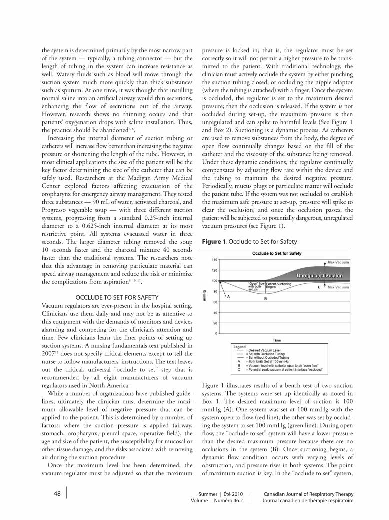
Canadian Journal of Respiratory TherapyJournal canadien de thérapie respiratoire
Summer | Été 2010Volume | Numéro 46.2
48
the system is determined primarily by the most narrow partof the system — typically, a tubing connector — but thelength of tubing in the system can increase resistance aswell. Watery fluids such as blood will move through thesuction system much more quickly than thick substancessuch as sputum. At one time, it was thought that instillingnormal saline into an artificial airway would thin secretions,enhancing the flow of secretions out of the airway.However, research shows no thinning occurs and thatpatients’ oxygenation drops with saline installation. Thus,the practice should be abandoned7, 8.
Increasing the internal diameter of suction tubing orcatheters will increase flow better than increasing the negativepressure or shortening the length of the tube. However, inmost clinical applications the size of the patient will be thekey factor determining the size of the catheter that can besafely used. Researchers at the Madigan Army MedicalCenter explored factors affecting evacuation of theoropharynx for emergency airway management. They testedthree substances — 90 mL of water, activated charcoal, andProgresso vegetable soup — with three different suctionsystems, progressing from a standard 0.25-inch internaldiameter to a 0.625-inch internal diameter at its mostrestrictive point. All systems evacuated water in three seconds. The larger diameter tubing removed the soup 10 seconds faster and the charcoal mixture 40 secondsfaster than the traditional systems. The researchers notethat this advantage in removing particulate material canspeed airway management and reduce the risk or minimizethe complications from aspiration9, 10, 11.
OCCLUDE TO SET FOR SAFETYVacuum regulators are ever-present in the hospital setting.Clinicians use them daily and may not be as attentive tothis equipment with the demands of monitors and devicesalarming and competing for the clinician’s attention andtime. Few clinicians learn the finer points of setting up suction systems. A nursing fundamentals text published in200712 does not specify critical elements except to tell thenurse to follow manufacturers’ instructions. The text leavesout the critical, universal “occlude to set” step that is recommended by all eight manufacturers of vacuum regulators used in North America.
While a number of organizations have published guide-lines, ultimately the clinician must determine the maxi-mum allowable level of negative pressure that can beapplied to the patient. This is determined by a number offactors: where the suction pressure is applied (airway, stomach, oropharynx, pleural space, operative field), theage and size of the patient, the susceptibility for mucosal orother tissue damage, and the risks associated with removingair during the suction procedure.
Once the maximum level has been determined, the vacuum regulator must be adjusted so that the maximum
pressure is locked in; that is, the regulator must be set correctly so it will not permit a higher pressure to be trans-mitted to the patient. With traditional technology, the clinician must actively occlude the system by either pinchingthe suction tubing closed, or occluding the nipple adaptor(where the tubing is attached) with a finger. Once the systemis occluded, the regulator is set to the maximum desiredpressure; then the occlusion is released. If the system is notoccluded during set-up, the maximum pressure is thenunregulated and can spike to harmful levels (See Figure 1and Box 2). Suctioning is a dynamic process. As cathetersare used to remove substances from the body, the degree ofopen flow continually changes based on the fill of thecatheter and the viscosity of the substance being removed.Under these dynamic conditions, the regulator continuallycompensates by adjusting flow rate within the device andthe tubing to maintain the desired negative pressure.Periodically, mucus plugs or particulate matter will occludethe patient tube. If the system was not occluded to establishthe maximum safe pressure at set-up, pressure will spike toclear the occlusion, and once the occlusion passes, thepatient will be subjected to potentially dangerous, unregulatedvacuum pressures (see Figure 1).
Figure 1. Occlude to Set for Safety
Figure 1 illustrates results of a bench test of two suction systems. The systems were set up identically as noted inBox 1. The desired maximum level of suction is 100mmHg (A). One system was set at 100 mmHg with thesystem open to flow (red line); the other was set by occlud-ing the system to set 100 mmHg (green line). During openflow, the “occlude to set” system will have a lower pressurethan the desired maximum pressure because there are noocclusions in the system (B). Once suctioning begins, adynamic flow condition occurs with varying levels ofobstruction, and pressure rises in both systems. The pointof maximum suction is key. In the “occlude to set” system,

Canadian Journal of Respiratory TherapyJournal canadien de thérapie respiratoire
Summer | Été 2010Volume | Numéro 46.2 49
REFERENCES1. Czarnik RE, Stone KS, Everhart CC, Preusser BA: Differential
effects of continuous versus intermittent suction on tracheal tissue. Heart & Lung 1991;20(2):144-151.
2. Duncan C, Erickson R: Pressures associated with chest tube stripping.Heart & Lung 1992;11(2):166-171.
3. Altimier L: Editorial [Evidence-based neonatal respiratory managementpolicy]. Newborn and Infant Nursing Reviews 2006;6(2):43-51.
4. Wilson R: Bacteria and airway inflammation in chronic obstructive pulmonary disease: more evidence. American Journal of Respiratory and Critical Care Medicine 2005;172(2):147-148.
5. Dowling RB, Johnson M, Cole PJ, Wilson R: Effect of fluticasone propionate and salmeterol on Pseudomonas aeruginosa infection of the respiratory mucosa in vitro. European Respiratory Journal1999;14:363-369.
6. Rutman A, Dowling R, Wills P, Feldman C, Cole PJ, Wilson R: Effect of dirithromycin on Haemophilus influenzae infection on the respiratory mucosa. Antimicrobial Agents and Chemotherapy 1998;42(4):772-778.
7. Ackerman MH, Ecklund MM, Abu-Jumah: A review of normal saline installation: implications for practice. Dimensions of Critical Care Nursing 1996;15(1):31-38.
8. Raymond SJ: Normal saline instillation before suctioning: helpful orharmful? A review of the literature. American Journal of Critical Care1995;4(4):267-271.
9. Vandenberg JT, Rudman NT, Burke TF, Ramos DE: Large-diametersuction tubing significantly improves evacuation time of simulated vomitus. American Journal of Emergency Medicine 1998;16(3):242-244.
10. Vandenberg JT, Lutz RH, Vinson DR: Large-diameter suction systemreduces oropharyngeal evacuation time. Journal of Emergency Medicine 1999;17(6):941-944.
11. Vandenberg JT, Vinson DR: The inadequacies of contemporary oropharyngeal suction. American Journal of Emergency Medicine1999;17(6):611-613.
12. Wilkinson JM, VanLeuven K: Fundamentals of nursing: thinking and doing vol. 2. 2007. FA Davis Company, Philadelphia.
13. Morrow BR, Futter MJ, Argent AC: Endotracheal suctioning: from principles to practice. Neonatal and Pediatric Intensive Care2004;30(6):1167-1174.
the pressure never rises above the desired maximum pressureof 100 mmHg. In the other system, pressure in this benchtest spiked to 125 mmHg of unregulated suction. Without“occlude to set,” the pressure can rise to 25% higher thanthe desired maximum level or more, exposing the patient toa safety hazard when regulated suction is needed.
Higher negative pressure is a particular hazard for patientswith friable mucosa in the airway or stomach, making it moresusceptible to traumatic tears. It is also a hazard for infantswho have small lung volumes. When all other variables arestable, a 25% increase in negative pressure will increase theamount of air pulled through the system by 25%. Thatincrease could result in a significant loss of lung volume inintubated neonates and infants13.
gency strikes and rapid evacuation is essential. An additionalunique concept introduced by Ohio Medical is the dual-spring design of the regulating module contained withinthe vacuum regulator. This feature provides the clinicianwith the ability to control vacuum levels more precisely inthe clinical range of 0-200 mmHg as well as the ability toachieve full vacuum when needed with only 2 turns of theknob on the regulator. In other regulators, six or more knobturns are needed to achieve “full vacuum,” and “full-vacuum”capability may be limited to the clinical range, not the fullsystem vacuum provided by the Ohio Medical ISU. Sincefull vacuum is needed in emergency conditions, thisenhanced responsiveness saves time when seconds are critical.
While vacuum regulators are often considered basicequipment in the hospital, research and innovation fromOhio Medical Corporation has shown vacuum regulatorsdo have a role in enhancing patient safety in clinical settings. Clinicians should advocate for technology thatprovides passive safety protection, enhanced control of vacuum pressures, rapid response, and ease of use — all ofwhich contribute to a culture of safety around the patient.
Box 1. Suction System Set-up.
Box 2. Case study.A nurse passing the bedside of an infant in the ICU saw bloodinside the tube used for airway suction. After checking the child’scondition, it was evident that bleeding was not expected.
Further investigation determined the maximum level of negative pressure set on the wall regulator was -200mmHg;far more than recommended suction levels for infants. Thenurse performing the suctioning did not occlude the tubing to set a safe maximum level of negative pressure.(personal communication to Ohio Medical Corporation)
Breakthrough Technologies Enhances SafetyAn ideal patient safety device removes clinician variables
as much as possible by providing the added safety passivelywhile the clinician carries out the procedure. Traditionally,the optimal safety of regulated vacuum pressure has dependedon the clinician’s action to occlude the system to set maxi-mum pressure. Now a breakthrough technology from OhioMedical Corporation in its new Intermittent Suction Unit(ISU), occludes the system automatically when the clinicianadjusts the pressure level. This creates a highly effective, passive safety system that removes the clinician variable andprotects the patient from unintended, unregulated pressurespikes during suction procedures. The “push to set” innovationassures the clinician that the patient will not be subjected topressure higher than that set on the regulator.
Another key safety aspect of any vacuum regulator is theability to quickly adjust to full vacuum mode when emer-
Suction System Set-Up
Vacuum regulator
12-inch connecting tube
1500cc (empty) collection bottle
6-foot standard connecting tubing
14 Fr. Suction catheter
áá
áá
á
Canadian Journal of Respiratory TherapyJournal canadien de thérapie respiratoire
Summer | Été 2010Volume | Numéro 46.2
50
ABSRTACTS FROM POSTER PRESENTATIONS
A Collaborative Inter-professional Approach toReducing Ventilator Associated Pneumonia (VAP)
in the Critically Ill Child
Geoff Flannagan,The Hospital for Sick Children, Toronto, ON
Winner of the Poster Presentations, CSRT Education Conferenceand Trade Show, St. John’s NL 2010
Coauthors: Leanne Davidson,The Hospital for Sick Children, Toronto, ON;
Kim Streitenberger, The Hospital for Sick Children,Toronto, ON
INTRODUCTIONVentilator associated pneumonia (VAP) is the second mostcommon nosocomial infection in the pediatric intensive careunit (PICU), accounting for 20% of such infections in thispatient population. VAP prolongs time spent on the ventila-tor, ICU length of stay (LOS), and hospital LOS following discharge from ICU .
The PICU and Cardiac Critical Care Unit (CCCU) at The Hospital for Sick Children have been making improve-ments to reduce our incidence of VAP. This poster willdescribe the successes, challenges & lessons learned during theimplementation of pediatric modifications to the evidencebased 'Safer Healthcare Now' VAP 'bundle'.
METHODSAn inter-professional team was assembled to research andimplement a pediatric VAP 'bundle' of practices including:• Elevation of the head of bed• Draining ventilator tubing away from the patient &
removing circuit condensation• Mouth care q2-4h
Education was provided to staff during the implementationperiod. Results were shared with all staff on an ongoing basisthrough bulletin boards and Quality & Safety committeeforums.
RESULTSThe main quality indicators used to track the effect of thebundle implementation on the VAP rate were: • VAP Rate• Compliance to VAP bundle components
We observed an overall increase in staff compliance to thebundle components while the VAP rate initially dropped thenremained stable.
CONCLUSIONAn overall increase in staff compliance to the VAP bundle resultedin an initial drop of VAP rate but a lack of further decrease ininfection rate warrants further evaluation of the bundle.
Mechanical Insufflation Exsufflation: Practice pat-terns among respiratory therapists in Ontario
Shelley Prevost,St. Joseph's Care Group & Thunder Bay Regional Health
Sciences Center
Co-authors: Dina Brooks, University of Toronto; MichelBedard, Lakehead University;
Philip Bwititi,Charles Sturt University
INTRODUCTIONThe mechanical insufflator exsufflator (MIE) is effective inassisting cough and in helping to avoid unplanned hospitaliza-tions, tracheostomy and long-term ventilation in patients withneuromuscular disease or spinal cord injury. In spite of this,the availability and usage of the device in Canada is not known.
OBJECTIVETo investigate practice patterns and availability of the MIE inOntario hospitals.
METHODSA cross-sectional, self-administered mail survey was sent to arandom sample of 400 respiratory therapists practicing within96 Ontario hospitals.
RESULTSA total of 114 (28%) completed surveys were returned from62 (65%) hospitals. Twenty hospitals (32%) had a MIE.Predominantly the respiratory therapist was the health careprovider using the MIE. The device was most commonly usedin the intensive care unit and medical/surgical units in patientswith neuromuscular diseases or spinal cord injuries. Optimalpressure spans of 35 cmH2O to 40 cmH2O were used by54% of respondents. Fourteen of the 20 hospitals with a MIEhad policies or guidelines in place and 4 of those hospitals hadestablished staff competencies. Measurements of peak coughflow, maximal inspiratory/expiratory pressure and vital capac-ity were reported to be infrequently performed.
Abstracts presented at the CSSRT Education Conference and Trade Show held in St. John’s NL, May 2010Geoff Flannagan, is the winner of the Poster Presentations.
Canadian Journal of Respiratory TherapyJournal canadien de thérapie respiratoire
Summer | Été 2010Volume | Numéro 46.2 51
CONCLUSIONSThis study demonstrated the MIE device is not widely avail-able in Ontario hospitals and there are variations in how thedevices are applied possibly resulting in suboptimal therapy. Acomprehensive educational program on MIE devices thatincorporates best practices and a practical component is rec-ommended for current providers as well as for inclusion in stu-dent curriculum.
Developing dynamic guidelines for Tobacco Control in Canada: Bridging research and
practice with CAN-ADAPTT
Katie Hunter,Centre for Addiction and Mental Health
Co-authors: Tamar Meyer, Organization:Centre for Addiction and Mental Health, Toronto ON;
Janet Ngo, Centre for Addiction and Mental Health, Toronto ON;
Peter Selby, Centre for Addiction and Mental Health,University of Toronto, Toronto ON
BACKGROUNDDespite the existence of clinical practice guidelines, only 50% of smokers report being asked about smoking by theirhealthcare provider. There are a number of reasons why practitioners have not yet embraced the guidelines: the guide-lines don’t reflect local circumstances, are quickly out of date,and reflect gaps between the perspectives of experts and theday-to-day experiences of practitioners.
The Canadian Action Network for the Advancement,Dissemination and Adoption of Practice-informed TobaccoTreatment (CAN-ADAPTT) is designed to meet these chal-lenges.
METHODSCAN-ADAPTT’s goal is to facilitate research and knowledgeexchange to inform the development of a dynamic set ofguidelines for use in both clinical practice and population-based cessation.
RESULTSThe establishment of a practice-based research network(PBRN) will ensure that end-users define and frame researchquestions that are informed by their practices and produceresearch results that are readily applicable. The use of anadapted wiki model (a collaborative website which can bedirectly edited by anyone with access to it) for updates willensure that the guidelines are up-to-date.
CONCLUSIONCollaboration through CAN-ADAPTT’s network will informthe development of a dynamic set of evidence-based guidelineswith the goal of helping to reduce tobacco initiation, use anddependence.
Perceived level of familiarity following a community NICU experience
Lori Peppler-Beechey,Conestoga College, Kitchener ON
Co-authors: Gino DePinto, Conestoga College, KitchenerON;
Gail Lang, Credit Valley Hospital;Celina Rogers, Credit Valley Hospital;Sophia Cabral, Credit Valley Hospital
OBJECTIVEThis project assessed the benefits and perceived level of famil-iarity for practice within the Neonatal Intensive Care Unit(NICU) following a 2 week clinical experience in a communi-ty based level 2 advanced NICU.
METHOD4 Respiratory Therapy students from one clinical site spent 2weeks with a dedicated NICU Respiratory Therapy preceptorin a community based NICU. The same students then spent 3weeks at a tertiary care level 3 NICU as part of their requiredclinical rotations. A questionnaire was administered to each ofthe 4 students once both NICU experiences were complete.The questionnaire used a 7 point Likert scale to assess the stu-dents perceived level of preparedness for the level 3 NICUexperience.
RESULTSOverall the students reported an above average level of famil-iarity for practice within the NICU environment after com-pleting the 2 week community based NICU. This level offamiliarity was seen in both documentation procedures andrespiratory care procedures with median scores between 5.5and 6.5 and averages between 5.5 and 6.25 on a 7 point Likertscale. The clinical site, Credit Valley Hospital, observed thatthe students had a greater level of comfort in the NICU envi-ronment, than in previous years when students did not havethis opportunity.
CONCLUSIONThe results suggest that in this small group of students the 2week community based NICU experience provided an aboveaverage level of familiarity for a level 3 NICU experience.Community based level 2 advanced NICU settings may alsooffer students additional opportunities for "hands on" experi-ence. While this is a very small sample of students these pre-liminary results indicate that there may be benefits to exposingstudents to a community based NICU prior to level 3 NICUexperience.

Canadian Journal of Respiratory TherapyJournal canadien de thérapie respiratoire
Summer | Été 2010Volume | Numéro 46.2
52
Mechanical ventilation protocols in intensive careunits: a survey of Ontario hospitals
Sandra Ellis, RRT MAppSc,Sunnybrook Health Sciences Centre
Co-authors: Katie Dainty, MSc, CRPC, Centre for HealthSciences Services, Toronto, ON;
Graham Munro, MHSM, BHSc CCP, MACAP, School ofBiomedical Sciences, Charles Sturt University, Bathurst, NSW,Australia; Damon C. Scales MD PhD, Department of Critical
Care Medicine, Sunnybrook Health Sciences Centre, Toronto,ON
INTRODUCTIONMechanical ventilation protocols for treating intensive careunit (ICU) patients may increase adoption of evidence-basedventilation strategies and thereby reduce duration of mechan-ical ventilation (MV) and rates of ventilator associated pneu-monia. We sought to ascertain ICU and hospital characteris-tics associated with adoption of MV protocols in Ontario, anddetermine factors associated with incorporation of lung pro-tective ventilation strategies (LPV) and spontaneous breathingtrials (SBTs) into these protocols.
METHODSWe conducted a postal survey of Respiratory Therapy (RT)
leaders in all 97 Ontario hospitals capable of providingmechanical ventilation.
RESULTSWe received responses from 70 (72.2%) hospitals. Only 28
hospitals (41.8%) reported having an intensivist-led staffingmodel. Most hospitals (48 [70.6%]) used MV protocols, butLPV was incorporated into only half of these (24; 54.6%),while SBTs were present in most (46; 80.5%). Factors associ-ated with reported use of MV protocols were intensivist-ledstaffing model (89.3% vs. 56.4%, odds ratio (OR) 6.44, [95%confidence interval (CI) 1.66 - 25.0, p = 0.007]), presence ofdaily multidisciplinary rounds (84.4% vs. 42.9%, OR 7.24[95% CI 2.22 - 23.6, p= 0.001]), and continuous RT cover-age (87.0% vs. 36.4%, OR 11.7 [95% CI 3.44 - 39.6, p<0.001]). Attending physician-to-patient ratio was associatedwith adoption of MV protocols; each increase of one patientto the ratio increased the likelihood of having a MV protocolby 1.17 (95% CI 1.01 - 1.35, p = 0.034).
CONCLUSIONThe majority of Ontario hospitals reported using MV proto-cols, but only half of these incorporated LPV. However, SBTswere commonly integrated into MV protocols. Increasingadoption of MV protocols incorporating best available evi-dence could be considered a target for future quality improve-ment initiatives.
Early Extubation of Paediatric Cardiac Patients Post Surgery
Leanne Davidson,Hospital for Sick Children
Co-authors: Steven Schwartz, Hospital for Sick Children;David Edgell, Hospital for Sick Children;
Geoff Flannagan, Hospital for Sick Children
INTRODUCTIONAn early extubation approach towards post operative paedi-atric cardiac surgery patients was developed at the Hospital forSick Children during 2009. The goal was to increase efficien-cies and decrease wait lists for paediatric cardiac surgery byfacilitating shorter lengths of hospital stay, decreasing ICUlengths of stay, and increasing overall patientsatisfaction.There was a fundamental shift in philosophy froman assumption of patient sickness postoperatively, to anassumption of wellness.
METHODSProtocols were developed to identify patients that would becandidates for early extubation, either in the operating room,or shortly after admission to the Cardiac Critical Care Unit.Five different routine cardiac surgeries were targeted as a start-ing point for the change implementation. Protocols were thendeveloped from many different disciplines: Changes weremade to the anaesthetic approach, the cardiac bypass manage-ment, anticoagulation, sedation, pain control and ventilationstrategies, parent education and discharge planning. Thesechanges were implemented over the course of 2009 and datawas collected on admission numbers, average census, intuba-tion time, readmission rates to the ICU, deferral rates, ORcancellation rates, infection rates and patient satisfaction. Thisdata was collected from February 2009 to January 2010 andwas compared to data from previous years.
RESULTSOverall, the average patient length of stay was decreased byone day for all patients in the Cardiac Critical Care Unit.Those patients with the identified surgical interventions hadan overall reduction in ICU length of stay by an average ofthree days, without an increase in readmission rate. Deferral sto our Cardiac Critical Care Unit dropped from a high of 18in October 2009 to an average of 2/month from June -December 2009.
CONCLUSIONSAn assumption of "wellness" for many post-operative condi-tions and an early extubation protocol can decrease length ofICU stay, decrease ventilator days, decrease cost and increasepatient satisfaction.

Canadian Journal of Respiratory TherapyJournal canadien de thérapie respiratoire
Summer | Été 2010Volume | Numéro 46.2 53
The Preceptorship Experience in RespiratoryTherapy Clinical Education: A Qualitative Analysis
Andrew West,Department of Respiratory Therapy, Faculty of Medicine,
University of Manitoba
Co-authors: Elenore Haywood, Department of RespiratoryTherapy, Faculty of Medicine, University of Manitoba;
Phucquoc Mai, Department of Respiratory Therapy, Facultyof Medicine, University of Manitoba
INTRODUCTIONUnderstanding the experiences of preceptors and students isnecessary to identify attributes, skills, and knowledge that arekey to successful preceptorship. Literature describing the pre-ceptorship experience exists for various health disciplines butthere is a paucity of information describing preceptorship inRespiratory Therapy (RT).
The Department of Respiratory Therapy, University ofManitoba, identified the need for novel research into precep-torship in order to support establishment of its clinical precep-tor training program. The purpose of this study was to identi-fy the unique skills, attributes, and knowledge relevant to pre-ceptorship in this context.
METHODSThis qualitative study employed an interpretive descriptiveframework. Data was collected through focus group interviewswith 22 Respiratory Therapist preceptors and 16 respiratorytherapy students. Interview questions were semi-structuredand open ended. Focus groups were digitally recorded andtranscribed verbatim. Content analysis of the transcripts wasperformed to determine themes, concepts, or ideas thatdemonstrated perceptions or attitudes.
RESULTSSix themes emerged from the data that speak to the preceptor-ship experience in Respiratory Therapy: (1) limitations in for-mal preparation for the preceptor role, (2) uncertainty inassigning value to clinical experience and teaching skill, (3)communication amongst the participants, (4) existing barriersto effective evaluation and feedback, (5) desires relating to thestudent-preceptor relationship, and (6) a vignette of the pre-ferred preceptor's characteristics. Within each of these themesthe unique perspectives of preceptors and students are com-paratively illustrated.
CONCLUSIONThis investigation serves as a first step to understanding thepreceptorship experience in Respiratory Therapy, as well as thelearning needs of Respiratory Therapists who provide clinicalpreceptorship for RT students. The themes identified in thisstudy underscore the complex nature of the preceptorshipexperience in RT. They illuminate the need for increased edu-cational supports for preceptors, and offer a better under-standing of the preceptorship experience for educational pro-grammers.
Canadian Journal of Respiratory TherapyJournal canadien de thérapie respiratoire
Summer | Été 2010Volume | Numéro 46.2
54
RÉSUMÉS DES PRÉSENTATIONS D’AFFICHES
Une approche de collaboration interprofessionnelle dela réduction de la pneumonie sous ventilation assistée
(PVA) chez les enfants gravement malades
Geoff Flannagan,The Hospital for Sick Children, Toronto (Ont.)
Geoff Flannagan a remporté le prix de la présentation desaffiches au Congrès éducatif et salon professionnel
Coauteurs : Leanne Davidson, The Hospital for SickChildren, Toronto (Ont.); Kim Streitenberger,The Hospital for Sick Children, Toronto (Ont.)
INTRODUCTIONLa pneumonie sous ventilation assistée (PVA) est la deuxièmeinfection nosocomiale la plus commune dans l’unité de soinsintensifs pédiatrique (USIP), représentant 20 % de ces infectionsdans cette population. PVA prolonge la durée du passage sous ventilation assistée, la durée du séjour en USI et la durée del’hospitalisation après le congé de l’USI.
L’USIP et l’Unité de soins intensifs en cardiologie (USIC) del’Hospital for Sick Children ont apporté des améliorations afin deréduire l’incidence de PVA. Cette présentation décrira les succès,les défis et les leçons apprises durant la mise en ?uvre des modifi-cations pédiatriques à la suite de mesures PVA « Safer HealthcareNow » fondée sur les données probantes.
MÉTHODESUne équipe interprofessionnelle a été constituée afin d’effectuerdes recherches et de mettre en place une « suite » PVA comprenant : . l’élévation de la tête de lit;. le drainage de la canalisation de ventilation loin du patient et
l’élimination de la condensation dans les canalisations; . soins de la bouche q2-4h.Une formation a été donnée au personnel durant la période d’implantation. Les résultats ont été communiqués à l’ensembledu personnel sur une base continue par les tableaux d’affichage etles rencontres trimestrielles sur la qualité et la sécurité.
RÉSULTATSLes principaux indicateurs de qualité utilisés pour faire le suivi deseffets de la mise en ?uvre de la suite de mesures du taux de PVAétaient :. le taux de PVA;. le respect des éléments de la suite PVA.Nous avons observé une augmentation générale de la conformitédu personnel aux différents éléments de la suite alors que le tauxde PVA a d’abord diminué pour ensuite se stabiliser.
CONCLUSIONUne augmentation générale de la conformité du personnel auxdifférents éléments de la suite PVA s’est traduite par une baisseinitiale du taux de PVA mais le fait que la baisse ne se soit paspoursuivie justifie une évaluation plus approfondie de la suite.
Insufflation et exsufflation mécaniques : modèles de pratique chez les thérapeutes respiratoires en Ontario
Shelley Prevost,St. Joseph's Care Group et Thunder Bay Regional Health
Sciences Center
Coauteurs : Dina Brooks, University of Toronto;Michel Bedard, Lakehead University;Philip Bwititi, Charles Sturt University
INTRODUCTIONL’appareil d’insufflation et d’exsufflation mécanique (IEM) estefficace pour aider à la toux et éviter les hospitalisationsimprévues, les trachéotomies et la ventilation de longue duréechez les patients atteints de maladies neuromusculaires ou deblessures à la colonne vertébrale. Malgré cela, la disponibilité etl’usage de l’appareil au Canada sont inconnues.
OBJECTIF Étudier les modèles de pratique et la disponibilité des appareilsd’IEM dans les hôpitaux de l’Ontario.
MÉTHODESUn sondage transversal autoadministré a été envoyé par la poste àun échantillon aléatoire de 400 thérapeutes respiratoires dans 96hôpitaux de l’Ontario.
RÉSULTATS Au total, 114 questionnaires (28 %) remplis ont été retournés par62 hôpitaux (65%). Vingt hôpitaux (32 %) disposent d’unappareil d’IEM. Dans la majorité des cas, le thérapeute respira-toire est le professionnel de la santé qui utilise l’appareil, qui estprincipalement utilisé dans les unités de soins intensifs et lesunités médicales/chirurgicales chez les patients atteints de mal-adies neuromusculaires ou de blessures à la colonne vertébrale.Des pressions optimales de 35 cmH2O à 40 cmH2O sont util-isées par 54 % des répondants. Quatorze des 20 hôpitaux qui dis-posent d’un appareil d’IEM ont une politique ou des directives enplace pour son utilisation et quatre de ces hôpitaux ont établi lescompétences du personnel. Selon les données recueillies, lesmesures du débit de pointe de la toux, de la pression maximale àl’inspiration/expiration et de la capacité vitale sont prises de façoninfréquente.
Résumés des présentations d’affiches au Congrès éducatif et salon professionnel de St.John’s (T.-N.-L.), mai 2010.Geoff Flannagan a remporté le prix de la présentation des affiches.
Canadian Journal of Respiratory TherapyJournal canadien de thérapie respiratoire
Summer | Été 2010Volume | Numéro 46.2 55
CONCLUSIONS Cette étude démontre que l’appareil d’IEM n’est pas largementdisponible dans les hôpitaux de l’Ontario et qu’il y a des écartsdans les modalités d’utilisation de l’appareil, entraînant possible-ment une thérapie sous-optimale. La mise en place d’un pro-gramme complet d’éducation sur les appareils d’IEM, incluant lespratiques exemplaires et un volet pratique, est recommandée pourles fournisseurs actuels et pour inclusion dans le programme deformation des étudiants.
Établissement de lignes directrices dynamiques pour le contrôle du tabac au Canada : jeter des ponts entre la
recherche et la pratique avec CAN-ADAPTT
Katie Hunter,Centre for Addiction and Mental Health
Coauteurs : Tamar Meyer, Organization: Centre forAddiction and Mental Health, Toronto (Ont.);
Janet Ngo, Centre for Addiction and Mental Health,Toronto (Ont.); Peter Selby, Centre for Addiction and Mental
Health, University of Toronto, Toronto (Ont.)
CONTEXTEMalgré l’existence de lignes directrices sur la pratique clinique, 50 % des fumeurs seulement indiquent que leur fournisseur desoins de santé leur pose des questions sur le tabac. Plusieursraisons expliquent que les praticiens n’appliquent pas encore leslignes directrices : elles ne reflètent pas les conditions locales, sontrapidement périmées et reflètent les écarts entre le point de vuedes experts et l’expérience quotidienne des praticiens.
CAN_ADAPTT (Réseau d’action canadien pour l’avance-ment, la diffusion et l’adoption de pratiques éclairées dans letraitement du tabagisme) a été conçu pour relever ces défis.
MÉTHODESCAN-ADAPTT vise à faciliter la recherche et le partage des con-naissances afin de documenter à l’échelle nationale l’élaborationde lignes directrices dynamiques pour la pratique dans le domaineclinique et l’abandon du tabac dans la population.
RÉSULTATSLa création d’un réseau de recherche basé sur la pratique permettraaux utilisateurs de définir et de cadrer des questions de rechercheéclairées par l’expérience clinique et produira des résultats derecherche directement applicables. L’utilisation d’un modèle wikiadapté (un site Web collaboratif pouvant être modifié directementpar toutes les personnes qui y ont accès) pour les mises à jour permettra de s’assurer que les lignes directrices sont tenues à jour.
CONCLUSIONLa collaboration au sein du réseau CAN-ADAPTT permettra d’éclairer l’établissement d’un ensemble dynamique de lignesdirectrices fondées sur des faits, dans le but de diminuer l’initiationau tabac, son usage et la dépendance
Niveau de familiarité perçu après une expérience dans une USIN communautaire
Lori Peppler-Beechey,Conestoga College, Kitchener (Ont.)
Coauteurs : Gino DePinto, Conestoga College,Kitchener (Ont.);
Gail Lang, Credit Valley Hospital;Celina Rogers, Credit Valley Hospital;Sophia Cabral, Credit Valley Hospital
OBJECTIFCe projet évalue les avantages et le degré de familiarité perçu pourla pratique dans l’Unité de soins intensifs en néonatalité (USIN)après une expérience clinique de deux semaines dans une USINcommunautaire avancée de niveau 2.
MÉTHODEQuatre étudiants en thérapie respiratoire d’un site clinique ontpassé deux semaines avec un précepteur spécialisé en thérapie respiratoire néonatale dans une USIN communautaire. Lesmêmes étudiants ont ensuite passé trois semaines dans une USINde soins tertiaires de niveau 3 dans le cadre de leur rotation clinique imposée. Un questionnaire a été remis à chacun des quatre étudiants après qu’ils aient terminé leurs deux stages enUSIN. Le questionnaire applique une échelle Likert en sept pointspour évaluer le degré de préparation perçu des étudiants en vue del’expérience en USIN niveau 3.
RÉSULTATS Globalement, les étudiants signalent une degré de familiaritésupérieur à la normale envers la pratique dans un environnementUSIN après avoir terminé le stage de deux semaines en USINcommunautaire. Ce degré de familiarité a été perçu dans lesprocédures de documentation et dans les procédures de soins res-piratoires, avec des notes médianes de 5.5 à 6.5 et des moyennesentre 5.5 et 6.25 sur l’échelle Likert qui en compte sept. Le siteclinique, Credit Valley Hospital, note que les étudiants présen-taient un plus grand niveau de confort face à l’environnementUSIN que les stagiaires des années précédentes, qui n’avaient paseu cette possibilité.
CONCLUSIONLes résultats semblent indiquer que dans ce groupe restreint destagiaires, les deux semaines d’expérience en USIN communautaireont permis d’acquérir un degré de familiarité supérieur en prévisiond’un stage en USIN Niveau 3. Le milieu d’USIN communautaireavancée de niveau 2 peut aussi offrir aux étudiants des occasionsadditionnelles d’expérience pratique. Bien que l’échantillon soittrès restreint, ces résultats préliminaires indiquent qu’il pourraitêtre avantageux d’exposer les étudiants à un environnementd’USIN communautaire avant le stage en USIN Niveau 3.

Canadian Journal of Respiratory TherapyJournal canadien de thérapie respiratoire
Summer | Été 2010Volume | Numéro 46.2
56
Niveau de familiarité perçu après une expérience dans une USIN communautaire
Lori Peppler-Beechey,Conestoga College, Kitchener (Ont.)
Coauteurs : Gino DePinto, Conestoga College, Kitchener(Ont.);
Gail Lang, Credit Valley Hospital;Celina Rogers, Credit Valley Hospital;Sophia Cabral, Credit Valley Hospital
OBJECTIFCe projet évalue les avantages et le degré de familiarité perçu pourla pratique dans l’Unité de soins intensifs en néonatalité (USIN)après une expérience clinique de deux semaines dans une USINcommunautaire avancée de niveau 2.
MÉTHODEQuatre étudiants en thérapie respiratoire d’un site clinique ontpassé deux semaines avec un précepteur spécialisé en thérapie res-piratoire néonatale dans une USIN communautaire. Les mêmesétudiants ont ensuite passé trois semaines dans une USIN de soinstertiaires de niveau 3 dans le cadre de leur rotation cliniqueimposée. Un questionnaire a été remis à chacun des quatre étudi-ants après qu’ils aient terminé leurs deux stages en USIN. Le ques-tionnaire applique une échelle Likert en sept points pour évaluerle degré de préparation perçu des étudiants en vue de l’expérienceen USIN niveau 3.
RÉSULTATS Globalement, les étudiants signalent une degré de familiaritésupérieur à la normale envers la pratique dans un environnementUSIN après avoir terminé le stage de deux semaines en USINcommunautaire. Ce degré de familiarité a été perçu dans lesprocédures de documentation et dans les procédures de soins res-piratoires, avec des notes médianes de 5.5 à 6.5 et des moyennesentre 5.5 et 6.25 sur l’échelle Likert qui en compte sept. Le siteclinique, Credit Valley Hospital, note que les étudiants présen-taient un plus grand niveau de confort face à l’environnementUSIN que les stagiaires des années précédentes, qui n’avaient paseu cette possibilité.
CONCLUSIONLes résultats semblent indiquer que dans ce groupe restreint destagiaires, les deux semaines d’expérience en USIN communau-taire ont permis d’acquérir un degré de familiarité supérieur enprévision d’un stage en USIN Niveau 3. Le milieu d’USIN com-munautaire avancée de niveau 2 peut aussi offrir aux étudiants desoccasions additionnelles d’expérience pratique. Bien que l’échan-tillon soit très restreint, ces résultats préliminaires indiquent qu’ilpourrait être avantageux d’exposer les étudiants à un environ-nement d’USIN communautaire avant le stage en USIN Niveau 3.
Protocoles de ventilation mécanique dans les unités desoins intensifs : enquête dans les hôpitaux de l’Ontario
Sandra Ellis, RRT MAppSc,Sunnybrook Health Sciences Centre
Coauteurs : Katie Dainty, MSc, CRPC, Centre for HealthSciences Services, Toronto, ON;
Graham Munro, MHSM, BHSc CCP, MACAP, School ofBiomedical Sciences, Charles Sturt University,
Bathurst, NSW, Australie; Damon C. Scales MD PhD,Department of Critical Care Medicine, Sunnybrook
Health Sciences Centre, Toronto, ON
INTRODUCTIONLes protocoles de ventilation mécanique pour le traitement despatients en unité de soins intensifs (USI) peuvent augmenterl’adoption de stratégies de ventilation fondées sur des élémentsprobants et ainsi diminuer la durée de la ventilation mécanique(VM) et donc diminuer l’incidence de pneumonie associée à laventilation. Nous souhaitons établir les caractéristiques des USI etdes hôpitaux associées à l’adoption des protocoles de VM enOntario et recenser les facteurs associés à l’incorporation desstratégies de ventilation protégeant les poumons (VPM) et desessais de respiration spontanée (ERS) dans ces protocoles.
MÉTHODESNous avons mené une enquête postale auprès des responsables dela thérapie respiratoire (TR) dans les 97 hôpitaux de l’Ontariooffrant des services de ventilation mécanique.
RÉSULTATSNous avons reçu des réponses de 70 hôpitaux (72,2 %).Seulement 28 hôpitaux (41,8 %) disent appliquer un modèledirigé par un intensiviste. La plupart des hôpitaux (48 [70,6 %])appliquent des protocoles de VM, mais la VPM n’est incorporéeque dans la moitié de ces protocoles (24; 54,6 %), alors que lesERS étaient présents dans la plupart des protocoles (46; 80,5 %).Les facteurs associés à l’utilisation recensée des protocoles de VMétaient les modèles de dotation dirigés par un intensiviste (89,3 %c, 56,4 %, rapport de cote (RC) 6,44, [95 % intervalle de confiance (IC) 1,66 - 25,0, p = 0,007]), la présence de visites multidisciplinaires quotidiennes (84,4 % c. 42,9 %, RC 7,24 [95 % IC 2,22 - 23,6, p= 0,001]), et la couverture continue en TR(87,0 % c. 36,4 %, RC 11,7 [95 % IC 3,44 - 39,6, p< 0,001]).Le ratio de patients par médecin traitant a été associé à l’adoptionprotocoles de VM; chaque augmentation d’un patient du ratioaugmentait la probabilité d’avoir un protocole de VM par 1,17(95 % IC 1,01 - 1,35, p = 0,034).
CONCLUSION La majorité des hôpitaux de l’Ontario indiquent recourir aux protocoles de VM, mais la moitié d’entre eux seulement y incorporent la VPM; les ERS sont cependant régulièrement intégrés aux protocoles de VM. L’adoption accrue de protocolesde VM incorporant les meilleures données probantes pourrait être envisagée comme un objectif pour les initiatives futuresd’amélioration de la qualité.
Canadian Journal of Respiratory TherapyJournal canadien de thérapie respiratoire
Summer | Été 2010Volume | Numéro 46.2 57
Extubation hâtive des patients en pédiatriecardiaque après la chirurgie
Leanne Davidson,Hospital for Sick Children
Coauteurs : Steven Schwartz, Hospital for Sick Children;David Edgell, Hospital for Sick Children;
Geoff Flannagan, Hospital for Sick Children
INTRODUCTIONUne approche d’extubation postopératoire hâtive chez les patientsde chirurgie cardiaque pédiatrique a été mise au point à l’Hospitalfor Sick Children en 2009. Le but visé était d’augmenter l’efficac-ité et de diminuer le temps d’attente pour la chirurgie cardiaquepédiatrique en favorisant des séjours plus courts, en diminuant leséjour à l’unité de soins intensifs et en augmentant la satisfactiongénérale des patients. Il s’agissait d’un changement de philosophiefondamental, passant d’une hypothèse de maladie du patient àune hypothèse de bonne santé postopératoire.
MÉTHODESDes protocoles ont été établis afin de recenser les patients candidatsà une extubation hâtive, que ce soit dans la salle d’opération ou peu de temps après l’admission à l’Unité de soins intensifs encardiologie. Cinq chirurgies cardiaques de routine ont été cibléescomme point de départ du changement. Des protocoles ontensuite été élaborés à partir de plusieurs disciplines différentes :des changements ont été apportés à l’approche d’anesthésie, à lagestion de la dérivation cardiaque, aux stratégies anticoagulatoire,de sédation, de contrôle de la douleur et de ventilation, à l’éduca-tion des patients et à la planification du congé. Ces changementsont été implantés progressivement tout au long de 2009 et desdonnées ont été recueillies sur le nombre d’admissions, le recense-ment moyen, la durée d’intubation, le taux de réadmission à l’USI, les taux de report, les taux d’annulation de salle d’opéra-tion, les taux d’infection et la satisfaction des patients. Ces données ont été recueillies entre février 2009 et janvier 2010 etont été comparées aux données des années antérieures.
RÉSULTATSGlobalement, la durée moyenne du séjour en USI a été réduited’une journée pour tous les patients de l’Unité de soins intensifsen cardiologie. La réduction du séjour en ISU pour les patientsdes interventions chirurgicales visées a été de trois jours enmoyenne, sans augmentation du taux de réadmission. Les reportsde notre Unité de soins intensifs en cardiologie sont passés d’unsommet de 18 en octobre 2009 à une moyenne de deux par moisentre juin et décembre 2009.
CONCLUSIONSL’adoption d’une hypothèse de « santé » pour plusieurs conditionspostopératoires et un protocole d’extubation hâtive peuventdiminuer la durée du séjour en USI, le nombre de jours sous ventilation et les coûts tout en augmentant le degré de satisfactiondes patients.
L’expérience du préceptorat dans la formation cliniqueen thérapie respiratoire : une analyse qualitative
Andrew West,Département de thérapie respiratoire, Faculté de médecine,
Université du Manitoba
Coauteurs : Elenore Haywood, Département de thérapierespiratoire, Faculté de médecine, Université du Manitoba;
Phucquoc Mai, Département de thérapie respiratoire, Facultéde médecine, Université du Manitoba
INTRODUCTIONIl est nécessaire de comprendre l’expérience du précepteur et desétudiants pour recenser les attributs, les compétences et les compétences requises pour assurer le succès du préceptorat. Lestextes scientifiques décrivant l’expérience de préceptorat existentpour différentes disciplines des sciences de la santé mais il y a peud’information sur le préceptorat en thérapie respiratoire.
Le Département de thérapie respiratoire de l’Université duManitoba a déterminé qu’il y avait un besoin pour des recherchesinnovantes sur le préceptorat afin de soutenir la création de sonprogramme de formation de précepteurs cliniques. L’étude visait àdéfinir les compétences, les attributs et les connaissances uniquespertinentes pour les précepteurs dans ce contexte.
MÉTHODESCette étude qualitative fait appel à un cadre descriptif interpré-tatif. Des données ont été recueillies dans le cadre de groupes dediscussion avec 22 précepteurs et 16 étudiants en thérapie respira-toire. Les questions étaient semi-structurées et ouvertes. Lesgroupes de discussion ont fait l’objet d’un enregistrementnumérique et d’une transcription. Le contenu des transcriptions aété analysé afin de recenser les thèmes, les concepts ou les idéesmontrant des perceptions ou des attitudes.
RÉSULTATSLes données ont permis de déterminer six thèmes relatifs à l’ex-périence de préceptorat en thérapie respiratoire : (1) limitationsdans la préparation formelle au rôle de précepteur, (2) incertitudedans l’attribution de valeur à l’expérience clinique et aux compé-tences d’enseignement; (3) communications entre les participants,(4) obstacles à l’évaluation et au feedback efficaces; (5) souhaitsrelatifs à la relation entre le précepteur et l’étudiant; (6) unevignette des caractéristiques souhaitées du précepteur. Sous chacun de ces thèmes, les perspectives propres aux précepteurs etaux étudiants sont illustrées de façon comparative.
CONCLUSIONCette enquête est une première étape vers la compréhension del’expérience de préceptorat en thérapie respiratoire et des besoinsd’apprentissage des thérapeutes respiratoires qui agissent commeprécepteurs cliniques pour les étudiants. Les thèmes recensés dansl’étude soulignent la nature complexe de l’expérience de précep-torat en thérapie respiratoire. Ils illustrent le besoin de soutienséducatifs accrus pour les précepteurs et offrent une meilleure compréhension de l’expérience de préceptorat pour les program-mateurs en formation.