Embed Size (px)
Citation preview

Revista Facultad de Odontología Universidad de Antioquia - Vol. 23 N.º 1 - Segundo semestre, 2011 37
CAMBIOS ESQUELÉTICOS, FACIALES Y DENTOALVEOLARES PRODUCIDOS POR EL TRATAMIENTO CON PLACAS DE ACETATO Y ELÁSTICOS DE CLASE III
EN PACIENTES CON MESIORRELACIÓN ESQUELÉTICA Y DENTAL EN DENTICIÓN DECIDUA O MIXTA TEMPRANA
SKELETAL, FACIAL AND DENTOALVEOLAR CHANGES PRODUCED BY TREATMENT WITH ACETATE PLATES AND CLASS III ELASTICS IN PATIENTS
WITH SKELETAL AND DENTAL MESIORELATION IN EARLY DECIDOUS OR MIXED TEETHING
GABRIEL EMILIO ESPINAL BOTERO1, JOHN JAIRO BETANCUR PÉREZ2, ANA MARÍA BLANDÓN URIBE3, PAOLA ALEJANDRA ORTIZ BURGOS4
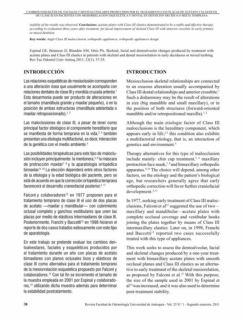
RESUMEN. Introducción: el propósito fue establecer los cambios clínicos y radiográficos con el uso durante un año de placas de acetato bimaxilares con elásticos de clase III para el tratamiento de la clase III esquelética en pacientes con dentición decidua o mixta temprana. Métodos: se hizo un estudio clínico prospectivo cuasi-experimental en 26 sujetos entre tres y nueve años. Se tomaron radiografías cefálicas laterales y modelos de estudio al inicio, a los seis meses y al año de tratamiento, se evaluaron cambios postratamiento después de tres años en cinco sujetos. En el análisis estadístico se obtuvieron medidas descriptivas y se utilizó la prueba Anova de medidas repetidas para determinar los cambios en el tiempo y se utilizó la prueba no paramétrica de Wilcoxon, con nivel de significancia de 0,005. Resultados: se obtuvieron cambios esqueléticos favorables a nivel mandibular en los ángulos SNB, facial y goníaco, la distancia pog. perpendicular a N, altura facial anterior inferior, y eje Y; dentoalveolarmente una mejoría en la sobremordida horizontal y el Witts, mejoría en la estética facial evidenciada por la disminución del ángulo FH plano E y de la protrusión del labio inferior. Después de tres años de finalizado el tratamiento se encontró estabilidad en los resultados. Conclusiones: las placas de acetato con elásticos de clase III fueron una terapia efectiva y estable a tres años de evaluación postratamiento para el mejoramiento facial, de la clase III esquelética y la mordida cruzada anterior.
Palabras clave: maloclusión de angle clase III, aparatos ortopédicos, diseño de aparatos ortopédicos.
Espinal GE, Betancur JJ, Blandón AM, Ortiz PA. Cambios esqueléticos, faciales y dentoalveolares producidos por el tratamiento con placas de acetato y elásticos de clase III en pacientes con mesiorrelación esquelética y dental en dentición primaria o mixta temprana. Rev Fac Odontol Univ Antioq 2011; 23(1): 37-55.
ABSTRACT. Introduction: the purpose of this study was to establish the clinical and radiographic changes produced by using bimaxillary acetate plates with Class III elastics during one year for treatment of skeletal Class III in patients with early deciduous or mixed dentition. Methods: a quasi-experimental prospective clinical study was carried out in 26 subjects, ages three to nine years. Lateral cephalometric radiographs and study models were taken at the beginning, six months, and one year of treatment; post-treatment changes were evaluated after three years in five subjects. During the statistical analysis descriptive measures were obtained, and the Anova test for repeated measures was used to determine changes over time; also, the nonparametric Wilcoxon test was used with a significance level of 0.005. Results: favorable skeletal changes were achieved in the mandible, especially at the SNB, facial and gonial angles, the pog distance perpendicular to N, the lower anterior facial height, and the Y axis; dentoalveolarly, there were improvements in both horizontal overbite and Witts, as well as in facial aesthetics, as evidenced by the decrease of the E-plane FH angle and lower lip protrusion. After three years of treatment,
1 Odontólogo, especialista en Odontopediatría, profesor asociado Universidad de Antioquia. Correo electrónico: [email protected]
2 Odontólogo, especialista en Odontología Integral del Adolescente, especialista en Administración de Servicios en Salud: Mercadeo, especialista en Didáctica Universitaria, profesor asociado Facultad de Odontología Universidad de Antioquia, Medellín, Colombia. Correo electrónico: [email protected]
3 Odontólogo, especialista en Odontología Integral del Niño y Ortopedia Maxilar, estudiante posgrado: Odontología Integral del Adolescente y Ortodoncia.
4 Odontólogo, estudiante del posgrado Odontología Integral del Niño y Ortopedia Maxilar.
RECIBIDO: OCTUBRE 6/2010- ACEPTADO: MAYO 3/2011 SUBMITTED: OCTOBER 6, 2009-ACCEPTED: MAY 3, 2011
1 Dentist. Specialist in Odontopediatrics, associated professor at Universidad de Antioquia. E-mail: [email protected]
2 Dentist. Specialist in Teenager Integral Dentistry. Specialist in Health Services Management: Marketing. Specialist in University Didactics. Associated professor of the School of Dentistry at Universidad de Antioquia, Medellín Colombia. E-mail: [email protected]
3 Dentist. Specialist in Children Integral Dentistry and Maxillary Orthopedics, Graduate student: Teenager Integral Dentistry and Orthodontics.
4 Dentist. Graduate student to the Program Integral Dentistry in Children and Maxillary Orthopedics.
Revista Facultad de Odontología Universidad de Antioquia - Vol. 23 N.º 1 - Segundo semestre, 201138
INTRODUCCIÓN
Las relaciones esqueléticas de mesioclusión corresponden a una alteración ósea que usualmente se acompaña con relaciones dentales de clase III y mordida cruzada anterior.1 Esta desarmonía puede ser producto de alteraciones en el tamaño (mandíbula grande y maxilar pequeño), o en la posición de ambas estructuras (mandíbula adelantada o maxilar retroposicionado).1, 2
Las maloclusiones de clase III, a pesar de tener como principal factor etiológico el componente hereditario que se manifiesta de forma temprana en la vida,2, 3 también presentan una etiología multifactorial, es decir, interacción de la genética con el medio ambiente.4
Las posibilidades terapéuticas para este tipo de maloclu-sión incluyen principalmente: la mentonera,5, 6 la máscara de protracción maxilar7, 8 y la aparatología ortopédica bimaxilar.9, 10 La elección dependerá entre otros factores de la etiología y la edad biológica del paciente, pero se está de acuerdo en que la corrección ortopédica temprana favorecerá el desarrollo craneofacial posterior.2, 11
Falconi y colaboradores12 en 1977 proponen para el tratamiento temprano de clase III el uso de dos placas de acetato —maxilar y mandibular— con cubrimiento oclusal completo y ganchos vestibulares que unen las placas por medio de elásticos intermaxilares de clase III. Posteriormente, Franchi y Baccetti13 en 1998 hicieron el reporte de dos casos tratados exitosamente con este tipo de aparatología.
En este trabajo se pretende evaluar los cambios den-toalveolares, faciales y esqueléticos producidos por el tratamiento durante un año con placas de acetato bimaxilares con planos oclusales lisos y elásticos de clase III como alternativa para el tratamiento temprano de la mesiorrelación esquelética propuesto por Falconi y colaboradores.12 Con tal fin se incrementó el tamaño de la muestra empleada en 2001 por Espinal y colaborado-res,14 utilizando dicha muestra además para determinar la estabilidad postratamiento.
stability of the results was observed. Conclusions: acetate plates with Class III elastics demonstrated to be a stable and effective therapy, according to evaluation three years after treatment, for facial improvement of skeletal Class III with anterior crossbite in early primary or mixed dentition.
Key words: Angle Class III malocclusion, orthopedic appliances, orthopedic appliances design.
Espinal GE, Betancur JJ, Blandón AM, Ortiz PA. Skeletal, facial and dentoalveolar changes produced by treatment with acetate plates and Class III elastics in patients with skeletal and dental mesiorelation in early deciduous or mixed teething. Rev Fac Odontol Univ Antioq 2011; 23(1): 37-55.
INTRODUCTION
Mesiocclusion skeletal relationships are connected to an osseous alteration usually accompanied by Class III dental relationships and anterior crossbite.1 Such a disharmony may be the result of alterations in size (big mandible and small maxillary), or in the position of both structures (forward-oriented mandible and/or retropositioned maxilla).1, 2
Although the main etiologic factor of Class III malocclusions is the hereditary component, which appears early in life,2,3 this condition also exhibits a multifactorial etiology, that is, an interaction of genetics and environment.4
Therapy alternatives for this type of malocclusion include mainly: chin cup treatment,5, 6 maxillary protraction face mask,7, 8 and bimaxillary orthopedic apparatus.9,10 The choice will depend, among other factors, on the etiology and the patient’s biological age, but researchers generally agree that early orthopedic correction will favor further craniofacial development.2,11
In 1977, seeking early treatment of Class III maloc-clusions, Falconi et al12 suggested the use of two—maxillary and mandibular—acetate plates with complete occlusal coverage and vestibular hooks joining the plates together by means of Class IIIintermaxillary elastics. Later on, in 1998, Franchi and Baccetti13 reported two cases successfully treated with this type of appliances.
This work seeks to assess the dentoalveolar, facial and skeletal changes produced by a one-year treat-ment with bimaxillary acetate plates with smooth occlusal planes and Class III elastics as an alterna-tive to early treatment of the skeletal mesiorelation, as proposed by Falconi et al.12 With this purpose, the size of the sample used in 2001 by Espinal et al14 was increased, and it was also used to determine post-treatment stability.
CAMBIOS ESQUELÉTICOS, FACIALES Y DENTOALVEOLARES PRODUCIDOS POR EL TRATAMIENTO CON PLACAS DE ACETATO Y ELÁSTICOS DE CLASE III EN PACIENTES CON MESIORRELACIÓN ESQUELÉTICA Y DENTAL EN DENTICIÓN DECIDUA O MIXTA TEMPRANA

Revista Facultad de Odontología Universidad de Antioquia - Vol. 23 N.º 1 - Segundo semestre, 2011 39
MATERIALES Y MÉTODOS
Se llevó a cabo un estudio de tipo clínico prospectivo cuasi experimental, en el que se hizo una intervención con placas de acetato bimaxilares que sirvieron como anclaje para el uso de elásticos intermaxilares con un vector de clase III en una muestra de 26 sujetos de ambos sexos, entre los tres y nueve años de edad, seleccionados por conveniencia del Servicio de odontología de la Facultad de Odontología de la Universidad de Antioquia bajo los siguientes criterios de inclusión: edad entre tres y nueve años con mesiorrelación esquelética cefalométricamente confirmada, mordida cruzada anterior o borde a borde, con o sin mordida cruzada posterior (uni- o bilateral). Se consideraron como criterios de exclusión: tratamiento ortopédico maxilar previo, oclusión seudoclase III, mor-dida abierta esquelética o dental, pérdida prematura de dientes o extracciones tempranas, signos y síntomas de disfunción de la articulación temporomandibular, ante-cedentes de trauma maxilofacial, anomalías congénitas y síndromes relacionados con el desarrollo de los maxi-lares. Veintiuno fueron pacientes nuevos y cinco fueron retomados del estudio hecho en 2001 por Espinal y co-laboradores,14 para evaluar la estabilidad postratamiento.
Después de hacer una historia clínica completa y definido el diagnóstico, se firmó un consentimiento informado, se tomaron impresiones superiores e inferiores utilizando cubetas perforadas tipo Coe American Style®, se preparó material de impresión Jeltrate Kerr® según las recomen-daciones de la casa comercial (7 g de polvo en 19 ml de agua) y se obtuvieron modelos en yeso piedra (100 mg de polvo en 28 ml de agua) sobre los cuales se confec-cionaron las placas removibles en acetato de 0,8 mm de espesor de la casa comercial Dentsplay® (los modelos a los 6 y 12 meses de tratamiento se tomaron siguiendo las mismas especificaciones).
El diseño de las placas correspondió al propuesto por Franchi y Baccetti,13 fueron fabricadas por el mismo laboratorista y verificadas por el investigador principal, cada una con un gancho confeccionado en alambre de acero 0,03” marca Ormco® ubicado por vestibular para anclar el elástico en posición distal al segundo molar deciduo superior, y mesial al canino inferior. Las placas se diseñaron con las superficies oclusales lisas para permitir el libre desplazamiento de los maxilares exten-diéndose desde distal de los segundos molares deciduos
MATERIALS AND METHODS
A quasi-experimental prospective clinical study was performed, making interventions with bimaxil-lary acetate plates (which worked as anchorage for intermaxillary elastics with a Class III vector) in a sample of 26 individuals of both sexes, between 3 and 9 years of age, who were selected for conve-nience of the Dentistry Care Center of the Dentistry Department at Universidad de Antioquia following these inclusion criteria: between 3 and 9 years of age with skeletal mesiorelation cephalometrically confirmed, anterior crossbite or edge-to-edge bite, with or without posterior crossbite (uni- or bi-lateral). The criteria for exclusion were: previous maxillary orthopedic treatment, pseudo-Class III oc-clusion, skeletal or dental open bite, premature loss of teeth or early extractions, signs and symptoms of temporomandibular joint dysfunction, track record of maxillofacial trauma, congenital anomalies and maxillary development-related syndromes. Twenty one of them were new patients and five were taken from the study made in 2001 by Espinal et al,14 in order to assess their post-treatment stability.
After preparing a complete medical record, and once the diagnostics had been defined, an informed consent was signed, and the following steps were completed: upper and lower arch impressions were takenusing Coe American Style® perforated trays, Jeltrate Kerr® impression material was prepared following the distributor´s directions (7 g of powder in 19 ml of water), and stone gypsum models were obtained (100 mg of powder in 28 ml of water) on which removable acetate plates were made on Dentsplay® acetate 0.8 mm thick (the models were taken after 6 and 12 months of treatment following the same specifications).
Construction of the plates followed the design proposed by Franchi and Baccetti;13 they were made by the same laboratory operator and veri-fied by the main researcher. Each plate contained a hook made on Ormco® steel wire 0.03” located on vestibular in order to anchor the elastic in a posi-tion distal to the second upper deciduous molar, and mesial to the lower canine. The plates were made on smooth occlusal surfaces in order to al-low free movement of the maxillaries; they were located distal to the second deciduous molars,
SKELETAL, FACIAL AND DENTOALVEOLAR CHANGES PRODUCED BY TREATMENT WITH ACETATE PLATES AND CLASS III ELASTICS IN PATIENTS WITH SKELETAL AND DENTAL MESIORELATION IN EARLY DECIDOUS OR MIXED TEETHING

Revista Facultad de Odontología Universidad de Antioquia - Vol. 23 N.º 1 - Segundo semestre, 201140
cubriendo todos los dientes por la superficie vestibular y lingual con el fin de lograr un anclaje máximo y evitar cambios verticales o anteroposteriores individuales en las estructuras dentarias (figura 1).
Una vez instaladas las placas, se les dio instrucción a los pacientes y sus padres para utilizar elásticos intermaxilares con vector de clase III desde el gancho ubicado en la región molar de la placa superior hasta el gancho ubicado en la zona canina de la placa inferior. Los elásticos se renova-ron cada 24 horas y la aplicación de fuerzas se aumentó progresivamente: tres y medio onzas durante las primeras tres semanas; cuatro y medio onzas las siguientes tres semanas y seis onzas de la semana nueve en adelante. Se dieron indicaciones para usar la aparatología durante la mayor parte del día y durante toda la noche (en promedio 16 horas diarias) y se hicieron controles cada mes para evaluar la higiene, motivar y corroborar el uso adecuado de los aparatos y hacer ajustes en la retención de las placas rebasándolas con acrílico, de ser necesario.
Evaluación radiográfica. Se tomaron tres radiografías cefálicas laterales en tres momentos: T1 al inicio del tratamiento con fines diagnósticos, T2 a los seis meses de instalada la aparatología y T3 al año de tratamiento (figura 2). Entre T2 y T3 se perdieron tres pacientes de los 21 y la muestra se redujo a 18 sujetos. Para la valoración de estabilidad postratamiento se tomó una radiografía cefálica lateral, tres años después de terminado el tra-tamiento T4, a cinco pacientes tratados de igual manera por Espinal y colaboradores en 2001.14
covering all the teeth along the vestibular and lingual surface in order to achieve maximum anchorage and to prevent individual vertical or antero-posterior changes in dental structures (figure 1).
Once the plates had been installed, the patients and their parents were given directions on how to use intermaxillary elastics with a Class III vector, from the hook located in the molar region of the upper plate up to the hook located in the canine zone of the lower plate. The elastics were replaced each 24 hours and force application grew increasingly: three and a half ounces during the first three weeks; four and a half ounces the next three weeks, and six ounces from the ninth week on. Directions were given on how to use the appliances during most part of the day and all through the night (16 hours a day on average) and monthly controls were made to evaluate hygiene, motivate, verify the adequate use of the appliances, and to make adjustments in plate retention rebasing them with acrylic, if needed.
Radiographic evaluation: three lateral cephalic X-rays were taken in three different moments: T1 at the beginning of treatment for diagnostic purposes, T2 after six months of installation of the appliances, and T3 after one year of treatment (figure 2). Between T2 and T3, three of the 21 patients withdrew; there-fore, the sample lowered to 18 individuals. For the post-treatment stability estimation, lateral cephalic X-rays were taken three years after finishing treat-ment (T4) to five patients with the same treatment conducted by Espinal et al in 2001.14
Figura 1. Diseño de las placas de acetato con ganchos para elásticos de clase III
Figure 1. Design of the acetate plates with hooks for Class III elastics
CAMBIOS ESQUELÉTICOS, FACIALES Y DENTOALVEOLARES PRODUCIDOS POR EL TRATAMIENTO CON PLACAS DE ACETATO Y ELÁSTICOS DE CLASE III EN PACIENTES CON MESIORRELACIÓN ESQUELÉTICA Y DENTAL EN DENTICIÓN DECIDUA O MIXTA TEMPRANA

Revista Facultad de Odontología Universidad de Antioquia - Vol. 23 N.º 1 - Segundo semestre, 2011 41
Se utilizó un equipo Panorex INSTRUMENTARIUM Mod. LL100 serie 910 bajo los siguientes parámetros: 77 kW, 12 mA. Distancia foco-película 165 m. Distancia foco-objeto 150 m. Tiempo de exposición 0,5 s. Película AGFA ORTHO CPG plus 20 x 25. Se verificó el paralelismo entre el plano de Frankfort y el piso teniendo en cuenta el mé-todo del espejo y verificando la colocación de los pines auriculares en posición natural de la cabeza. La magni-ficación calculada para este equipo es cercana al 8%.
Las radiografías se procesaron en un revelador automáti-co Air techniques 2000® plus utilizando líquidos revelador y fijador Kodak RPX, Omat de acuerdo con las instruccio-nes del fabricante. Se estandarizó el personal técnico para lograr resultados uniformes con respecto al contraste de estructuras y tejidos en el momento de la exposición y en los procedimientos para la obtención de datos.
Se aplicó un trazado mixto con medidas tomadas de diferentes análisis (tabla 1, figura 3). Todas las radiogra-fías fueron trazadas y medidas por el mismo operador al cual se le practicó la prueba de correlación de Pearson para determinar el grado de error, se seleccionó al azar la segunda radiografía en todos los pacientes y el ángulo SNB, hallando que la calibración fue estadísticamente significativa (coeficiente r = 0,996, p < 0,05). Para una aproximación más adecuada se aplicó el coeficiente de correlación intraclase (CCI) que permite evaluar la concordancia general entre dos o más métodos de medida u observaciones diferentes en variables conti-nuas, el CCI fue 0,994 lo que permite asegurar que la fiabilidad fue adecuada. Los trazados se hicieron con portaminas 0,5 mm sobre papel de acetato de 0,03” de espesor (Truvision Cephalometric Tracing Acetate)
A Panorex equipment INSTRUMENTARIUM Mod. LL 100 910 series with the following features was used: 77 kW, 12 mA. Distance focus-film 165 mt. Distance focus-target 150 mt. Exposure time 0.5 sec. Film AGFA ORTHO CPG plus 20 x 25. Parallelism between the Frankfort plane and the floor was verified, taking into account the mirror method and verifying auricular pins placement on the head’s natural position. Estimated magnification for this equipment is close to 8%.
The radiographs were processed on an automatic developer Air Techniques 2000® Plus using develop-ing/fixing liquid Kodak RPX, Omat, following the manufacturer’s instructions. The technical staff was standardized in order to achieve similar results in terms of structure and tissue contrast at the time of exposure and in data gathering procedures.
A mixed tracing was applied with measurements taken from different analyses (table 1, figure 3). All the radiographs were drawn and measured by the same operator to whom the Pearson’s correlation test was applied in order to establish the error rate. The second radiographs from all patients were ran-domly selected as well as the SNB angle, resulting in a statistically significant calibration (coefficient r = 0.996, p < 0.05). For a more adequate approxima-tion, the inter-class correlation coefficient (ICC) was applied, as it helps to assess general concordance between two or more measurement methods of different observations in continuous variables. The ICC was of 0.994, which allowed ensuring that re-liability was adequate. The traces were drawn with a 0.5 mm propelling pencil on acetate paper 0.03” thick (Truvision Cephalometric Tracing Acetate).
Figura 2. Radiografías cefálicas laterales para control del tratamiento con placas de acetato en tres momentos diferentes: T1, T2 y T3
Figure 2. Lateral cephalic X-rays to control treatment with acetate plates at three different times: T1, T2 and T3
SKELETAL, FACIAL AND DENTOALVEOLAR CHANGES PRODUCED BY TREATMENT WITH ACETATE PLATES AND CLASS III ELASTICS IN PATIENTS WITH SKELETAL AND DENTAL MESIORELATION IN EARLY DECIDOUS OR MIXED TEETHING
Revista Facultad de Odontología Universidad de Antioquia - Vol. 23 N.º 1 - Segundo semestre, 201142
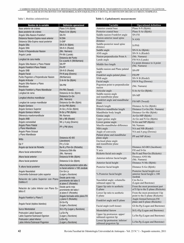
Tabla 1. Medidas cefalométricas
Nombre de la variable Definición operacionalBase anterior de cráneo Plano S-N (Björk)Base posterior de cráneo Plano S-Ar (Björk)Ángulo Silla-Nasion-Frankfort SN-FHDistancia Nasion-Espina nasal anterior N ENADistancia Silla-Espina nasal posterior S-ENPÁngulo Silla SN-S Ar (Björk)Ángulo SNA SN-N A (Riedel)Punto A Perpendicular Nasion (Mc. Namara)Ángulo Lande FH-NA (Lande)
Longitud de cara mediaDistancia del Punto Co a punto A (McNamara)
Ángulo Silla-Nasion y Plano Palatal SN-PPÁngulo Frankfort-Plano Palatal FH-PPÁngulo SNB SN-N B (Riedel)Ángulo Facial FH-N pog (Downs)Punto Pogonion a Perpendicular Nasion (McNamara)Ángulo Articular S Ar-Ar Go (Björk)Ángulo Silla-Nasion y Plano Mandibular
SN-PM
Ángulo Frankfort y Plano Mandibular FH-PM (Tweed)Longitud de rama Distancia Ar-Go (Björk)
Longitud efectiva mandibularDistancia Co-Gn (Mc. Namara)
Longitud de cuerpo mandibular Distancia Go-Me (Björk)Ángulo Goníaco Ar-Go-PM (Björk)Ángulo Goníaco Superior Ar-Go y N-Go (Björk)Ángulo Goníaco Inferior N-Go y PM (Björk)Diferencia maxilomandibular Mc NamaraÁngulo ANB NA y NB (Riedel)Ángulo convexidad NA y A pog (Downs)Ángulo Plano Palatal y Plano Mandibular
PP y PM (Kim)
Ángulo Plano Oclusal y Plano Mandibular
WittsDistancia AO-BO (Jacobson)
Eje Y FH y S-GnÁngulo eje facial de Ricketts Ba-N y Ptm-Gn (Ricketts)
Altura facial anteroinferiorDistancia ENA-Me (Mc. Namara)
Altura facial anterior Distancia N-Me (Björk)
Altura facial posterior Distancia S-Go (Björk)
% altura facial posteriorAltura facial posterior sobre altu-ra facial anterior x 100 Jarabak
Ángulo Nasolabial: Columnilla-Subnasal-Labio superior
Cm-Sn-Ls (Legan y Burstone)
Relación de Labio Superior con Plano Estético E
Desde parte más prominente del labio S al plano E (Ricketts)
Relación de Labio Inferior con Plano Es-tético E
Desde parte más prominente del labio I al plano E (Ricketts)
Ángulo Frankfort y Plano EÁngulo formado entre plano FH y plano E (Ricketts)
Ángulo Facial (tejidos blandos)Gl-Sn-Pg (Legan y Burstone)
Surco Mentolabial Si/Li-Pg (Legan y Burstone)
Protrusión Labial Superior: Labio Superior/Subnasal-Ogonion
Ls/Sn-Pg (Legan y Burstone)
Protrusión Labial Inferior: Labio Inferior/Subnasal-Pogonion
Li/Sn-Pg (Legan y Burstone)
Table 1. Cephalometric measurements
Variable Operational definitionAnterior cranial base Plane S-N (Björk)Posterior cranial base Plane S-Ar (Björk)Saddle-nasion-Frankfort angle SN-FHNasion-anterior nasal spine distance N ANS
Saddle-posterior nasal spine distance S-PNS
Saddle angle SN-S Ar (Björk)ANS angle SN-N A (Riedel)Nasion perpendicular Point A (Mc. Namara)Lande angle FH-NA (Lande)
Middle face length Co point distance to A point (Mc. Namara)
Saddle-nasion and Plane palatal angle SN-PP
Frankfort angle-palatal plane FH-PPSNB angle SN-N B (Riedel)Facial angle FH-N Pog (Downs)Pogonion point to perpendicular nasion (Mc. Namara)
Articular angle S Ar-Ar Go (Björk)Saddle-nasion angle and mandibular plane SN-MP
Frankfort angle and mandibular plane FH-MP (Tweed)
Branch length Distance Ar-Go (Björk)Effective mandibular length Distance Co-Gn (Mc. Namara)Mandibular body length Distance Go-Me (Björk)Goniac angle Ar-Go-MP (Björk)Upper goniac angle Ar-Go and N-Go (Björk)Lower goniac angle N-Go and MP (Björk)Maxillo-mandibular difference Mc NamaraNBA angle NA and NB (Riedel)Angle of convexity NA and A pog (Downs)Palatal plane and mandibular plane angle PP and MP (Kim)
Occlusal plane angle and mandibular planeWitts Distance AO-BO (Jacobson)Y axis FH and S-GnRicketts facial axis angle Ba-N and Ptm-Gn (Ricketts)
Anterior-inferior facial height Distance ANS-Me (Mc. Namara)
Anterior facial height Distance N-Me (Björk)
Posterior facial height Distance S-Go (Björk)
% Posterior facial heightPosterior facial height over anterior facial height x 100 Jarabak
Nasolabial angle: columella-subnasal-upper lip
Cm-Sn-Ls (Legan and Burstone)
Upper lip ratio to aesthetic E-plane
From the most prominent part of S lip to the E-plane (Ricketts)
Lower lip ratio to aesthetic E-plane
From the most prominent I lip point to the E-plane (Ricketts)
Frankfort angle and E-plane Angle formed between FH plane and E-plane (Ricketts)
Facial angle (soft tissue) Gl-Sn-Pg (Legan and Burstone)
Mentolabial furrow Si/Li-Pg (Legan and Burstone)Upper lip protrusion: upper/subnasal-ogonion lip Ls/Sn-Pg (Legan and Burstone)
Lower lip protrusion: inferior/subnasal-pogonion lip Li/Sn-Pg (Legan and Burstone)
CAMBIOS ESQUELÉTICOS, FACIALES Y DENTOALVEOLARES PRODUCIDOS POR EL TRATAMIENTO CON PLACAS DE ACETATO Y ELÁSTICOS DE CLASE III EN PACIENTES CON MESIORRELACIÓN ESQUELÉTICA Y DENTAL EN DENTICIÓN DECIDUA O MIXTA TEMPRANA

Revista Facultad de Odontología Universidad de Antioquia - Vol. 23 N.º 1 - Segundo semestre, 2011 43
Para las mediciones lineales y angulares se utilizó una regla cefalométrica Ormco cepha (Ormco-SYBRON). Tanto las medidas lineales como las angulares fueron aproximadas al 0,5 mayor.
Para determinar el patrón de crecimiento general se hizo una superimposición cefalométrica de la radiografía inicial, a los seis meses y final sobre el plano Silla Nasión (figura 4). Para determinar el comportamiento individual del maxilar y la mandíbula se hizo superim-posición sobre el plano palatal; y sobre el conducto del nervio dentario inferior y la sínfisis respectivamente (figuras 5 y 6).
Finalizado el periodo de tratamiento, a los pacientes se les colocó como retención aparatología tipo bionator, para continuar bajo supervisión en las clínicas de la Facultad, de acuerdo con la especificidad de cada caso.
Para el análisis estadístico de los datos se emplearon los software Excel y SPSS 15.0. Se hizo un análisis descriptivo de las variables radiográficas y una repre-sentación en forma independiente del comportamiento de cada una utilizando la prueba de Anova de medidas repetidas para determinar los cambios en el tiempo. Se aplicó un diseño de muestras relacionadas utilizando la prueba no paramétrica de Wilcoxon para determinar los cambios a los 6 meses y al año de tratamiento con respecto al inicio. El nivel de significancia considerado para las pruebas fue de 0,005.
For linear and angular measurements, an Ormco cepha (Ormco-SYBRON) cephalometric ruler was used. Both linear and angular measurements were approximate to the larger 0.5.
In order to establish the general growth pattern, a cephalometric superimposition of the initial radio-graph was made after six months and at the end of treatment on the saddle-nasion plane (fig. 4). In order to establish the individual behavior of maxillary and mandible, a superimposition on the palatal plane was made, as well as on the inferior dental nerve conduct and on the symphysis, respectively (figs. 5 and 6).
Once the period of treatment ended, bionator-like devices were placed on the patients as a retention aid, to continue under supervision at the School’s clinics, depending on the specific characteristics of each case.
For the statistical data analysis, the Excel and SPSS 15.0 software were used. A descriptive analysis of the radiographic variables was made, as well as an independent representation of the behavior of each variable by means of the Anova test of repeated measurements to establish changes through time. A design of related samples was applied using the non-parametric Wilcoxon test to determine changes after six months and one year of treatment in relation to the beginning. The significance level considered for the tests was 0.005.
Figura 3. Variables cefalométricas utilizadas para la evaluación de los cambios producidos por las placas de acetato y los elásticos de clase III utilizadas para el tratamaiento de la mesiorrelación esquelética
Figure 3. Cephalometric variables used to assess the changes produced by acetate plates and Class III elastics employed for treatment of skeletal mesiorelation
SKELETAL, FACIAL AND DENTOALVEOLAR CHANGES PRODUCED BY TREATMENT WITH ACETATE PLATES AND CLASS III ELASTICS IN PATIENTS WITH SKELETAL AND DENTAL MESIORELATION IN EARLY DECIDOUS OR MIXED TEETHING

Revista Facultad de Odontología Universidad de Antioquia - Vol. 23 N.º 1 - Segundo semestre, 201144
Figura 4. Superimposición del trazado cefalométrico al inicio, 6 meses y un año de tratamiento
Figure 4. Cephalometric superimposition traces at the beginning and after 6 months and one year of treatment
Figura 5. Cambios en el maxilar de pacientes con mesiorrelación esquelética y dental después de 6 meses de tratamiento con elásticos intermaxilares anclados en placas de acetato bimaxilares
Figure 5. Changes in the maxillary of patients with skeletal and dental mesiorelation after 6 months of treatment with intermaxillary elastics anchored on bimaxillary acetate plates
Figura 6. Cambios en la mandíbula de pacientes con mesiorrelación esquelética y dental después de 6 meses de tratamiento con elásticos intermaxilares anclados en placas de acetato bimaxilares
Figure 6. Changes in the mandible of patients with skeletal and dental mesiorelation after 6 months of treatment with intermaxillary elastics anchored on bimaxillary acetate plates.
CAMBIOS ESQUELÉTICOS, FACIALES Y DENTOALVEOLARES PRODUCIDOS POR EL TRATAMIENTO CON PLACAS DE ACETATO Y ELÁSTICOS DE CLASE III EN PACIENTES CON MESIORRELACIÓN ESQUELÉTICA Y DENTAL EN DENTICIÓN DECIDUA O MIXTA TEMPRANA

Revista Facultad de Odontología Universidad de Antioquia - Vol. 23 N.º 1 - Segundo semestre, 2011 45
RESULTADOS
En la tabla 2 se presentan las variaciones en la base de cráneo, se observa como único cambio estadísticamente significativo (con p < 0,005) el incremento promedio (3,1º) en el ángulo Silla a expensas de la base craneal posterior entre T1 y T2 presente en 21 pacientes obser-vados. El ángulo SN/FH disminuye o permanece igual en los 21 pacientes de la muestra (tabla 2).
En cuanto al maxilar en el plano sagital entre T1 y T2 (tabla 3) se encontró un incremento estadísticamente significati-vo en la longitud de cara media (tamaño) con promedio de 2,85 mm en 20 pacientes. Los demás cambios no fueron estadísticamente significativos e incluyeron disminución de la distancia de A a la perpendicular de N en 16 pa-cientes (-1,8), disminución del ángulo de Lande (3,43º) en 17 pacientes e incremento del ángulo SNA (1,7º) en 11 de los 21 pacientes. Al año de tratamiento T1/T3 no se presentan cambios estadísticamente significativos.
El maxilar en el plano vertical presentó incremento de todas las medidas entre T1 y T2 (tabla 3) sin tendencia a la rotación en un sentido definido. Sin embargo el único cambio estadísticamente significativo fue el incremento en la distancia N-ENA presente en 16 pacientes con promedio de 2,93. Entre T1 y T3 continúa el incremento en el desarrollo vertical, más significativo en la parte posterior (distancia S-ENP) con incremento de 2,43 mm en 16 pacientes.
Tabla 2. Cambios estadísticamente significativos en la base de cráneo de pacientes con mesiorrelación esquelética y dental después de 6 meses de tratamiento con placas de acetato bimaxilares y elásticos intermaxilares (T1, T2)
VariableAumento Disminución No cambio Sig. estadística
(p)*Promedio N.º Promedio N.º N.ºS-N 1,75 18 3,43 3 5 0,006S-Ar 1,45 11 1,38 5 10 0,105
S-N/FH 1,30 5 2,40 15 6 0,009Ángulo S 3,10 21 1,25 4 1 0,001
*(p): comparación de las medianas de las mediciones entre los tiempos de análisis.n = 26.
RESULTS
Table 2 displays the variations in the cranial base. The only statistically significant change observed (being p < 0.005) is the average increase (3.1º) in the saddle angle at the expense of posterior cranial base between T1 and T2 in 21 observed patients. The SN/FH angle was reduced or remained unchanged in all the 21 patients of the sample (table 2).
Concerning the maxillary in the sagittal plane, between T1 and T2 (table 3) a statistically sig-nificant increase was found in the middle face length (size) averaging 2.85 mm in 20 patients. The remaining changes were not statistically significant, and included lowered distance from A to the perpendicular of N in 16 patients (-1.8), Lande’s angle reduction (3.43º) in 17 patients and SNA angle increase (1.7º) in 11 out of 21 patients. After one year of treatment (T1/T3) there are no statistically significant changes.
The maxillary showed an increase in the vertical plane in all the measurements between T1 and T2 (table 3) without a tendency to rotate within a defined direction. However, the only statistically significant change was the increase in the N-ANS distance observed in 16 patients with an average of 2.93. Between T1 and T3 the increase in vertical development continues, being more significant in the posterior region (distance S-PNS) with an increase of 2.43 mm in 16 patients.
Table 2. Statistically significant changes on the cranial base in patients with skeletal and dental mesiorelation after 6 months of treatment with bimaxillary acetate plates and intermaxillary elastics (T1-T2)
Variable Increase Reduction No change Statistical significance (p)*Average N.º Average N.º N.º
S-N 1.75 18 3.43 3 5 0.006S-Ar 1.45 11 1.38 5 10 0.105
S-N/FH 1.30 5 2.40 15 6 0.009S angle 3.10 21 1.25 4 1 0.001
*(p): comparison of the measurement medians between times of analysis.n = 26.
SKELETAL, FACIAL AND DENTOALVEOLAR CHANGES PRODUCED BY TREATMENT WITH ACETATE PLATES AND CLASS III ELASTICS IN PATIENTS WITH SKELETAL AND DENTAL MESIORELATION IN EARLY DECIDOUS OR MIXED TEETHING

Revista Facultad de Odontología Universidad de Antioquia - Vol. 23 N.º 1 - Segundo semestre, 201146
Tabla 3. Cambios estadísticamente significativos del maxilar superior en pacientes con mesiorrelación esquelética y dental después de 6 meses de tratamiento con placas de acetato bimaxilares y elásticos intermaxilares (T1, T2)
VariableAumento Disminución No cambio
(p)*Promedio N.º Promedio N.º N.º
Longitud cara media 2,85 20 1,66 3 3 0,001N-ENA 2,93 16 1,00 5 5 0,004
*(p): comparación de las medianas de las mediciones entre los tiempos de análisis.n = 26
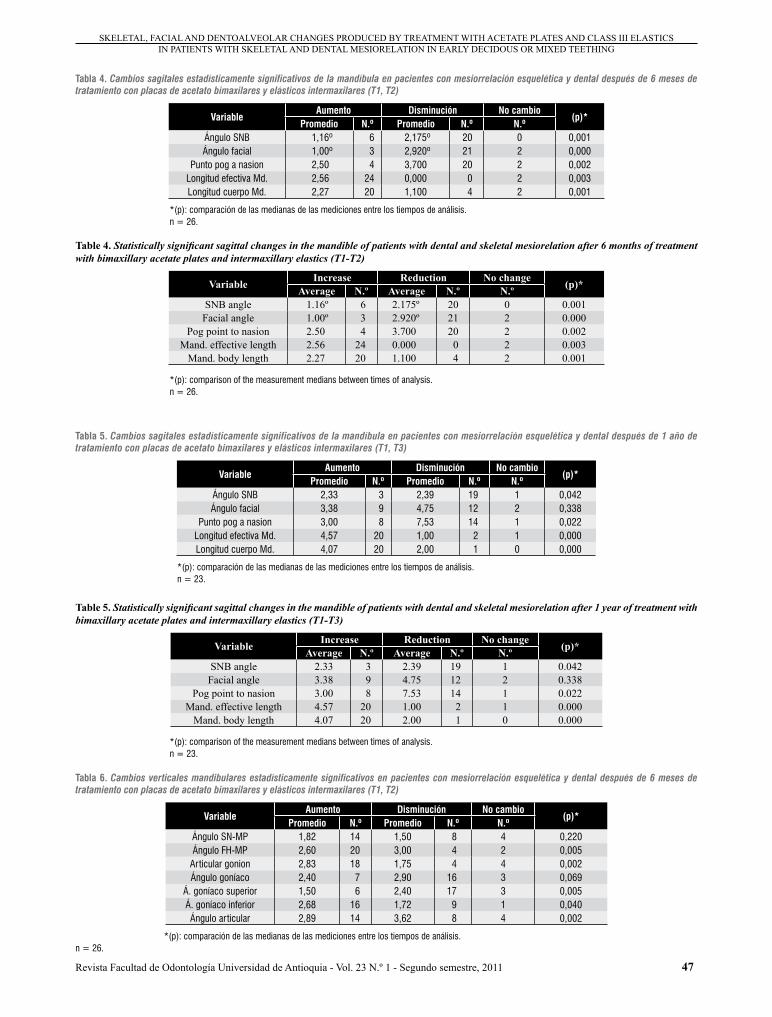
Concerning the mandible in the sagittal plane, all of the measurements showed statistically significant changes between T1 and T2 (table 4); an increase in effective length and in mandibular body length was observed along with a reduction in SNB angle, facial angle and pog point perpendicular to N. Be-tween T1 and T3 (table 5), even though it was not statistically significant, facial angle decreased in 12 patients (4.75º on average) and the SNB angle was reduced in 19 of the 23 patients of the sample (2.39º on average) with an average increase of 4.57 mm in mandibular effective length and 4.07 mm in mandibular body length (20 patients).
At the mandibular level in the vertical plane (table 6), statistically significant changes occurred during the first 6 months of treatment in the FH-MP angle measurements (clockwise rotation), the Ar-Go distance (branch length), the upper goniac angle and the Ar angle.
Concerning maxillomandibular relationships in the sagittal direction (table 7) an increase in Witts (3,4) wasobserved between T1 and T2 in the 21 patients, and between T1 and T3 (4,02) in 20 of the 21 patients, both being statistically significant and expressing a better relationship between maxillary and mandible. In the vertical plane (table 7), statistically significant changes are observed only between T1 and T2, given by an increase in the Y axis measurement, anterior facial height and lower antero-facial height.
Table 3. Statistically significant changes of the upper maxillary in patients with dental and skeletal mesiorelation after 6 months of treatment with bimaxillary acetate plates and intermaxillary elastics (T1-T2)
VariableIncrease Reduction No change
(p)*Average N.º Average N.º N.º
Middle face length 2.85 20 1.66 3 3 0.001
N-ANS 2.93 16 1.00 5 5 0.004
*(p): comparison of the measurement medians between times of analysis.n = 26
Con relación a la mandíbula en el plano sagital todas las medidas mostraron cambios estadísticamente significa-tivos entre T1-T2 (tabla 4), se presentó incremento en la longitud efectiva y la longitud del cuerpo mandibular con una disminución del ángulo SNB, ángulo facial y punto pog. perpendicular a N. Entre T1 y T3 (tabla 5), aun que no fue estadísticamente significativo, el ángulo facial disminuyó en 12 pacientes (4,75º en promedio) y el ángulo SNB disminuyó en 19 de los 23 pacientes de la muestra (2,39º en promedio) con un incremento promedio de 4,57 mm en la longitud efectiva mandibular y 4,07 mm en la longitud del cuerpo mandibular (20 pacientes).
A nivel mandibular en el plano ver tical (tabla 6) se presentaron cambios estadísticamente significativos durante los primeros 6 meses de tratamiento en las me-didas del ángulo FH-PM (rotación en sentido horario), la distancia Ar-Go (longitud de la rama), el ángulo goníaco superior y el ángulo Ar.
En cuanto a las relaciones maxilomandibulares en sentido sagital (tabla 7) se observó incremento en el Witts (3,4) entre T1 y T2 en los 21 pacientes y entre T1 y T3 (4,02) en 20 de los 21pacientes siendo am-bos estadísticamente significativos lo que representa una mejor relación entre el maxilar y la mandíbula. En el plano ver tical (tabla 7) se observan cambios estadísticamente significativos solo entre T1 y T2 dados por el incremento en la medida del eje Y, la altura facial anterior y la altura facial anteroinferior.
CAMBIOS ESQUELÉTICOS, FACIALES Y DENTOALVEOLARES PRODUCIDOS POR EL TRATAMIENTO CON PLACAS DE ACETATO Y ELÁSTICOS DE CLASE III EN PACIENTES CON MESIORRELACIÓN ESQUELÉTICA Y DENTAL EN DENTICIÓN DECIDUA O MIXTA TEMPRANA
Revista Facultad de Odontología Universidad de Antioquia - Vol. 23 N.º 1 - Segundo semestre, 2011 47
Table 5. Statistically significant sagittal changes in the mandible of patients with dental and skeletal mesiorelation after 1 year of treatment with bimaxillary acetate plates and intermaxillary elastics (T1-T3)
Variable Increase Reduction No change (p)*Average N.º Average N.º N.ºSNB angle 2.33 3 2.39 19 1 0.042Facial angle 3.38 9 4.75 12 2 0.338
Pog point to nasion 3.00 8 7.53 14 1 0.022Mand. effective length 4.57 20 1.00 2 1 0.000
Mand. body length 4.07 20 2.00 1 0 0.000
*(p): comparison of the measurement medians between times of analysis.n = 23.
Table 4. Statistically significant sagittal changes in the mandible of patients with dental and skeletal mesiorelation after 6 months of treatment with bimaxillary acetate plates and intermaxillary elastics (T1-T2)
Variable Increase Reduction No change (p)*Average N.º Average N.º N.ºSNB angle 1.16º 6 2.175º 20 0 0.001
Facial angle 1.00º 3 2.920º 21 2 0.000Pog point to nasion 2.50 4 3.700 20 2 0.002
Mand. effective length 2.56 24 0.000 0 2 0.003Mand. body length 2.27 20 1.100 4 2 0.001
*(p): comparison of the measurement medians between times of analysis.n = 26.
Tabla 4. Cambios sagitales estadísticamente significativos de la mandíbula en pacientes con mesiorrelación esquelética y dental después de 6 meses de tratamiento con placas de acetato bimaxilares y elásticos intermaxilares (T1, T2)
VariableAumento Disminución No cambio
(p)*Promedio N.º Promedio N.º N.º
Ángulo SNB 1,16º 6 2,175º 20 0 0,001Ángulo facial 1,00º 3 2,920º 21 2 0,000
Punto pog a nasion 2,50 4 3,700 20 2 0,002Longitud efectiva Md. 2,56 24 0,000 0 2 0,003Longitud cuerpo Md. 2,27 20 1,100 4 2 0,001
*(p): comparación de las medianas de las mediciones entre los tiempos de análisis.n = 26.
Tabla 5. Cambios sagitales estadísticamente significativos de la mandíbula en pacientes con mesiorrelación esquelética y dental después de 1 año de tratamiento con placas de acetato bimaxilares y elásticos intermaxilares (T1, T3)
VariableAumento Disminución No cambio
(p)*Promedio N.º Promedio N.º N.º
Ángulo SNB 2,33 3 2,39 19 1 0,042Ángulo facial 3,38 9 4,75 12 2 0,338
Punto pog a nasion 3,00 8 7,53 14 1 0,022Longitud efectiva Md. 4,57 20 1,00 2 1 0,000Longitud cuerpo Md. 4,07 20 2,00 1 0 0,000
*(p): comparación de las medianas de las mediciones entre los tiempos de análisis.n = 23.
Tabla 6. Cambios verticales mandibulares estadísticamente significativos en pacientes con mesiorrelación esquelética y dental después de 6 meses de tratamiento con placas de acetato bimaxilares y elásticos intermaxilares (T1, T2)
VariableAumento Disminución No cambio
(p)*Promedio N.º Promedio N.º N.º
Ángulo SN-MP 1,82 14 1,50 8 4 0,220Ángulo FH-MP 2,60 20 3,00 4 2 0,005
Articular gonion 2,83 18 1,75 4 4 0,002Ángulo goníaco 2,40 7 2,90 16 3 0,069
Á. goníaco superior 1,50 6 2,40 17 3 0,005Á. goníaco inferior 2,68 16 1,72 9 1 0,040Ángulo articular 2,89 14 3,62 8 4 0,002
*(p): comparación de las medianas de las mediciones entre los tiempos de análisis.n = 26.
SKELETAL, FACIAL AND DENTOALVEOLAR CHANGES PRODUCED BY TREATMENT WITH ACETATE PLATES AND CLASS III ELASTICS IN PATIENTS WITH SKELETAL AND DENTAL MESIORELATION IN EARLY DECIDOUS OR MIXED TEETHING

Revista Facultad de Odontología Universidad de Antioquia - Vol. 23 N.º 1 - Segundo semestre, 201148
Es de anotar, aunque no alcanzó significancia estadística, que se presenta incremento promedio mayor en la altura facial posterior que en la anterior, tanto a los seis meses como al año de tratamiento (figuras 1 y 2).
La comparación cefalométrica de las medidas postra-tamiento entre T3 y T4 mostró aumento sin significancia estadística (p < 0,005) en las medidas SN, longitud efectiva mandibular y diferencia maxilomandibular en todos los pacientes. En la superimposición se observó en el maxilar superior mayor desarrollo en sentido sagital, mientras que en la mandíbula el desarrollo fue principalmente en sentido vertical.
It is worth noting that, even though it didn’t reach statistical significance, a higher average increase occurs in the posterior facial height compared to the anterior facial height, both at six months and after one year of treatment (figures. 1 and 2).
The cephalometric comparison of the post-treatment measurements between T3 and T4 revealed an in-crease with no statistical significance (p < 0.005) in SN, as well as mandibular effective length and maxillomandibular difference measurements in all the patients. On the superimposition, greater development in the sagittal direction was observed in the upper maxillary, while in the mandible the development was mainly in the vertical direction.
Table 7. Statistically significant maxillomandibular changes in patients with dental and skeletal mesiorelation after 6 months of treatment with bimaxillary acetate plates and intermaxillary elastics (T1/T2)
Variable Increase Reduction No change (p)*Average N.º Average N.º N.ºWitts 3.40 21 1.62 4 1 0.000Y axis 2.92 21 1.83 3 2 0.000AFH 4.00 20 6.50 3 3 0.001
LAFH 2.22 21 3 1 0.003PFH 2.37 17 1.86 6 3 0.053
PFH-AFH X 100 1.56 10 1.72 13 3 0.301
*(p): comparison of the measurement medians between times of analysis.n = 26.
Table 6. Statistically significant mandibular vertical changes in patients with dental and skeletal mesiorelation after 6 months of treatment with bimaxillary acetate plates and intermaxillary elastics (T1-T2)
Variable Increase Reduction No change (p)*Average N.º Average N.º N.ºSN-MP angle 1.82 14 1.50 8 4 0.220FH-MP angle 2.60 20 3.00 4 2 0.005
Articular gonion 2.83 18 1.75 4 4 0.002Goniac angle 2.40 7 2.90 16 3 0.069
Upper goniac angle 1.50 6 2.40 17 3 0.005Lower goniac angle 2.68 16 1.72 9 1 0.040
Articular angle 2.89 14 3.62 8 4 0.002
*(p): comparison of the measurement medians between times of analysis.n = 26.
Tabla 7. Cambios maxilomandibulares estadísticamente significativos en pacientes con mesiorrelación esquelética y dental después de 6 meses de tratamiento con placas de acetato bimaxilares y elásticos intermaxilares (T1, T2)
VariableAumento Disminución No cambio
(p)*Promedio N.º Promedio N.º N.º
Witts 3,40 21 1,62 4 1 0,000Eje Y 2,92 21 1,83 3 2 0,000AFA 4,00 20 6,50 3 3 0,001AFAI 2,22 21 3 1 0,003AFP 2,37 17 1,86 6 3 0,053
AFP-AFA X 100 1,56 10 1,72 13 3 0,301
*(p): comparación de las medianas de las mediciones entre los tiempos de análisis.n = 26.
CAMBIOS ESQUELÉTICOS, FACIALES Y DENTOALVEOLARES PRODUCIDOS POR EL TRATAMIENTO CON PLACAS DE ACETATO Y ELÁSTICOS DE CLASE III EN PACIENTES CON MESIORRELACIÓN ESQUELÉTICA Y DENTAL EN DENTICIÓN DECIDUA O MIXTA TEMPRANA

Revista Facultad de Odontología Universidad de Antioquia - Vol. 23 N.º 1 - Segundo semestre, 2011 49
En cuanto a los tejidos blandos (tabla 8), después de 6 meses de tratamiento (T1-T2) se encontró disminución estadísticamente significativa en la protrusión del labio inferior (1,64 mm) y en el ángulo FH-plano E (2,75º), y después de un año de tratamiento (T1-T3) incremento estadísticamente significativo del ángulo facial (4,15º). Es importante mencionar además que durante el periodo de observación se presentó en la mayoría de los pacientes aumento en el ángulo nasolabial (8,23º) y en la protrusión del labio superior (2,36 mm), y disminución del surco men-tolabial, que no lograron significancia estadística (figura 7).
Los cambios en la sobremordida vertical y horizontal medidos clínicamente mostraron significancia estadística entre T1 y T3 hallando aumento del overjet con promedio de 2,79 mm en 22 de 23 pacientes e incremento del overbite de 2,05 mm en 16 de los 23 pacientes (figura 8).
En las relaciones caninas se encontró un cambio promedio de 2,51 mm pasando de relaciones de clase III a relaciones de clase II en 12 pacientes y a relaciones de clase I en10 pacientes. En cuanto a las relaciones molares se observó mejoría en 20 pacientes que pasaron de escalón mesial a plano terminal recto con promedio de 2,43 mm.
En los pacientes en los cuales se evaluó la fase de reten-ción (T3/T4), se encontró en sentido vertical aumento en la sobremordida en 3 pacientes en promedio de 0,9 mm y en 2 pacientes disminuyó en promedio 0,75 mm.
En sentido sagital entre T3 y T4 hubo mejor relación en la sobremordida horizontal en 4 de los 5 pacientes con aumento promedio de 1,25 mm. En general se encontró estabilidad en las relaciones molares y caninas después de terminado el tratamiento, coincidiendo con lo encon-trado en el estudio anterior.
Concerning soft tissue (table 8), after 6 months of treatment (T1-T2) a statistically significant reduc-tion was found in lower lip protrusion (1.64 mm) and in FH- E-Plane angle (2.75º), and a statistically significant increase in facial angle (4,15º) was ob-served after a year of treatment (T1-T3). It is worth mentioning that during the period of observation most patients exhibited an increase in the nasolabial angle (8.23º) and on upper lip protrusion (2.36 mm), as well as a reduction of mentolabial fold, which failed to achieve statistical significance (figure 7).
Changes in vertical and horizontal overbite clinically measured showed statistical significance between T1 and T3, exhibiting an overjet increase with 2.79 mm average in 22 of 23 patients and an overbite increase of 2.05 mm in 16 of 23 patients (figure 8).
In terms of canine relationships, an average change of 2.51 mm was found, moving from Class III rela-tionships to Class II relationships in 12 patients and to Class I relationships in 10 patients. Concerning molar relationships, improvement was observed in 20 patients, moving from a mesial scale to a straight terminal plane averaging 2.43 mm.
In patients evaluated during the retention phase (T3/T4), a vertical overbite increase was found in 3 of them in 0.9 mm average, and a lower increase in vertical overbite of 0.75 mm on average was found in 2 patients.
Sagittaly, between T3 and T4 a better relationship of horizontal overbite was achieved in 4 of 5 patients, with an average increase of 1.25 mm. Generally, stability of molar and canine relationships was ob-served after the treatment was finished; this agrees with the findings of the previous study.
Tabla 8. Cambios en tejidos blandos estadísticamente significativos en pacientes con mesiorrelación esquelética y dental después de 6 meses de tratamiento con placas de acetato bimaxilares y elásticos intermaxilares (T1-T2)
VariableAumento Disminución No cambio
(p)*Promedio N.º Promedio N.º N.º
Á. nasolabial 11,07 13 7,83 12 1 0,492Labio superior-plano E 1,63 15 3,10 5 6 0,119Labio inferior-plano E 1,35 7 2,11 13 6 0,077
Á. FH-plano E 1,33 3 2,75 20 3 0,000Ángulo facial 3,31 11 3,10 10 5 0,392
Surco mentolabial 0,83 3 1,79 12 11 0,008Protrusión labial superior 1,73 13 1,50 8 5 0,206Protrusión labial inferior 1,12 4 1,64 17 5 0,005
*(p): comparación de las medianas de las mediciones entre los tiempos de análisis.n = 26.
SKELETAL, FACIAL AND DENTOALVEOLAR CHANGES PRODUCED BY TREATMENT WITH ACETATE PLATES AND CLASS III ELASTICS IN PATIENTS WITH SKELETAL AND DENTAL MESIORELATION IN EARLY DECIDOUS OR MIXED TEETHING

Revista Facultad de Odontología Universidad de Antioquia - Vol. 23 N.º 1 - Segundo semestre, 201150
Table 8. Statistically significant changes of soft tissue in patients with mesial dental and skeletal mesiorelation after 6 months of treatment with bimaxillary acetate plates and intermaxillary elastics (T1-T2)
Variable Increase Reduction No change (p)*Average N.º Average N.º N.ºNasolabial angle 11.07 13 7.83 12 1 0.492
Upper lip- E-plane 1.63 15 3.10 5 6 0.119Lower lip- E-plane 1.35 7 2.11 13 6 0.077FH angle-E-plane 1.33 3 2.75 20 3 0.000
Facial angle 3.31 11 3.10 10 5 0.392Mentolabial fold 0.83 3 1.79 12 11 0.008
Upper lip protrusion 1.73 13 1.50 8 5 0.206Lower lip protrusion 1.12 4 1.64 17 5 0.005
*(p): comparison of the measurement medians between times of analysis.n = 26.
Figura 7. Cambios faciales en pacientes con mesiorrelación esquelética y dental después de un año de tratamiento (T1-T3-T4) con placas de acetato bimaxilares y elásticos intermaxilares de clase III
Figure 7. Facial changes in patients with dental and skeletal mesiorelation after a year of treatment (T1-T3-T4) with bimaxillary acetate plates and Class III intermaxillary elastics
Figura 8. Cambios dentoalveolares en pacientes con mesiorrelación esquelética y dental después de un año de tratamiento (t1,t3) con placas de acetato bimaxilares y elásticos intermaxilares de clase III
Figure 8. Dentoalveolar changes in patients with dental and skeletal mesiorelation after a year of treatment (T1-T3) with bimaxillary acetate plates and Class III intermaxillary elastics
DISCUSIÓN
La relación esquelética de clase III es una de las alteracio-nes oclusales más complejas de diagnosticar y de tratar. De acuerdo con la edad del paciente, es posible hacer diferentes tipos de tratamiento, cada uno con posibilida-des y limitaciones.
El enfoque del tratamiento temprano ha sido propuesto por varios autores entre ellos: Mc Namara,9 Petit,15 Franchi y Baccetti.13 Estos últimos recomiendan que el periodo óptimo para mejorar las relaciones esqueléticas de cla-se III con aparatología funcional debería ser durante la dentición decidua y mixta temprana. En algunos casos,
DISCUSSION
Class III skeletal relationship is one of the occlusal diseases with the most complex diagnostic and treat-ment. Depending on the patient’s age, different kinds of treatment may be implemented, each featuring possibilities and drawbacks.
The early treatment approach has been recom-mended by several authors such as Mc Namara,9 Petit,15 Franchi and Baccetti.13 The latter suggest that the optimal period to improve Class III skeletal relationships with functional appliances is during early deciduous and mixed dentition. In some cases,
CAMBIOS ESQUELÉTICOS, FACIALES Y DENTOALVEOLARES PRODUCIDOS POR EL TRATAMIENTO CON PLACAS DE ACETATO Y ELÁSTICOS DE CLASE III EN PACIENTES CON MESIORRELACIÓN ESQUELÉTICA Y DENTAL EN DENTICIÓN DECIDUA O MIXTA TEMPRANA

Revista Facultad de Odontología Universidad de Antioquia - Vol. 23 N.º 1 - Segundo semestre, 2011 51
incluso, se considera que el tratamiento temprano puede disminuir la necesidad de intervenciones quirúrgicas en edades avanzadas.10, 12, 16, 17
La dificultad en la valoración de los resultados de este estudio esta dada por el periodo de crecimiento en el que se encuentra la población sujeta a observación y por la escasez de estudios de crecimiento en pacientes con desarmonías en desarrollo que permitan hacer una com-paración. Al respecto, el estudio de Reyes3 en 2006 que compara pacientes en crecimiento clase III no tratados, con respecto a pacientes con relaciones esqueléticas de clase I en desarrollo reveló que no hubo diferencias en la posición sagital del maxilar de unos respecto a los otros, sin embargo en la mandíbula los pacientes clase IIIpresentaron un incremento en la longitud efectiva y en la altura facial anteroinferior significativamente mayor que los clase I.
En el presente estudio el cambio estadísticamente signi-ficativo en el ángulo Silla (tabla 2), y el incremento en la distancia S-N muestra que la población sujeta a estudio se encontraba en un periodo de crecimiento más activo en promedio durante los primeros 6 meses de observación, lo cual puede explicar que los cambios encontrados sean principalmente en posición más que en tamaño.
De acuerdo con Franchi y Baccetti,13 el efecto ortopédico de los elásticos de clase III utilizados con las placas de acetato genera una línea de acción de la fuerza en direc-ción retrusiva sobre la mandíbula que pasa a través o justo detrás del centro del cóndilo mandibular, esto produce, a largo plazo, una rotación en sentido horario de la mandí-bula, acompañada de una remodelación y rotación antiho-raria del componente dentoalveolar inferior debido a que el vector de fuerza pasa superior al centro de resistencia del componente dental. En el maxilar la línea de acción tiene igual magnitud pero sentido protrusivo generando una rotación antihoraria del plano palatino. En el presente estudio esta rotación (dada por la disminución del ángu-lo FH-PP) se presenta solo en 11 pacientes y no logra significancia estadística en los primeros seis meses de tratamiento, paradójicamente la medida N-ENA es la única que se incrementa con significancia estadística (opuesto a la rotación antihoraria), lo cual puede explicarse por el periodo de desarrollo vertical que atraviesan los sujetos de la muestra y que se manifiesta también en el sector posterior (incremento S-ENP que muestra tendencia rota-cional antihoraria después de los 6 meses de tratamiento).
it is even considered that early treatment may reduce the need of surgical interventions at later ages.10, 12, 16, 17.
The difficulties in assessing the findings of this study are related to the stages of growth of the population under observation, and to the lack of studies on the growth of patients with development disharmonies, which would allow comparisons. In this sense, Reyes’ study in 2006,3 in which untreated Class III growing patients were compared to patients with developing Class I skeletal relationships, revealed no differences in the maxillary sagittal position among the patients. However, the mandible of Class III patients displayed an effective length and anterior-inferior facial height increase significantly higher than those of class I.
In this study, the statistically significant change in the saddle angle (table 2), and the increase in the S-N distance prove that the population under study was undergoing, on average, a more active period of growth during the first 6 months of observation; this may explain why the changes found were mainly in terms of position rather than size.
According to Franchi and Baccetti,13 the orthopedic effect of Class III elastics used with acetate plates generates a line of action of a retrusive force on the mandible, passing through or right behind the center of the mandibular condyle. This produces in the long term a clockwise rotation in the mandible, accompa-nied by a reshaping and counterclockwise rotation of the lower dentoalveolar component because of the force vector passing over the centre of resistance of the dental component. In the maxillary, the line of action has the same magnitude, but a protrusive direction, which generates a counterclockwise rota-tion on the palatal plane. In the present study, this rotation (given by the reduction in FH-PP angle) was observed only in 11 patients and did not reach statistical significance in the first six months of treat-ment; paradoxically the N-ANS measurement is the only one showing a statistically significant increase (in opposition to the counterclockwise rotation), which may be explained by the period of vertical development undergone by the individuals of the sample and which is also observed in the posterior sector (S-PNS increase, showing a counterclockwise rotational tendency after six months of treatment).
SKELETAL, FACIAL AND DENTOALVEOLAR CHANGES PRODUCED BY TREATMENT WITH ACETATE PLATES AND CLASS III ELASTICS IN PATIENTS WITH SKELETAL AND DENTAL MESIORELATION IN EARLY DECIDOUS OR MIXED TEETHING

Revista Facultad de Odontología Universidad de Antioquia - Vol. 23 N.º 1 - Segundo semestre, 201152
Por el contrario, en pacientes tratados con disyuntor y máscara de protracción, autores como Nartallo-Turley,18 Da Silva y colaboradores,8 Turley,19 Ferro,20 y Major21 encontraron una rotación en sentido horario del maxilar durante el tratamiento, la cual se explica, en este caso, por la localización del vector de fuerza del elástico en una posición anterior al centro de resistencia. Estos hallazgos sugieren que el uso de este tipo de terapia no debería hacerse en pacientes con tendencia a mordida abierta anterior.
El efecto protrusivo sobre el maxilar se manifiesta con significancia estadística en los primeros seis meses a nivel de la longitud de cara media que se incrementa en 20 de los 26 pacientes con promedio de 2,85 mm.
Con respecto al tamaño mandibular se esperaría que la longitud efectiva se mantuviera estable como consecuen-cia del tratamiento en condiciones ideales; sin embargo, se observó incremento tanto en la longitud efectiva mandibular (2,56 mm a los 6 meses y 4,57 mm al año de tratamiento), como en la longitud del cuerpo (2,27 mm a los 6 meses y 4,07 mm al año); esto es similar a lo reportado por Saadia y colaboradores17 y McNamara22 en el estudio de pacientes clase III tratados con máscara de protracción que tampoco logró mantener la longitud de la mandíbula, y esta tuvo un incremento promedio de 4 mm.
En el presente estudio se encontró un retroposicionamien-to mandibular en sentido sagital estadísticamente signifi-cativo en los primeros seis meses de tratamiento (SNB, ángulo facial y punto pog perpendicular a N). Esta posición más posterior de la mandíbula continuó a lo largo del estudio, pero con un incremento menor entre T2 y T3, lo que explica la falta de significancia estadística al final del periodo de observación y establece una pauta de logro de los objetivos de tratamiento a corto plazo (6 meses).
En el plano vertical, se presentó aumento de la altura facial anteroinferior, el eje Y de crecimiento, y en el ángulo goníaco, lo cual indica una rotación mandibular abajo y atrás lo cual esta de acuerdo con los hallazgos reporta-dos por Franchi y Baccetti,13 y Espinal y colaboradores.14 Sin embargo, el incremento en la altura facial posterior presente en 17 pacientes a los 6 meses de tratamiento y en 12 pacientes al final del periodo de observación muestra que los pacientes se encuentran en un periodo de desarrollo vertical activo no solo anterior, como lo mencionan Reyes3 y Mitani,23 sino también posterior, importante clínicamente para mantener la proporción, aunque no sea estadísticamente significativo.
On the contrary, in patients treated with a disjunctor and a protraction mask, authors like Nartallo-Turley,18 Da Silva et al.,8 Turley,19 and Major21 have found a clockwise rotation of the maxillary during treatment, which is explained, in this case, by the position of the elastic force vector located anterior to the center of resistance. These findings suggest that the use of this type of therapy should not be performed in patients with a tendency to anterior overbite.
The protrusive effect on the maxillary presents statistical significance in the first six months in the middle face length level, which increased in 20 out of 26 patients with a 2.85 mm on average.
Regarding the mandibular size, one would expect that the effective length remained stable as a con-sequence of the treatment under ideal conditions. However, an increase was observed in both the mandibular effective length (2.56 mm after 6 months and 4.57 mm after one year of treatment), and body length (2.27 mm after 6 months and 4.07 mm after one year); this is similar to the reports by Saadia et al17 and McNamara,22 who studied Class III patients treated with a protraction mask, which also failed to keep the length of the mandible stable, having an average increase of 4 mm.
In this study, a mandibular retro-positioning in the sagittal direction was found to be statistically sig-nificant in the first six months of treatment (SNB, facial angle and pog point perpendicular to N). This more posterior position of the mandible persisted all throughout the study, but with a small increase between T2 and T3, which explains the lack of statistical significance at the end of the observation period, and establishes a guideline for the achieve-ment of goals in short-term treatments (6 months).
In the vertical plane, an increase of anteroinferior facial height occurred, as well as in the Y growth axis, and the goniac angle. This is an indication of downwards and backwards mandibular rotation, agreeing with the findings reported by Franchi and Baccetti,13 and Espinal et al.14 However, the increase in posterior facial height occurring in 17 patients after 6 months of treatment and in 12 patients at the end of the observation period demonstrates that patients are in a period of active vertical develop-ment not only anterior —as Reyes3 and Mitani23 mention—, but also posterior, which is clinically important to keep the proportions, even though it is not statistically significant.
CAMBIOS ESQUELÉTICOS, FACIALES Y DENTOALVEOLARES PRODUCIDOS POR EL TRATAMIENTO CON PLACAS DE ACETATO Y ELÁSTICOS DE CLASE III EN PACIENTES CON MESIORRELACIÓN ESQUELÉTICA Y DENTAL EN DENTICIÓN DECIDUA O MIXTA TEMPRANA

Revista Facultad de Odontología Universidad de Antioquia - Vol. 23 N.º 1 - Segundo semestre, 2011 53
En cuanto a las relaciones maxilomandibulares en el plano sagital, el incremento en el ángulo ANB, reportado también por Franchi y Baccetti,13 y el incremento en el Witts favorecen desde el primer periodo la corrección del perfil facial que se mantiene a lo largo del tratamiento.
En concordancia con lo anterior, se encontró además aumento del ángulo del eje Y, de la altura facial anterior y altura facial anteroinferior que indican una compensa-ción vertical de la displasia sagital similar a la reportada por Saadia17 en un estudio en 112 pacientes entre 6 y 12 años tratados con expansión maxilar y máscara facial en los que se encontró aumento en los ángulos FH-PM, SN-PM y en el del eje facial de Ricketts, a pesar de que, como se explicó antes, el efecto or to-pédico debería ser diferente. Esto podría explicarse por el patrón de crecimiento ver tical propio de los pacientes clase III común a ambas muestras, a pesar de los rangos de edad diferentes (3-9 años versus 6-12 años).
Los cambios dentoalveolares fueron de gran magnitud con mejoría de la relación canina y molar pasando en la mayoría de los casos (96%) de relaciones clase III a clase I. En cuanto a la sobremordida horizontal se ob-servó en todos los pacientes la corrección de la mordida cruzada anterior con cambios en promedio de 2,79 mm pasando de valores negativos a positivos en 22 de los 23 pacientes. En cuanto a la sobremordida vertical se encontró incremento en 16 de los 23 pacientes.
Los reportes de Nartallo-Turley,18 Saadia17 y Hickham,24 con respecto a los tejidos blandos incluyen un movimiento hacia adelante del labio superior, y un movimiento del mentón hacia abajo y hacia atrás. En este estudio los tejidos blandos mostraron mejoría en el perfil facial demostrada clínica y cefalométricamente, dada por una posición más posterior del punto pogonion y del labio inferior y mejor expresión del surco mentolabial y ángulo nasolabial, todo lo cual contribuye a la rehabilitación de la estética facial (figura 4).
En los resultados a largo plazo de los pacientes ma-nejados con este tipo de aparatología, se encontró en términos generales estabilidad esquelética, dentoalveo-lar y facial.
Regarding maxillomandibular relationships in the sagittal plane, both ANB angle increase—also reported by Franchi and Baccetti—13 and Witts in-crease favor correction of the facial profile from the first period, and it is kept throughout the treatment.
Accordingly, an increase in the Y axis angle was also found, as well as in the anterior facial height and the lower-anterior facial height, indicating a verti-cal compensation of sagittal dysplasia similar to the one reported by Saadia17 in a study in 112 patients between 6 and 12 years of age, who were treated with maxillary expansion and face mask showing an increase in FH-MP angle, SN-MP angle, and in Ricketts facial axis, although, as explained above, the orthopedic effect might be different. This may be explained by the vertical growth pattern character-istic of Class III patients, which is common to both samples, regardless of the different age ranges (3-9 years and 6-12 years).
Dentoalveolar changes were significant, with an improvement of canine-molar relationship, moving in most cases (96%) from Class III to Class I rela-tionships. In regards to horizontal overbite, all the patients experienced correction of anterior crossbite with changes of 2.79 mm on average, moving from negative to positive values in 22 of 23 patients. Regarding vertical overbite, an increase was found in 16 of 23 patients.
The reports by Nartallo-Turley,18 Saadia17 and Hick-ham24 concerning soft tissues include an upper lip forward movement, and a chin downward/back-ward movement. In this study, soft tissues showed improvement of the facial profile clinically and cephalometrically demonstrated, given by a more posterior position of the pogonion point and the lower lip and by a better expression in mentolabial fold and nasolabial angle, all of which contributes to facial aesthetic rehabilitation (figure 4).
In relation to long-term results in patients treated with this kind of appliances, dentoalveolar and facial skeletal stability was generally observed.
SKELETAL, FACIAL AND DENTOALVEOLAR CHANGES PRODUCED BY TREATMENT WITH ACETATE PLATES AND CLASS III ELASTICS IN PATIENTS WITH SKELETAL AND DENTAL MESIORELATION IN EARLY DECIDOUS OR MIXED TEETHING

Revista Facultad de Odontología Universidad de Antioquia - Vol. 23 N.º 1 - Segundo semestre, 201154
The most significant changes were represented by an increase in both anterior cranial base length and mandibular effective length, which agrees with the studies by Ferro et al.,20 who pointed out that long-term stability of this kind of treatment was related to the degree of intercuspidation achieved, and that recurrence was directly related to mandibular ramus growth after treatment.
It is also important to point out that during the obser-vation period an increase in nasolabial angle (8.23º) and in upper lip protrusion (2.36 mm) was observed in most patients (13-15), as well as a reduction in mentolabial fold, which, even if it did not reached statistical significance, may have significantly con-tributed in a clinical way to improve the aspect of soft tissues of the population under study at the end of the observation period.
CONCLUSIONS
Acetate plates with Class III elastics proved to be an effective and stable therapy after three years of post-treatment assessment for both facial and dental-skeletal mesiorelation improvement in patients with deciduous and mixed dentition from the sample evaluated.
The main results are achieved in the first six months of treatment and are due to changes in mandibular position that are expressed both in the sagittal and the vertical planes (SNB and facial angle reduc-tions accompanied by clockwise rotation) and an increase in middle face length, which is reflected in some Witts increase, higher vertical and horizontal overbites and a reduction in lower lip protrusion. Soft tissue facial angle increase is achieved after a year of treatment.
This therapy does not suppress the increase in man-dibular effective length characteristic of patients with Class III skeletal relationships.
ACKNOWLEDGEMENTS
To the Universidad de Antioquia’s Facultad de Odontología. To Doctor Mónica Jones for her meth-odological contributions. To the patients for their participation and contributions.
Los cambios más significativos estuvieron representados por el incremento tanto en la longitud de base de cráneo anterior como en la longitud efectiva mandibular, lo cual está de acuerdo con los estudios hechos por Ferro y colaboradores,20 quienes determinaron que la estabi-lidad a largo plazo de este tipo de tratamiento estaba relacionada con el grado de intercuspidación lograda, y que la recidiva estaba directamente relacionada con el crecimiento, postratamiento, de la rama mandibular.
Es importante mencionar además que durante el periodo de observación se presentó en la mayoría de los pacientes (13-15) aumento en el ángulo nasolabial (8,23º) y en la protrusión del labio superior (2,36 mm), y la disminución del surco mentolabial, que aunque no lograron signifi-cancia estadística, pueden haber contribuido de manera clínicamente significativa a mejorar el aspecto de los tejidos blandos al final del periodo de observación en la población de estudio.
CONCLUSIONES
Las placas de acetato con elásticos de clase III fueron una terapia efectiva y estable a 3 años de evaluación postratamiento para el mejoramiento facial y de la mesiorrelación esquelética y dental en los pacientes con dentición decidua y mixta de la muestra evaluada.
Los principales resultados se logran dentro de los prime-ros seis meses de tratamiento y se deben a cambios en la posición mandibular que se expresan tanto en el plano sagital como en el vertical (disminución de SNB y del án-gulo facial acompañadas por rotación en sentido horario) e incremento en la longitud de cara media, lo cual se refleja en el incremento del Witts, el incremento en la sobremor-dida vertical y horizontal y la disminución de la protrusión del labio inferior. El incremento del ángulo facial de tejidos blandos se logra después de un año de tratamiento.
Este tipo de terapia no elimina el incremento en la longi-tud efectiva mandibular característica de pacientes con relaciones esqueléticas de clase III.
AGRADECIMIENTOS
A la Facultad de Odontología Universidad de Antioquia. A la Doctora Mónica Jones por sus aportes metodológicos. A los pacientes por su participación y compromiso en el estudio.
CAMBIOS ESQUELÉTICOS, FACIALES Y DENTOALVEOLARES PRODUCIDOS POR EL TRATAMIENTO CON PLACAS DE ACETATO Y ELÁSTICOS DE CLASE III EN PACIENTES CON MESIORRELACIÓN ESQUELÉTICA Y DENTAL EN DENTICIÓN DECIDUA O MIXTA TEMPRANA

Revista Facultad de Odontología Universidad de Antioquia - Vol. 23 N.º 1 - Segundo semestre, 2011 55
CORRESPONDENCIA
Gabriel Espinal BoteroFacultad de OdontologíaUniversidad de AntioquiaCorreo electrónico: [email protected]
CORRESPONDING AUTHOR:
Gabriel Espinal BoteroFacultad de OdontologíaUniversidad de AntioquiaE-mail address: [email protected]
REFERENCIAS / REFERENCES
1. Delaire J. Maxillary development revisited: relevance to the orthopedic treatment of Class III malocclusions. Eur J Orthod 1997; 19(3): 289-311.
2. Nakasima A, Ichinose M, Nakata S, Takahama Y. Hereditary factors in the craniofacial morphology of Angle’s Class II and Class III malocclusions. Am J Orthod 1982; 82(2): 150-156.
3. Reyes BC, Baccetti T, McNamara JA Jr. An Estimate of craniofacial growth in Class III malocclusion. Angle Orthod 2006; 76(4): 577-584.
4. Nakasima A, Ichinose M, Nakata S. Genetic and environ-mental factors in the development of so-called pseudo- and true mesiocclusions. Am J Orthod Dentofacial Orthop 1986; 90(2): 106-116.
5. Graber LW. Chin cup therapy for mandibular prognathism. Am J Orthod 1977; 72(1): 23-41.
6. Sakamoto T, Iwase I, Uka A, Nakamura S. A roentgeno-cephalometric study of skeletal changes during and after chin cup treatment. Am J Orthod Dentofacial Orthop 1984; 85(4): 341-350.
7. Takada K, Petdachai S, Sakuda M. Changes in dentofacial morphology in skeletal Class III children treated by a modified maxillary protraction headgear and a chin cup: a longitudinal cephalometric appraisal. Eur J Orthod 1993; 15(3): 211-221.
8. Da Silva Filho OG, Macro AC, Capelozza Filho L. Early treatment of the Class III malocclusion with rapid maxillary expansion and maxillary protraction. Am J Orthod Dento-facial Orthop 1998; 113(2): 196-203.
9. McNamara JA Jr., Brudon WL. Tratamiento ortodóncico y ortopédico en la dentición mixta. 4.ª ed. Michigan: Needham Press; 1995.
10. Baccetti T, Tollaro I. A retrospective comparison of functio-nal appliance treatment of Class III malocclusion in the deciduous and mixed dentitions. Eur J Orthod 1998; 20(3): 309-317.
11. Litton SF, Ackermann LV, Isaacson RJ, Shapiro BL. A ge-netic study of Class III malocclusion. Am J Orthod 1970; 58(6): 565-577.
12. Falconi P, Tenti FV, Melis MT. Precocious correction of cross bite of incisors by intermaxillary traction on remova-ble masticatory plates. Mondo Ortod 1977; 19(3): 14-23.
13. Franchi L, Baccetti T. Splint therapy for skeletal class III maloclusión in the primary dentition. J Clin Pediatr Dent 1998; 22(2): 93-98.
14. Espinal G, Betancur JJ, Carrillo N, Piedrahita MT. Eva-luación de los cambios esqueléticos y dentales producidos con el tratamiento con placas de acetato y elásticos de clase III en pacientes con mesio-relación esquelética y dental en dentición primaria o mixta temprana. Rev Fac Odontol Univ Antioq 2005; 16(1): 70-80.
15. Petit H. Adaptation following accelerated facial mask thera-py. En: McNamara JA Jr., Ribbens KA, Howe RP. Clinical alterations of the growing face monograph. Craneofacial growth series. Ann Arbor: Center for Human Growth and development. University of Michigan; 1983.
16. Kapust AJ, Sinclair PM, Turley PK. Cephalometric effects of facemask/ expansion therapy in Class III children: a comparison of three age groups. Am J Orthod Dentofacial Orthop 1998; 113(2): 204-212.
17. Saadia M, Torres E. Sagittal changes after maxillary protrac-tion with expansion in Class III patients in the primary, mixed, and late mixed dentitions: a longitudinal retrospective study. Am J Orthod Dentofacial Orthop 2000; 117(6): 669-680.
18. Nartallo-Turley PE, Turley PK. Cephalometric effects of combined palatal expansion and facemask therapy on Class III malocclusion. Angle Orthod 1998; 68(3): 660-680.
19. Turley P. Orthopedic correction of Class III malocclusion: retention and phase II therapy. J Clin Orthod 1996; 39(3): 313-324.
20. Ferro A, Perillo N L, Ferro F, Gallo C. Long-term stability of skeletal Class III patients treated with splints, Class III elastics and chincup. Am J Orthod Dentofacial Orthop 2003; 123(4): 423-434.
21. Major PW, Elbadrawy HE. Maxillary protraction for early orthopedic correction of a skeletal Class III maloclussion. Pediatr Dent 1993; 15(3): 203-207.
22. McNamara JA Jr. An orthopedic approach to the treatment of Class III maloclussion in young patients. J Clin Orthod 1987; 21(9): 598-608.
23. Mitani H, Sakamoto T. Chin cup force to a growing mandible long-term clinical reports. Angle Orthod 1984; 54(2): 93-122.
24. Hickham JH. Maxillary protraction therapy: Diagnosis and treatment. J Clin Orthod 1991; 25(2): 102-104.
SKELETAL, FACIAL AND DENTOALVEOLAR CHANGES PRODUCED BY TREATMENT WITH ACETATE PLATES AND CLASS III ELASTICS IN PATIENTS WITH SKELETAL AND DENTAL MESIORELATION IN EARLY DECIDOUS OR MIXED TEETHING