Embed Size (px)
Citation preview
Journal of Neurosciences in Rural Practice | July - December 2010 | Vol 1 | Issue 2 89
Address for correspondence:Dr. Subodh Hiran, Department of Neurosurgery, JLN Hospital & Research Centre, Bhilai Steel Plant, Bhilai, Durg, Chhattisgarh, India. E-mail: [email protected]
A Trivedi, S HiranDepartment of Neurosurgery, JLN Hospital & Research Centre, Bhilai Steel Plant, Bhilai, Durg, Chhattisgarh, India
Calcifi ed epidural hematoma in pediatric age group: A report of two cases
DOI: 10.4103/0976-3147.71716
Case Report
Introduction
Calcification or ossification in a chronic subdural hematoma or cephalhematoma has been already reported. However, ossifi cation of epidural hematoma has been reported earlier.[1-6] Nagane et al[4] reported incidental fi nding of calcifi ed epidural hematoma aft er severe head injury. Other authors also reported similar lesions aft er decompressive brain surgery.[5,6] We report here two rare cases of ossifi ed epidural hematoma: Case 1—a six-and-a-half-year-old girl operated for right cerebellar pilocytic astrocytoma 6 months earlier, with no history of any trauma; and Case 2: a 9-year-old boy presenting with a mild headache with a history of fall 5 months earlier.
Case Reports
Case 1 A six-and-a-half-year-old right-handed female, an operated case of right cerebellar astrocytoma 6 months earlier in our department, presented in the outpatient department for follow-up, with complaints of mild headache located in the left parietal region without
vomiting; she took analgesics that were prescribed for headache. On examination, the child was conscious, alert, afebrile, obeyed verbal commands, had mild right cerebellar signs with normal cranial nerves, and had no motor or sensory defi cits.
Computed tomography (CT) scan revealed isodense chronic biconvex type lesion enclosed in a thick and regular (3–4 mm) layer of ossifi cation along the inner border of hematoma. The skull over the outer surface of hematoma was normal. A left parietal osteoplastic fl ap craniotomy was performed, and aft er the bone flap was removed, a yellowish, thick, elliptical capsule was noted. The capsule was easily dissected from the surrounding bone. The bisected hematoma capsule revealed a watery fluid. A newly formed bone at the inner border of the hematoma capsule was completely covering the underlying dura. The bone that was att ached to the dura was 3 mm thick. The bone was excised by Kerrison punch in piecemeal. Aft er removal of the bone, the dura started coming up. The postoperative course was uneventful. The patient was asymptomatic when discharged. The operative procedure and CT imaging pictures are shown in Figures 1 and 2 in detail.
The authors present a rare case of calcifi ed (ossifi ed) chronic epidural hematoma developed in a six-and-a-half-year-old female patient who was operated for cerebellar astrocytoma 6 months earlier. There was no history of trauma. Ossifi ed epidural hematoma was seen as an incidental fi nding in the follow-up in computed tomography scan after 6 months of primary glioma surgery. Ossifi ed chronic epidural hematoma with thick collagenous wall and newly formed bone on dura was excised. The development of calcifi ed chronic subdural hematoma after decompressive intracranial surgery is a well-known occurrence, but the fact that a calcifi ed epidural hematoma, which is rare and which can also develop after decompressive surgery, and the occurrence of calcifi ed (ossifi ed) epidural hematoma after postfossa a glioma surgery is not yet reported. The second case is a 9-year-old male anemic child with a history of fall while playing 5 months earlier who presented with headache of 3 months duration. He had bifrontal calcifi ed epidural hematoma operated by craniotomy and excision of calcifi ed dural edge.
Key words:Key words: Epidural hematoma, pediatric epidural hematoma, calcifi ed
ABSTRACT
www.ruralneuropractice.com
Published online: 2019-09-25
90 Journal of Neurosciences in Rural Practice | July - December 2010 | Vol 1 | Issue 2
Trivedi and Hiran: Calcifi ed epidural hematoma in pediatric age group
Case 2A 9-year-old boy presented to us with holocranial headache with dullness since 4 months and was unable to walk properly since 3 months. There was a history of fall at home, while playing 5 months earlier for which no treatment was off ered.
Figure 1: CT scan showing postoperative posterior fossa craniectomy defect
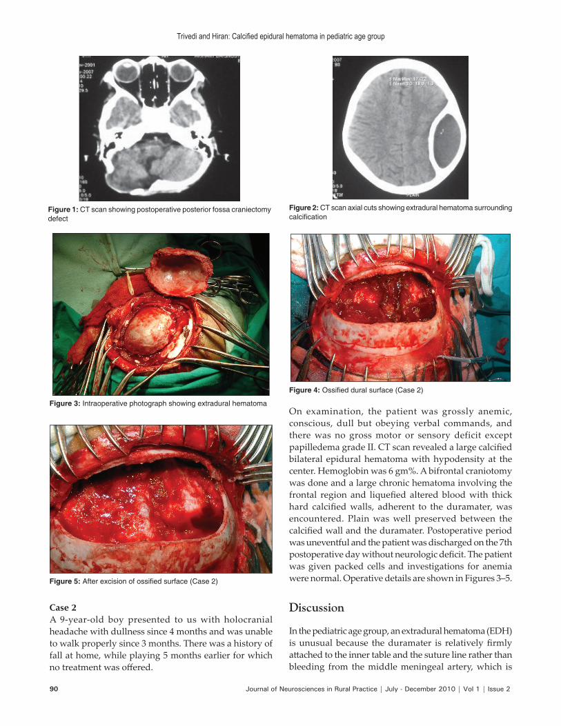
Figure 4: Ossifi ed dural surface (Case 2)
Figure 5: After excision of ossifi ed surface (Case 2)
Figure 2: CT scan axial cuts showing extradural hematoma surrounding calcifi cation
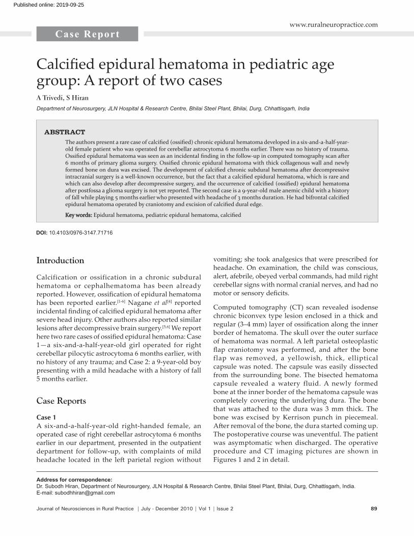
Figure 3: Intraoperative photograph showing extradural hematomaOn examination, the patient was grossly anemic, conscious, dull but obeying verbal commands, and there was no gross motor or sensory deficit except papilledema grade II. CT scan revealed a large calcifi ed bilateral epidural hematoma with hypodensity at the center. Hemoglobin was 6 gm%. A bifrontal craniotomy was done and a large chronic hematoma involving the frontal region and liquefi ed altered blood with thick hard calcifi ed walls, adherent to the duramater, was encountered. Plain was well preserved between the calcifi ed wall and the duramater. Postoperative period was uneventful and the patient was discharged on the 7th postoperative day without neurologic defi cit. The patient was given packed cells and investigations for anemia were normal. Operative details are shown in Figures 3–5.
Discussion
In the pediatric age group, an extradural hematoma (EDH) is unusual because the duramater is relatively fi rmly att ached to the inner table and the suture line rather than bleeding from the middle meningeal artery, which is

Journal of Neurosciences in Rural Practice | July - December 2010 | Vol 1 | Issue 2 91
Trivedi and Hiran: Calcifi ed epidural hematoma in pediatric age group
common in adults. In children, the cause of a hematoma can be a hemorrhage of venous blood, which takes longer to accumulate before it causes a signifi cant mass eff ect, and this could be the reason why most of the children and infants with epidural hematoma show no deterioration of consciousness, and presented with chronic form.[7,8]
In our case, the patient was operated for right cerebellar astrocytoma 6 months earlier. The patient was asymptomatic for 2 months and then developed mild headache without any neurologic defi cits indicating that the fi rst surgery reduced the intracranial pressure and some venous blood oozed out and accumulated at the left parietal region and became chronic. Consequently, ossifi cation developed in a short period before the natural absorption of blood.
Reports have shown that cephalhematoma in neonates is commonly absorbed within 1 month and if it persists for more than a month, it begins to calcify The subperiosteal osteogenesis is responsible for ossifi cation and deformity of the skull in cephalhematoma.[9-12]
Ikakuma and Brunngraber[13] reported that encapsulated epidural hematoma and microscopic ossifi cation were found in the thin layer of granulation tissue on the dural surface. Nakamura et al suggested that a fi broblast layer emerges adjacent to the dura as early as 4 days aft er bleeding, which develops into sinusoidal channel layers in 2 or 3 weeks. These fi brous layers then extend toward the cranial vault from the hematoma margin and subsequently form a connective tissue layer to form the fi brous capsule around the hematoma.
Iwakuma and Brunngraber[3] also suggested that active bone formation starts at the junction between dura and hematoma, but Nagane et al[4] reported that epidural hematoma ossified in both active and regressive manner, however, require more time. In our case, the ossifi cation developed because of an active process. Osteogenesis occurred at periosteal layer of dura that was in contact with epidural hematoma. The mechanism of ossifi cation in our case is similar to the ossifi cation of cephalhematoma in neonates. The present case is unique in terms of ossifi cation of EDH in contralateral supratentorial space aft er posterior fossa surgery.
Head injuries are a public health problem worldwide. Incidence of surgical and nonsurgical EDH among patients with traumatic brain injury has been reported to be in the range of 2.7%–4% with mortality around 5% in children and 7%–12.5% in adults. The precise mechanism of an osseous transformation is still not well understood,
and it has been hypothesized that damage to vascularized tissues, such as bone and dura provokes infl ammation, repair, and remodeling, in the tissues.[14] This natural sequence of healing is more rapid in children than in adults. Moreover, expansion of an EDH may result from repeated bleeding in the inner table of the skull[14] or because of oozing of blood from the dural surface veins.[15]
In conclusion, it can be stated that the blood accumulates slowly from a venous source and becomes chronic EDH and becomes ossifi ed, especially in the pediatric age group. Therefore, close surveillance with serial CT scans are mandatory and urgent removal of EDH should be considered for children with progressive neurologic defi cits.
We recommend a detailed examination of the history and serial CT scan aft er decompressive intracranial surgery and head injury, especially in the pediatric population to identify the early formation of epidural hematoma and removal before it gets ossifi ed.
References
1. Cambria S, Marra GA, Di Perri R, Bramanti P. Ossifi ed epidural hematoma: Report of case with epilepsy. J Neurosurg Sci 1985;29:285-8.
2. Grant WT. Chronic extradural hematoma: Report of a case of hematoma in anterior cranial fossa. Bull Los Angeles Neurol Soc 1944;9:156-62.
3. Iwakuma T, Brunngraber CV. Extradural ossifi cation following an extradural hematoma. J Neurosurg 1974;41:104-6.
4. Nagane M, Oyama H, Shibui S, Nomura K, Nakanishi Y, Kamiya M. Ossifi ed and calcifi ed epidural hematoma incidentally found 40 years after head injury: Case report. Surg Neurol 1994;42:65-9.
5. Parkinson D, Reddy V, Taylor J. Ossifi ed epidural hematoma: Case report. Neurosurgery 1980;7:171-3.
6. Whisler WW, Voris HC. Ossifi ed epidural hematoma following posterior fossa exploration. Report of case. J Neurosurg 1965;23:214-6.
7. Aldrich EF, Levin HS, Eisenberg HM. Mild head injury in children. In: Youmans JR, editor. Neurological surgery. Philadelphia: Saunders; 1996. p. 1719-29.
8. King AB, Chambers JW. Delayed onset of symptoms due to extradural hematomas. Surgery 1952;31:839-44.
9. Drake JM, Siddiqi SN. Birth trauma. In: Youmans JR, editor. Neurological surgery. Philadelphia: Saunders; 1996. p. 1767-76.
10. Firlik KS, Adelson PD. Large chronic cephalohematoma without calcifi cation. Pediatr Neurosurg 1999;30:39-42.
11. Kaufman HH, Hochberg J, Anderson RP, Schochet SS Jr, Simmons GM Jr. Treatment of calcifi ed cephalhematoma. Neurosurgery 1993;32:1037-40.
12. Morgan JE. Calcifi cation in cephalhematoma of the newborn infant. Am J Obstet Gynecol 1944;48:702-5.
13. Iwakima T, Brunngraber CV. Chronic extradural hematomas: A study of 21 cases. J Neurosurg 1973;38:488-93.
14. Buckwalter JA, Cruess RL. Healing of musculoskeletal tissues. In: Rockwood CA, Green DP, Bucholz RW, editors. Fracture in adults. 3rd ed, Vol. 1. Philadelphia: Lippincott; 1991. p. 181-222
15. Kaye EM, Cass PR, Dooling E, Rosman NP. Chronic epidural haematoma in childhood: Increased recognition and non surgical management. Pediatr Neurol 1985;1:255-9.
Source of Support: Nil, Confl ict of Interest: None declared.