Embed Size (px)
Citation preview
347Vol. 68, September-October 2011
Spontaneous cerebral infarction in a full-term neonate
clinical case
Bol Med Hosp Infant Mex 2011;68(5):347-351
Roberto Flores Santos,1 Cindy Viviana Veloz Serrano,1 Ricardo Jorge Hernández Herrera,1 and Francisco García Quintanilla2
ABSTRACTBackground. Cerebral vascular accident (CVA) is an important cause of hemorrhagic or ischemic cerebral injury and increases neonatal morbidity and mortality. It occurs in 1/4000 term neonates. We report a case of a neonate with a spontaneous CVA. Case report. We present the case of a newborn (NB) who was delivered from a 31-year old mother. It was the second pregnancy with 41.5 weeks of gestation. The mother presented gestational diabetes controlled only by dietary therapy. A 3640-g, apparently healthy female was obtained by cesarean delivery that was indicated due to cephalopelvic disproportion. Apgar scores were 8-9 according to the conven-tional time points. The pediatrician used only initial steps of reanimation. During the first day of life, the infant presented a deviation of the right mouth commissure and tonic-clonic movements on the right half of the body two times. The newborn was treated with phenobarbital intravenous infusion. Laboratory tests were all normal, and cultures of cerebrospinal fluid and blood were considered negative. A head sonogram showed no evidence of hemorrhage or ventricular distortion but a cranial CT reported a low-density zone suggesting a cerebral infarction in the left parietal and temporal regions. Conclusions. We continued to treat with phenobarbital and acetylsalicylic acid and the patient remained asymptomatic prior to discharge at the 7th day of life, recommending follow-up with a pediatric neurologist.Key words: spontaneous cerebral infarction, full term neonate.
1 Unidad Médica de Alta Especialidad No. 23, Instituto Mexicano del Seguro Social, Monterrey, Nuevo Leon, Mexico
2 Centro de Radiodiagnóstico e Imagen, Monterrey, Nuevo León, México
Correspondence: Dr. Ricardo Jorge Hernández Herrera1Unidad Médica de Alta Especialidad No. 23 Instituto Mexicano del Seguro Social Monterrey, Nuevo Leon, MexicoE-mail: [email protected]
Received for publication: 3-2-10Accepted for publication: 7-3-10
INTRODUCTION
The most common and important cause of neurological disorders in the neonatal period is hypoxia-ischemia asso-ciated with perinatal asphyxia. It presents with a frequency of 1/1.5% of newborns (NB) during the antenatal period in 20% of the cases, during the intrapartum period in 30% of cases and during the postnatal period in 45% of cases.1-4 The primary cause is lack of oxygen to the brain, which determines hypoxia or ischemia.1 Gestational age and birth weight are factors to consider because it affects 0.5% of full-term newborns and 9% of premature babies.1 The etio-logy and pathogenesis of hypoxia-ischemia are essentially related with intrauterine and intrapartum asphyxia, hyaline
membrane disease, cyanotic heart disease or persistence of fetal circulation or sepsis with secondary cardiovascular collapse. Other maternal causes are toxemia, diabetes, drug abuse, infections and thrombotic placental events or ma-ternal autoimmune disease and hypercoagulable states.1,2,5,6 The prevalence of neonatal cerebral infarction is relatively common and occurs in 1/4,000 births.3,6 The pathogenesis of cerebral infarction begins with the abrupt cessation of cerebral arterial blood flow and is secondary to thrombo-sis.7 Vascular thrombotic processes are rare in childhood, although half of these occur during the neonatal period. Its cause has been associated with secondary conditions such as metabolic acidosis, dehydration, sepsis, polycythemia, or congenital defects of the fibrinolytic system present in 50% of cases, such as antithrombin III deficiency of C and S proteins as well as resistance to C-activated protein (also known as Leiden factor).1,3 We report the case of a neonate with spontaneous cerebral infarction.
CLINICAL CASE
We report the case of a full-term newborn with a cere-brovascular accident (CVA) of apparently spontaneous origin. We obtained information of the imaging studies that corresponded to two computed axial tomography (CAT)
348 Bol Med Hosp Infant Mex
Roberto Flores Santos, Cindy Viviana Veloz Serrano, Ricardo Jorge Hernández Herrera and Francisco García Quintanilla
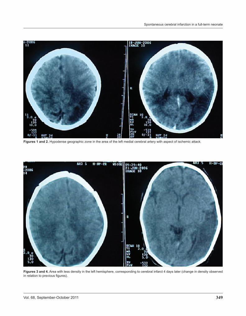
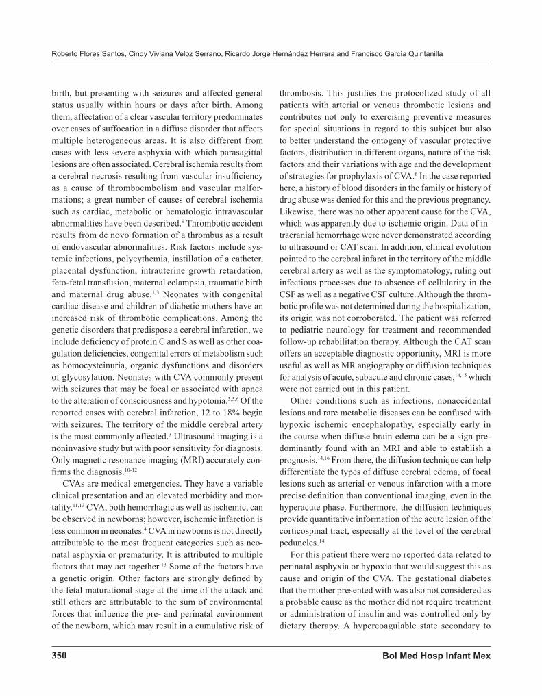
scans of the brain that were taken on the 3rd and 5th days of life. The 31-year-old mother demonstrated blood group A positive. HIV and VDRL results were negative. This was her second, full-term pregnancy with a history of previous birth. The mother had gestational diabetes for which she received only dietary treatment. Labor started at 41.5 wee-ks of gestation measuring from the last menstrual period; cesarean section was indicated because of cephalopelvic disproportion and the outcome was a single, live female with Apgar of 8-9, weighing 3,640 g, length 50 cm, and head circumference 36 cm. The infant received initial procedures and was moved to the accommodation with the mother. During the infant’s stay, she presented with an event with deviation of the right corner of the mouth accompanied by tonic-clonic movements in the right he-misphere. The neonate was transferred to the pathological nursery. Upon her admission, she presented a new seizure event with the same characteristics. The patient was trea-ted with phenobarbital. Physical examination showed anterior normotensive fontanel, facies with epicanthal folds, ocular hypertelorism, and slightly depressed nasal bridge. The remainder of the examination was normal. Samples were taken for laboratory tests that reported blood glucose 49 mg/dl, sodium 143 mg/dl, potassium 4.8 mg/dl, calcium 8.4 mg/dl, and creatinine 0.6 mg/dl. Clotting times and blood count were normal. Blood cultu-res and cerebrospinal fluid (CSF) cultures were negative. Chest x-ray was normal. Lumbar puncture was traumatic. Benzodiazepines were administered due to the presence of a third seizure event without quantifying the duration time. A transfontanel ultrasound showed no hemorrhage or ventricular dysfunction and a color Doppler showed a probable decrease in the flow of the left middle cerebral artery. Cerebral infarction was suspected and confirmed with a CAT scan conducted on the third day of life. A hypodense area was reported with geographic aspect of an ischemic event in the territory of the middle cerebral artery in the temporoparietal region (Figures 1 and 2). The patient was assessed by a pediatric neurologist who or-dered continuation with phenobarbital and acetylsalicylic acid; no seizure or neurological events were reported after that. We performed control CAT scan on the 5th day of life. It showed the same described lesion without increase in size or with the presence of new lesions (Figures 3 and 4). There was no documentation of an electroencephalogram or an MRI having been conducted or evidence of hyper-
coagulability tests. Imaging studies were performed after obtaining informed consent of the family. The patient was discharged on the 7th day of life with anticonvulsant and antiplatelet management and with an outpatient follow-up appointment with a pediatric neurologist.
DISCUSSION The physiopathogenesis of cerebral infarction in a full-term newborn is primarily associated with generalized hypoxia-ischemia or secondary to lack of oxygen or lack of appropriate blood flow that conditions an anaerobic me-chanism with reduction of the phosphorus-rich compounds in energy and the production and accumulation of toxic elements, eventually causing additional neuronal damage. Among the circulatory factors, altered cerebral blood flow plays a major role in the genesis of hypoxia-ischemia; hence, the importance of maintaining an adequate cere-bral perfusion. Cerebral vessels have the characteristic of self-regulating this flow, independent of the systemic blood pressure, through intrinsic mechanisms regulating arterioles.1,3 After the hypoxic-ischemic insult, a phenome-non occurs of flow redistribution to preserve the brain and heart, with a reduction of irrigation to other organs such as kidneys and lungs. There is increased sympathetic activity and release of substances such as arginine-vasopressin that increase peripheral resistance and blood flow. This response is enhanced by self-regulatory mechanisms such as arterial PCO2 and perivascular pH1 that decrease the cerebral glycogen reserves and phosphocreatinine. Lactic acid is produced, and glucose and ATP are reduced, indu-cing anaerobic glucose utilization and increased acidosis. This results in neuronal damage.1 The presence of seizures, pointing to the debut of the lesion, is the result of edema that occurs later and appears to contribute to the severity of the lesion, especially if seizures are prolonged.5,8 Type of seizure may be focal, associated with or without apnea with impaired consciousness and hypotonia. In some cases they may be asymptomatic or show an asymmetric motor function.7,8 Treatment requires admission to an intensive care unit (ICU) for appropriate treatment with a ventilator, metabolic correction, infusion maintenance, and mana-gement of seizures and complications that may arise.1 Outside the context of acute neonatal encephalopathy due to asphyxia, CVA occurs mostly in full-term newborns often of first-time mothers with a usually anodyne child-
349Vol. 68, September-October 2011
Spontaneous cerebral infarction in a full-term neonate
Figures 3 and 4. Area with less density in the left hemisphere, corresponding to cerebral infarct 4 days later (change in density observed in relation to previous figures).
Figures 1 and 2. Hypodense geographic zone in the area of the left medial cerebral artery with aspect of ischemic attack.
350 Bol Med Hosp Infant Mex
Roberto Flores Santos, Cindy Viviana Veloz Serrano, Ricardo Jorge Hernández Herrera and Francisco García Quintanilla
birth, but presenting with seizures and affected general status usually within hours or days after birth. Among them, affectation of a clear vascular territory predominates over cases of suffocation in a diffuse disorder that affects multiple heterogeneous areas. It is also different from cases with less severe asphyxia with which parasagittal lesions are often associated. Cerebral ischemia results from a cerebral necrosis resulting from vascular insufficiency as a cause of thromboembolism and vascular malfor-mations; a great number of causes of cerebral ischemia such as cardiac, metabolic or hematologic intravascular abnormalities have been described.9 Thrombotic accident results from de novo formation of a thrombus as a result of endovascular abnormalities. Risk factors include sys-temic infections, polycythemia, instillation of a catheter, placental dysfunction, intrauterine growth retardation, feto-fetal transfusion, maternal eclampsia, traumatic birth and maternal drug abuse.1,3 Neonates with congenital cardiac disease and children of diabetic mothers have an increased risk of thrombotic complications. Among the genetic disorders that predispose a cerebral infarction, we include deficiency of protein C and S as well as other coa-gulation deficiencies, congenital errors of metabolism such as homocysteinuria, organic dysfunctions and disorders of glycosylation. Neonates with CVA commonly present with seizures that may be focal or associated with apnea to the alteration of consciousness and hypotonia.3,5,6 Of the reported cases with cerebral infarction, 12 to 18% begin with seizures. The territory of the middle cerebral artery is the most commonly affected.3 Ultrasound imaging is a noninvasive study but with poor sensitivity for diagnosis. Only magnetic resonance imaging (MRI) accurately con-firms the diagnosis.10-12
CVAs are medical emergencies. They have a variable clinical presentation and an elevated morbidity and mor-tality.11,13 CVA, both hemorrhagic as well as ischemic, can be observed in newborns; however, ischemic infarction is less common in neonates.4 CVA in newborns is not directly attributable to the most frequent categories such as neo-natal asphyxia or prematurity. It is attributed to multiple factors that may act together.13 Some of the factors have a genetic origin. Other factors are strongly defined by the fetal maturational stage at the time of the attack and still others are attributable to the sum of environmental forces that influence the pre- and perinatal environment of the newborn, which may result in a cumulative risk of
thrombosis. This justifies the protocolized study of all patients with arterial or venous thrombotic lesions and contributes not only to exercising preventive measures for special situations in regard to this subject but also to better understand the ontogeny of vascular protective factors, distribution in different organs, nature of the risk factors and their variations with age and the development of strategies for prophylaxis of CVA.6 In the case reported here, a history of blood disorders in the family or history of drug abuse was denied for this and the previous pregnancy. Likewise, there was no other apparent cause for the CVA, which was apparently due to ischemic origin. Data of in-tracranial hemorrhage were never demonstrated according to ultrasound or CAT scan. In addition, clinical evolution pointed to the cerebral infarct in the territory of the middle cerebral artery as well as the symptomatology, ruling out infectious processes due to absence of cellularity in the CSF as well as a negative CSF culture. Although the throm-botic profile was not determined during the hospitalization, its origin was not corroborated. The patient was referred to pediatric neurology for treatment and recommended follow-up rehabilitation therapy. Although the CAT scan offers an acceptable diagnostic opportunity, MRI is more useful as well as MR angiography or diffusion techniques for analysis of acute, subacute and chronic cases,14,15 which were not carried out in this patient.
Other conditions such as infections, nonaccidental lesions and rare metabolic diseases can be confused with hypoxic ischemic encephalopathy, especially early in the course when diffuse brain edema can be a sign pre-dominantly found with an MRI and able to establish a prognosis.14,16 From there, the diffusion technique can help differentiate the types of diffuse cerebral edema, of focal lesions such as arterial or venous infarction with a more precise definition than conventional imaging, even in the hyperacute phase. Furthermore, the diffusion techniques provide quantitative information of the acute lesion of the corticospinal tract, especially at the level of the cerebral peduncles.14
For this patient there were no reported data related to perinatal asphyxia or hypoxia that would suggest this as cause and origin of the CVA. The gestational diabetes that the mother presented with was also not considered as a probable cause as the mother did not require treatment or administration of insulin and was controlled only by dietary therapy. A hypercoagulable state secondary to
351Vol. 68, September-October 2011
Spontaneous cerebral infarction in a full-term neonate
polycythemia was ruled out because the initial hematocrit was normal. It is obvious that due to the complexity of the heterogeneous physiopathological mechanisms involved in cerebral ischemia in different groups of neonates, ma-nagement should be based on monitoring of the newborn during the acute phase of the ischemic accident. During early diagnosis of the physiopathological situation and determination of the “therapeutic window,” its action can be blocked by rechanneling vessels using thrombolytic therapy and neuroprotective treatment on cerebral tissue capable of recovery.1 We suggest that when facing a pa-tient with seemingly ischemic CVA, we should consider the hypercoagulable states mentioned above, keeping in mind the possible causes of genetic alteration of coagu-lation factors.
REFERENCES
1. Campos-Castelló J, Canelón de López MS, Santiago-Gómez R. Accidentes vasculares isquémicos y hemorrágicos cere-brales del recién nacido a término. Protocolo de estudio y orientaciones terapéuticas. Rev Neurol 2000;31:632-644.
2. Kirton A, deVeber G. Cerebral palsy secondary to perinatal ischemic stroke. Clin Perinatol 2006;33:367-386.
3. Akman Y, Özek E, Yýlmaz Y, Bilgen H. Cerebral infarcts in full term neonates. Turk J Pediatrics 2003;45:141-147.
4. Estan J, Hope P. Unilateral neonatal cerebral infarction in full term infants. Arch Dis Child Fetal Neonatal Ed 1997;76:F88-F93.
5. Mercuri E, Cowan F, Gupte G, Manning R, Laffan M, Rutherford M, et al. Prothrombotic disorders and abnormal neurodevelop-
mental outcome in infants with neonatal cerebral infarction. Pediatrics 2001;107:1400-1404.
6. Vergara E, Busselo S, Cortajarena M, Collado V, Echeverría J, Gaztañaga R, et al. Infarto cerebral arterial tras traumatismo al nacimiento. An Pediatr 2006;64:379-384.
7. Dunn DW, Austin JK. Differential diagnosis and treatment of psychiatric disorders in children and adolescents with epilepsy. Epilepsy Behav 2004;5(suppl 3):S10-S17.
8. Mercuri E, Rutherford M, Cowan F, Pennock J, Counsell S, Papadimitriou M, et al. Early prognostic indicators of outcome in infants with neonatal cerebral infarction: a clinical, elec-troencephalogram, and magnetic resonance imaging study. Pediatrics 1999;103:39-46.
9. Volpe JJ. Hypoxic ischemic encephalopathy: clinical aspects. In: Volpe JJ, ed. Neurology of the Newborn. Philadelphia: Saunders; 2000. pp. 315-330.
10. Lövblad KO, Ruoss K, Guzman R, Schroth G, Fusch C. Diffusion-weighted MRI of middle cerebral artery stroke in a newborn. Pediatr Radiol 2001;31:374-376.
11. Galicia PG, Aragón GM. Convulsiones neonatales. Bol Pediatr 2006;46(suppl 1):145-150.
12. Cowan F, Mercuri E, Groenendaal F, Bassi L, Ricci D, Ruther-ford M, et al. Does cranial ultrasound imaging identify arterial cerebral infarction in term neonates? Arch Dis Child Fetal Neonatal Ed 2005;90:F252-F256.
13. Govaert A, Matthys E, Zecic A, Roelens F, Oostra A, Vanzieleg-hem B. Perinatal cortical infarction within middle cerebral artery trunks. Arch Dis Child Fetal Neonatal Ed 2000;82:F59-F63.
14. Triulzi F, Parazzini C, Righini A. Patterns of damage in the mature neonatal brain. Pediatr Radiol 2006;36:608-620.
15. Sandberg DI, Lamberti-Pasculli M, Drake JM, Humphreys RP, Rutka JT. Spontaneous intraparenchymal hemorrhage in full-term neonates. Neurosurgery 2001;48:1042-1048.
16. Chang LW, Liu J, Li WB, Zhu WZ. Value of magnetic resonan-ce imaging in the early evaluation of prognosis for hypoxic-ischemic encephalopathy in full-term infants. Zhongguo Dang Dai Er Ke Za Zhi 2007;9:407-410.