Embed Size (px)
Citation preview

c h a p t e r
The Muscular System
Scanning electronmicrograph of motorneurons terminating atmuscle fibers. A musclefiber receives thestimulus to contract at aneuromuscular junction.
chapter outline & learning objectives After you have studied this chapter, you should be able to:
7.1 Functions and Types of Muscles(p. 114) ■ Distinguish between the three types of
muscles, and tell where they are located inthe body.
■ Describe the connective tissues of a skeletalmuscle.
■ Name and discuss five functions of skeletalmuscles.
7.2 Microscopic Anatomy andContraction of Skeletal Muscle (p. 116)■ Name the components of a skeletal muscle
fiber, and describe the function of each.■ Explain how skeletal muscle fibers are
innervated and how they contract.■ Describe how ATP is made available for
muscle contraction.
7.3 Muscle Responses (p. 122)■ Contrast the responses of a muscle fiber and
whole muscle in the laboratory with theirresponses in the body.
■ Contrast slow-twitch and fast-twitch musclefibers.
7.4 Skeletal Muscles of the Body(p. 124)■ Discuss how muscles work together to
achieve the movement of a bone.■ Give examples to show how muscles are
named.■ Describe the locations and actions of the
major skeletal muscles of each body region.
7.5 Effects of Aging (p. 134)■ Describe the anatomical and physiological
changes that occur in the muscular system aswe age.
7.6 Homeostasis (p. 136)■ Describe how the muscular system works with
other systems of the body to maintainhomeostasis.
■ Describe some common muscle disorders andsome of the serious diseases that can affectmuscles.
Visual FocusAnatomy of a Muscle Fiber (p. 117)
Medical FocusBenefits of Exercise (p. 135)
113
mad64372_ch07pg113_139 4/9/03 6:28 PM Page 113 seema Seema-QXP-4:Inprocess-4:Rakesh(GAJ)03-09:mad5ch07:

7.1 Functions and Typesof MusclesAll muscles, regardless of the particular type, can contract—that is, shorten. When muscles contract, some part of thebody or the entire body moves. Humans have three types ofmuscles: smooth, cardiac, and skeletal (Fig. 7.1). The contrac-tile cells of these tissues are elongated and therefore are calledmuscle fibers.
Smooth MuscleSmooth muscle is located in the walls of hollow internal or-gans, and its involuntary contraction moves materialsthrough an organ. Smooth muscle fibers are spindle-shapedcells, each with a single nucleus (uninucleated). The cells areusually arranged in parallel lines, forming sheets. Smoothmuscle does not have the striations (bands of light and dark)seen in cardiac and skeletal muscle. Although smooth muscleis slower to contract than skeletal muscle, it can sustain pro-longed contractions and does not fatigue easily.
Cardiac Muscle Cardiac muscle forms the heart wall. Its fibers are uninucle-ated, striated, tubular, and branched, which allows the fibersto interlock at intercalated disks. Intercalated disks permit
contractions to spread quickly throughout the heart. Cardiacfibers relax completely between contractions, which preventsfatigue. Contraction of cardiac muscle fibers is rhythmical; itoccurs without outside nervous stimulation or control. Thus,cardiac muscle contraction is involuntary.
Skeletal MuscleSkeletal muscle fibers are tubular, multinucleated, and stri-ated. They make up the skeletal muscles attached to the skele-ton. Skeletal muscle fibers can run the length of a muscle andtherefore can be quite long. Skeletal muscle is voluntary be-cause its contraction is always stimulated and controlled bythe nervous system. In this chapter, we will explore why skele-tal muscle (and cardiac muscle) is striated.
Connective Tissue Coverings
Muscles are organs, and as such they contain other types of tis-sues, such as nervous tissue, blood vessels, and connective tis-sue. Connective tissue is essential to the organization of thefibers within a muscle (Fig. 7.2). First, each fiber is surroundedby a thin layer of areolar connective tissue called the endomy-sium. Blood capillaries and nerve fibers reach each musclefiber by way of the endomysium. Second, the muscle fibers aregrouped into bundles called fascicles. The fascicles have asheath of connective tissue called the perimysium. Finally, the
114 Part II Support, Movement, and Protection
Figure 7.1 Types of muscles. The three types of muscles in the body have the appearance and characteristics shown here.
Smooth muscle• has spindle-shaped, nonstriated, uninucleated fibers.• occurs in walls of internal organs.• is involuntary.
Cardiac muscle• has striated, tubular, branched, uninucleated fibers.• occurs in walls of heart.• is involuntary.
Skeletal muscle• has striated, tubular, multinucleated fibers.• is usually attached to skeleton.• is voluntary.
mad64372_ch07pg113_139 9/1/03 4:26 PM Page 114

muscle itself is covered by a connective tissue layer called theepimysium. The epimysium becomes a part of the fascia, a layerof fibrous tissue that separates muscles from each other (deepfascia) and from the skin (superficial fascia). Collagen fibersof the epimysium continue as a strong, fibrous tendon that at-taches the muscle to a bone. The epimysium merges with theperiosteum of the bone.
Functions of Skeletal Muscles
This chapter concerns the skeletal muscles, and therefore it isfitting to consider their functions independent of the othertypes of muscles:
Skeletal muscles support the body. Skeletal muscle contractionopposes the force of gravity and allows us to remain
upright. Some skeletal muscles are serving this purposeeven when you think you are relaxed.
Skeletal muscles make bones and other body parts move. Musclecontraction accounts not only for the movement oflimbs but also for eye movements, facial expressions,and breathing.
Skeletal muscles help maintain a constant body temperature.Skeletal muscle contraction causes ATP to break down,releasing heat that is distributed about the body.
Skeletal muscle contraction assists movement in cardiovascularand lymphatic vessels. The pressure of skeletal musclecontraction keeps blood moving in cardiovascular veinsand lymph moving in lymphatic vessels.
Skeletal muscles help protect internal organs and stabilize joints.Muscles pad the bones that protect organs, and theyhave tendons that help hold bones together at joints.
115Chapter 7 The Muscular System
Figure 7.2 Connective tissue of a skeletal muscle. a. Trace the connective tissue of a muscle from the endomysium to the perimysiumto the epimysium, which becomes a part of the deep fascia and from which the tendon extends to attach a muscle to the periosteum of abone. b. Cross section of the arm showing the arrangement of the muscles, which are separated from the skin by fascia. The superficialfascia contains adipose tissue. c. Photomicrograph of muscle fascicles from the tongue where the fascicles run in different directions. (c.s. = cross section; l.s. = longitudinal section.)
skeletalmuscle
perimysium
epimysium
a.
articularcartilage
endomysium
fascicle
musclefibers
osseoustissue
periosteum(cut)
tendon
deep fascia
skinsuperficial fascia(adipose tissue)
nerve
artery
deep fascia
vein
fascicles
humerus
individualmuscle
b.
Lateral Medial
perimysium
muscle fiber, c.s.
fascicle, c.s.
fascicle, l.s.
endomysium
c.
mad64372_ch07pg113_139 9/1/03 4:26 PM Page 115

7.2 Microscopic Anatomy andContraction of Skeletal Muscle We have already examined the structure of skeletal muscle asseen with the light microscope. As you know, skeletal muscletissue has alternating light and dark bands, giving it a striatedappearance. The electron microscope shows that these bandsare due to the arrangement of myofilaments in a muscle fiber.
Muscle Fiber A muscle fiber contains the usual cellular components, butspecial names have been assigned to some of these compo-nents (Table 7.1 and Figure 7.3). The plasma membrane iscalled the sarcolemma; the cytoplasm is the sarcoplasm; andthe endoplasmic reticulum is the sarcoplasmic reticulum. Amuscle fiber also has some unique anatomical characteristics.One feature is its T (for transverse) system; the sarcolemmaforms T (transverse) tubules that penetrate, or dip down,into the cell so that they come into contact—but do notfuse—with expanded portions of the sarcoplasmic reticulum.The expanded portions of the sarcoplasmic reticulum are cal-cium storage sites. Calcium ions (Ca2!), as we shall see, areessential for muscle contraction.
The sarcoplasmic reticulum encases hundreds and some-times even thousands of myofibrils, each about 1 "m in
diameter, which are the contractile portions of the musclefibers. Any other organelles, such as mitochondria, are locatedin the sarcoplasm between the myofibrils. The sarcoplasmalso contains glycogen, which provides stored energy for mus-cle contraction, and the red pigment myoglobin, which bindsoxygen until it is needed for muscle contraction.
Myofibrils and Sarcomeres
Myofibrils are cylindrical in shape and run the length of themuscle fiber. The striations of skeletal muscle fibers are formedby the placement of myofilaments within units of myofibrilscalled sarcomeres. A sarcomere extends between two darklines called the Z lines. A sarcomere contains two types ofprotein myofilaments. The thick filaments are made up of aprotein called myosin, and the thin filaments are made up ofa protein called actin. Other proteins are also present. The Iband is light colored because it contains only actin filamentsattached to a Z line. The dark regions of the A band containoverlapping actin and myosin filaments, and its H zone hasonly myosin filaments.
Myofilaments
The thick and thin filaments differ in the following ways:
Thick Filaments A thick filament is composed of severalhundred molecules of the protein myosin. Each myosin mol-ecule is shaped like a golf club, with the straight portion ofthe molecule ending in a double globular head, or cross-bridge. Cross-bridges are slanted away from the middle of asarcomere.
Thin Filaments Primarily, a thin filament consists of two in-tertwining strands of the protein actin. Two other proteins,called tropomyosin and troponin, are also present, as we willdiscuss later in this section.
Sliding Filaments We will also see that when muscles areinnervated, impulses travel down a T tubule, and calcium isreleased from the sarcoplasmic reticulum. Now the musclefiber contracts as the sarcomeres within the myofibrilsshorten. When a sarcomere shortens, the actin (thin) fila-ments slide past the myosin (thick) filaments and approachone another. This causes the I band to shorten and the H zoneto almost or completely disappear. The movement of actin fil-aments in relation to myosin filaments is called the slidingfilament theory of muscle contraction. During the slidingprocess, the sarcomere shortens even though the filamentsthemselves remain the same length. ATP supplies the energyfor muscle contraction. Although the actin filaments slidepast the myosin filaments, it is the myosin filaments that dothe work. Myosin filaments break down ATP and have cross-bridges that pull the actin filaments toward the center of thesarcomere.
116 Part II Support, Movement, and Protection
Table 7.1 Microscopic Anatomy of a Muscle
Name Function
Sarcolemma Plasma membrane of a muscle fiber that forms T tubules
Sarcoplasm Cytoplasm of a muscle fiber thatcontains organelles, including myofibrils
Glycogen A polysaccharide that stores energyfor muscle contraction
Myoglobin A red pigment that stores oxygen for muscle contraction
T tubule Extension of the sarcolemma thatextends into the muscle fiber and conveys impulses that cause Ca2!
to be released into the sarcoplasmicreticulum
Sarcoplasmic reticulum The smooth ER of a muscle fiber thatstores Ca2!
Myofibril A bundle of myofilaments thatcontracts
Myofilament Actin filaments and myosin filaments whose structure and functions account for muscle striations and contractions
mad64372_ch07pg113_139 25/9/03 6:49 PM Page 116 seema Seema-QXP-4:Desktop Folder:mad5ch07:

117Chapter 7 The Muscular System
Figure 7.3 Anatomy of a muscle fiber. A muscle fiber contains many myofibrils with the components shown. A myofibril has manysarcomeres that contain myosin and actin filaments whose arrangement gives rise to the striations so characteristic of skeletal muscle.Muscle contraction occurs when sarcomeres contract and actin filaments slide past myosin filaments.
bundle ofmuscle fibers
musclefiber
T tubules
nucleus sarcoplasm
sarcolemma
cross-bridge
myosin
actin
sarcoplasmicreticulum
calciumstorage sites
one sarcomere
Z line Z line
H zone
Z line
Muscle fiber hasmany myofibrils.
skeletalmusclefiber
one myofibril
A band I band
Myofibril hasmany sarcomeres.
Sarcomereis relaxed.
Sarcomereis contracted.
mad64372_ch07pg113_139 19/9/03 5:45 PM Page 117 seema Seema-QXP-4:Desktop Folder:Rakesh(GAJ)19-09:mad5ch07:

Skeletal Muscle ContractionMuscle fibers are innervated—that is, they are stimulated tocontract by motor neurons whose axons are found in nerves.The axon of one motor neuron has several branches and canstimulate from a few to several muscle fibers of a particularmuscle. Each branch of the axon ends in an axon terminalthat lies in close proximity to the sarcolemma of a musclefiber. A small gap, called a synaptic cleft, separates the axonbulb from the sarcolemma. This entire region is called a neu-romuscular junction (Fig. 7.4).
Axon terminals contain synaptic vesicles that are filledwith the neurotransmitter acetylcholine (ACh). When nerveimpulses traveling down a motor neuron arrive at an axon ter-minal, the synaptic vesicles release a neurotransmitter intothe synaptic cleft. It quickly diffuses across the cleft and bindsto receptors in the sarcolemma. Now the sarcolemma gener-ates impulses that spread over the sarcolemma and down Ttubules to the sarcoplasmic reticulum. The release of calciumfrom the sarcoplasmic reticulum causes the filaments withinthe sarcomeres to slide past one another. Sarcomere contrac-tion results in myofibril contraction, which in turn results inmuscle fiber, and finally muscle, contraction.
118 Part II Support, Movement, and Protection
Figure 7.4 Neuromuscular junction. The branch of an axon ends in an axon terminal that meets but does not touch a muscle fiber. Asynaptic cleft separates the axon terminal from the sarcolemma of the muscle fiber. Nerve impulses traveling down an axon cause synapticvesicles to discharge acetylcholine, which diffuses across the synaptic cleft. When the neurotransmitter is received by the sarcolemma of amuscle fiber, impulses begin and lead to muscle fiber contractions.
axon terminal
synaptic vesicle
branch of motor nerve fiber
mitochondria
nucleus
synaptic cleft
neurotransmitter
axon terminalmyofibril
folded sarcolemma
receptor
mad64372_ch07pg113_139 9/1/03 4:26 PM Page 118

The Role of Actin and MyosinFigure 7.5 shows the placement of two other proteins associ-ated with an actin filament, which you will recall is com-posed of a double row of twisted actin molecules. Threads oftropomyosin wind about an actin filament, and troponin oc-curs at intervals along the threads. Calcium ions (Ca2!) thathave been released from the sarcoplasmic reticulum com-bine with troponin. After binding occurs, the tropomyosinthreads shift their position, and myosin binding sites areexposed.
The double globular heads of a myosin filament haveATP binding sites. The heads function as ATPase enzymes,splitting ATP into ADP and P!. This reaction activates thehead so that it will bind to actin. The ADP and P! remain on
the myosin heads until the heads attach to actin, forming across-bridge. Now, ADP and P! are released, and this causesthe cross-bridges to change their positions. This is the powerstroke that pulls the thin filaments toward the middle of thesarcomere. When another ATP molecule binds to a myosinhead, the cross-bridge is broken as the head detaches fromactin. The cycle begins again; the actin filaments movenearer the center of the sarcomere each time the cycle isrepeated.
Contraction continues until nerve impulses cease andcalcium ions are returned to their storage sites. The mem-branes of the sarcoplasmic reticulum contain active transportproteins that pump calcium ions back into the sarcoplasmicreticulum.
119Chapter 7 The Muscular System
Figure 7.5 The role of calcium and myosin in muscle contraction. a. Upon release, calcium binds to troponin, exposing myosin bindingsites. b. After breaking down ATP, myosin heads bind to an actin filament, and later, a power stroke causes the actin filament to move.
a.
b.
actin filament troponin
Troponin-Ca2+ complex pulls tropomyosin away,exposing myosin binding sites.
myosin bindingsites
tropomyosin
Ca2+
Ca2+
ADP+ release causes head to changeposition and actin filament to move.
P3.
ADP+ are bound to myosin asmyosin head attaches to actin.
P
ATP is hydrolyzed when myosinhead is unattached.
P
2.
actinfilament
ADP
Binding of ATP causes myosinhead to return to resting position.
4.
myosinfilament
myosin headcross-bridgeATP
1.
ATP
mad64372_ch07pg113_139 9/1/03 4:26 PM Page 119

Energy for Muscle ContractionATP produced previous to strenuous exercise lasts a few sec-onds, and then muscles acquire new ATP in three differentways: creatine phosphate breakdown, cellular respiration, andfermentation (Fig. 7.6). Creatine phosphate breakdown andfermentation are anaerobic, meaning that they do not requireoxygen.
Creatine Phosphate Breakdown
Creatine phosphate is a high-energy compound built upwhen a muscle is resting. Creatine phosphate cannot partici-pate directly in muscle contraction. Instead, it can regenerateATP by the following reaction:
This reaction occurs in the midst of sliding filaments, andtherefore is the speediest way to make ATP available to mus-cles. Creatine phosphate provides enough energy for onlyabout eight seconds of intense activity, and then it is spent.Creatine phosphate is rebuilt when a muscle is resting bytransferring a phosphate group from ATP to creatine.
Cellular Respiration
Cellular respiration completed in mitochondria usually pro-vides most of a muscle’s ATP. Glycogen and fat are stored inmuscle cells. Therefore, a muscle cell can use glucose fromglycogen and fatty acids from fat as fuel to produce ATP if oxy-gen is available:
Myoglobin, an oxygen carrier similar to hemoglobin, issynthesized in muscle cells, and its presence accounts for thereddish-brown color of skeletal muscle fibers. Myoglobinhas a higher affinity for oxygen than does hemoglobin.Therefore, myoglobin can pull oxygen out of blood andmake it available to muscle mitochondria that are carryingon cellular respiration. Then, too, the ability of myoglobinto temporarily store oxygen reduces a muscle’s immediateneed for oxygen when cellular respiration begins. The end
glucose + oxygen carbon dioxide + water
ADP ATP
creatine phosphate creatine
ADP ATP
products (carbon dioxide and water) are usually no prob-lem. Carbon dioxide leaves the body at the lungs, and watersimply enters the extracellular space. The by-product, heat,keeps the entire body warm.
Fermentation
Fermentation, like creatine phosphate breakdown, suppliesATP without consuming oxygen. During fermentation, glu-cose is broken down to lactate (lactic acid):
The accumulation of lactate in a muscle fiber makes the cyto-plasm more acidic, and eventually enzymes cease to functionwell. If fermentation continues longer than two or three min-utes, cramping and fatigue set in. Cramping seems to be dueto lack of the ATP needed to pump calcium ions back into thesarcoplasmic reticulum and to break the linkages between theactin and myosin filaments so that muscle fibers can relax.
Oxygen DeficitWhen a muscle uses fermentation to supply its energy needs,it incurs an oxygen deficit. Oxygen deficit is obvious when aperson continues to breathe heavily after exercising. Theability to run up an oxygen deficit is one of muscle tissue’sgreatest assets. Brain tissue cannot last nearly as long with-out oxygen as muscles can.
Repaying an oxygen deficit requires replenishing crea-tine phosphate supplies and disposing of lactic acid. Lacticacid can be changed back to pyruvic acid and metabolizedcompletely in mitochondria, or it can be sent to the liver toreconstruct glycogen. A marathon runner who has justcrossed the finish line is not exhausted due to oxygendeficit. Instead, the runner has used up all the muscles’, andprobably the liver’s, glycogen supply. It takes about twodays to replace glycogen stores on a high-carbohydrate diet.
People who train rely more heavily on cellular respirationthan do people who do not train. In people who train, thenumber of muscle mitochondria increases, and so fermenta-tion is not needed to produce ATP. Their mitochondria canstart consuming oxygen as soon as the ADP concentrationstarts rising during muscle contraction. Because mitochondriacan break down fatty acid, instead of glucose, blood glucose isspared for the activity of the brain. (The brain, unlike otherorgans, can only utilize glucose to produce ATP.) Because lesslactate is produced in people who train, the pH of the bloodremains steady, and there is less of an oxygen deficit.
glucose lactate
ADP ATP
120 Part II Support, Movement, and Protection
mad64372_ch07pg113_139 9/1/03 4:26 PM Page 120

121Chapter 7 The Muscular System
Figure 7.6 Energy sources for muscle contraction.
In resting muscle, creatinephosphate is built up.
O2 available:
To continue contracting, muscles either carry on cellular respiration(preferred) or carry on fermentation, which can lead to fatigue.
To start contracting, muscles break down creatine phosphate.
no O2 available:
lactateH2OCO2
pyruvate
Cellular respirationCreatine phosphatebreakdown Fermentation
ATP
ATP
ATP
+
+
muscle contraction
P
+ P
In a contracting muscle, ATP is broken down to ADP + P
ADP
creatine
creatinephosphate
glucose
P
+ P
ADP
creatine
creatinephosphate
.
mad64372_ch07pg113_139 9/1/03 4:26 PM Page 121

7.3 Muscle ResponsesMuscles can be studied in the laboratory in an effort to un-derstand how they respond when in the body.
In the LaboratoryWhen a muscle fiber is isolated, placed on a microscopeslide, and provided with ATP plus the various electrolytes itrequires, it contracts completely along its entire length. Thisobservation has resulted in the all-or-none law: A musclefiber contracts completely or not at all. In contrast, a wholemuscle shows degrees of contraction. To study whole musclecontraction in the laboratory, an isolated muscle is stimulatedelectrically, and the mechanical force of contraction isrecorded as a visual pattern called a myogram. When thestrength of the stimulus is above a threshold level, the musclecontracts and then relaxes. This action—a single contractionthat lasts only a fraction of a second—is called a muscletwitch. Figure 7.7 is a myogram of a muscle twitch, which iscustomarily divided into three stages: the latent period, or theperiod of time between stimulation and initiation of contrac-tion; the contraction period, when the muscle shortens; andthe relaxation period, when the muscle returns to its formerlength. It’s interesting to use our knowledge of muscle fibercontraction to understand these events. From our study thusfar, we know that a muscle fiber in an intact muscle contractswhen calcium leaves storage sacs and relaxes when calcium re-turns to storage sacs.
But unlike the contraction of a muscle fiber, a muscle hasdegrees of contraction, and a twitch can vary in height(strength) depending on the degree of stimulation. Whyshould that be? Obviously, a stronger stimulation causesmore individual fibers to contract than before.
If a whole muscle is given a rapid series of stimuli, it canrespond to the next stimulus without relaxing completely.Summation is increased muscle contraction until maximalsustained contraction, called a tetanic contraction, is achieved(Fig. 7.8). The myogram no longer shows individual twitches;rather, the twitches are fused and blended completely into astraight line. Tetanus continues until the muscle fatigues due todepletion of energy reserves. Fatigue is apparent when a mus-cle relaxes even though stimulation continues.
In the BodyIn the body, muscles are innervated to contract by nerves. Asmentioned, each axon within a nerve stimulates a number ofmuscle fibers. A nerve fiber together with all of the musclefibers it innervates is called a motor unit. A motor unit obeysthe all-or-none law. Why? Because all the muscle fibers in amotor unit are stimulated at once, and they all either contractor do not contract. A variable of interest is the number of mus-cle fibers within a motor unit. For example, in the ocular mus-cles that move the eyes, the innervation ratio is one motor axonper 23 muscle fibers, while in the gastrocnemius muscle of thelower leg, the ratio is about one motor axon per 1,000 musclefibers. No doubt, moving the eyes requires finer control thanmoving the legs.
122 Part II Support, Movement, and Protection
Figure 7.7 A myogram showing a single muscle twitch.
Forc
e of
Con
trac
tion
Timetime of
stimulation
contractionperiod
relaxationperiod
latentperiod
Figure 7.8 Myograms showing (a) a series of twitches, (b) summation, and (c) a tetanic contraction. Note that anincreased frequency of stimulations has resulted in these differentresponses.
Time
Forc
e of
Con
trac
tion
c.
Forc
e of
Con
trac
tion
b.
Forc
e of
Con
trac
tion
a.
mad64372_ch07pg113_139 9/1/03 4:26 PM Page 122

Slow-twitch muscle fiber• is aerobic• has steady power• has endurance
Fast-twitch muscle fiber• is anaerobic• has explosive power• fatigues easily
slow-twitchfibers
fast-twitchfibers
Tetanic contractions ordinarily occur in the body because,as the intensity of nervous stimulation increases, more andmore motor units are activated. This phenomenon, known asrecruitment, results in stronger and stronger muscle contrac-tions. But while some muscle fibers are contracting, others arerelaxing. Because of this, intact muscles rarely fatigue com-pletely. Even when muscles appear to be at rest, they exhibittone, in which some of their fibers are always contracting.Muscle tone is particularly important in maintaining posture.If all the fibers within the muscles of the neck, trunk, and legswere to suddenly relax, the body would collapse.
Athletics and Muscle Contraction
Athletes who excel in a particular sport, and much of the gen-eral public as well, are interested in staying fit by exercising.The Medical Focus on page 135 gives suggestions for exerciseprograms according to age.
Exercise and Size of Muscles Muscles that are not used orthat are used for only very weak contractions decrease in size,or atrophy. Atrophy can occur when a limb is placed in a castor when the nerve serving a muscle is damaged. If nerve stim-ulation is not restored, muscle fibers are gradually replaced byfat and fibrous tissue. Unfortunately, atrophy can cause mus-cle fibers to shorten progressively, leaving body parts con-tracted in contorted positions.
Forceful muscular activity over a prolonged period causesmuscle to increase in size as the number of myofibrils withinthe muscle fibers increases. Increase in muscle size, called hy-pertrophy, occurs only if the muscle contracts to at least 75%of its maximum tension.
Some athletes take anabolic steroids, either testosteroneor related chemicals, to promote muscle growth. This practicehas many undesirable side effects as discussed in the MedicalFocus on page 199.
Slow-Twitch and Fast-Twitch Muscle Fibers We have seenthat all muscle fibers metabolize both aerobically and anaer-obically. Some muscle fibers, however, utilize one methodmore than the other to provide myofibrils with ATP. Slow-twitch fibers tend to be aerobic, and fast-twitch fibers tend tobe anaerobic (Fig. 7.9).
Slow-twitch fibers have a steadier tug and more endurance,despite having motor units with a smaller number of fibers.These muscle fibers are most helpful in sports such as long-distance running, biking, jogging, and swimming. Becausethey produce most of their energy aerobically, they tire onlywhen their fuel supply is gone. Slow-twitch fibers have manymitochondria and are dark in color because they contain myo-globin, the respiratory pigment found in muscles. They arealso surrounded by dense capillary beds and draw more bloodand oxygen than fast-twitch fibers. Slow-twitch fibers have alow maximum tension, which develops slowly, but these mus-cle fibers are highly resistant to fatigue. Because slow-twitchfibers have a substantial reserve of glycogen and fat, theirabundant mitochondria can maintain a steady, prolongedproduction of ATP when oxygen is available.
Fast-twitch fibers tend to be anaerobic and seem to be de-signed for strength because their motor units contain manyfibers. They provide explosions of energy and are most helpfulin sports activities such as sprinting, weight lifting, swinging agolf club, or throwing a shot. Fast-twitch fibers are light incolor because they have fewer mitochondria, little or no myo-globin, and fewer blood vessels than slow-twitch fibers do.Fast-twitch fibers can develop maximum tension more rapidlythan slow-twitch fibers can, and their maximum tension isgreater. However, their dependence on anaerobic energy leavesthem vulnerable to an accumulation of lactic acid that causesthem to fatigue quickly.
123Chapter 7 The Muscular System
Figure 7.9 Slow- and fast-twitch fibers. If your muscles contain many slow-twitch fibers (dark color), you would probably do better at asport like cross-country running. But if your muscles contain many fast-twitch fibers (light color), you would probably do better at a sport likeweight lifting.
mad64372_ch07pg113_139 9/1/03 4:26 PM Page 123

7.4 Skeletal Muscles of the BodyThe human body has some 600 skeletal muscles, but this textwill discuss only some of the most significant of these. First,let us consider certain basic principles of muscle contraction.
Basic PrinciplesWhen a muscle contracts, one bone remains fairly station-ary, and the other one moves. The origin of a muscle is onthe stationary bone, and the insertion of a muscle is on thebone that moves.
Frequently, a body part is moved by a group of musclesworking together. Even so, one muscle does most of thework, and this muscle is called the prime mover. For exam-ple, in flexing the elbow, the prime mover is the bicepsbrachii (Fig. 7.10) The assisting muscles are called the syn-ergists. The brachialis (see Fig. 7.12) is a synergist thathelps the biceps brachii flex the elbow. A prime mover canhave several synergists.
When muscles contract, they shorten. Therefore, mus-cles can only pull; they cannot push. However, muscles haveantagonists, and antagonistic pairs work opposite one an-other to bring about movement in opposite directions. Forexample, the biceps brachii and the triceps brachii are antag-onists; one flexes the forearm, and the other extends the fore-arm (Fig. 7.10). Later on in our discussion, we will encounterother antagonistic pairs.
Naming MusclesWhen learning the names of muscles, considering what thename means will help you remember it. The names of thevarious skeletal muscles are often combinations of the fol-lowing terms used to characterize muscles:
1. Size. For example, the gluteus maximus is the largestmuscle that makes up the buttocks. The gluteus minimusis the smallest of the gluteal muscles. Other terms usedto indicate size are vastus (huge), longus (long), andbrevis (short).
2. Shape. For example, the deltoid is shaped like a delta, ortriangle, while the trapezius is shaped like a trapezoid.Other terms used to indicate shape are latissimus (wide)and teres (round).
3. Direction of fibers. For example, the rectus abdominis is alongitudinal muscle of the abdomen (rectus meansstraight). The orbicularis is a circular muscle around theeye. Other terms used to indicate direction are transverse(across) and oblique (diagonal).
4. Location. For example, the frontalis overlies the frontalbone. The external obliques are located outside theinternal obliques. Other terms used to indicate locationare pectoralis (chest), gluteus (buttock), brachii (arm),and sub (beneath). You should also review thesedirectional terms: anterior, posterior, lateral, medial,proximal, distal, superficial, and deep.
124 Part II Support, Movement, and Protection
tendon
biceps brachii(contracted)
biceps brachii(relaxed)
radius
ulna
a. Flexion of forearm
b. Extension of forearm
insertion
insertionhumerus
triceps brachii(relaxed)
triceps brachii(contracted)
origin
origin
Figure 7.10 The origin of a muscle is on a bone that remains stationary, and the insertion of a muscle is on a bone that moves when amuscle contracts. Two of the muscles shown here are antagonistic. a. When the biceps brachii contracts, the lower arm flexes. b. When thetriceps brachii contracts, the lower arm extends.
mad64372_ch07pg113_139 25/9/03 6:49 PM Page 124 seema Seema-QXP-4:Desktop Folder:mad5ch07:

5. Attachment. For example, the sternocleidomastoid isattached to the sternum, clavicle, and mastoid process.The brachioradialis is attached to the brachium (arm)and the radius.
6. Number of attachments. For example, the biceps brachiihas two attachments, or origins (and is located on thearm). The quadriceps femoris has four origins (and islocated on the anterior femur).
7. Action. For example, the extensor digitorum extends thefingers or digits. The adductor magnus is a large musclethat adducts the thigh. Other terms used to indicateaction are flexor (to flex), masseter (to chew), andlevator (to lift).
With every muscle you learn, try to understand its name.
Skeletal Muscle GroupsIn our discussion, the muscles of the body (Figs. 7.11 and7.12) will be grouped according to their location and their ac-tion. After you understand the meaning of a muscle’s name,try to correlate its name with the muscle’s location and the ac-tion it performs. Knowing the origin and insertion will alsohelp you remember what the muscle does. Why? Because theinsertion is on the bone that moves. You should review thevarious body movements listed and illustrated in Chapter 6(see page 106). Only then will you be able to understand theactions of the muscles listed in Tables 7.2–7.5. Scientific ter-minology is necessary because it allows all persons to knowthe exact action being described for that muscle. Also reviewthe meaning of the terms arm and leg.
125Chapter 7 The Muscular System
Figure 7.11 Anterior view of the body’s superficial skeletalmuscles.
Figure 7.12 Posterior view of the body’s superficial skeletalmuscles.
orbicularis oris
sternocleido-mastoid
masseterzygomaticusorbicularis oculifrontalis
deltoid
pectoralismajor
brachialis
biceps brachii
gastrocnemius
trapezius
serratusanterior
tibialis anterior
external oblique
rectusabdominis
sartorius
rectusfemoris
vastuslateralis
peroneus longus
extensordigitorum
vastusmedialis
quadricepsfemorisgroup gracilis
adductorgroup
adductorlongus
pectineus
hamstringgroup
gracilis
latissimusdorsi
infraspinatus
externaloblique
gluteusmedius
adductormagnus
vastuslateralis
sartorius
peroneuslongus
brachialis
temporalis
occipitalis
semi-membranosus
sternocleidomastoid
trapezius
teres major
tricepsbrachii
bicepsfemorissemi-tendinosus
gastrocnemius
calcaneal tendon
gluteusmaximus
deltoid
mad64372_ch07pg113_139 9/1/03 4:26 PM Page 125

Muscles of the HeadThe muscles of the head and neck are the first group of mus-cles we will study. The muscles of the head and neck are illus-trated in Figure 7.13 and listed in Table 7.2. The muscles ofthe head are responsible for facial expression and mastication(chewing). One muscle of the head and several muscles of theneck allow us to swallow. The muscles of the neck also movethe head.
Muscles of Facial Expression
The muscles of facial expression are located on the scalp andface. These muscles are unusual in that they insert into andmove the skin. Therefore, we expect them to move the skinand not a bone. The use of these muscles communicates toothers whether we are surprised, angry, fearful, happy, and soforth.
Frontalis lies over the frontal bone; it raises the eyebrowsand wrinkles the brow. Frequent use results in furrowingof the forehead.
Orbicularis oculi is a ringlike band of muscle that encircles(forms an orbit about) the eye. It causes the eye to closeor blink, and is responsible for “crow’s feet” at the eyecorners.
Orbicularis oris encircles the mouth and is used to puckerthe lips, as in forming a kiss. Frequent use results in linesabout the mouth.
Buccinator muscles are located in the cheek areas. When abuccinator contracts, the cheek is compressed, as when aperson whistles or blows out air. Therefore, this muscleis called the “trumpeter’s muscle.” Important to everydaylife, the buccinator helps hold food in contact with theteeth during chewing. It is also used in swallowing, asdiscussed next.
Zygomaticus extends from each zygomatic arch (cheekbone)to the corners of the mouth. It raises the corners of themouth when a person smiles.
Muscles of Mastication
The muscles of mastication are used when we chew food orbite something. Although there are four pairs of muscles forchewing, only two pairs are superficial and shown in Figure7.13. As you might expect, both of these muscles insert on themandible.
Each masseter has its origin on the zygomatic arch and itsinsertion on the mandible. The masseter is a muscle ofmastication (chewing) because it is a prime mover forelevating the mandible.
Each temporalis is a fan-shaped muscle that overlies thetemporal bone. It is also a prime mover for elevating themandible. The masseter and temporalis are synergists.
126 Part II Support, Movement, and Protection
Figure 7.13 Muscles of the head and neck. Some of these muscles account for our facial expressions and the ability to chew our food;others move the head.
masseter
sternocleidomastoid
trapezius
orbicularis oculi
zygomaticus
buccinator
orbicularis oris
temporalis
frontalis
mad64372_ch07pg113_139 9/1/03 4:26 PM Page 126

Muscles of the NeckDeep muscles of the neck (not illustrated) are responsible forswallowing. Superficial muscles of the neck move the head(see Table 7.2 and Figure 7.13).
Swallowing
Swallowing is an important activity that begins after we chewour food. First, the tongue (a muscle) and the buccinatorssqueeze the food back along the roof of the mouth toward thepharynx. An important bone that functions in swallowing isthe hyoid (see page 92). The hyoid is the only bone in thebody that does not articulate with another bone.
Muscles that lie superior to the hyoid, called the suprahy-oid muscles, and muscles that lie inferior to the hyoid, calledthe infrahyoid muscles, move the hyoid. These muscles liedeep in the neck and are not illustrated in Figure 7.13. Thesuprahyoid muscles pull the hyoid forward and upward to-ward the mandible. Because the hyoid is attached to the lar-ynx, this pulls the larynx upward and forward. The epiglottisnow lies over the glottis and closes the respiratory passages.Small palatini muscles (not illustrated) pull the soft palatebackward, closing off the nasal passages. Pharyngeal constric-tor muscles (not illustrated) push the bolus of food into thepharynx, which widens when the suprahyoid muscles movethe hyoid. The hyoid bone and larynx are returned to theiroriginal positions by the infrahyoid muscles. Notice that thesuprahyoid and infrahyoid muscles are antagonists.
Muscles That Move the Head
Two muscles in the neck are of particular interest: The ster-nocleidomastoid and the trapezius are listed in Table 7.2 andillustrated in Figure 7.13. Recall that flexion is a movementthat closes the angle at a joint and extension is a movementthat increases the angle at a joint. Recall that abduction is amovement away from the midline of the body, while adduc-tion is a movement toward the midline. Also, rotation is themovement of a part around its own axis.
Sternocleidomastoid muscles ascend obliquely from theirorigin on the sternum and clavicle to their insertion onthe mastoid process of the temporal bone. Which part ofthe body do you expect them to move? When bothsternocleidomastoid muscles contract, flexion of thehead occurs. When only one contracts, the head turns tothe opposite side. If you turn your head to the right, youcan see how the left sternocleidomastoid shortens,pulling the head to the right.
Each trapezius muscle is triangular, but together, they takeon a diamond or trapezoid shape. The origin of atrapezius is at the base of the skull. Its insertion is on aclavicle and scapula. You would expect the trapeziusmuscles to move the scapulae, and they do. They adductthe scapulae when the shoulders are shrugged or pulledback. The trapezius muscles also help extend the head,however. The prime movers for head extension areactually deep to the trapezius and not illustrated inFigure 7.13.
127Chapter 7 The Muscular System
Table 7.2 Muscles of the Head and Neck
Name Function Origin/Insertion
Muscles of Facial Expression
Frontalis (frun-ta!lis) Raises eyebrows Cranial fascia/skin and muscles around eye
Orbicularis oculi (or-bik!yu-la-ris ok!yu-li) Closes eye Maxillary and frontal bones/skin around eye
Orbicularis oris (or-bik!yu-la-ris o!ris) Closes and protrudes lips Muscles near the mouth/skin around mouth
Buccinator (buk!si-na"tor) Compresses cheeks inward Outer surfaces of maxilla and mandible/orbicularis oris
Zygomaticus (zi"go-mat!ik-us) Raises corner of mouth Zygomatic bone/skin and muscle around mouth
Muscles of Mastication
Masseter (mas-se!ter) Closes jaw Zygomatic arch/mandible
Temporalis (tem-po-ra!lis) Closes jaw Temporal bone/mandibular coronoid process
Muscles That Move the Head
Sternocleidomastoid Flexes head and rotates head Sternum and clavicle/mastoid process of temporal(ster"no-kli"do-mas!toid) bone
Trapezius (truh-pe!ze-us) Extends head and adducts scapula Occipital bone and all cervical and thoracicvertebrae/spine of scapula and clavicle
mad64372_ch07pg113_139 19/9/03 5:45 PM Page 127 seema Seema-QXP-4:Desktop Folder:Rakesh(GAJ)19-09:mad5ch07:

128 Part II Support, Movement, and Protection
Figure 7.14 Muscles of the anterior shoulder and trunk. The right pectoralis major is removed to show the deep muscles of the chest.
trapezius
deltoid
pectoralis major
linea alba(band of connective tissue)
external oblique
sternocleidomastoid
internal intercostal
serratus anterior
rectus abdominis
internal oblique
transversus abdominis
external oblique
external intercostal
Table 7.3 Muscles of the Trunk
Name Function Origin/Insertion
Muscles of the Trunk
External intercostals Elevate rib cage for inspiration Superior rib/inferior rib
Internal intercostals Depress rib cage for expiration Inferior rib/superior rib
External oblique Tenses abdominal wall; lateral rotation of trunk Lower eight ribs/iliac crest
Internal oblique Tenses abdominal wall; lateral rotation of trunk Iliac crest/lower three ribs
Transversus abdominis Tenses abdominal wall Lower six ribs/pubis
Rectus abdominis Flexes and rotates the vertebral column Pubis, pubic symphysis/xiphoid processof sternum, fifth to seventh costal cartilages
Muscles of the TrunkThe muscles of the trunk are listed in Table 7.3 and illustratedin Figure 7.14. The muscles of the thoracic wall are primarilyinvolved in breathing. The muscles of the abdominal wallprotect and support the organs within the abdominal cavity.
Muscles of the Thoracic Wall
External intercostal muscles occur between the ribs; theyoriginate on a superior rib and insert on an inferior rib.
These muscles elevate the rib cage during the inspirationphase of breathing.
The diaphragm is a dome-shaped muscle that, as you know,separates the thoracic cavity from the abdominal cavity(see Fig. 1.5). Contraction of the diaphragm also assistsinspiration.
Internal intercostal muscles originate on an inferior rib andinsert on a superior rib. These muscles depress the ribcage and contract only during a forced expiration.Normal expiration does not require muscular action.
mad64372_ch07pg113_139 9/1/03 4:27 PM Page 128

Muscles of the Abdominal Wall
The abdominal wall has no bony reinforcement (Fig. 7.14).The wall is strengthened by four pairs of muscles that run atangles to one another. The external and internal obliques andthe transversus abdominis occur laterally, but the fasciae ofthese muscle pairs meet at the midline of the body, forming atendinous area called the linea alba. The rectus abdominis isa superficial medial pair of muscles.
All of the muscle pairs of the abdominal wall compressthe abdominal cavity and support and protect the organswithin the abdominal cavity.
External and internal obliques occur on a slant and are atright angles to one another between the lower ribs andthe pelvic girdle. The external obliques are superior tothe internal obliques. These muscles also aid trunkrotation and lateral flexion.
Transversus abdominis, deep to the obliques, extendshorizontally across the abdomen. The obliques and thetransversus abdominis are synergistic muscles.
Rectus abdominis has a straplike appearance but takes itsname from the fact that it runs straight (rectus meansstraight) up from the pubic bones to the ribs andsternum. These muscles also help flex and rotate thelumbar portion of the vertebral column.
Muscles of the ShoulderMuscles of the shoulder are shown in Figures 7.14 and 7.15.They are also listed in Table 7.4 on page 130. The muscles ofthe shoulder attach the scapula to the thorax and move thescapula; they also attach the humerus to the scapula andmove the arm.
Muscles That Move the Scapula
Of the muscles that move the scapula, we have already dis-cussed the trapezius (see page 127).
Serratus anterior is located below the axilla (armpit) on thelateral chest. It runs between the upper ribs and thescapula. It depresses the scapula and pulls it forward, aswhen we push something. It also helps to elevate thearm above the horizontal level.
Muscles That Move the Arm
Deltoid is a large, fleshy, triangular muscle (deltoid in Greekmeans triangular) that covers the shoulder and causes abulge in the arm where it meets the shoulder. It runsfrom both the clavicle and the scapula of the pectoralgirdle to the humerus. This muscle abducts the arm tothe horizontal position.
129Chapter 7 The Muscular System
Figure 7.15 Muscles of the posterior shoulder. The right trapezius is removed to show deep muscles that move the scapula and therotator cuff muscles.
trapezius
deltoid
latissimus dorsi
rotatorcuffmuscles
mad64372_ch07pg113_139 9/1/03 4:27 PM Page 129

Pectoralis major (Fig. 7.14) is a large anterior muscle of theupper chest. It originates from a clavicle, but also fromthe sternum and ribs. It inserts on the humerus. Thepectoralis major flexes the arm (raises it anteriorly) andadducts the arm, pulling it toward the chest.
Latissimus dorsi (Fig. 7.15) is a large, wide, triangular muscleof the back. This muscle originates from the lower spineand sweeps upward to insert on the humerus. Thelatissimus dorsi extends and adducts the arm (brings itdown from a raised position). This muscle is veryimportant for swimming, rowing, and climbing a rope.
Rotator cuff (Fig. 7.15). This group of muscles is so namedbecause their tendons help form a cuff over the proximalhumerus. These muscles lie deep to those alreadymentioned, and they are synergists to them.
Muscles of the ArmThe muscles of the arm move the forearm. They are illustratedin Figure 7.16 and listed in Table 7.4.
Biceps brachii is a muscle of the proximal anterior arm(Fig. 7.16a) that is familiar because it bulges when theforearm is flexed. It also supinates the hand when adoorknob is turned or the cap of a jar is unscrewed. Thename of the muscle refers to its two heads that attach tothe scapula, where it originates. The biceps brachiiinserts on the radius.
Brachialis originates on the humerus and inserts on theulna. It is a muscle of the distal anterior humerus andlies deep to the biceps brachii. It is synergistic to thebiceps brachii in flexing the forearm.
Triceps brachii is the only muscle of the posterior arm(Fig. 7.16b). It has three heads that attach to the scapulaand humerus, and it inserts on the ulna. The tricepsextends the forearm. It is sometimes called the “boxer’smuscle” because it extends the elbow when a punch isthrown. The triceps is also used in tennis to do abackhand volley.
Muscles of the ForearmThe muscles of the forearm move the hand and fingers. Theyare illustrated in Figure 7.16c,d and listed in Table 7.4. Notethat extensors of the wrists and fingers are on the lateral fore-arm and flexors are on the medial forearm.
Flexor carpi and extensor carpi muscles originate on thebones of the forearm and insert on the bones of thehand. The flexor carpi flex the wrists and hands, and theextensor carpi extend the wrists and hands.
Flexor digitorum and extensor digitorum muscles alsooriginate on the bones of the forearm and insert on thebones of the hand. The flexor digitorum flexes the wristand fingers, and the extensor digitorum extends the wristand fingers (i.e., the digits).
130 Part II Support, Movement, and Protection
Table 7.4 Muscles of the Shoulder and Upper Limb
Name Function Origin/Insertion
Muscles That Move the Scapula and Arm
Serratus anterior Depresses scapula and pulls it forward; Upper nine ribs/vertebral border of scapulaelevates arm above horizontal
Deltoid Abducts arm to horizontal Acromion process, spine of scapula, and clavicle/deltoid tuberosity of humerus
Pectoralis major Flexes and adducts arm Clavicle, sternum, second to sixth costal cartilages/ intertubular groove of humerus
Latissmus dorsi Extends or adducts arm Iliac crest/intertubular groove of humerus
Rotator cuff Angular and rotational movements Scapula/humerusof arm
Muscles That Move the Forearm
Biceps brachii Flexes forearm, and supinates hand Scapula/radial tuberosity
Triceps brachii Extends forearm Scapula, proximal humerus/olecranon process of ulna
Brachialis Flexes forearm Anterior humerus/coronoid process of ulna
Muscles That Move the Hand and Fingers
Flexor carpi and extensor carpi Move wrist and hand Humerus/carpals and metacarpals
Flexor digitorum and Move fingers Humerus, radius, ulna/phalangesextensor digitorum
mad64372_ch07pg113_139 19/9/03 5:45 PM Page 130 seema Seema-QXP-4:Desktop Folder:Rakesh(GAJ)19-09:mad5ch07:

131Chapter 7 The Muscular System
Figure 7.16 a. Muscles of the anterior arm and shoulder. b. Muscles of the posterior arm and shoulder. c. Muscles of the anteriorforearm. d. Muscles of the posterior forearm.
trapezius
deltoid
short head ofbiceps brachii
long head ofbiceps brachii
a.
clavicle
medial borderof scapula
brachialis
rotator cuff muscle
rotator cuff muscles
spine of scapula
deltoid
long head oftriceps brachii
lateral head oftriceps brachii
b.
c. d.
biceps brachii
brachialis
extensor carpi
flexor carpi
triceps brachii
flexor carpi
extensor digitorum
extensor carpi
mad64372_ch07pg113_139 9/1/03 4:27 PM Page 131
Muscles of the Hip and Lower LimbThe muscles of the hip and lower limb are listed in Table 7.5and shown in Figures 7.17 to 7.20. These muscles, particularlythose of the hips and thigh, tend to be large and heavy be-cause they are used to move the entire weight of the body andto resist the force of gravity. Therefore, they are important formovement and balance.
Muscles That Move the Thigh
The muscles that move the thigh have at least one origin onthe pelvic girdle and insert on the femur. Notice that the il-iopsoas is an anterior muscle that moves the thigh, while thegluteal muscles (“gluts”) are posterior muscles that move thethigh. The adductor muscles are medial muscles (Fig. 7.17and Fig. 7.18). Before studying the action of these muscles, re-view the movement of the hip joint when the thigh flexes, ex-tends, abducts, and adducts.
Iliopsoas (includes psoas major and iliacus) originates at theilium and the bodies of the lumbar vertebrae, and insertson the femur anteriorly (Fig. 7.17). This muscle is theprime mover for flexing the thigh and also the trunk, aswhen we bow. As the major flexor of the thigh, theiliopsoas is important to the process of walking. It alsohelps prevent the trunk from falling backward when aperson is standing erect.
The gluteal muscles form the buttocks. We will consideronly the gluteus maximus and the gluteus medius, both ofwhich are illustrated in Figure 7.18.
Gluteus maximus is the largest muscle in the body andcovers a large part of the buttock (gluteus means buttocksin Greek). It originates at the ilium and sacrum, andinserts on the femur. The gluteus maximus is a primemover of thigh extension, as when a person is walking,climbing stairs, or jumping from a crouched position.Notice that the iliopsoas and the gluteus maximus areantagonistic muscles.
Gluteus medius lies partly behind the gluteus maximus(Fig. 7.18). It runs between the ilium and the femur, andfunctions to abduct the thigh. The gluteus maximusassists the gluteus medius in this function. Therefore,they are synergistic muscles.
Adductor group muscles (pectineus, adductor longus,adductor magnus, gracilis) are located on the medialthigh (Fig. 7.17). All of these muscles originate from thepubis and ischium, and insert on the femur; the deepadductor magnus is shown in Figure 7.17. Adductormuscles adduct the thigh—that is, they lower the thighsideways from a horizontal position. Because they pressthe thighs inward, these are the muscles that keep a rideron a horse. Notice that the gluts and the adductor groupare antagonistic muscles.
132 Part II Support, Movement, and Protection
Table 7.5 Muscles of the Hip and Lower Limb
Name Function Origin/Insertion
Muscles That Move the Thigh
Iliopsoas (il!e-o-so!us) Flexes thigh Lumbar vertebrae, ilium/lesser trochanter of femur
Gluteus maximus Extends thigh Posterior ilium, sacrum/proximal femur
Gluteus medius Abducts thigh Ilium/greater trochanter of femur
Adductor group Adducts thigh Pubis, ischium/femur and tibia
Muscles That Move the Leg
Quadriceps femoris group Extends leg Ilium, femur/patellar tendon that continues as a ligamentto tibial tuberosity
Sartorius Flexes, abducts, and rotates Ilium/medial tibialeg laterally
Hamstring group Flexes and rotates leg medially, Ischial tuberosity/lateral and medial tibiaand extends thigh
Muscles That Move the Ankle and Foot
Gastrocnemius Plantar flexion and eversion of foot Condyles of femur/calcaneus by way of Achilles tendon(gas"trok-ne!me-us)
Tibialis anterior Dorsiflexion and inversion of foot Condyles of tibia/tarsal and metatarsal bones(tib"e-a!lis an-te!re-or)
Peroneus group (per"o-ne-us) Plantar flexion and eversion of foot Fibula/tarsal and metatarsal bones
Flexor and extensor Moves toes Tibia, fibula/phalangesdigitorum longus
mad64372_ch07pg113_139 25/9/03 6:49 PM Page 132 seema Seema-QXP-4:Desktop Folder:mad5ch07:

Muscles That Move the Leg
The muscles that move the leg originate from the pelvic girdleor femur and insert on the tibia. They are listed in Table 7.5and illustrated in Figures 7.17 and 7.18. Before studying thesemuscles, review the movement of the knee when the leg ex-tends and when it flexes.
Quadriceps femoris group (rectus femoris, vastus lateralis,vastus medialis, vastus intermedius), also known as the“quads,” is found on the anterior and medial thigh. Therectus femoris, which originates from the ilium, isexternal to the vastus intermedius, and therefore thevastus intermedius is not shown in Figure 7.17. Thesemuscles are the primary extensors of the leg, as whenyou kick a ball by straightening your knee.
Sartorius is a long, straplike muscle that has its origin on theiliac spine and then goes across the anterior thigh toinsert on the medial side of the knee (Fig. 7.17). Becausethis muscle crosses both the hip and knee joint, it acts
on the thigh in addition to the leg. The insertion of thesartorius is such that it flexes both the leg and the thigh.It also rotates the thigh laterally, enabling us to sit cross-legged, as tailors were accustomed to do in another era.Therefore, it is sometimes called the “tailor’s muscle,”and in fact, sartor means tailor in Latin.
Hamstring group (biceps femoris, semimembranosus,semitendinosus) is located on the posterior thigh(Fig. 7.18). Notice that these muscles also cross the hipand knee joint because they have origins on the ischiumand insert on the tibia. They flex and rotate the legmedially, but they also extend the thigh. Their strongtendons can be felt behind the knee. These sametendons are present in hogs and were used by butchersas strings to hang up hams for smoking—hence, thename. Notice that the quadriceps femoris group and thehamstring group are antagonistic muscles in that thequads extend the leg and the hamstrings flex the leg.
133Chapter 7 The Muscular System
Figure 7.17 Muscles of the anterior right hip and thigh. Figure 7.18 Muscles of the posterior right hip and thigh.
psoas major
iliacus
rectus femoris
sartorius
vastus lateralisquadriceps
femorisgroup vastus
medialis
patella
adductor longus adductor
group
pectineus
adductor magnusgracilis
iliopsoas
adductor magnus
gracilis
semi-tendinosus
semimem-branosus
sartorius
gastrocnemius
gluteus medius
gluteus maximus
vastus lateraliscovered by fascia
bicepsfemorishamstring
group
mad64372_ch07pg113_139 9/1/03 4:27 PM Page 133
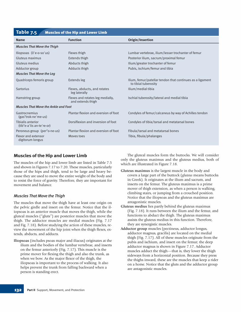
Muscles That Move the Ankle and Foot
Muscles that move the ankle and foot are shown in Figures7.19 and 7.20.
Gastrocnemius is a muscle of the posterior leg, where itforms a large part of the calf. It arises from the femur;distally, the muscle joins the strong calcaneal tendon,which attaches to the calcaneus bone (heel). Thegastrocnemius is a powerful plantar flexor of the footthat aids in pushing the body forward during walking orrunning. It is sometimes called the “toe dancer’s muscle”because it allows a person to stand on tiptoe.
Tibialis anterior is a long, spindle-shaped muscle of theanterior leg. It arises from the surface of the tibia andattaches to the bones of the ankle and foot. Contractionof this muscle causes dorsiflexion and inversion of thefoot.
Peroneus muscles (peroneus longus, peroneus brevis) arefound on the lateral side of the leg, connecting the fibulato the metatarsal bones of the foot. These muscles evertthe foot and also help bring about plantar flexion.
Flexor (not shown) and extensor digitorum longus musclesare found on the lateral and posterior portion of the leg.They arise mostly from the tibia and insert on the toes.They flex and extend the toes, respectively, and assist inother movements of the feet.
7.5 Effects of AgingMuscle mass and strength tend to decrease as people age.How much of this is due to lack of exercise and a poor diethas yet to be determined. Deteriorated muscle elements arereplaced initially by connective tissue and, eventually, by fat.With age, degenerative changes take place in the mitochon-dria, and endurance decreases. Also, changes in the nervousand cardiovascular systems adversely affect the structure andfunction of muscles.
Muscle mass and strength can improve remarkably ifelderly people undergo a training program. Exercise at anyage appears to stimulate muscle buildup. As discussed in theMedical Focus on page 135, exercise has many other bene-fits as well. For example, exercise improves the cardiovascu-lar system and reduces the risk of diabetes and glycation.During glycation, excess glucose molecules stick to bodyproteins so that the proteins no longer have their normalstructure and cannot function properly. Exercise burns glu-cose and, in this way, helps prevent muscle deterioration.
134 Part II Support, Movement, and Protection
Figure 7.19 Muscles of the anterior right leg. Figure 7.20 Muscles of the lateral right leg.
tibialis anterior
peroneus longus
extensor digitorumlongus
peroneus brevis
patella
patellar ligament
gastrocnemius
tibia
biceps femoris
gastrocnemius
peroneus longus
calcaneal tendon
vastus lateralis
head of fibula
tibialis anterior
extensor digitorumlongus
peroneus tertius
peroneus brevis
mad64372_ch07pg113_139 9/1/03 4:27 PM Page 134

135Chapter 7 The Muscular System
Benefits of ExerciseExercise programs improve muscular strength, muscular en-durance, and flexibility. Muscular strength is the force a musclegroup (or muscle) can exert against a resistance in one maximal ef-fort. Muscular endurance is judged by the ability of a muscle to con-tract repeatedly or to sustain a contraction for an extended period.Flexibility is tested by observing the range of motion about a joint.
As muscular strength improves, the overall size of the muscle,as well as the number of muscle fibers and myofibrils in the mus-cle, increases. The total amount of protein, the number of capil-laries, and the amounts of connective tissue, including tissuefound in tendons and ligaments, also increase. Physical trainingwith weights can improve muscular strength and endurance in alladults, regardless of their age. Over time, increased musclestrength promotes strong bones.
A surprising finding, however, is that health benefits also ac-company less strenuous programs, such as those described inTable 7A. A study of 12,000 men by Dr. Arthur Leon at the Uni-versity of Minnesota showed that even moderate exercise loweredthe risk of a heart attack by one-third. People with arthritis re-ported much less pain, swelling, fatigue, and depression afteronly four months of attending a twice-weekly, low-impact aero-bics class. Increasing daily activity by walking to the corner storeinstead of driving and by taking the stairs instead of the elevatorcan improve a person’s health.
The benefits of exercise are most apparent with regard to car-diovascular health. Brisk walking for 2.5–4 hours a week can raisethe blood levels of high-density lipoprotein (HDL), a chemical
that promotes healthy blood vessels (see Chapter 12). Exercisealso helps prevent osteoporosis, a condition in which the bonesare weak and tend to break. The stronger the bones are when aperson is young, the less chance of osteoporosis as a person ages.Exercise promotes the activity of osteoblasts (as opposed to os-teocytes) in young people, as well as older people. An increasedactivity level can also keep off unwanted pounds, which is aworthwhile goal because added body weight contributes to nu-merous conditions, such as type II diabetes (see page 197). In-creased muscle activity is also helpful by causing glucose to betransported into muscle cells and making the body less depen-dent on the presence of insulin.
People in chronic pain are often diagnosed as having fi-bromyalgia, characterized by achy pain, tenderness, and stiffnessof muscles. Substance P has been found in the bloodstream ofthese patients. Exercise (more frequent and longer periods of ex-ercise, not increased intensity) decreases the concentration ofsubstance P. Stretching exercises, such as yoga, and massages (twoto three a week) also decrease the amount of substance P. Moreinformation on this subject is currently being sought.
Cancer prevention and early detection involve eating prop-erly, not smoking, avoiding cancer-causing chemicals and radia-tion, undergoing appropriate medical screening tests, and know-ing the early warning signs of cancer. However, evidence indicatesthat exercise also helps prevent certain kinds of cancer. Studiesshow that people who exercise are less likely to develop colon,breast, cervical, uterine, and ovarian cancer.
Table 7A A Checklist for Staying Fit
Children, 7=12 Teenagers, 13=18 Adults, 19=55 Seniors, 56 and Up
Vigorous activity 1–2 hours Vigorous activity 1 hour Vigorous activity 1 hour Moderate exercise 1 hour dailydaily 3–5 days a week; otherwise, 3 days a week; otherwise, 3 days a week; otherwise,
1–2 hour daily moderate activity 1
–2 hour daily moderate activity 1–2 hour daily moderate activity
Free play Build muscle with calisthenics Exercise to prevent lower back Take a daily walkpain: aerobics, stretching,yoga
Build motor skills through Do aerobic exercise to Take active vacations: hike, Do daily stretching exercisesteam sports, dance, swimming control buildup of fat cells bicycle, cross-country ski
Encourage more exercise Pursue tennis, swimming, Find exercise partners: join a Learn a new sport or activity: outside of physical education horseback riding—sports that running club, bicycle club, golf, fishing, ballroom dancingclasses can be enjoyed for a lifetime outing group
Initiate family outings: bowling, Continue team sports, Try low-impact aerobics.boating, camping, hiking dancing, hiking, swimming Before undertaking new exercises,
consult your doctor
mad64372_ch07pg113_139 9/1/03 4:27 PM Page 135

7.6 HomeostasisThe illustration in Human Systems Work Together on page137 tells how the muscular system works with other systemsof the body to maintain homeostasis.
Cardiac muscle contraction accounts for the heartbeat,which creates blood pressure, the force that propels blood inthe arteries and arterioles. The walls of the arteries and arteri-oles contain smooth muscle. Constriction of arteriole walls isregulated to help maintain blood pressure. Arterioles branchinto the capillaries where exchange takes place that createsand cleanses tissue fluid. Blood and tissue fluid are the inter-nal environment of the body, and without cardiac andsmooth muscle contraction, blood would never reach the cap-illaries for exchange to take place. Blood is returned to theheart in cardiovascular veins, and excess tissue fluid is re-turned to the cardiovascular system within lymphatic vessels.Skeletal muscle contraction presses on the cardiovascularveins and lymphatic vessels, and this creates the pressure thatmoves fluids in both types of vessels. Without the return ofblood to the heart, circulation would stop, and without thereturn of lymph to the blood vessels, normal blood pressurecould not be maintained.
The contraction of sphincters composed of smooth mus-cle fibers temporarily prevents the flow of blood into a capil-lary. This is an important homeostatic mechanism because intimes of emergency it is more important, for example, forblood to be directed to the skeletal muscles than to the tissuesof the digestive tract. Smooth muscle contraction also ac-counts for peristalsis, the process that moves food along the di-gestive tract. Without this action, food would never reach allthe organs of the digestive tract where digestion releases nutri-ents that enter the bloodstream. Smooth muscle contractionassists the voiding of urine, which is necessary for ridding thebody of metabolic wastes and for regulating the blood volume,salt concentration, and pH of internal fluids.
Skeletal muscles protect internal organs, and theirstrength protects joints by stabilizing their movements. Skele-tal muscle contraction raises the rib cage and lowers the di-aphragm during the active phase of breathing. As we breathe,oxygen enters the blood and is delivered to the tissues, in-cluding the muscles, where ATP is produced in mitochondriawith heat as a by-product. The heat produced by skeletal mus-cle contraction allows the body temperature to remain withinthe normal range for human beings.
Finally, skeletal muscle contraction moves bones and al-lows us to perform those daily activities necessary to ourhealth and benefit. Although it may seem as if movement ofour limbs does not affect homeostasis, it does so by allowingus to relocate our bodies to keep the external environmentwithin favorable limits for our existence.
Muscular DisordersWhen spasms or injuries occur, homeostasis is challenged,and when disease is present, homeostasis may be overcome tothe point of death.
Spasms and Injuries
Spasms are sudden and involuntary muscular contractions,most often accompanied by pain. Spasms can occur in bothsmooth and skeletal muscles. A spasm of the intestinal tract isa type of colic sometimes called a “bellyache.” Multiplespasms of skeletal muscles are called a seizure or convulsion.Cramps are strong painful spasms, especially of the leg andfoot, usually due to strenuous activity. Cramps can even occurwhen sleeping after a strenuous workout. Facial tics, such asperiodic eye blinking, head turning, or grimacing, are spasmsthat can be controlled voluntarily but only with great effort.
A strain is the overstretching of a muscle near a joint. Asprain is the twisting of a joint, leading to swelling and to in-jury not only of muscles but also of ligaments, tendons, bloodvessels, and nerves. The ankle is often subject to sprains.
Myalgia refers to inflammation of muscle tissue. Tendini-tis is inflammation of a tendon due to the strain of repeatedathletic activity. The tendons most commonly affected arethose associated with the shoulder, elbow, hip, and knee.
Diseases
In persons who have not been properly immunized, the toxinof the tetanus bacterium can cause muscles to lock in a tetaniccontraction. A rigidly locked jaw is one of the first signs of aninfection known as tetanus. Like other bacterial infections,tetanus is curable with the administration of an antibiotic.
Muscular dystrophy is a broad term applied to a group ofdisorders characterized by progressive degeneration andweakening of muscles. As muscle fibers die, fat and connectivetissue take their place. Duchenne muscular dystrophy, themost common type, is inherited through a flawed gene carriedby the mother. It is now known that the lack of a proteincalled dystrophin causes the condition. When dystrophin isabsent, calcium leaks into the cell and activates an enzymethat dissolves muscle fibers. In an attempt to treat the condi-tion, muscles have been injected with immature muscle cellsthat do produce dystrophin.
Myasthenia gravis is an autoimmune disease character-ized by weakness that especially affects the muscles of the eye-lids, face, neck, and extremities. Muscle contraction isimpaired because the immune system mistakenly produces an-tibodies that destroy acetylcholine receptors. In many cases,the first signs of the disease are drooping eyelids and doublevision. Treatment includes drugs that are antagonistic to theenzyme acetylcholinesterase.
136 Part II Support, Movement, and Protection
mad64372_ch07pg113_139 9/1/03 4:27 PM Page 136

137Chapter 7 The Muscular System
Human Systems Work Together MUSCULAR SYSTEM
Cardiovascular System
Lungs provide oxygen for,and rid the body of, carbondioxide from contractingmuscles.
Muscle contractionprovides heat to warmskin. Muscle movesskin of face.
mad64372_ch07pg113_139 9/1/03 4:28 PM Page 137

138 Part II Support, Movement, and Protection
Basic Key Termsactin (ak’tin), p. 116all-or-none law, p. 122antagonist (an-tag’o-nist), p. 124cardiac muscle (kar’de-ak mus’el), p. 114creatine phosphate (kre’uh-tin fos’fat), p. 120insertion (in-ser’shun), p. 124motor unit (mo’tor yu’nit), p. 122muscle fiber (mus’el fi’ber), p. 114muscle twitch (mus’el twich), p. 122myofibril (mi”o-fi’bril), p. 116myoglobin (mi”o-glo’bin), p. 116myosin (mi’o-sin), p. 116neuromuscular junction (nu”ro-mus’kyu-ler junk’shun), p. 118origin (or’I-jin), p. 124oxygen deficit (ok’sI-jen def’I-sit), p. 120prime mover (prim mu’ver), p. 124recruitment (re-krut’ment), p. 123sarcomere (sar’ko-mer), p. 116
skeletal muscle (skel’E-tal mus’el), p. 114sliding filament theory (sli’ding fil’uh-ment the’o-re), p. 116smooth muscle (smuth mus’el), p. 114synergist (sin’er-jist), p. 124T (transverse) tubules (tranz-vers’ tu’byul), p. 116tendon (ten’don), p. 115tone (ton), p. 123
Clinical Key Termsatrophy (at’ro-fe), p. 123hypertrophy (hi-per’tro-fe), p. 123lockjaw (lok’jaw), p. 136muscular dystrophy (mus’kyu-ler dis’trE-fe), p. 136myalgia (mi-al’juh), p. 136myasthenia gravis (mi”as-the’ne-uh grah’vis), p. 136spasm (spazm), p. 136sprain (spran), p. 136 strain (stran), p. 136tendinitis (ten”dE-ni’tis), p. 136tetanus (tet’uh-nus), p. 136
Selected New Terms
7.1 Functions and Types of MusclesA. Muscular tissue is either smooth,
cardiac, or skeletal. Skeletal muscleshave tubular, multinucleated, andstriated fibers that contractvoluntarily.
B. Skeletal muscles support the body,make bones move, help maintain aconstant body temperature, assistmovement in cardiovascular andlymphatic vessels, and help protectinternal organs and stabilize joints.
7.2 Microscopic Anatomy andContraction of Skeletal MuscleA. The sarcolemma, which extends into
a muscle fiber, forms T tubules; thesarcoplasmic reticulum has calciumstorage sites. The placement of actinand myosin in the contractilemyofibrils accounts for the striationsof skeletal muscle fibers.
B. Skeletal muscle innervation occurs atneuromuscular junctions. Impulsestravel down the tubules of the Tsystem and cause the release ofcalcium from calcium storage sites.
The presence of calcium and ATP inmuscle cells prompts actinmyofilaments to slide past myosinmyofilaments, shortening the lengthof the sarcomere.
C. ATP, required for muscle contraction,can be generated by way of creatinephosphate breakdown andfermentation. Lactic acid fromfermentation represents an oxygendeficit, because oxygen is required tometabolize this product. Cellularrespiration, an aerobic process, is thebest source of ATP.
7.3 Muscle Responses A. In the laboratory, muscle fibers obey
the all-or-none law, but wholemuscles do not. The occurrence of amuscle twitch, summation, ortetanic contraction depends on thefrequency with which a muscle isstimulated.
B. In the body, muscle fibers belong tomotor units that obey the all-or-none law. The strength of musclecontraction depends on the
recruitment of motor units. A musclehas tone because some fibers arealways contracting.
7.4 Skeletal Muscles of the BodyA. When muscles cooperate to achieve
movement, some act as primemovers, others as synergists, and stillothers as antagonists.
B. The skeletal muscles of the body aredivided into those that move: thehead and neck (see Table 7.2); thetrunk (see Table 7.3); the shoulderand arm (see Table 7.4); the forearm(see Table7.4); the hand and fingers(see Table 7.4); the thigh (see Table7.5); the leg (see Table 7.5); and theankle and foot (see Table 7.5).
7.5 Effects of AgingAs we age, muscles become weaker, butexercise can help retain vigor.
7.6 HomeostasisSmooth muscle contraction helps movethe blood; cardiac muscle contractionpumps the blood. Skeletal musclecontraction produces heat and isneeded for breathing.
Summary
mad64372_ch07pg113_139 9/1/03 4:28 PM Page 138

139Chapter 7 The Muscular System
1. Name and describe the three types ofmuscles, and give a general location foreach type. (p. 114)
2. List and discuss five functions ofmuscles. (p. 115)
3. Describe the anatomy of a muscle, fromthe whole muscle to the myofilamentswithin a sarcomere. Name the layers offascia that cover a skeletal muscle anddivide the muscle interior. (pp. 116–17)
4. List the sequential events that occurafter a nerve impulse reaches a muscle.(pp. 118–19)
5. How is ATP supplied to muscles? Whatis oxygen deficit? (pp. 120–21)
6. What is the all-or-none law? What is thedifference between a single muscle twitch,summation, and a tetanic contraction?(p. 122)
7. What is muscle tone? How does musclecontraction affect muscle size? (p. 123)
8. Describe how muscles are attached tobones. Define the terms prime mover,synergist, and antagonist. (p. 124)
9. How do muscles get their names? Givean example for each characteristic usedin naming muscles. (pp. 124–25)
10. Which of the muscles of the head areused for facial expression? Which areused for chewing? (p. 126)
11. Which muscles of the neck flex andextend the head? (p. 127)
12. What are the muscles of the thoracicwall? What are the muscles of theabdominal wall? (pp. 128–29)
13. Which of the muscles of the shoulderand upper limb move the arm andforearm, and what are their actions?Name the muscles that move the handand fingers. (p. 130)
14. Which of the muscles of the hip movethe thigh, and what are their actions?Which of the muscles of the thigh movethe leg, and what are their actions?Which of the muscles of the leg movethe feet? (pp. 132–34)
Study Questions
I. Fill in the blanks.1. muscle is
uninucleated, nonstriated, and located in the walls of internalorgans.
2. The fascia called separates muscle fibers from oneanother within a fascicle.
3. When a muscle fiber contracts, anmyofilament slides
past a myosin myofilament.4. The energy molecule
is needed for muscle fibercontraction.
5. Whole muscles have ,a condition in which some fibersare always contracted.
6. When muscles contract, thedoes most of the
work, but the help.7. The is a muscle in
the arm that has two origins.8. The acts as the
origin of the latissimus dorsi, andthe acts as theinsertion during most activities.
II. For questions 9-12, name the muscleindicated by the combination oforigin and insertion shown.
Origin Insertion9. temporal bone mandibular
coronoid process10. scapula, clavicle humerus11. scapula, proxi- olecranon process
mal humerus of ulna12. posterior ilium, proximal femur
sacrum
III. Match the muscles in the key to theactions listed in questions 13-20.Key:
a. orbicularis oculib. zygomaticusc. deltoidd. serratus anteriore. rectus abdominisf. iliopsoasg. gluteus maximush. gastrocnemius
13. Allows a person to stand on tiptoe14. Tenses abdominal wall15. Abducts arm16. Flexes thigh17. Raises corner of mouth18. Closes eyes
Objective Questions
Consult Appendix B for help inpronouncing and analyzing the meaningof the terms that follow.
1. hyperkinesis (hi”per-ki-ne’sis)
2. dystrophy (dis’tro-fe)3. electromyogram (e-lek’’tro-mi’-o-gram)4. menisectomy (men’’i-sek’to-me)5. tenorrhaphy (te-nor’uh-fe)6. myatrophy (mi-at’ro-fe)
7. leiomyoma (li’’o-mi-o’muh)8. kinesiotherapy (ki-ne’’se-o-thEr’uh-pe)9. myocardiopathy
(mi’’o-kar’’de-op’ uh-the)10. myasthenia (mi’’as-the’ne-uh)
Medical Terminology Reinforcement Exercise
Visit the Student Edition of the Online Learning Center at http://www.mhhe.com/maderap5 for additional quizzes, interactive learningexercises, and other study tools.
Website Link
mad64372_ch07pg113_139 22/9/03 4:49 PM Page 139 seema 309:GQ164:mad5ch07:layouts: