Embed Size (px)
Citation preview

1
UNIQUE DELETIONS IN PRADER-WILLI SYNDROME
By
SOO-JEONG KIM
A THESIS PRESENTED TO THE GRADUATE SCHOOL OF THE UNIVERSITY OF FLORIDA IN PARTIAL FULFILLMENT
OF THE REQUIREMENTS FOR THE DEGREE OF MASTER OF SCIENCE
UNIVERSITY OF FLORIDA
2010

2
© 2010 Soo-Jeong Kim

3
ACKNOWLEDGMENTS
I gratefully acknowledge Krista A. Schwenk and Christy L. Hollywood for their
expert assistance in clinical assessments, Eve Johnson for her organization and
editorial support, Drs. Karin Buiting and Bernhard Horsthemke for sharing their MLPA
normalization protocol and Dr. Robert Nicholls for critical comments on this manuscript.
I extend my gratitude to my wonderful mentors, Drs. Mark H. Lewis, Daniel J. Driscoll
and Edwin H. Cook for their guidance and support. I am especially thankful to all
participating patients with Prader-Willi syndrome (PWS) and their families. This work
was supported in part by Hayward FoundationRare Disease Clinical Research Network,
Prader-Willi syndrome Association (USA) and grant K23MH082883 from the National
Institute of Mental Health.

4
TABLE OF CONTENTS page
ACKNOWLEDGMENTS .................................................................................................. 3
LIST OF TABLES ............................................................................................................ 5
LIST OF FIGURES .......................................................................................................... 6
ABSTRACT ..................................................................................................................... 7
CHAPTER
1 INTRODUCTION ...................................................................................................... 9
2 MATERIALS AND METHODS ................................................................................ 12
Samples .................................................................................................................. 12 The Methylation-Specific Multiplex Ligation-dependent Probe Amplification (MS-
MLPA).................................................................................................................. 12 The Array-based Comparative Genome Hybridization (aCGH): ............................. 13 Clinical Evaluation .................................................................................................. 13
3 RESULTS ............................................................................................................... 17
Copy Number Analyses .......................................................................................... 17 DNA Methylation Analyses ..................................................................................... 17 Agreement Rate Between the MS-MLPA and aCGH: ............................................. 17 Case Studies .......................................................................................................... 18
4 DISCUSSION ......................................................................................................... 34
LIST OF REFERENCES ............................................................................................... 39
BIOGRAPHICAL SKETCH ............................................................................................ 43

5
LIST OF TABLES
Table page 2-1 Demographic information of the 74 PWS subjects with deletion......................... 14
2-2 The MS-MLPA probes ........................................................................................ 15
2-3 DNA methylation analyses from 5 methylation sensitive probes ........................ 16
3-1 Subjects with unique deletions ........................................................................... 31

6
LIST OF FIGURES
Figure page 3-1 Seven unique PWS deletions ............................................................................. 30

7
Abstract of Thesis Presented to the Graduate School of the University of Florida in Partial Fulfillment of the Requirements for the Degree of Master of Science
UNIQUE DELETIONS IN PRADER-WILLI SYNDROME
By
Soo-Jeong Kim
May 2010
Chair: Daniel Driscoll Major: Medical Sciences – Clinical and Translational Science
Prader-Willi syndrome (PWS) is a rare genetic disorder caused by an absence of
paternally expressed genes within the 15q11-q13 region via one of the three genetic
mechanisms: deletion of paternally inherited genes, maternal uniparental disomy (UPD)
and imprinting defect (ID). Deletion is often subdivided into Type 1 and Type 2, while
UPD is subdivided into heterodisomy and isodisomy. Despite PWS being a well-
characterized genetic disorder, specific genetic factors contributing to specific PWS
phenotypes are not fully understood.
The methylation-specific multiplex ligation-dependent probe amplification (MS-
MLPA) is a newly developed technique that detects copy number changes and aberrant
DNA methylation. In this study, we applied the MS-MLPA to further elucidate the genetic
subtypes of deletion in 74 PWS subjects with deletion. Sixty-seven of 74 subjects had
either Type 1 (n=25) or Type 2 (n=42) deletions. The remaining seven subjects had
unique deletions other than Type 1 or Type 2. Subsequently, we verified the unique
breakpoints with the microarray-based comparative genome hybridization (CGH).

8
Additionally, we reviewed the available medical records to examine the clinical
characteristics of these seven subjects with unique deletions.
In conclusion, we identified unique deletions from approximately 9.5% of PWS
subjects with deletion in our laboratory using the MS-MLPA, which was verified by the
CGH. Case studies of the subjects with unique deletions may help identify the specific
genetic factors contributing to the specific PWS phenotypes.

9
CHAPTER 1 INTRODUCTION
Prader–Willi syndrome (PWS) is the most frequently diagnosed genetic cause of
obesity, characterized by pronounced hyperphagia (often described as “voracious
appetite”) and early-onset morbid obesity (Cassidy and Driscoll 2009). Other most
consistent major manifestations include hypotonia with poor suck and poor weight gain
in infancy, mild to moderate mental retardation, hypogonadism, growth hormone
insufficiency causing short stature, characteristic facial appearance, and behavioral and
sometimes psychiatric disturbance (Cassidy 1997; Holm et al. 1993; State and Dykens
2000). Furthermore, the majority of PWS individuals suffer from various forms of
abnormal restricted repetitive behavior, such as stereotypies, repetitive forms of self-
injurious behavior, obsession, compulsion, ritualistic behavior, sameness behavior and
restricted interests. For example, 69 to 100% of individuals with PWS showed skin-
picking (Dykens et al. 1999; Thompson and Gray 1994; Torrado et al. 2006; Veltman et
al. 2004; Webb et al. 2002; Whitman and Accardo 1987) and 37 to 58% of individuals
with PWS manifested prominent obsessive compulsive symptoms, such as hoarding,
ordering/arranging, concerns with symmetry/exactness, rewriting, need to tell/know/ask
(Dykens et al. 1996).
PWS is a highly variable genetic disorder affecting multiple body systems with an
estimated incidence rate of 1 in 15,000 (Butler 1990). PWS is caused by an absence of
paternal contribution in the chromosome 15 q11-q13 region via three distinct genetic
mechanisms including deletion of paternally inherited genes, maternal uniparental
disomy (inheritance of two copies of maternal chromosomes) and imprinting defect
(genetic mutation leading to abnormal silencing of gene expression from the paternally

10
inherited chromosome) (Cassidy and Driscoll 2009; Glenn et al. 1997; Nicholls and
Knepper 2001). Deletion is responsible for approximately 70% of PWS cases, while
maternal uniparental disomy (UPD) causes about 25% of PWS cases. Less than 5% of
PWS cases are caused by imprinting defect (ID). Deletion is typically subdivided into
two subgroups. Type 1 deletion encompasses a larger portion of the 15q11-q13 region
between common breakpoints BP1 and BP3, whereas Type 2 deletion involves a
shorter distance between BP2 and BP3 (Bittel and Butler 2005; Butler et al. 2004;
Milner et al. 2005). Studies found at least two more rare breakpoints (BP4 and BP5)
distal to BP3 (Sahoo et al. 2005; Sharp et al. 2006; Wang et al. 2004). In addition, the
15q11-q13 region may be deleted as a result of an unbalanced translocation in a few
patients (Buiting and Horsthemke 2006).
Despite PWS being a well-characterized genetic disorder, specific genetic factors
contributing to specific PWS phenotypes are not yet clearly understood (State and
Dykens 2000). Interestingly, several studies have investigated the phenotypic
characteristics across these genetic subtypes and reported that individuals with Type 1
deletion have more typical PWS phenotypes including higher levels of restricted
repetitive behavior (RRB) than those with Type 2 deletion or UPD/ID (Bittel et al. 2006;
Butler et al. 2004; Dykens et al. 1999; Torrado et al. 2006). Contrary to these reports,
Milner et al. (2005) did not find differences in RRB scores across the genetic subtypes
(Milner et al. 2005). This conflicting result may have been due to genetic heterogeneity.
For example, patients with unique deletions other than Type 1 or Type 2 deletions may
have been categorized as either subgroup.

11
The methylation-specific multiplex ligation-dependent probe amplification (MS-
MLPA) is a relatively new technique to detect changes of copy number and DNA
methylation (Bittel et al. 2007; Nygren et al. 2005; Procter et al. 2006). Compared to the
traditional diagnostic methods, such as chromosomal analysis or Fluorescent in situ
hybridization (FISH), the MS-MLPA provides more detailed information on the size of
deletion as well as methylation status. The MS-MLPA is a labor and cost-effective
alternative to the array based comparative genome hybridization (aCGH).
In the present study, we utilized the MS-MLPA to elucidate the size of deletions
in our PWS subjects. Once we identified several subjects with unique deletions, we
reviewed the available medical records to examine the genotype-phenotype
relationships. Additionally, we applied aCGH to the subset of our subjects including
those with unique deletions to verify the MS-MLPA findings.

12
CHAPTER 2 MATERIALS AND METHODS
Samples
We obtained DNA samples from 74 PWS individuals (Table 2-1) with deletion
whose subtypes were unknown. In addition, we examined additional 128 DNA samples
to compare the methylation patterns across the diagnostic categories. These include 14
PWS subjects with maternal UPD, 4 PWS subjects with ID, 8 Angelman syndrome (AS)
subjects with deletion, 1 AS subjects with paternal UPD, 2 AS subjects with ID, 4 family
members of the known maternal interstitial duplication of the 15q11-q13, 1 subject with
maternal isodicentric duplication of the chromosome 15, 90 subjects with a history of
early-onset morbid obesity (EMO) and 4 normal control subjects. The research protocol
and informed consents were approved by the University of Florida Institutional Review
Board (IRB).
The Methylation-Specific Multiplex Ligation-dependent Probe Amplification (MS-MLPA)
A commercial MS-MLPA kit (ME028-A1) for Prader-Willi/Angelman syndrome
(MRC-Holland, Amsterdam, The Netherlands) was utilized. This kit contained 25 probes
specific for sequence in the 15q11-q13 region to detect copy number changes of the
major genes. Among these 25 probes, five also contained methylation sensitive
restriction enzyme HhaI recognition site, thereby amplifying only methylated sequence
after digestion process (Procter et al. 2006) (Table 2-2). The manufacturer’s protocol
was followed for the DNA preparation, ligation, enzyme digestion and multiplex PCR
reaction (www.mrc-holland.com). After the final PCR step, the Applied Biosystems
3730xl DNA Analyzer (Applied Biosystems, Foster City, USA) and Peak Scanner
Software (version 1.0) (Applied Biosystems) were used for capillary electrophoresis and

13
fragment analysis. The peak heights of the 15q11-q13 specific probe-amplified products
were normalized using the averaged peak heights of neighboring control probe-
amplified products. By comparing normalized peak heights of specific probe-amplified
products with those of control subjects, we were able to identify copy number changes
(e.g., deletion or duplication). To detect aberrant methylation, peak heights of the
15q11-q13 specific probes before and after digestion were normalized using global
normalization process suggested by the manufacturer. Comparison of normalized peak
heights before digestion with those after digestion provided information on quantity of
methylation (Table 2-3). All MS-MLPA results were reviewed independently by three
investigators. Whenever the results were inconclusive, the MS-MLPA reaction was
repeated.
The Array-based Comparative Genome Hybridization (aCGH):
The MS-MLPA results from a subset of 24 PWS subjects were verified by an
enhanced version of chromosome 15-specific genomic microarray, which included a
total of 175 genomic bacterial artificial chromosome (BAC) clones across the 15q, with
the higher density of clones spanning approximately 10 Mb of the 15q11-q14 interval in
addition to 160 clones specific for the subtelomeric regions of all other chromosomes at
the Baylor College of Medicine (Sahoo et al. 2007).
Clinical Evaluation
Clinical evaluations for 7 unique subjects were done as part of typical clinic visits
and/or during clinical research encounters.

14
Table 2-1. Demographic information of the 74 PWS subjects with deletion Type 1 Type 2 Unique
Number of subjects 25 40 (+2-brain sample)
7
Gender (M:F) 16:9 22:18 4:3
Race/ethnicity
White 24 35 5 Black 1 3 1 Hispanic 0 0 1 Asian 0 1 Other1 0 1 0
1. Other includes the subjects who did not report their ethnicity or are biracial.

15
Table 2-2. The MS-MLPA probes
Methylation1 Probe Gene Position2 Distance3 1 TUBGCP5 20,398,305 122,299 2 CYFIP1 20,520,604 841,839 100% 3 MKRN3 21,362,443 376 4 MKRN3 21,362,819 78,685 5 MAGEL2 21,441,504 966 100% 6 MAGEL2 21,442,470 40,021 7 NDN 21,482,491 929 50% 8 NDN 21,483,420 1,136,387 50% 9 SNRPN U1B exon 22,619,807 96,908 10 SNRPN U3 exon 22,716,715 34,499 50% 11 SNRPN alt exon 1 22,751,214 269 50% 12 SNRPN (CpG) 22,751,483 290 50% 13 SNRPN (CpG) 22,751,773 12,482 14 SNRPN exon 3 22,764,255 372,148 15 UBE3A, exon 15 23,136,403 31,346 16 UBE3A, exon 9 23,167,749 67,104 0% 17 UBE3A, exon 1 23,234,853 332 0% 18 UBE3A, exon 1 23,235,185 342,162 19 ATP10A, exon 2 23,577,347 82,014 0% 20 ATP10A, exon 1 23,659,361 684,889 21 GABRB3, exon 9 24,344,250 224,854 0% 22 GABRB3, exon 1 24,569,104 1,201,013 23 OCA2, exon 22 25,770,117 247823 0% 24 OCA2, exon 1 26,017,940 1,178,810 25 APBA2 27,196,750 1. Percentage of methylated sequence in blood-derived DNA 2. Base-pair position on March 2006 assembly on the UCSD genome browser
(http://genome.ucsc.edu/) 3. Distance to the next probe in base pairs
16
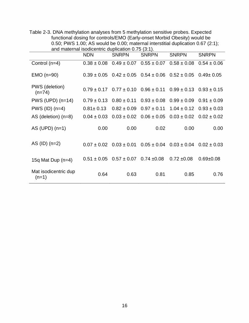
Table 2-3. DNA methylation analyses from 5 methylation sensitive probes. Expected functional dosing for controls/EMO (Early-onset Morbid Obesity) would be 0.50; PWS 1.00; AS would be 0.00; maternal interstitial duplication 0.67 (2:1); and maternal isodicentric duplication 0.75 (3:1).
NDN SNRPN SNRPN SNRPN SNRPN
Control (n=4) 0.38 ± 0.08 0.49 ± 0.07 0.55 ± 0.07 0.58 ± 0.08 0.54 ± 0.06
EMO (n=90) 0.39 ± 0.05 0.42 ± 0.05 0.54 ± 0.06 0.52 ± 0.05 0.49± 0.05
PWS (deletion) (n=74) 0.79 ± 0.17 0.77 ± 0.10 0.96 ± 0.11 0.99 ± 0.13 0.93 ± 0.15
PWS (UPD) (n=14) 0.79 ± 0.13 0.80 ± 0.11 0.93 ± 0.08 0.99 ± 0.09 0.91 ± 0.09 PWS (ID) (n=4) 0.81± 0.13 0.82 ± 0.09 0.97 ± 0.11 1.04 ± 0.12 0.93 ± 0.03 AS (deletion) (n=8) 0.04 ± 0.03 0.03 ± 0.02 0.06 ± 0.05 0.03 ± 0.02 0.02 ± 0.02
AS (UPD) (n=1) 0.00 0.00 0.02 0.00 0.00
AS (ID) (n=2) 0.07 ± 0.02 0.03 ± 0.01 0.05 ± 0.04 0.03 ± 0.04 0.02 ± 0.03
15q Mat Dup (n=4) 0.51 ± 0.05 0.57 ± 0.07 0.74 ±0.08 0.72 ±0.08 0.69±0.08
Mat isodicentric dup (n=1) 0.64 0.63 0.81 0.85 0.76

17
CHAPTER 3 RESULTS
Copy Number Analyses
Among 74 PWS subjects with deletion, we identified 25 subjects with Type 1
deletion (33.78%) and 42 subjects with Type 2 deletion (56.76%). The remaining seven
PWS subjects (9.46%) had unique deletions other than Type 1 or Type 2 (Figure 3-1).
In eight AS subjects with deletion, five subjects (62.5%) had Type 1 deletion and three
subjects (37.5%) had Type 2 deletion. We also observed microdeletions within SNRPN
in one of four PWS subjects with ID and two of two AS subjects with ID. We incidentally
identified two unique deletions among 90 individuals with EMO. One subject with EMO
had a deletion of the 15q26 region and the other subject with EMO had deletion
between BP1 and BP2. The four subjects with maternal interstitial duplication of the
15q11-q13 as well as the subject with maternal isodicentric duplication of the
chromosome 15 showed increased amplification pattern consistent with their molecular
diagnoses.
DNA Methylation Analyses
DNA methylation analyses for NDN and SNRPN confirmed characteristic
methylation patterns for PWS, AS and normal control subjects, respectively (Table 2-3).
However, we noted individual variations in some methylation-sensitive probes (Table 2-
3).
Agreement Rate Between the MS-MLPA and aCGH:
We conducted both MS-MLPA and aCGH in a subset of our PWS subjects (n=24).
Among these 24 subjects, seven subjects had unique deletions identified by MS-MLPA.
The remaining 17 subjects were the study participants of the Rare Disease Clinical

18
Research Network (RDCRN). Not surprisingly, the agreement rate between MS-MLPA
and aCGH was 100%. However; the aCGH revealed more precise location of
breakpoints.
Case Studies
We reviewed the available medical records of the seven subjects with unique
deletion (Figure 3-1 and Table 3-1).
PW235P: The patient was a 21 year-old white male who was last seen just prior to
his accidental death. He was born at 43 weeks gestation with a birth weight of 3.73Kg.
He was noted to be very hypotonic and have a poor sucking as a neonate. He was fed
via nasogastric tube for the first three months of life and thereafter by a bottle with a
widened nipple. He fed slowly and poorly for the first nine months. However, by 18
months his weight started going up quickly and by 24 months he was obese with a body
mass index (BMI) standard deviation score (SDS) = 2.05. His developmental history
indicated that he sat at 10 months, was late to walk at 2 ½ years, and his first words
were at 2 ½ years.
He was first seen by the genetics service at 4 11/12 years of age to evaluate for
PWS. His BMI SDS was +4.12 and hand length was at approximately 65th percentile
(%ile). High resolution chromosomal analysis at that time was reportedly normal. In his
follow up at 5 11/12 years, his geneticist noted that he had “some but not all of the
features of PWS.” The reluctance to label him as PWS at that time was partly due to his
macrocephaly (95th %ile) in relation to his younger brother’s (60th %ile) and mother’s
(60th %ile) head sizes. In addition, his birth weight (3.73Kg) and his height (30th %ile)
were higher than expected, and he lacked a voracious appetite. He did, however, have

19
obesity and hypogonadism. At age 10 ½ years, he had been started on growth hormone
(GH) therapy by an endocrinologist to improve his muscle mass. His height was noted
at the 40th %ile prior to starting GH.
He was seen again by the genetics service at 11 ½ years of age to re-evaluate for
PWS. At that time, his height was at the 75th %ile and head circumference was >95th
%ile, with a BMI SDS = 2.38. His voracious appetite had begun at 8 years of age but he
did not develop food stealing problems yet. Skin picking was also noted at that time.
Academically, he was in regular classes and achieving good grades, but had speech
difficulties. On exam, he did have almond shaped eyes and bitemporal narrowing. His
saliva was viscous. He had hypogonadism. He was not hypopigmented relative to his
family. He had large hands (85th %ile) and feet (85th %ile). This time, the SNRPN FISH
analysis and DNA methylation analysis using the SNRPN probe (Glenn et al., 1996)
revealed a deletion and aberrant methylation compatible with a diagnosis of PWS.
By 14 years of age, he started having severe behavioral problems and he had
physically threatened family members several times. He was arrested for breaking into
an occupied home and stealing food. He was subsequently placed into a psychiatric
facility for his out of control behavior. His GH was stopped at this time and not restarted
again till 5 years later at 19 years. He was last seen at 21 years on our inpatient clinical
research unit. At that time he was noted to have mild bitemporal narrowing but
otherwise he lacked the facial gestalt of PWS. His height was at the 25th %ile, head
circumference was 98th %ile, and BMI SDS was 2.65. Cognitive testing revealed an
IQ=63 on the Woodcock-Johnson III and a total achievement score of 78.

20
Unique features not typical of PWS include macrocephaly, large hands and feet,
tall stature for PWS (before the start of GH), higher than average birth weight, normal
skin pigmentation, and lower than average pain tolerance. The MS-MLPA showed a
deletion of MKRN3 to intron 2 of ATP10A, sparing exons 1 and 2 of ATP10A. Array
CGH showed a deletion of 2.46 Mb.
HBTB_113: The subject was a 42 year-old deceased white male who came to our
attention after his brain was donated to the Human Brain Tissue Bank (HBTB) program
at the University of Florida. Therefore, we obtained minimal records on him which
included his autopsy and his hospital records prior to his death. At autopsy he was
found to have chronic right ventricle hypertrophy (RVH), right sided heart failure,
pulmonary hypertension, chronic gastritis and acute renal tubular necrosis. The
pathologist attributed the cause of his death as “pulmonary hypertension leading to RVH
and right sided heart failure resulting in hypotensive injury to the liver and to the
kidneys. A probable contributing factor was stress secondary to his recent surgery.” Of
note, he had undergone a full dental extraction for multiple abscessed carries. He went
into acute renal failure for unclear reasons 48 hours after his dental surgery, and died 3
days later. He was not tested for central adrenal insufficiency (de Lind van Wijngaarden
et al. 2009), but his adrenal glands were described as within normal limits for size and
sectioning showed normal architecture.
On physical exam he was 149.9cm in height (SDS = -4.21) and weighed 88.5 Kg
(SDS = 4.54). He was short and obese (BMI SDS = 3.41) with small hands and feet with
a small phallus and testes in the inguinal canal. Spermatogenesis was not present.
Preoperative notes revealed that he had “profound mental retardation.” We have no

21
information regarding behavior, psychiatric or pigmentation status. He was on no
medications prior to his dental surgery.
The MS-MLPA found a deletion between BP1 and distal to ATP10A sparing
GABRB3 and the more distal genes. This was confirmed by array CGH that showed a
deletion size of 3.60 Mb.
PW173P: The patient is a 49 year-old white male, who was first seen in our clinic
at 39 years. He was 3.18Kg, full term produced to a 37 year-old G3P2 mother by
spontaneous vaginal delivery. Early feedings were problematic, and he needed to be
fed via an eyedropper. He was noted to have poor muscle tone. He was discharged
from the hospital at a few months of age. The family history revealed that he was one of
five children born from the union of his parents, and the only one with a birth defect. He
was diagnosed at 22 years with PWS after a chromosome report in 1982 was
interpreted as “abnormal and consistent with PWS.” We have no further information on
that report. At 24 years the Wechsler Adult Intelligence Scale-Revised (WAIS-R)
revealed a full scale IQ of 62 (verbal IQ=63 and performance IQ=62).
He moved to a PWS group home at 39 years and was seen shortly after in our
clinic. At his first clinic visit with us, his height was <5th %ile, weight was >95th %ile,
head circumference was <3rd %ile with a BMI SDS = 4.11. He had a pleasant
personality with a mild PWS facial gestalt, but not classical. For example, he did not
have striking bitemporal narrowing. He did have esotropia, dry and viscous saliva and a
hypernasal voice. He was very obese with decreased muscle mass and hanging skin.
He had hypogonadism as well as small hands and feet measuring at <5th %ile. His skin
pigmentation was darker than his younger brother and his mother (his father is

22
deceased). He had no acute skin lesion and no evidence of post skin picking (i.e.,
scarring). The PWS group home staff, his mother and his brother all stated that he was
“good natured” and pleasant. The PWS group home staff further stated that he was a
“model client.” They rarely have any difficulties with him. However, he shows rigid
behavior and does not tolerate changes well. He has no psychiatric history and he has
never done any active skin picking. Since being in the group home, he has lost weight
and his last BMI was 26.2 (BMI SDS = 1.00) at 49 years.
Chromosomal and FISH analyses done by our service revealed a chromosomal
translocation involving chromosomes 6 and 15 with a karyotype that was interpreted as
the following: 45,XY,der(6)t(6;15)(p25;q12),-15. FISH analysis confirmed a 15q11.2
deletion with the SNRPN probe. The MS-MLPA revealed a deletion of gene interval
extending to the probe for GABRB3, sparing OCA2 (there were no MS-MLPA probes for
GARBA5 and GABRG3). The research array CGH confirmed this finding and further
delineated the location of distal BP, which was within the intron 3 of GABRG3, sparing
exons and genes telomeric to intron 3 of GABRG3 including OCA2. Furthermore, our
assumption of proximal BP being further upstream than BP1 in light of the chromosomal
analysis finding of translocation was confirmed by the array CGH with a deletion of 5.09
Mb. Of note, aCGH demonstrated no deletion of unique 6p material. Unique features
not typical of PWS include normal pigmentation, pleasant and mild mannered behavior,
microcephaly, not argumentative, no history of skin picking, and chromosomal
translocation.
PW246P: The patient is now 17 year-old white female who was first seen in our
genetics clinic at 12 years. She was the 2.1Kg full-term product to a 31 year G3P2 who

23
was delivered by a C-section due to fetal distress. She was in the neonatal intensive
care unit for 3 weeks. It is unknown if she received assisted feeding. She was
reportedly seen by a geneticist in infancy and diagnosed with PWS; however it is
unknown if genetic testing was ever done at that time.
Her weight began to climb significantly at 4 years and by 5 ½ years she was obese
(BMI>97th %ile). Her weight continued to increase abnormally and at 11 ½ years, the
child protection team recommended that she be removed from the mother’s custody
and be placed in a medical foster home. At 12 years she entered a PWS group home
weighing 136Kg (SDS = 5.07) and was shortly thereafter seen in our genetics clinic. On
examination she was morbidly obese, but did not have the typical PWS facial gestalt.
Her height was 151.7cm (SDS = 0.27) with a BMI 59.1 (SDS = 4.68). Her head
circumference was at the 2nd %ile. She had mild bitemporal narrowing, dry and
viscous saliva, esotropia, a hypernasal voice, and a mild pectus excavatum. She did not
have heart murmur. Breast and pubic hair were at Tanner stage II. She had an
abdominal obesity and lacked appropriate tapering at the ankles and wrists. There were
old healed skin excoriations, but no new, open lesions. Hand length was 16cm and foot
length was 21.5cm. She did not appear hypopigmented, but there were no family
member available for comparison. The karyotype and FISH analysis were reported as
chromosomal translocation involving the chromosomes 14 and 15 with a karyotype of
45,XX,der(14),t(14;15)(p11.1;q13).
She was started on growth hormone therapy at 15 years. She lost a great deal of
weight in the group home and at her last visit at 17 years her BMI was 21.2 (SDS =
0.131). She occasionally has inappropriate verbal behavior and bed wetting. She was

24
reported by the group home staff as “higher functioning” than many other PWS clients in
the group home. She rarely engaged in skin picking. She does not have any specific
psychiatric issues but has mild “autistic-like” behaviors including some self-stimulatory
behavior and keeping to herself. Unique features, not typical of PWS, include the
autistic-like behavior, microcephaly, smaller than average birth weight for PWS and
chromosomal translocation.
The MS-MLPA revealed a deletion of all the proximal loci extending to GABRB3.
The research aCGH revealed a deletion extending from the 15 centromeric region to
intron 5 of GABRG3. The size of the deletion was 5.58 Mb. The aCGH did not
demonstrate any loss of unique 14q material.
PW133P: This is now a 47-old white female living in a PWS group home. She was
the 2.4Kg, 42 weeks gestational product to a 26 year old G3P2 who was delivered
vaginally. Hypotonia and feeding difficulties were noted at birth. She was fed through an
nasogastric tube (NG-tube) for the first 4 months of life. She was obese by 3 years. She
was seen by a geneticist at 13 years when the possible diagnosis of PWS was raised;
however, the chromosomal analysis at that time was inconclusive.
She was first seen by our genetics service at 30 years of age. She was
accompanied by her parents to the research study, but she had been living for many
years in a group home specifically designed for individuals with PWS. She was weighed
daily in the group home and her diet was well controlled. She had oligomenorrhea.
She had no specific psychiatric illnesses. Previous IQ testing was reported to be 55-60,
but we do not have a copy of the report.

25
On physical examination at 30 years, she was a pleasant, smiling adult female
with a typical PWS facial gestalt. Length was 150.1cm (SDS = -2.28); weight 56.0Kg
(SDS = -0.29); BMI=24.9 (SDS = 0.82) and head circumference at just <2nd %ile
(versus the father who was ~75th %ile and the mother at ~40th %ile). She had
bitemporal narrowing and dry, sticky saliva. She had mild, moderate skin picking and
her skin pigmentation was lighter than either parent. Her hands and feet were small at
<3rd %ile. Genetic testing done as part of our research study at 30 years showed that
her DNA methylation was positive for PWS at the DN34 locus (Driscoll et al., 1992) and
a deletion was detected by dosing for the 189-1, 34, 3-21, and GABRB3 loci but intact
at IR10-1 and biparental at the CMW-1 loci (Robinson et al. 1993). She was
subsequently shown several years later in the lab to also be PWS positive at the PW71
and SNRPN methylation sensitive loci (Glenn et al., 1997). Unique feature, not typical of
PWS, was the lower than average birth weight for PWS (Butler et al. 2009).
The MS-MLPA revealed a deletion of the gene interval from BP1 to within the
OCA2 gene which was confirmed by the aCGH. Specifically the aCGH revealed the
distal BP within intron 18 of OCA2, sparing exons (1 to 18 of OCA2) with a deletion size
of 5.55 Mb. Interestingly, the aCGH also identified another small deletion of less than
300 Kb which was approximately 300 Kb, distal to BP5. At this time we do not know
whether this deletion is on the same (paternally inherited) chromosome and/or whether
this second deletion has any clinical implications in this patient. We are in the process of
obtaining the parents’ DNA to determine if this is a copy number variant. In addition, we
plan to obtain the patient’s blood to do FISH studies to determine if they are on the
same chromosome.

26
PW231P: The patient is now a 7 2/12 year-old Hispanic female. She was born at
40 weeks gestation with a birth weight of 3.18 Kg. Her mother recalled decreased fetal
movements during her pregnancy. Even though she was a poor breast feeder and
therefore the mother needed to bottle feed with a widened nipple, she was in the
nursery for only four days. She never needed a feeding tube placed. She was referred
to a neurologist at 2 1/2 months of age because of the hypotonia. She was diagnosed
with a 15q11.2-q13 deletion and PWS at 3 months of age by FISH and DNA methylation
analyses. By 5 months of age, she no longer had difficulty feeding. She was first seen
by our service at 9 months of age with a length of 68 cm (25th %ile), weight 8.26Kg
(46th %ile) and a head circumference 45.0cm (80th %ile) and weight/length at the
75th %ile.
Developmentally she sat at 8 1/2 months, first words at 12 months and walked
independently at 20 months. At 7 2/12 years she underwent cognitive testing using the
Woodcock-Johnson III tests of cognitive and achievement. Her general IQ was 89 and
her achievement IQ was 109.
She started growth hormone treatment at 19 months. Her BMI exceeded the 97th
%ile at 3 years of age, but her appetite did not become greater than average until 3 3/12
years. By 6 years she started exhibiting a very aggressive appetite typical of PWS. By
7 2/12 years her head circumference was 98th %ile, weight was 40.7 Kg (SDS=+2.68),
and height was 126.2 cm (SDS=0.35) with a BMI SDS=+2.48.
On physical examination, she lacks the PWS typical gestalt. She has frontal
bossing, almond shaped eyes, esotropia and a hypernasal voice. She has inverted
nipples, bilateral transverse palmar creases, genu valgum and pes plantar. She has

27
mild hypoplasia of her labia major and minor and clitoris. She has the typical weight
distribution of PWS at the buttocks, hips, thighs and legs. Her skin pigmentation is
appropriate for her family background, but her irises are lighter than her two sibs and
both parents. Of note, no one in her family or school noted any seizure activities.
However, given the recent report of 15q13 microdeletion syndrome and epilepsy (Sharp
et al. 2008), we administered an EEG during her most recent visit at our inpatient
clinical research unit. During the EEG session, she was noted to have absence seizure
that corresponded to the abnormal EEG recording.
Unique features not typical of PWS include the frontal bossing, macrocephaly,
inverted nipples, bilateral transverse palmar creases large hands and feet, higher than
typical IQ, and abnormal EEG with subclinical seizure activities. The MS-MLPA
revealed a deletion from MKRN3 to APBA2. Array CGH demonstrated a 9.06 Mb
deletion extending from MKRN3 to CHRNA7.
PW259P: The patient is now a 4 11/12 year-old black male. He was born at 40
weeks gestation and weighed 2.36 Kg at birth. He was very hypotonic which prompted
his neonatologist to suspect PWS. This was confirmed by chromosomal, FISH and DNA
methylation analysis at 6 weeks of age. In addition, a maternally inherited paracentric
inversion of chromosome 12 was noted with a karyotype of
46,XY,inv(12)(q13.1q22),del(15)(q11.2q13).
He spent 2 months in the NICU. Initially he was fed by an NG-tube and then
received a gastric feeding tube (G-tube) at 1 ½ months of age. He was first seen by our
genetics service at 11 months of age with severe failure-to-thrive despite continuous
feeding through the G-tube. His weight was 4.83 Kg (SDS = -6.64), length was 64.0 cm

28
(SDS = -4.20), head circumference was 41.5 cm (much less than 3rd %ile) and
weight/length was also much less than 3rd %ile. Growth hormone treatment was begun
at 13 months of age and was discontinued at 39 months of age due to insurance issues.
He received a Nissen fundoplication at 14 months of age for gatroesophageal reflux
disease (GERD) and was fed through his G-tube until 2.5 years of age. He appeared
well-nourished at 24 months of age with a weight for length at the tenth percentile.
Developmentally he did not sit unsupported until 18 months and did not begin
saying his first words until 3 years. At 4 11/12 years, he was still not walking
independently and was speaking in three word sentences. At 7 months of age, an EEG
was done and showed high amplitude background rhythm, but no specific epileptiform
activities. An EEG repeated at 4 11/12 years revealed runs of medium-high amplitude
delta activities over the left parietal region. While the findings were non-specific it raised
the concern for potential focal lower seizure threshold over the left parietal region,
although to date he has had no frank seizures.
His appetite began to abnormally increase at 3 9/12 years and by 4 11/12 years
his BMI SDS was +3.48. His height was 101 cm (SDS = -1.78), weight was 24.3 Kg
(SDS = 2.12) and head circumference SDS was -2.0. On physical examination, he is
microcephalic and lacked the PWS facial gestalt with no bitemporal narrowing. He is
obese. He has a round face and dry and sticky saliva. He has estropia bilaterally and a
very hypoplastic scrotal sac with undescended testes on his most recent exam. He does
have self-stimulatory behavior and moderate hypotonia. Unique features not typical of
PWS include greater than expected severity of developmental delay, the microcephaly,
atypical facial features, increased hypotonia and the prolonged failure-to-thrive which

29
lasted until 24 months of age. The MS-MLPA detected a deletion from MRKN3 to
APBA2. Array CGH demonstrated a 9.31 Mb deletion extending from MKRN3 through
CHRNA7.

30
Figure 3-1. Seven unique PWS deletions. The position of genes and genetic markers (circles) in the chromosomal 15q11-q13 region are shown. In the PWS region (shown in blue), there are six paternal-only (PWS region) expressed unique copy genes (MKRN3, MAGEL2, NECDIN, C15ORF2 and SNURF- SNRPN and a family of 5 paternal-only expressed snoRNA genes). Only UBE3A and ATP10A (shown in red), related to Angelman syndrome (AS), have maternal-only expression in mouse and humans, and this imprinted expression is limited to certain tissue specific regions (i.e., mostly regions in the brain). The bipartite imprinting center (IC) lies proximal to SNURF-SNRPN and within the 3 Mb PWS/AS imprinted region. The cluster of GABA receptor genes (GABRB3, GABRA5 and GABRG3), OCA2 (Type 2 albinism) and HERC2 are not imprinted and have biparental expression (shown in open black circle). The jagged vertical lines denote the common PWS deletion breakpoints, which lie within the segmental duplications associated with BP1 to BP5. Type 1 deletions extend from BP1 to BP3 and type 2 deletions extend from BP2 to BP3. The 7 unique deletions (i.e., neither type 1 or 2) identified from this study are shown in solid lines with base pair positions of breakpoints confirmed by array CGH. These base pair positions are derived from the UCSC genome browser March 2006 (hg18) freeze (http://www.genome.ucsc.edu/). There is a lack of agreement in the literature regarding the order of the genes between BP1 and BP2 (Chai et al. 2003; Makoff and Flomen 2007). Note that there are more copies of the HBII-85 and HBII-52 snoRNA genes than are shown and map distances are not drawn exactly to scale.

31
Table 3-1. Subjects with unique deletions PW235P_DG HBTB_113 PW173P_PB PW246P_EM PW133P_BL PW231P_IB PW259P_AH Molecular genetics Age of PWS diagnosis
11 years Unknown 22 years “Infancy” 12 years 3 months 6 weeks
Base pair positions of breakpoints
21,194,486 to 23,658,124
20,307,873 to 23,907,683
19,790,000 to 24,884,422
19,790,000 to 25,368,815
20,307,873 to 25,860,385 (2nd deletion 31.2Mb to 31.5Mb)
21,194,486 to 30,249,986
21,194,486 to 30,504,250
Deleted genes MRKN3 to ATP10A
GCP5 to ATP10A
Centromeric to GABRG3
Centromeric to GABRG3
GCP5 to OCA2 MKRN3 to CHRNA7
MKRN3 to DKFZp434L187
Demographics
Gender Male Male Male Female Female Female Male
Race/ethnicity White White White White White Hispanic / White Black
Age of subject for clinical features
Died at 21 years
Died at 42 years
39 years 12 years 30 years 7.2 years 4.9 years
Pregnancy/neonate Reduced fetal movement
Yes Unknown Unknown Unknown Yes Yes Yes
Gestational age 43 weeks Unknown Full term Full term 42 weeks 40 weeks 40 weeks Birth weight 3.73 kg Unknown 3.18 kg 2.1 kg 2.4 kg 3.18 kg 2.36 kg Hypotonia Yes Unknown Yes Yes Yes Yes - mild Yes - severe
Feeding difficulty as a neonate
Yes Unknown Yes Yes Yes Yes - mild Yes - severe
Assisted feeding Yes - NG tube
Unknown Yes - eye dropper
Unknown Yes - NG tube No Yes - G tube until 2.5 years
Age range of assisted feeding
0 to 3weeks Unknown Several months
Unknown 0 to 4 months NA 0 to 2.5 years
Duration of poor feeding
0 to 9 months Unknown Unknown Unknown 0 to 4 months 0 to 5 months 0 to 2.5 years

32
Table 3-1. Continued. PW235P_DG HBTB_113 PW173P_PB PW246P_EM PW133P_BL PW231P_IB PW259P_AH Developmental milestones Sitting 10 months Unknown Unknown Unknown 11 months 8 months 18 months Walking 30 months Unknown 36 months 24 months 30 months 20 months Not walking at
59 months First words 30 months Unknown Unknown Unknown Unknown 12 months 36 months Intellectual disability
WJ-III at 19 years; GIA = 63, TIA=78
Described as “very mentally retarded”
WAIS-R at age 31 years; full scale = 62, verbal = 63
Yes IQ 55-60 per parents
WJ-III at 7.2 years, GIA = 89, TIA = 109
Moderate/severe MR
Growth Growth hormone treatment
10 to 14 and 19 to 21 years
NA NA 15 years to present
30’s to 40’s 19 months to present
13 to 39 months
Weight (SDS) + 2.65 + 1.28 + 2.07 + 3.75 - 0.24 + 2.47 + 1.88 Height (SDS) - 0.76 - 3.72 - 3.72 + 0.07 - 2.04 + 0.65 - 1.59
BMI (SDS) + 2.65 + 2.53 + 2.93 + 3.08 + 0.76 + 2.48 + 3.51
Head circumference in percentile
98% Unknown <2% <2% <2% 98% 2%
Small hands and feet before GH treatment
No
Yes Yes Yes
Yes No
Yes
Obesity and appetite
Childhood obesity
Yes Yes Yes Yes Yes Yes Yes
Subject became obese (BMI >97%)
2 years Unknown Unknown 4.5 years 3 years 3 years 3.9 years
Voracious appetite began
8 years Unknown Unknown Unknown 3 years 6 years 4.9 years

33
Table 3-1. Continued. PW235P_DG HBTB_113 PW173P_PB PW246P_EM PW133P_BL PW231P_IB PW259P_AH Other clinical issues Facial gestalt suggestive of PWS
Yes at 5 years before GH, no at 21 years
Unknown Mild No Yes No No
Dry/sticky saliva Yes Unknown Yes Yes Yes No Yes Hypo-pigmentation
No Unknown No No Yes Only in iridies No
Skin picking Moderate Unknown No Very mild Mild to moderate No No
Scoliosis Moderate Unknown No No Mild Mild No Hypogonadism Yes Yes Yes Yes Yes Yes Yes Menstruation NA NA NA No Rarely NA NA Seizure activity No No No No No Yes No clinical seizures,
but abnormal EEG Other medical problems
Hypothyroidism
Right ventricular hypertrophy & chronic gastritis
Hypertension, Inguinal hernia
Enuresis NA Hypothyroidism, Premature adrenarche Hip dysplasia
Reflux Hip dysplasia
Psychiatric & behavioral problems
Destructive behavior, Rigid thinking
Unknown Resistance to change
Imaginary friends still at 14 years
No issues now Nail biting, resistance to change, stubborn, labile
Plays with feces, Self stimulatory behavior
Pain threshold Lower than average
Unknown Higher than average
Unknown Higher than average
Higher than average
Higher than average
Vomiting Much lower than average
Unknown Much lower than average
Much lower than average
Never vomited Much lower than average
NA due to Nissen fundoplication
SDS: Standard deviation score WJ-III: Woodcock-Johnson Tests of Cognitive Abilities and Achievement, Third Edition WAIS-R: Wechsler Adult Intelligence Scale-Revised NA: not applicable Center for Disease Control curves (2000) used for weight, height and BMI SDS Nellhaus (1968) used for head circumference percentages

34
CHAPTER 4 DISCUSSION
PWS is the most frequently diagnosed genetic cause of obesity, and also an
example of genomic imprinting disorders. It is a contiguous genomic disorder most
frequently caused by deletion of the 15q11-q13 region. This 15q11-q13 region is highly
vulnerable to structural rearrangements due to repeated sequences.
Several efforts to identify phenotypic characteristics across the genetic subtypes of
PWS (Type 1 vs. Type 2 vs. UPD/ID) were not conclusive, although some studies
provided evidence for more severe and typical PWS phenotypes in Type 1 deletion
compared to Type 2 deletion or UPD/ID. The lack of consistent findings across
genotype-phenotype studies may have arisen from heterogeneous sample set, for
example, patients with unique deletions may have been categorized as either Type 1 or
Type 2, skewing the data one way or another.
The primary focus of this study was to examine the copy number changes and
aberrant methylation patterns in our PWS subjects using the MS-MLPA method. Among
74 PWS subjects with deletion, we found 25 Type 1, 42 Type 2 and 7 unique deletions.
The prevalence of unique deletions (~9.5%) was higher than our expectation, which
may provide an explanation as to conflicting results across the genotype-phenotype
studies. During the course of the study, we also applied this method to additional study
participants to compare the results across the diagnostic categories. Interestingly, we
incidentally identified unique deletions in 2 EMO subjects, whose clinical implications
would require further investigation.
To find clues for underlying genetic factors for specific PWS phenotypes, we
reviewed the available medical records of these seven PWS subjects with unique

35
deletions. Because of the nature of retrospective chart review, our description of clinical
phenotypes was not comprehensive. Despite this limitation, we were able to confirm
that intact OCA2 was associated with absence of hypopigmentation, consistent with the
function of OCA2 in skin pigmentation (cases of PW235P, PW173P and PW246P).
From the case study of PW133P, we also learned that even with majority of exons of
OCA2 (exons 1 to 18) spared, disruption of distal part of this gene still could cause
hypopigmentation. In addition, we learned that smaller sizes of deletion (PW235P) may
have resulted in more atypical features of PWS, thereby, contributing to the delay of
diagnosis. For example, this subject had above average birth weight, macrocephaly,
normal-sized hands and feet, normal stature and delayed onset of voracious appetite
and food-stealing behavior. The atypical features may be explained by the sparing of
genes distal to ATP10A, which include the GABAA receptor subunit gene clusters and
OCA2. However, the prominent skin-picking behavior in this subject was rather
puzzling. Given the clinical efficacy of Topiramate on skin-picking behavior supposedly
mediated via GABA pathway (Shapira et al. 2004), we anticipated that sparing the
GABAA receptor subunit gene clusters may lessen this behavior. The likely explanation
for this phenomenon may involve the positional effects of proximal deletion acting
epigenetically preventing proper expression of the genes distal to the deletion.
Unfortunately, detailed behavioral phenotypic information on HBTB_113 was not
available, therefore, we could not make further hypothesis regarding behavioral effects
of sparing the GABAA receptor subunit gene clusters at this time.
The most interesting finding from this study was the similarity and differences
between two subjects, PW231P and PW259P, who had similar pattern of deletion. Both

36
subjects had a large deletion spanning over 9Mb between MKRN3 and CHRNA7, but
PW259P had additional 250 Kb deletion telomeric to CHRNA7 involving
DKFZp434L187. Interestingly, CHRNA7 encodes the alpha-7 subunit of neuronal
nicotinic acetylcholine receptor (Agulhon et al. 1999). Recently several groups have
implicated CHRNA7 as a candidate gene for the 15q13.3 microdeletion syndrome
whose clinical manifestations include mental retardation (MR), seizures, facial and
digital dysmorphology, expressive language deficit, various neuropsychiatric disorders,
such as schizophrenia or autism (Agulhon et al. 1999; Dibbens et al. 2009; Erdogan et
al. 2007; Freedman et al. 2001; Helbig et al. 2009; Martin et al. 2007; Miller et al. 2009;
Sharp et al. 2008; Stefansson et al. 2008; Xu et al. 2001).
In line with these reports, during the most recent visit, we noted that PW231P had
absence seizures documented on her EEG. No one noted any clinical seizure activities
from PW259P, but his EEG revealed abnormal waves, suggesting the possibility of
unrecognized seizure disorder. However, other than the fact both subjects had
abnormal EEG recordings, the clinical presentation of these two subjects appeared
widely different. For example, PW231P had much milder postnatal course, less severe
developmental delay, higher intellectual and academic functioning, whereas PW259P
had much more severe failure to thrive requiring feeding tube over 9 months, markedly
delayed gross and fine motor as well as speech development.
This wide difference in their clinical features despite similar genetic findings
including deletion of CHRNA7 led us consider the following possibilities: (1) It is
plausible that genetic variation in the 15q13-q14 on the maternal chromosome is
“unmasked” by the paternally-derived deletion, which may have contributed to more

37
severer clinical phenotype in PW259P. (2) Although it is less likely, the gene(s)
downstream to CHRNA7, such as DKFZp434L187 may require further evaluation for its
potential implication in developmental delay and failure to thrive, given much severer
clinical course of PW259P who has additional deletion beyond CHRNA7. (3) Genetic
variation elsewhere in the genome may have contributed to the composite clinical
features in PW259P. As described earlier, PW259P has another chromosomal
rearrangement (12q inversion inherited from his mother) that may have influenced on
his phenotype. However, given the normal phenotype of his mother, it is less likely that
this 12q inversion resulted in disruption of essential genes. (4) In addition to genetic
abnormality, modifying environmental factors, such as the level of prenatal care,
maternal health, and family support system may also have contributed to the clinical
course, although their effect sizes are probably modest at most.
We also identified several strengths and weaknesses of the MS-MLPA methods.
The MS-MLPA reliably found the presence of deletion in our subject (100% agreement
between our MS-MLPA results and aCGH). It also provided more detailed information
than fluorescent in situ hybridization (FISH), as it has 25 probes across the 15q11-q13
region, thereby allowing us to make educated guesses in regards to the position of
breakpoints. Compared to the aCGH, the MS-MLPA was much more labor and cost-
effective, although the aCGH provides much more detailed information regarding extent
of deletion than the MS-MLPA. In addition, the methylation analysis component of the
MS-MLPA allows us to differentiate PWS vs. AS (and vs. normal control). Therefore,
this method can be a good screening tool when a patient has clinical features of PWS,
especially because it also detects aberrant methylation status. The main weaknesses

38
include lack of probes proximal to BP1 and distal to APBA2 and relative paucity of
probes between GABRB3 and OCA2, thereby, making it difficult to identify more
accurate position of proximal or distal BP.
Additionally, we have learned several lessons through this study. First, we should
not categorize all deletions to either Type 1 or Type 2 for genotype-phenotype studies,
as a small but significant percentage of deletions (~9.5% in present study) may have
unique breakpoints. Genotype-phenotype studies on individuals with unique deletions
would be very important in further elucidating specific genetic factors for specific PWS
associated phenotypes. Second, mechanisms for unique deletions need further
clarification, as it does not appear at the typical low copy repeat (LCR) regions. Third,
we need to continuously monitor clinical courses of subjects with larger deletion
extending to the 15q13.3 region, as the 15q13.3 microdeletion syndrome has been
implicated in several neuropsychiatric disorders including schizophrenia and epilepsy
(Sharp et al. 2008; Stefansson et al. 2008). Fourth, at this point, it may not be
necessary to distinguish subtypes of deletion in all cases until we know what Type 1 vs.
Type 2 deletion means. However, it may be worthwhile clinically to investigate the types
of deletions if phenotype is milder or severer than expected.
In our future studies, we plan to obtain more detailed phenotypic information using
specific rating scales and tests, such as cognitive profile, behavioral profile, comorbid
psychiatric illnesses, and response to certain mediation, to examine genotype-
phenotype relationship in these subjects. We believe further elucidation of genetic
subtypes beyond the conventional classification system of Type 1 or Type 2 would be
necessary for successful genotype-phenotype studies.

39
LIST OF REFERENCES
Agulhon C, Abitbol M, Bertrand D, Malafosse A (1999). Localization of mRNA for CHRNA7 in human fetal brain. Neuroreport 10(11):2223-7.
Bittel DC, Butler MG (2005). Prader-Willi syndrome: clinical genetics, cytogenetics and molecular biology. Expert Rev Mol Med 7(14):1-20.
Bittel DC, Kibiryeva N, Butler MG (2006). Expression of 4 genes between chromosome 15 breakpoints 1 and 2 and behavioral outcomes in Prader-Willi syndrome. Pediatrics 118(4):e1276-83.
Bittel DC, Kibiryeva N, Butler MG (2007). Methylation-specific multiplex ligation-dependent probe amplification analysis of subjects with chromosome 15 abnormalities. Genet Test 11(4):467-75.
Buiting K, Horsthemke B. (2006). Molecular Genetic Findings in Prader-Willi Syndrome. In: Merlin G. Butler PDKL, Barbara Y. Whitman, Prader-Willi Syndrome Association, editor. Management of Prader-Willi Syndrome. 3rd ed. Birkhäuser: Springer. p 58-73.
Butler MG (1990). Prader-Willi syndrome: current understanding of cause and diagnosis. Am J Med Genet 35(3):319-32.
Butler MG, Bittel DC, Kibiryeva N, Talebizadeh Z, Thompson T (2004). Behavioral differences among subjects with Prader-Willi syndrome and type I or type II deletion and maternal disomy. Pediatrics 113(3 Pt 1):565-73.
Butler MG, Sturich J, Myers SE, Gold JA, Kimonis V, Driscoll DJ (2009). Is gestation in Prader-Willi syndrome affected by the genetic subtype? J Assist Reprod Genet 26(8):461-6.
Cassidy SB (1997). Prader-Willi syndrome. J Med Genet 34(11):917-23.
Cassidy SB, Driscoll DJ (2009). Prader-Willi syndrome. Eur J Hum Genet 17(1):3-13.
Chai JH, Locke DP, Greally JM, Knoll JH, Ohta T, Dunai J, et al. (2003). Identification of four highly conserved genes between breakpoint hotspots BP1 and BP2 of the Prader-Willi/Angelman syndromes deletion region that have undergone evolutionary transposition mediated by flanking duplicons. Am J Hum Genet 73(4):898-925.
de Lind van Wijngaarden RF, Joosten KF, van den Berg S, Otten BJ, de Jong FH, Sweep CG, et al. (2009). The relationship between central adrenal insufficiency and sleep-related breathing disorders in children with Prader-Willi syndrome. J Clin Endocrinol Metab 94(7):2387-93.

40
Dibbens LM, Mullen S, Helbig I, Mefford HC, Bayly MA, Bellows S, et al. (2009). Familial and sporadic 15q13.3 microdeletions in Idiopathic Generalized Epilepsy: Precedent for Disorders with Complex Inheritance. Hum Mol Genet.
Dykens EM, Cassidy SB, King BH (1999). Maladaptive behavior differences in Prader-Willi syndrome due to paternal deletion versus maternal uniparental disomy. Am J Ment Retard 104(1):67-77.
Dykens EM, Leckman JF, Cassidy SB (1996). Obsessions and compulsions in Prader-Willi syndrome. J Child Psychol Psychiatry 37(8):995-1002.
Erdogan F, Ullmann R, Chen W, Schubert M, Adolph S, Hultschig C, et al. (2007). Characterization of a 5.3 Mb deletion in 15q14 by comparative genomic hybridization using a whole genome "tiling path" BAC array in a girl with heart defect, cleft palate, and developmental delay. Am J Med Genet A 143(2):172-8.
Freedman R, Leonard S, Gault JM, Hopkins J, Cloninger CR, Kaufmann CA, et al. (2001). Linkage disequilibrium for schizophrenia at the chromosome 15q13-14 locus of the alpha7-nicotinic acetylcholine receptor subunit gene (CHRNA7). Am J Med Genet 105(1):20-2.
Glenn CC, Driscoll DJ, Yang TP, Nicholls RD (1997). Genomic imprinting: potential function and mechanisms revealed by the Prader-Willi and Angelman syndromes. Mol Hum Reprod 3(4):321-32.
Helbig I, Mefford HC, Sharp AJ, Guipponi M, Fichera M, Franke A, et al. (2009). 15q13.3 microdeletions increase risk of idiopathic generalized epilepsy. Nat Genet 41(2):160-2.
Holm VA, Cassidy SB, Butler MG, Hanchett JM, Greenswag LR, Whitman BY, et al. (1993). Prader-Willi syndrome: consensus diagnostic criteria. Pediatrics 91(2):398-402.
Makoff AJ, Flomen RH (2007). Detailed analysis of 15q11-q14 sequence corrects errors and gaps in the public access sequence to fully reveal large segmental duplications at breakpoints for Prader-Willi, Angelman, and inv dup(15) syndromes. Genome Biol 8(6):R114.
Martin LF, Leonard S, Hall MH, Tregellas JR, Freedman R, Olincy A (2007). Sensory gating and alpha-7 nicotinic receptor gene allelic variants in schizoaffective disorder, bipolar type. Am J Med Genet B Neuropsychiatr Genet 144(5):611-4.
Miller DT, Shen Y, Weiss LA, Korn J, Anselm I, Bridgemohan C, et al. (2009). Microdeletion/duplication at 15q13.2q13.3 among individuals with features of autism and other neuropsychiatric disorders. J Med Genet 46(4):242-8.

41
Milner KM, Craig EE, Thompson RJ, Veltman MW, Thomas NS, Roberts S, et al. (2005). Prader-Willi syndrome: intellectual abilities and behavioural features by genetic subtype. J Child Psychol Psychiatry 46(10):1089-96.
Nicholls RD, Knepper JL (2001). Genome organization, function, and imprinting in Prader-Willi and Angelman syndromes. Annu Rev Genomics Hum Genet 2:153-75.
Nygren AO, Ameziane N, Duarte HM, Vijzelaar RN, Waisfisz Q, Hess CJ, et al. (2005). Methylation-specific MLPA (MS-MLPA): simultaneous detection of CpG methylation and copy number changes of up to 40 sequences. Nucleic Acids Res 33(14):e128.
Procter M, Chou LS, Tang W, Jama M, Mao R (2006). Molecular diagnosis of Prader-Willi and Angelman syndromes by methylation-specific melting analysis and methylation-specific multiplex ligation-dependent probe amplification. Clin Chem 52(7):1276-83.
Robinson WP, Spiegel R, Schinzel AA (1993). Deletion breakpoints associated with the Prader-Willi and Angelman syndromes (15q11-q13) are not sites of high homologous recombination. Hum Genet 91(2):181-4.
Sahoo T, Bacino CA, German JR, Shaw CA, Bird LM, Kimonis V, et al. (2007). Identification of novel deletions of 15q11q13 in Angelman syndrome by array-CGH: molecular characterization and genotype-phenotype correlations. Eur J Hum Genet 15(9):943-9.
Sahoo T, Shaw CA, Young AS, Whitehouse NL, Schroer RJ, Stevenson RE, et al. (2005). Array-based comparative genomic hybridization analysis of recurrent chromosome 15q rearrangements. Am J Med Genet A 139(2):106-13.
Shapira NA, Lessig MC, Lewis MH, Goodman WK, Driscoll DJ (2004). Effects of topiramate in adults with Prader-Willi syndrome. Am J Ment Retard 109(4):301-9.
Sharp AJ, Hansen S, Selzer RR, Cheng Z, Regan R, Hurst JA, et al. (2006). Discovery of previously unidentified genomic disorders from the duplication architecture of the human genome. Nat Genet 38(9):1038-42.
Sharp AJ, Mefford HC, Li K, Baker C, Skinner C, Stevenson RE, et al. (2008). A recurrent 15q13.3 microdeletion syndrome associated with mental retardation and seizures. Nat Genet.
State MW, Dykens EM (2000). Genetics of childhood disorders: XV. Prader-Willi syndrome: genes, brain, and behavior. J Am Acad Child Adolesc Psychiatry 39(6):797-800.
Stefansson H, Rujescu D, Cichon S, Pietilainen OP, Ingason A, Steinberg S, et al. (2008). Large recurrent microdeletions associated with schizophrenia. Nature.

42
Thompson T, Gray DB, editors. 1994. Destructive Behavior in Developmental Disabilities: Diagnosis and Treatment. Thousand Oaks, CA: Sage Publishers.
Torrado M, Araoz V, Baialardo E, Abraldes K, Mazza C, Krochik G, et al. (2006). Clinical-etiologic correlation in children with Prader-Willi syndrome (PWS): An interdisciplinary study. Am J Med Genet A.
Veltman MW, Thompson RJ, Roberts SE, Thomas NS, Whittington J, Bolton PF (2004). Prader-Willi syndrome--a study comparing deletion and uniparental disomy cases with reference to autism spectrum disorders. Eur Child Adolesc Psychiatry 13(1):42-50.
Wang NJ, Liu D, Parokonny AS, Schanen NC (2004). High-resolution molecular characterization of 15q11-q13 rearrangements by array comparative genomic hybridization (array CGH) with detection of gene dosage. Am J Hum Genet 75(2):267-81.
Webb T, Whittington J, Clarke D, Boer H, Butler J, Holland A (2002). A study of the influence of different genotypes on the physical and behavioral phenotypes of children and adults ascertained clinically as having PWS. Clin Genet 62(4):273-81.
Whitman BY, Accardo P (1987). Emotional symptoms in Prader-Willi syndrome adolescents. Am J Med Genet 28(4):897-905.
Xu J, Pato MT, Torre CD, Medeiros H, Carvalho C, Basile VS, et al. (2001). Evidence for linkage disequilibrium between the alpha 7-nicotinic receptor gene (CHRNA7) locus and schizophrenia in Azorean families. Am J Med Genet 105(8):669-74.

43
BIOGRAPHICAL SKETCH
Soo-Jeong Kim, MD is an assistant professor in the departments of psychiatry and
pediatrics. She is board certified in general psychiatry and in child and adolescent
psychiatry. She currently serves as a director of the autism clinic at the Shands Medical
Plaza.
Dr. Soo-Jeong Kim obtained her M.D. degree at Seoul National University in South
Korea where she also received general psychiatry residency training (1993-1998). After
completing residency training in Korea, Dr. Kim studied the molecular genetics of
childhood-onset psychiatric disorders in Dr. Ed Cook’s laboratory at the University of
Chicago (1998-2001). To pursue further clinical training in both psychiatry and child
psychiatry, Dr. Kim completed a three-year general psychiatry residency and the first
year of Child Psychiatry fellowship at the University of Chicago (2001-2005). She
finished her second year of Child Psychiatry fellowship at the University of Illinois at
Chicago (2005-2006). During the clinical training period, Dr. Kim remained engaged in
Dr. Cook’s laboratory working on several interesting projects and publications.
Upon completion of clinical training in June 2006, Dr. Kim joined the University of
Florida faculty as a tenure-track assistant professor (2006-present). At this time, Dr.
Kim is most interested in studying the RRB genetics in two clinical populations, autism
spectrum disorders and Prader-Willi syndrome. To support her research programs, Dr.
Kim has attained external funding, including the NARSAD Young Investigator Award
(2007), PWSA (USA) Research Award (2008), NIMH R03 Award (2008) and NIMH K23
Award (2009), as well as support from the University of Florida’s NIH K30 program.