Embed Size (px)
Citation preview
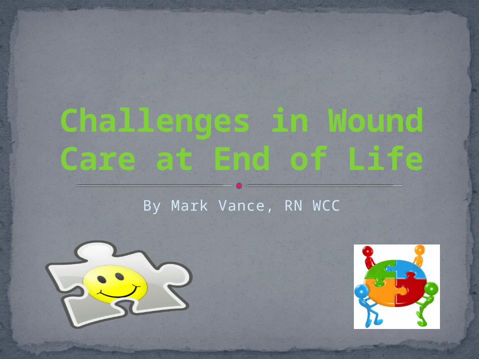
By Mark Vance, RN WCC
Challenges in Wound Care at End of Life

Non-healing woundsEscharOdorCaregiver supportEnvironmentNutrition and hydrationSupply management and understanding of
productsComorbidities
What Are the Challenges?

Assessment of Wound1. Location, type, and cause of wound2. Drainage3. Wound base4. Staging of wound, or determining partial
thickness vs. full thickness5. Surrounding skin6. Odor7. Pain
Building a Plan to Meet These Challenges

Assessment of Client and Caregiver Wishes1. What does client want? Aggressive care vs.
conservative care2. What are caregivers able to do in helping
hospice nurses and patient?3. What are caregivers willing to do?4. What type of environment are you dealing
with?5. What resources do you have?
Building a Plan to Meet Challenges

Assessment of Wound Potential for Healing
1. What time frame does client have? Days, weeks, or months?
2. Severity of wound, to include size of wound, type of wound, slough and eschar in wound.
3. Patient wants vs. needs 4. Available supplies
Building a Plan to Meet Challenges

Patient Centered Goals
1. Build trust and rapport with patient2. Understand impact of wound to patient and
caregivers3. Education on wound healing or decline and
outcome of wound4. Negotiate treatment plan, what works for
patient vs. best practice
Building a Plan to Meet Challenges

Types of Wounds and How They Develop Pressure ulcers caused by improper turning,
poor nutrition, less than ideal support surface Surgical wounds typically caused by infection,
poor nutrition, fragile skin Tumors caused by cancer Skin tears typically caused by falls or bumping
into furniture. Stasis ulcers caused by venous or arterial
insufficiency Radiation burns caused by radiation therapy
Understanding Wounds

1. Hemostasis: coagulation, clot formation, release growth factors
2. Inflammatory phase: leakage of plasma, neutrophils and macrophages
3. Proliferative phase: reformation of tissue4. Maturation phase: can take up to 1 year to
complete.
Phases of Wound Healing

Acute wounds are defined as disruptions in the integrity of the skin and underlying tissues that progress through the healing process in a timely and uneventful manner. The acute surgical wound is an example of a healthy wound in which healing can be maximized.
A chronic wound is a wound that does not heal in an orderly set of stages and in a predictable amount of time the way most wounds do; wounds that do not heal within three months are often considered chronic. Chronic wounds seem to be detained in one or more of the phases of wound healing.
Acute Wounds and Chronic Wounds

Type of wound and location Measurements to include length, width,
depth and tunneling or undermining Wound base; type of tissue, include
percentages Wound drainage and odor Periwound Age of wound Ability of wound to heal Nutrition
Wound Assessment
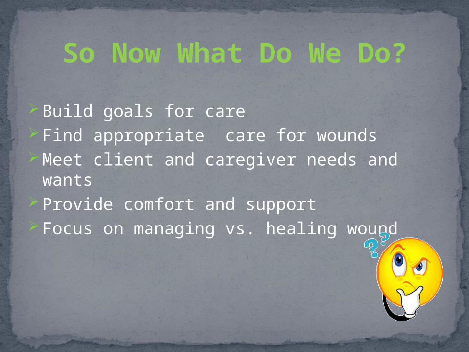
Build goals for careFind appropriate care for woundsMeet client and caregiver needs and wantsProvide comfort and supportFocus on managing vs. healing wound
So Now What Do We Do?

Wound Product Categories
Transparent Film
Hydrocolloid
Hydrogel
Calcium Alginate
Hydrofiber; Aquacel
Collagen
Foam
Antimicrobial dressings
Gauze

Transparent Film(Tegaderm)
Description: Polyurethane with porous adhesive layer
Indications:Primary &
Secondary dressingStage 1 & II ulcersNon-draining
Advantages:Ready wound
inspectionWater tightPrevents or reduces
frictionChange every 5-7
days Disadvantages:
Non-absorptiveMay adhere to woundNon-absorptive

Hydrocolloid(Duoderm CGF)
Description: Hydrophilic colloid particles bound to polyurethane foam
Indications:Stage I to IV ulcersPartial & full thicknessNecrotic woundsPreventive for high-
risk friction areasSecondary dressing or
under taping procedures
Advantages:Facilitate autolytic
debridementImpermeableConformableAbsorptive, minimal
to moderate drainageDisadvantages:
May be hard to remove
Shears off easilyNot recommended
for heavy drainage

Hydrogel(Curasol/wound gel)
Description: Water or glycerin based sheet or gel. Available with silver (SilvaSorb Gel)
Indications:Stage II to IV ulcersPartial & full
thicknessPainful woundsRadiation-damaged
tissueDermabrasion
Advantages:Non-adherentRehydrates wound bedReduces wound painCan use with topical
medsDisadvantages:
May require secondary dressing
Surrounding skin maceration
Not for heavy drainage

Calcium Alginate(Curasorb/Sorbsan)
Description: Nonwoven composite of fibers from calcium-sodium alginate (seaweed)
Indications:Partial & full
thickness Moderate to heavy
drainageStage III or IV ulcersDehisced woundsSinus tracts, tunnels,
or cavitiesInfected wounds
Advantages:Absorbent &
nonocclusiveTrauma-free removalUse with infected
woundsReduces change
frequencySheets & ropes
availableDisadvantages:
Not with dry eschar, burns, heavy bleeding
Need secondary dressing
May produce odorPossible bed damage

Hydrofiber(Aquacel)
Description: Sodium carboxymethylcellulose that interacts with wound exudate. Also in silver
Indications:Partial to full-
thicknessModerate to heavy
drainageDonor sitesDehisced woundsStage III to IV ulcersSinus tracts, tunnels,
or cavities
Advantages:Highly absorptiveTrauma-free removal
Disadvantages:Not with dry eschar,
non-exudating wounds, 3rd degree burns, or heavy bleeding
Requires secondary dressing to secure
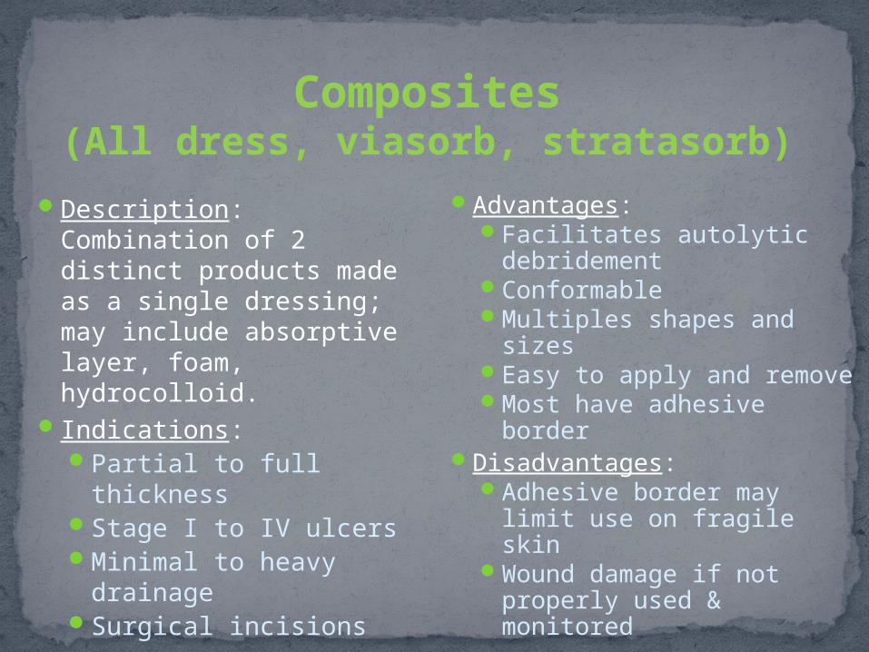
Composites(All dress, viasorb, stratasorb)
Description: Combination of 2 distinct products made as a single dressing; may include absorptive layer, foam, hydrocolloid.
Indications:Partial to full thicknessStage I to IV ulcersMinimal to heavy
drainageSurgical incisions
Advantages:Facilitates autolytic
debridementConformableMultiples shapes and
sizesEasy to apply and removeMost have adhesive
borderDisadvantages:
Adhesive border may limit use on fragile skin
Wound damage if not properly used & monitored

Collagen(Fibercol, Promogran matrix, Silver Prisma
matrix)
Description: Major body protein; stimulates cellular migration & contributes to new tissue development & wound debridement
Indications:Partial and full
thicknessStage III & some IV
ulcersDermal ulcersDonor sitesSurgical wounds
Advantages:Absorbent, nonadherentForms biodegradable gelConforms wellUse with topical agentsChange every 1-3 daysUse for minor slough
Disadvantages:Not for 3rd degree burnsNot for necrotic woundsNeeds secondary dressing

Foams
Description: A hydrophilic, polyurethane film coated foam, non occlusive nonadherent absorptive
Indications:Partial to full
thicknessMinimal to heavy
drainageStage II to IV ulcersSurgical woundsUlcersInfected & non
infected wounds
Advantages:NonadherentTrauma-free removalAbsorbs min to heavy Easy to apply and
removeChange every 3-5
daysDisadvantages:
Not for non-draining or dry eschar
Second dressing to secure
May macerate surrounding skin if not changed

Silver DressingsDescription: Immediate and
sustained release of ionic silver; effective barrier to bacterial penetration
Indications:All wounds except: Stage
I ulcer, 3rd degree burns, and non-draining
Infected woundsHighly colonized woundsOver grafts or skin
substitutesUnder compression
AdvantagesInhibits growth of
bacteria, especially antibiotic-resistant strains
Effective up to 7 daysDisadvantages
Secondary dressing required
Incompatible with oil-based products
Possible sensitivity to silver
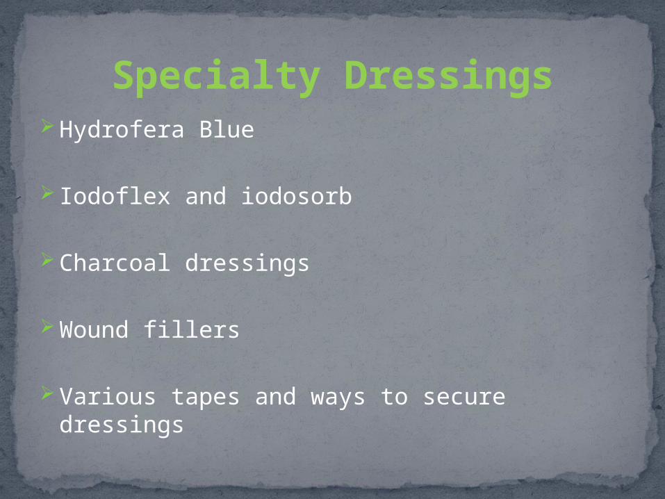
Hydrofera Blue
Iodoflex and iodosorb
Charcoal dressings
Wound fillers
Various tapes and ways to secure dressings
Specialty Dressings

If wound is too moist, then soak up drainage. If wound is too dry, then moisturize it. Use the right product for the right wound.Meet patient’s needs, not your own.
Basic Premise of Wound Care

Now let’s look at some pictures and decide on
wound care.

Arterial/Diabetic

Arterial Wounds

Necrotic/ Unstageable
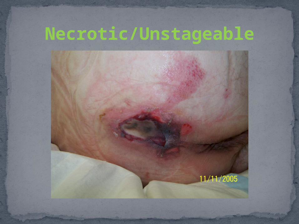
Necrotic/Unstageable

Necrotic/Unstageable

Stage 3 Pressure Ulcer

Stage 4 Pressure Ulcer

Venous Leg Wound

Diabetic Foot Wound

Stage 2 Pressure Ulcer

Stage 2 Pressure Ulcer

Necrotic Eschar Wound

Any Questions????
References
Bryant, Ruth A., and Denise P. Nix. Acute & Chronic Wounds: Current Management Concepts. St. Louis, MO: Elsevier/Mosby, 2012. Print.
Collier, Kyna Setsor, Bridget McCrate Protus, Connie L. Bohn, and Jason M. Kimbrel. Wound Care at End of Life: A Guide for Hospice Professionals. Montgomery: HospiScript Sevices, 2013. Print.
Morgan, Nancy, and Donna Sardina. Skin and Wound Mangement Course: Seminar Workbook. Stevensville: Wound Care Education Institute, 2012. Print.

![The Inventory of the John Holbrook Vance [Jack Vance ...archives.bu.edu/finding-aid/finding_aid_122912.pdf · Vance.JR Box 1 Box 2 Box 3 VANCE, JOHN HOLBROOK, 1916 - [Jack Vance]](https://img.dokumen.tips/doc/110x75/5e776f55989f9014e425e4a0/the-inventory-of-the-john-holbrook-vance-jack-vance-vancejr-box-1-box-2-box.jpg)