Embed Size (px)
Citation preview
Biofilm formation by Listeria monocytogenes. Resistance to industrial biocides and cross‐response caused by adaptation to benzalkonium chloride.
Facultade de Química
Departamento de Química Orgánica
Paula Saá Ibusquiza Tesis Doctoral
DOUTOR EUROPEUS Vigo, Marzo 2011

Biofilm formation by Listeria monocytogenes. Resistance to industrial biocides and cross‐response caused by adaptation to benzalkonium chloride.
Facultade de Química
Departamento de Química Orgánica
Memoria presentada en la Universidad de Vigo por Dña. Paula Saá Ibusquiza
para optar al Grado de Doctora por la Universidad de Vigo Modalidad de Doutor Europeus
Vigo, Marzo 2011

Facultade de Química
Departamento de Química Orgánica
Dña. Rosana Álvarez Rodríguez, Profesora Titular de Química Orgánica de la
Universidade de Vigo
INFORMA: que el trabajo titulado “Biofilm formation by Listeria monocytogenes.
Resistance to industrial biocides and cross-response caused by adaptation to
benzalkonium chloride”, que constituye la Memoria que presenta Dña. Paula Saá
Ibusquiza para optar al Grado de Doctor por la Universidade de Vigo, ha sido realizado
bajo la dirección de Dra. Marta López Cabo y Dr. Juan José Rodríguez Herrera en el
Instituto de Investigaciones Marinas (C.S.I.C.).
Vigo, Marzo 2011.
Fdo. Prof. Dra. Rosana Álvarez Rodríguez


Facultade de Química
Departamento de Química Orgánica
Dra. Marta López Cabo y Dr. Juan José Rodríguez Herrera, Científicos Titulares del
Instituto de Investigaciones Marinas (C.S.I.C.):
CERTIFICAN: que la memoria adjunta, titulada “Biofilm formation by Listeria
monocytogenes. Resistance to industrial biocides and cross-response caused by adaptation to
benzalkonium chloride”, que constituye el trabajo que presenta Dña. Paula Saá Ibusquiza para
optar al Grado de Doctor por la Universidad de de Vigo, ha sido realizada bajo nuestra
inmediata dirección en el Instituto de Investigaciones Marinas (C.S.I.C.).
Vigo, Marzo 2011.
Fdo. Dra. Marta López Cabo Fdo. Dr. Juan José Rodríguez Herrera


“Sorprendernos por algo es el primer paso de la mente hacia el descubrimiento"
Louis Pasteur (1822-1895)
Microbiólogo y químico orgánico francés.


XI
Acknowledgements
En primer lugar mi agradecimiento, por supuesto, a Marta, bueno Dra. Marta López Cabo.
¡He aprendido tanto contigo! Profesionalmente prácticamente todo lo que sé de
Microbiología, personalmente has sido mi amiga, gracias por tu cercanía, humanidad,
espontaneidad, cariño y protección. Siempre he tenido la tranquilidad de poder expresarme de
manera directa y sincera y me he sentido escuchada. Me has dado en todo momento la
seguridad de que conseguiríamos los objetivos marcados reflejados finalmente en esta
memoria que compartiremos siempre.
Ahora vas tú Juan, gracias al Dr. Juan José Rodríguez Herrera. Gracias por ser el
contrapunto entre Marta y yo por esas veces en las que Marta decía que somos iguales
simplemente por tener la misma opinión en algo puntual y diferente a la de ella. Me he
sentido en un entorno familiar mientras trabajábamos. Gracias por tu ironía, por las
conversaciones en que desmenuzamos cualquier asunto en profundidad independientemente
de su relevancia, por vanal que fuese. Gracias a los dos por hacer el equipo “cuasi” perfecto.
Gracias a la Dra. Laura Pastoriza por formar parte de la formación de Marta y Juan.
Gracia Sonia, por tu alegría, tu buen carácter por tu actitud positiva, por tu capacidad de
trabajo en equipo, de ti tendré siempre la imagen en la campana, codo con codo en esas
siembras interminables en las que teníamos tiempo de ponernos al día de nuestras cosas, de
las gracias de Diego, de lo que habíamos hecho el fin de semana. Has hecho el día a día
muuucho más llevadero.
Gracias Dani por tus ganas de querer hacer las cosas bien, independientemente de que sea
la manera de hacerla…jaja. Eres muy buen tío, generoso, siempre dispuesto a ayudar aunque
refunfuñes, jaja. Los dos supimos siempre que nuestras “pequeñas” discusiones eran por tener
que compartir la campana y que a los 5 minutos ya no podíamos evitar disculparnos por
nuestro carácter, es muy fácil cogerte cariño.

Gracias Alberto por tratarme como una reina aunque alguna vez me llevases la contraria,
ya se sabe, hasta las mejores parejas discuten cómo tú dices. Salir contigo de fiesta es noche
divertida asegurada.
Gracias Lola por el primer año de tesis que compartí contigo, cuando todo era nuevo,
gracias por tu apoyo cuando no podíamos seguir el ritmo que nos marcaban y por tus ganas de
establecer una buena relación que espero poder seguir manteniendo.
Gracias Gabi, cada día contigo hacía imposible no esbozar una sonrisa. De mayor quiero
ser como tú y disfrutar de la vida cómo tú sabes. Gracias por tu cariño.
Gracias Eva porque llegaste y al día siguiente parecía que llevabas aquí desde el principio,
por tu capacidad de adaptación, tu sencillez, tus ganas de aprender.
Gracias Lidia por tu energía y simpatía. Se notó tu falta cuando acabaste.
Gracias al resto de las personas del equipo: Vanesa, Graci, Marta B., Ana, Tere, Javi, Pilar.
Gracias chicas porque además de una tesis me he sacado un master: especialista en niños!!.
Solo me faltan las prácticas, ya llegarán, ¿verdad Eva?.
Gracias María Gregory por las comidas, los cafés, las clases de gimnasio que compartimos
después del trabajo…me encanta que seas tan activa, mantendremos nuestra amistad por
mucho tiempo. ¡Tú también me aportas mucho!. Gracias Cuchi a ti también.
Gracias a todo el personal del Instituto de Investigaciones Marinas por su ayuda durante
estos años. Todos me habéis hecho la vida más sencilla, gracias al personal de administración,
mantenimiento, gracias Juan Luis, y al resto de los equipos de investigación. Gracias
compañeros, por compartir conmigo el trabajo o medios de este instituto de cualquiera de las
maneras posibles, una charla, material prestado.
Gracias Sarah Jane, mi profe de inglés en el iim, por tu decisión de querer ser mi amiga.
Gracias a tu familia por su hospitalidad cuando estuve en Galway.
Thanks to Professor Colin Hill for allowing me working in his lab in Cork for 6 months. I
could discover a new world, like a child when learns how to cut with a pair of scissors.
Thanks for inviting me to spend a fantastic weekend with your family. I want to thank also to
XIII
Dr. Roy Sleator for his laboratory supervision and all the people in the lab for their help and
love.
Gracias a los miembros de CACTI por los experimentos realizados y a los miembros del
Departamento de Química Orgánica por protagonizar mi primer contacto con la investigación
materializado en el DEA. Gracias a Rosana y a Laura por resolver mis dudas burocráticas y a
Ángel como líder del grupo. Gracias Xenxo por los libros que me bajaste de L.
monocytogenes, las referencias están en la introducción.
Y ya fuera del ámbito professional, gracias a mis amigos de siempre porque parte de lo que
soy es por vosotros. Gracias, Iria, María, Xiana, Jessica, Rebeca P., Rebeca S., Marina,
Rubén, Jorge, os quiero.
Gracias Iago, por todo, siempre podrás contar conmigo.
Gracias Patri por ser mi amiga, celebrar e invitarme a tu cumple, como sino habría
conocido a Marcos. Gracias Marcos por darme luz durante el final del recorrido de esta tesis.
Mi familia, gracias mami ya he llegado a ti, te quiero muchíiiiiiiiiisimo!!. Eres un referente
en mi vida. Gracias a mi hermano, gracias Marcos, gracias por tu apoyo constante, tu
generosidad, tu amor, siempre sé que estarás ahí para mí, te admiro. Los dos llenáis allí a
donde vais. Gracias al resto de mi familia, a mi padre y a mi abuelo Gustavo especialmente.

XV
CONTENTS
ACKNOWLEDGEMENTS…………………………………………………………...…. …XI
LIST OF ORIGINAL PUBLICATIONS……………………………………...…………. XVII
INTRODUCTION..………………………………………..……...…………….........…..1
1.1. LISTERIA MONOCYTOGENES……………………………………...…....…... ..1
1.1.1. History, biological characteristics and classification………………...………... ..1
1.1.2. Natural niches………………………………………………….....……...…..... ..3
1.1.3. The disease: clinic, invasiveness and virulence……………………...….…...... ..4
1.1.4. Virulence mechanism…………………………………………………...……... ..6
1.2. BIOFILM FORMATION BY L. MONOCYTOGENES………….……..…….. ..9
1.2.1. What is a biofilm? History and definition ………………………………..…… ..9
1.2.2. Some important consequences of biofilm formation……………………....….. ..9
1.2.2.1. Transference of gases and nutrients…………………………………...…… ..9
1.2.2.2. Cell disposition in the biofilm............…………………………….……..… 10
1.2.2.3. Resistance to external stimulus: Implications in medicine and
in food industry……………………………………….............................…. 10
1.2.3. Biofilm development in L. Monocytogenes........................................................ 12
1.2.3.1. Initial attachment………………………………………………………...…. 12
1.2.3.1.1. Effect of external conditions: nutrients, type of surface, pH............…. 12
1.2.3.1.2. Effect of the conditioning film in the initial attachment………....…… 14
1.2.3.1.3. Effects of flagellar motility……………………………………..……... 14
1.2.3.2. Maturation of attached bacteria into a differenciated biofilm ………..……. 15
1.2.3.2.1. Effect of external medium…………........…………………...………… 15
1.2.3.2.2. Importance of the EPS matrix………………………………...……….. 16
1.2.3.3. Dettachment…………………………………………...……………....…… 17

1.2.4. Regulation of biofilms formation in L. Monocytogenes.......…………….....… 19
1.2.4.1. Cell-cell communication................................………………………...…… 19
1.2.4.2. General stress sigma factor…………………………………………..…….. 20
1.2.5. Mixed biofilms…....……………………………………………………...……. 20
1.2.6. Persistance…………………………………………………………………...… 22
1.3. INCIDENCE AND CONTROL OF L. MONOCYTOGENES…………...……. 23
1.3.1. Incidence of L. monocytogenes in foods and food processing plants................. 23
1.3.1.1. A brief overview of L. monocytogenes outbreaks………...…........…….. 23
1.3.1.2. Incidence of L. monocytogenes in food processing plants……………...….. 26
1.3.1.3. Incidence of L. monocytogenes in foods……………………………...……. 27
1.3.2. Control of L. monocytogenes in food processing plants …………………..…. 29
1.3.2.1. Prerrequisites-HACCP: importance for food safety…………………...…… 29
1.3.2.2. The problem of cross contamination…………………………….…...…….. 31
1.3.2.3. Disinfectants…………………………………………………………...…… 32
1.3.2.3.1. Classical disinfectants…………………………………………...…….. 32
1.3.2.3.2. Other compound with potencial applications in cleaning and disinfection
protocols……………………………………………….......................... 35
1.3.2.4. Resistance of L. monocytogenes biofilms to disinfectants……………...….. 38
1.3.2.5. Cross-resistance responses of L. monocytogenes…………………………... 39
1.3.2.5.1. Cross-resistance of acid-adapted L. monocytogenes…………………. 39
1.3.2.5.2. Cross-resistance of L. monocytogenes between bacteriocins………… 40
1.3.3. Control of Listeria monocytogenes in foods………………………………...… 40
1.3.3.1. Bacteriocins…………………………………………………………..…….. 40
1.3.3.2. Application of active packaging…………………………………..……….. 41
1.3.3.3. Ozone (O3)………………………………………………………...….…... 41

XVII
1.3.3.4. Electrolyzed water (EO)…………………………………………...……… ..41
1.3.3.5. Phages……………………………………………………………...………....41
1.3.3.6. Nanotechnology…………………………………………………...……….. ..42
1.3.4. Listeria monocytogenes as a tool..................…………………………...…...… ..42
References............................................................................................... .........................43
JUSTIFICATION AND OBJECTIVES..………………...…………………...……...…..69
RESULTS AND DICUSSION..…………….………….…...…………………….......….71
1.4. ADHERENCE: Effects of mussel processing soils on the adherence of Listeria
monocytogenes to polypropylene and stainless steel……………………………... ..75
1.4.1. Introduction………………………………………………………………...….. ..76
1.4.2. Material and Methods……………………………………………………..…... ..77
1.4.3. Results and discussion……………………………………………………….... ..80
References…………………………………………………………………………...... ..89
1.5. ADAPTATION: An efficient method to obtain benzalkonium chloride-adapted cells
of Listeria monocytogenes……………………...………….……………………... ..95
1.5.1. Introduction…………………………………………..…………………….….. ..96
1.5.2. Material and Methods………………………………………………………..... ..97
1.5.3. Results……………………………………………………...………………….. 100
1.5.4. Discussion……………………………………………………………………... 105
References……………………………...……………………...………………..….......109
1.6. MATURE BIOFILMS: Resistance to benzalkonium chloride, peracetic acid and
nisin during formation of mature biofilms by Listeria monocytogenes……...…... 115
1.6.1. Introduction…………………………………………..………………….…….. 116
1.6.2. Material and Methods………………………………………………………..... 117
1.6.3. Results……………………………………………………...………………….. 122

1.6.4. Discussion……………………………………………………………………... 128
References……………………………………………...………………………..…......132
1.7. MIXED BIOFILMS: Adherence kinetics, resistance to benzalkonium chloride
and microscopic analysis of mixed biofilms formed by L. monocytogenes and
Pseudomonas putida…………………………………….………………………... 139
1.7.1. Introduction…………………………………………..…………..…….…….. 140
1.7.2. Material and Methods………………………………………………………..... 141
1.7.3. Results……………………………………………………...………………….. 145
1.7.4. Discussion……………………………………………………………………... 152
References……………………...…………………………...………….………..…......157
1.8. MAP: Comparison between the resistance of benzalkonium chloride-adapted
and non-adapted biofilms of Listeria monocytogenes to modified atmosphere
packaging (MAP) and nisin once transferred to mussels…………………….…... 163
1.8.1. Introduction…………………………………………..……………………….. 164
1.8.2. Material and Methods………………………………………………………..... 165
1.8.3. Results……………………………………………………...………………….. 169
1.8.4. Discussion……………………………………………………………………... 174
References……………………...……...…………..………...………...………...…......178
CONCLUSIONS..…………………….…………………....…...………….....….......…185
SUPPORTING INFORMATION…………………………..…………..………….........189
PERSPECTIVES……………………..……………………….…..…………..…..........199
RESUMEN………………………………...……………………………...….…... 203

XIX
LIST OF ORIGINAL PUBLICATIONS
The thesis is based on the following original publications:
1. Effects of mussel processing soils on the adherence of Listeria monocytogenes to
polypropylene and stainless steel.
Authors: P. Saá, M. L. Cabo M. and J. Herrera
Reference: J. Food Prot 72(9), 1885-90 (2009).
2. An efficient method to obtain benzalkonium chloride-adapted cells of Listeria
monocytogenes CECT 5873.
Authors: P. Saá Ibusquiza, J. R. Herrera, D. Vázquez-Sánchez and M. L. Cabo
Reference: in preparation.
3. Resistance to benzalkonium chloride, peracetic acid and nisin during formation of mature
biofilms by Listeria monocytogenes
Authors: P. Saá Ibusquiza, J. R. Herrera and M. L. Cabo
Reference: Accepted in FoodMicrobiol. doi:10.1016/j.fm.2010.09.014
4. Adherence kinetics, resistance to benzalkonium chloride and microscopic analysis of mixed
biofilms formed by Listeria monocytogenes and Pseudomonas putida.
Authors: P. Saá Ibusquiza, J. R. Herrera, D. Vázquez-Sánchez and M. L. Cabo
Reference: submitted.
5. Comparison between the resistance of benzalkonium chloride-adapted and non-adapted
biofilms of Listeria monocytogenes to modified atmosphere packaging (MAP) and nisin once
transferred to mussels
Authors: P. Saá Ibusquiza, J. R. Herrera and M. L. Cabo
Reference: accepted in J. Food Prot.


11.. IInnttrroodduuccttiioonn


Introduction
1
1.1. LISTERIA MONOCYTOGENES
1.1.1. History, biological characteristics and classification
L. monocytogenes was discovered by EGD Murray in 1924 following an epidemiy that
affected rabbits and guinea pigs in animal care houses in Cambridge (Murray et al., 1926). It
was firstly described as a pathogen in 1929 and initially considered as an animal pathogen
with rare human cases, L. monocytogenes emerged as a “new” human pathogen in the 1980s,
with several food-associated listeriosis outbreaks. The case fatality rate is about 20-30%. In
the US, listeriosis accounts for approximately 28% of the deaths and the highest
hospitalization rate (91%) caused by known food-borne infections.
Prior to the 1980s the origins of human infections caused by Listeria monocytogenes were
uncertain. However, at the end of the 1970s and the start of the 1980s, the number of reports
on Listeria isolations began to increase and in 1983, the first human listeriosis outbreak
directly linked to the consumption of Listeria contaminated foodstuffs was reported (Schlech
et al., 1983). From 1983 onwards, a series of epidemia outbreaks in humans in North America
and Europe clearly established listeriosis as a severe food-borne infection (Farber and
Peterkin, 1991; Fleming et al., 1985; James et al., 1985; Schlech et al., 1983), and thereby L.
monocytogenes as a food-borne pathogen.
The genus Listeria consists of a group of Gram-positive bacteria of low G+C content
closely related to Bacillus and Staphylococcus (Sallen et al., 1996). It includes six species: L.
monocytogenes, L. ivanovii, L. seeligeri, L. innocua, L. welshimeri and L. grayi (Sallen et al.,
1996). Two of them, L. monocytogenes and L. ivanovii, are pathogenic. While L. ivanovii is
mainly pathogenic for animals, L. monocytogenes can infect humans, and domestic and wild
animals (Seeliger and Jones, 1986). A third species, L. seeliegeri, is considered avirulent,
although it has been isolated from at least one case of human listeriosis (Rocourt et al., 1986).
Listeria monocytogenes is a Gram positive facultative anaerobic bacterium (Seeliger and
Jones, 1986) that can resist acid pHs, low Aw, low O2 concentrations and low temperatures
(Kathariou, 2002; Ross et al., 2000). Morfologically, L. monocytogenes are non-spore
forming rods of 0.4 μm in diameter and 0.5 – 2 μm in length. They are motile by means of
peritrichous flagella when cultured below 25 °C (Seeliger and Jones, 1986). L.
2 Introduction
monocytogenes can grow over the temperature range of 0.5-45 °C with an optimum between
30 °C and 37 °C. However, its growth at temperatures below 4 °C is generally very slow and
the lag phase can be very long. As the temperature increases above 4 °C, the growth rate of L.
monocytogenes increases and the lag phase time decreases considerably; consequently,
storage at slightly abusive temperatures (e.g. 7 to 10 °C) of refrigerated ready-to-eat (RTE)
greatly increases the risk that L. monocytogenes, if present, will reach numbers that could
cause human disease (ILSI Research Foundation/Risk Science Institute Expert Panel on L.
monocytogenes in Foods, 2005; International Commission on Microbiological Specifications
for Foods, 1996). Freezing at −18 ºC, and even repeated freezing, has little effect on the
survival of L. monocytogenes; these conditions are more likely to injure than to inactivate this
organism.
L. monocytogenes is very well equipped to survive typical hurdles applied in food
preservation. It grows across a broad pHs range (4.3-9.8), but depends on the acid type and
temperature, L. monocytogenes can grow at pHs as low as 4.0 (Lado and Yousef, 2007,
Martin and Fisher, 1999). L. monocytogenes grow in complex medium containing up to 10%
(w/v) NaCl, but some strains can tolerate up to 20 % (w/v) NaCl, so it can resist very low
water activities (aw 0.91) (Lado and Yousef, 2007, Seeliger and Jones, 1986). Moreover, it
can grow in aerobic modified atmospheres also with competitive microorganisms
(Wimpfheimer et al., 1990).
L. monocytogenes has been classified under different criteria:
1. According to the presence of O (somatic) and H (flagelar) antigens L. monocytogenes is
classified in 13 serovars (Seeliger and Höhne, 1979; Seeliger and Jones, 1986): 1/2a, 1/2b,
1/2c, 3a, 3b, 3c, 4a, 4ab, 4b, 4c, 4d, 4e, 7. Although human listeriosis may be caused by all 13
serovars of L. monocytogenes, 1/2a, 1/2b, 1/2c and 4b serovar cause at least 95% of the cases
(Doumith et al., 2004; Farber and Peterkin, 1991; Swaminathan and Gerner-Smidt, 2007).
Among the outbreaks of invasive listeriosis, serovar 4b strains have caused the majority of the
outbreaks worldwide from 1980-2005, whereas strains of serovar 1/2 have caused the
majority of the non-invasive, gastrointestinal listeriosis outbreaks worldwide from 1993-2001
(Swaminathan and Gerner-Smidt, 2007). However, among food isolates, serotype 1/2 is the
most frequently found (Farber and Peterkin, 1991; Jacquet et al., 2002).

Introduction
3
2. According to the genotypic analyses: whereas enzyme electrophoresis (Piffaretti et al.,
1989), pulsed-field gel electrophoresis (PFGE) (Brosch et al., 1994) and ribotyping (Graves et
al., 1994) can divide L. monocytogenes into two major subgroups, virulence gene analysis
have grouped L. monocytogenes into three groups: lineage I, II and III (Rasmussen et al.,
1995; Wiedmann et al., 1997). At the same time, each lineage includes several serotypes:
lineage I, comprising serotypes 1/2b, 3b, 3c and 4b; lineage II, comprising serotypes 1/2a,
1/2c and 3a, and lineage III comprising serotypes 4a and 4c (Nadon et al., 2001). Invasive
listeriosis is primarily caused by lineage I strains, whereas lineage II strains are most
frequently isolated from food. In comparison, serotypes belonging to lineages II and III are
less significant, being rarely associated with foodborne listeriosis.
Typing studies involving L. monocytogenes isolates from clinical, food and food
processing sources led to the consensus of considering the division of L. monocytogenes in
those three lineages (Harvey and Gilmour, 1992).
1.1.2. Natural niches
Although L. monocytogenes is ubiquitous, its prevalence in the outdoor environment is not
high (Porsby et al., 2008). Listeria spp. are isolated from a diversity of environmental sources,
including soil, water, effluents, a large variety of foods, and the feces of humans and animals
(Barbuddhe et al., 2009). It is thought to be widespread, being a saprophytic organism adapted
to the plant-soil environment (Weis and Seeliger, 1975). The bacteria is widely present in
plants, soils, sediments and surface water samples, and has also been found in sewage, human
and animal faeces (MacGowan et al., 1994; Weis and Seeliger, 1975). Generally, the
proportion of positive samples is low in the outdoor environment, between 0 and 6%, but
studies indicate that the prevalence increases with the degree of human activity (Hansen et al.,
2006; MacGowan et al., 1994; El Marrackchi et al., 2005). Animals are susceptible to
listeriosis but can carry L. monocytogenes asymptomatically. Fecal carriage of L.
monocytogenes in livestock animals such as cattle beef, dairy, poultry and horses has been
found with varying frequency around 0-13%, and in wildlife up to 40% (Lyautey et al., 2007).
Prevalence of L. monocytogenes has also been reported in wild life animals like deer, moose
and birds (Hellström et al., 2008; Lyautey et al., 2007). Similarly, humans can be
4 Introduction
asymptomatic carriers of L. monocytogenes, but generally with prevalence below 1%
(MacGowan et al., 1994; Sauders et al., 2005).
1.1.3. The disease: clinic, invasiveness and virulence
The invasive disease is fatal in 30% of cases although it can be treated with amoxicillin
antibiotics if early caught (Williams and Nadel, 2001). Clinical manifestations range from
febrile gastroenteritis to more severe invasive forms, including sepsis, meningitis,
rhombencephalitis, perinatal infections, and abortions (Allerberger et al., 2010). In pregnant
women, listeriosis may lead to spontaneous abortion, stillbirth or fetal death.
Recent outbreaks demonstrated that L. monocytogenes can cause gastroenteritis in
otherwise healthy individuals and more severe invasive disease in immunocompromised
patients. Common symptoms include fever, watery diarrhea, nausea, headache, and pains in
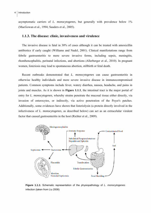
joints and muscles. As it is shown in Figure 1.1.1, the intestinal tract is the major portal of
entry for L. monocytogenes, whereby strains penetrate the mucosal tissue either directly, via
invasion of enterocytes, or indirectly, via active penetration of the Peyer's patches.
Additionally, some evidences have shown that listeriolysin (a protein directly involved in the
infectiveness of L. monocytogenes, as described below) can act as an extracelular virulent
factor that caused gastroenteritis in the host (Richter et al., 2009).
Figure 1.1.1: Schematic representation of the physiopathology of L. monocytogenes
infection (taken from Liu 2008)

Introduction
5
Although all the strains belonging to this species are assumed to be pathogenic,
epidemiological evidence indicates that strain-to-strain differences in virulence. Thus, only 4
of the 13 Listeria serotypes—namely, 1/2a, 1/2b, 1/2c, and 4b—account for 95–98% of all
cases of human and animal listeriosis worldwide. Of these, serovar 1/2c is found in a minority
of clinical isolates (2–4%) but predominates among food isolates. Similarly, serovar 4b,
belonging to one of the two major genetic lineages of L. monocytogenes, predominates among
clinical isolates (>50% of listeriosis cases), whereas it is much less frequently found among
food isolates than serogroup ½ (i.e., serovars 1/2a, 1/2b, and 1/2c) strains. Moreover, a
restricted number of 4b strains, representing distinct genotypes, are responsible for most food-
borne outbreaks of human listeriosis world-wide, which suggests that certain clones of L.
monocytogenes may be more pathogenic for humans (Liu, 2008).
Virulence heterogeneity among L. monocytogenes isolates—often associated to natural
attenuating mutations in key virulence loci—is also supported by experimental evidence.
However, the most critical factor is the underlying condition and immunological status of the
host as this determines the susceptibility to a given strain of L. monocytogenes. The vast
majority of listeriosis patients have a physiological or pathological condition that impairs the
capacity to mount an effective cellular immune response. Three major population groups at
risk for invasive listeriosis are the neonates, elderly (>60 years) and pregnant woman. In
neonates and elderly, that risk is due to the inmature or declining immune system. In pregnant
woman is associated with depression of cell-mediated immunity to prevent rejection of the
fetoplacental allograft. In nonpregnant adults, almost all cases of listeriosis have been found
in individuals with chronic, debilitating illnesses or subjected to immunosuppressive therapy.
Specific risk groups in the intermediate-age band include cancer and organ transplant patients,
HIV-infected and AIDS patients, and individuals with chronic liver disease (alcoholism and
cirrhosis), diabetes and lupus (Liu, 2008).
In immunocompetent individuals with no predisposing condition, ingestion of low to
moderate doses of L. monocytogenes (≤105 CFUs) has no effect other than boosting
antilisterial protective immunity, whereas ingestion of large doses of the bacteria (≥106 CFUs,
sometimes doses as high as 1011) may cause acute febrile gastroenteritis within 24 h of
consumption of the contaminated food due to massive invasion of the intestinal mucosa.
Depending on the pathogenicity of the strain, some healthy nonpregnant adults exposed to a
large L. monocytogenes inoculum may develop invasive listeriosis. In immunocompromised

6 Introduction
individuals, however, invasive disease is facilitated by the inefficient mobilization of the host
defenses and the blood-borne dissemination of L. monocytogenes from the primary infectious
foci in the liver and spleen (silent phase of infection). In those cases, bacteremia may lead to
meningoencephalitis if bacteria traverse the brain microcapillaries, to abortion or perinatal
septicemia if they traverse the placental barrier, or to septicemic disease in cases of severe
immunosuppression (Figure 1.1.2).
Figure 1.1.2: Clinical and pathological features of Listeria monocytogenes infection. (A)
Fetomaternal listeriosis. Stillborn fetus with septicemic invasion (“granulomatosis
infantiseptica”). (B) Liver from the stillborn fetus in (A) showing typical disseminated
pyogranulomatous necrotic foci. (C) Histopathological image of the liver from an
experimentally infected sheep with milliary listerial pyogranulomatous hepatitis
(hematoxilin/eosin-stained). (D) Meningoencephalitis due to L. monocytogenes in a cow.
(E) Section of the brainstem of a sheep with listerial rhombencephalitis showing
inflammatory lesions in the nerve tissue. (F) Parenchymal inflammatory infiltration of the
brainstem in (E) showing typical perivascular cuffing (arrow) indicative of blood-borne
invasion of the brain tissue by L. monocytogenes. Clinical and pathological
manifestations of listeriosis are essentially similar in humans and animals. (taken from Liu
2008 Pag. 99)
1.1.4. Virulence mechanism
Macrophages and epithelial cells are widely used to study the interaction of L.
monocytogenes with mammalian host cells. However, it was shown that also neutrophils,

Introduction
7
dendritic cells, hepatocytes, fibroblasts, endothelial cells, or glial cells may become infected
with, and serve as host cells for L. monocytogenes in vitro and in vivo.
Figure 1.1.3: Stages of listerial intracellular parasitism. (A) Scheme of the intracellular life
cycle of pathogenic Listeria spp. (B–H) Scanning and transmission electron micrographs
of cell monolayers infected with L. monocytogenes. (B) Numerous bacteria adhering to
the microvilli of a Caco-2 cell (30 min after infection). (C) Two bacteria in the process of
invasion (Caco-2 cell, 30 min postinfection). (D) Two intracellular bacteria soon after
phagocytosis, still surrounded by the membranes of the phagocytic vacuole (Caco-2 cell,
1 h postinfection). (E) Intracellular Listeria cells free in the host cell cytoplasm after
escape from the phagosome (Caco-2 cell, 2 h postinfection). (F) Pseudopod-like
membrane protrusion induced by moving Listeria cells, with the bacterium being evident
at the tip (brain microvascular endothelial cell, 4 h postinfection. (G) Section of a
pseudopod-like structure in which a thin cytoplasmic extension of an infected cell is
protruding into a neighboring noninfected cell, with the protrusion being covered by two
membrane layers (Caco-2 cell, 4 h postinfection). (H) Bacteria in a double membrane
vacuole formed during cell-to-cell spread (Caco-2 cell, 4 h postinfection). (Taken from
Vazquez-Boland et al., 2001.

8 Introduction
Macrophages actively engulf L. monocytogenes spontaneously, but internalization of the
bacterium by normally nonphagocytic cells is triggered by L. monocytogenes-specific factors.
Aside from the internalization step, the intracellular life cycle (Figure 1.1.3 A) of the
bacterium in phagocytes or normally nonphagocytic mammalian cells is, however, essentially
identical. L. monocytogenes induces its own internalization without an extensive remodeling
of the host cell surface. Entry occurs via zipper-like phagocytosis, characterized by the
emission of small pseudopods that firmly entrap the bacteria and the intimate contact of the
bacterial surface with the host cell plasma membrane. Upon uptake, the pathogen appears in a
membrane-bound vacuole (Figure 1.1.3 D), which is subsequently lysed by the combined
action of the pore-forming hemolysin, listeriolysin (LLO), and two phospholipases (see
below). The bacteria that are released into the cytoplasm begin to replicate while making use
of specific transporters to gain carbohydrates from the host cell, whereas those remaining in
the phagosome are killed and digested.
Concomitant with the onset of intracellular replication, L. monocytogenes induces the
expression of the surface protein ActA which, through the activation of the cellular rp2/3
complex, induces the nucleation of host actin filaments. The formation of a polar tail and the
permanent polymerization of F-actin at the interface between the bacteria and the actin tails
produce a propulsive force, which moves the bacteria through the cytoplasm. Those bacteria
that in their random movement reach the plasma membrane push outwards inducing the
formation of pseudopod-like structures with the bacterium at the tip. These invading
pseudopods or “listeriopods” are taken up by the neighboring cells, in which the bacteria
become entrapped within a double membrane. This vacuole is again lysed by LLO and the
phospholipases, a broad-specificity phospholipase, releasing the bacteria into the cytoplasm of
the newly infected host cell where they initiate a new cycle of replication and actin-based
motility. This direct cell-to-cell invasion mechanism allows the bacteria to spread through
host tissues without leaving the host cytosolic compartment, protected from the humoral
effectors of the immune system and phagocytosis (Liu, 2008)
L. monocytogenes has become not only an important paradigm for immunological
investigation but also an important model system for analysis of the molecular mechanisms of
intracellular parasitism (Vázquez-Boland, 2001)

Introduction
9
1.2. BIOFILM FORMATION BY L. MONOCYTOGENES
1.2.1. What is a biofilm? History and definition
Although bacteria grow preferentially in the biofilm mode in industrial and natural systems
(Blaschek et al., 2007), studies on bacteria in laboratories are still generally carried out in
planctonic cells. The vast majority of microorganisms tested end up by forming biofilms, on
practically any surface and any environmental conditions, although their adhesion and growth
rates, extracellular polymeric substances (EPS) yields and final structure is highly variable
(Watnick and Kolter, 2000).
During the last 25 years, definitions of biofilms have been proposed by several authors
(Marshall, 1976; Costerton et al., 1978; Costerton et al., 1987; Characklis and Marshall, 1990;
Costerton et al., 1995; Costerton and Lappin-Scott, 1995; Davies and Geesey, 1995; Prigent-
Combaret and Lejeune, 1999). Finally, the following definition is widely accepted: “Biofilm
is a microbially derived sessile community characterized by cells that are irreversively
attached to a substratum or interface or to each other, embedded in a matrix of extracellular
polymeric substances that they have produced, and with an altered phenotype with respect to
growth rate and gene transcription (Donlan and Costerton, 2002)”.
1.2.2. Some important consequences of biofilm formation
1.2.2.1. Transference of gases and nutrients
Diffusion limitations create gradients of nutrients and oxygen from the top to the bottom of
biofilms that are associated with decreased bacterial metabolic activity and increased doubling
times of the bacterial cells. Anaerobic conditions may be present in the centre of the biofilm.
Likewise, growth, protein synthesis and metabolic activity are stratified in biofilms, i.e. a high
level of activity at the surface and a low level and slow or no growth in the centre. In fact, this
is one of the explanations for the reduced susceptibility of biofilms to antibiotics (Høiby et al.,
2010). Such gradients will increase in extent as biofilms thicken and will become particularly
marked in aged biofilms (Gilbert et al., 2002). A major contributor towards the failure of
biofilms to rapidly succumb to antimicrobial treatments must, therefore, be associated with its
physiological heterogeneity (Allison et al., 2000; Gilbert et al., 2002).

10 Introduction
1.2.2.2. Cell disposition in the biofilm
Bacterial cells have the ability to aggregate into particular three-dimensional assemblages,
differentiate, divide labor within these assemblages, and then disperse as part of their life
cycle (Davey and O´Toole, 2000). The structures that form in biofilms contain channels in
which nutrients can circulate. The complexity of biofilm structure and metabolism has led to
the analogy of biofilms to tissues of higher organisms. The complex biofilm architecture
provides an opportunity for metabolic cooperation and could imply the generation of niches
where antimicrobial-resistant phenotypes are formed within the spatially well-organized
system (Davies, 2003; Klapper et al., 2007).
So, beside the individual cell changes, intercellular interactions and communication are
required for biofilm development and persistence. Dissecting these interactions provides one
of the future challenges in biofilm research. Particularly challenging is the attempt to
understand the complexity of the interactions within the biofilm community. Communication
between species may include extracellular compounds whose sole role is to influence gene
expression, metabolic cooperativity and competition (possibly encompassing global changes
in gene expression and metabolism), physical contact, and the production of antimicrobial
exoproducts. One or all of these interactions may be occurring simultaneously (Davey et al,
2000).
1.2.2.3. Resistance to external stimulus: implications in medicine and in food industry.
Biofilms constitute a protected mode of growth that allows survival in a hostile
environment (Costerton et al., 1999) under changing environmental conditions and hostile
chemical or physical agents: predators, host cells, innate defense compounds, antibiotics and,
in the food industry, antimicrobial additives and cleaning and disinfection agents. Biofilm-
embedded cells can stand up to 100-1000 times higher biocide concentrations than planktonic
cells. As a consequence, bacterial cells persist in biofilm form, causing problems mainly in
the medical ambit and in the food industry.
Concerning the medical ambit, the US National Institutes of Health estimates that upwards
of 75% of microbial infections that occur in the human body are underpinned by formation
and persistence of biofilms. Some examples are recopilated in Table 1.2.1. Biofilms increase
the tolerance to antibiotics and disinfectant chemicals in association with medical devices as
Introduction
11
well as resisting phagocytosis and other components of the body's defence system (Campanac
et al., 2002, Costerton et al., 1999, Høiby et al., 2010). Biofilms are the most likely
environmental nidus of resistance development and selection that might relate to active efflux
of the treatment agent or to others less well-understood mechanisms (Gilbert et al., 2002).
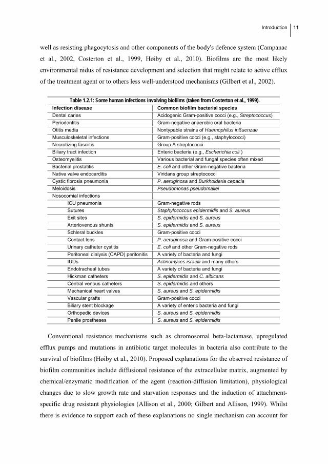
Table 1.2.1: Some human infections involving biofilms (taken from Costerton et al., 1999). Infection disease Common biofilm bacterial species
Dental caries Acidogenic Gram-positive cocci (e.g., Streptococcus)
Periodontitis Gram-negative anaerobic oral bacteria
Otitis media Nontypable strains of Haemophilus inßuenzae
Musculoskeletal infections Gram-positive cocci (e.g., staphylococci)
Necrotizing fasciitis Group A streptococci
Biliary tract infection Enteric bacteria (e.g., Escherichia coli )
Osteomyelitis Various bacterial and fungal species often mixed
Bacterial prostatitis E. coli and other Gram-negative bacteria
Native valve endocarditis Viridans group streptococci
Cystic fibrosis pneumonia P. aeruginosa and Burkholderia cepacia
Meloidosis Pseudomonas pseudomallei
Nosocomial infections
ICU pneumonia Gram-negative rods
Sutures Staphylococcus epidermidis and S. aureus
Exit sites S. epidermidis and S. aureus
Arteriovenous shunts S. epidermidis and S. aureus
Schleral buckles Gram-positive cocci
Contact lens P. aeruginosa and Gram-positive cocci
Urinary catheter cystitis E. coli and other Gram-negative rods
Peritoneal dialysis (CAPD) peritonitis A variety of bacteria and fungi
IUDs Actinomyces israelii and many others
Endotracheal tubes A variety of bacteria and fungi
Hickman catheters S. epidermidis and C. albicans
Central venous catheters S. epidermidis and others
Mechanical heart valves S. aureus and S. epidermidis
Vascular grafts Gram-positive cocci
Biliary stent blockage A variety of enteric bacteria and fungi
Orthopedic devices S. aureus and S. epidermidis
Penile prostheses S. aureus and S. epidermidis
Conventional resistance mechanisms such as chromosomal beta-lactamase, upregulated
efflux pumps and mutations in antibiotic target molecules in bacteria also contribute to the
survival of biofilms (Høiby et al., 2010). Proposed explanations for the observed resistance of
biofilm communities include diffusional resistance of the extracellular matrix, augmented by
chemical/enzymatic modification of the agent (reaction-diffusion limitation), physiological
changes due to slow growth rate and starvation responses and the induction of attachment-
specific drug resistant physiologies (Allison et al., 2000; Gilbert and Allison, 1999). Whilst
there is evidence to support each of these explanations no single mechanism can account for

12 Introduction
the general observation of resistance. Rather, these mechanisms act in concert within the
biofilm and amplify the effect of small variations in phenotype susceptibility.
In the food industry, it is generally admitted that L. monocytogenes can be present in food
processing environments and some strains are persistently present (see section 1.2.6). These
resident strains are expected to form biofilms on food processing equipment, on conveyor
belts, in pipes, on floors, and in drains. Since biofilms are generally more difficult to eradicate
than planktonic cells during disinfection treatments (Mah and O'Toole, 2001; Robbins et al.,
2005), the capability of L. monocytogenes to form biofilms poses a major concern for the food
industry. However, to our knowledge, none of the reported outbreaks have been related with
contamination from biofilms.
1.2.3. Biofilm development in L. monocytogenes
In general, as suggested by Donlan and Costerton (2002), several factors should be
considered in the development of a biofilm: i) the medium (composition, temperature,
presence of antimicrobial agents), ii) the inoculum (identity of organism, number of cells), iii)
hydrodynamics (flow rate, presence of shear, batch versus open system, retention time), and
iv) the type of substratum (roughness, chemistry, conditioning films). These factors can
influence the different steps of biofilm formation: initial attachment, maduration and
dettachment (Campanac et al., 2002).
1.2.3.1. Initial attachment
1.2.3.1.1. Effect of external conditions: nutrients, type ofsurface, pH.
The development of a biofilm is initiated by planctonic (freely moving) bacteria that
reversibly attach to a surface, which may be covered by a layer of, for example, proteins (a
pellicle). At this stage, the bacteria are still susceptible to antibiotics (Høiby et al., 2010). All
the factors previously enumerated are determinant in this initial phase of biofilm formation.
Here we describe the most relevant according with previous works:
Nutritional conditions: Contradictory results concerning the influence of nutritional
conditions on the initial adherence of L. monocytogenes were found. Whereas several authors
observed that attachment of L. monocytogenes ATCC 19111 to stainless steel surface was

Introduction
13
greater when cultivated in a rich medium (Hood and Zottola, 1997; Mai and Corner, 2007;
Stepanovic et al., 2004; Takhistov and George, 2004), the studies of Jaradat and Bhunia
(2002), observed that nutrient-rich media and high glucose concentration suppressed Listeria
adhesion protein (LAP) expression. Folsom et al. (2006) demonstrated that the formation of
biofilms at different nutrient levels in tryptic soy broth (TSB) or in a 1:10 dilution of TSB
(DTSB) for 24 h at 32 ºC on stainless steel by Listeria monocytogenes was genotype
dependent. They found that serotype 4b strains produced more biofilm in TSB than did
serotype 1/2a strains, whereas serotype 1/2a strains produced more biofilm in DTSB than did
serotype 4b strains. These results indicate that strains of serotype 1/2a and serotype 4b differ
in the regulation of their biofilm phenotype. By the way, the poor biofilm accumulation of
serotype 4b isolates when grown in DTSB could be a factor in the predominance of serogroup
1/2 strains in food processing plants, where nutrients may be limited. Finally, the contribution
of nutrients to biofilm formation may differ from their roles in attachment, reinforcing the
distinction between attachment and biofilm growth.
Type of surface: Most of the published results reached the consensus that material nature
determines the level of adherence in bacteria (Blackman and Frank, 1996; Di Bonaventura et
al., 2008; Krysinski and Brown 1992; Meylheuc et al., 2001; Rodríguez et al. 2008; Saá et al.,
2009; Sinde and Carballo, 2000; Somers and Wong, 2004; Smoot and Pierson, 1998; Teixeira
et al., 2008). In fact, we could only found one previous work (Gamble and Muriana, 2007) in
which strongly adherent strains of L. monocytogenes adhered equally well to four different
abiotic surfaces (glass, plastic, rubber, and stainless steel).
pH: Several authors have demonstrated that pH has a great influence on the adhesion
capacity of L. monocytogenes, although with some discrepancies between them. Whereas
Herald and Zottola (1988) concluded saying that a decrease in the adhesion capacity of L.
monocytogenes at acid pH in comparison with alkaline pH, Poimenidou et al., 2009 pointed
out that low pH stimulates initial adhesion of L. monocytogenes to stainless steel due to the
protonation of the negative groups on the cell surface. In the same line, Smoot and Pierson
(1998) showed that after short contact times (less than 30 min) the levels of attached cells
were lower under alkaline conditions. However, the maximum levels of adhered cells to
Buna-N rubber were not affected by adjustments of pH between 4 and 9. Stopforth et al.,
2002 observed no significant differences in attachment and biofilm formation between acid-
adapted and nonadapted L. monocytogenes.

14 Introduction
Concerning the effect of temperature on initial attachment, it has been stated in several
works that attachment of L. monocytogenes increase with the temperature (Mai and Corner,
2007; Poimenidou et al., 2009).
1.2.3.1.2. Effect of the presence of conditioning film in the initial bacterial attachment
During the first stage of biofilm formation, molecules present in a bulk flow, both organic
and inorganic, are carried toward the surface either by diffusion or turbulent flow. This
accumulation of molecules at the solid–liquid interface on surfaces found in many food
industries is commonly called conditioning film and it leads to a higher concentration of
nutrients at the surface than in the liquid phase. The adsorption of organic molecules such as
proteins to surfaces could play an important role in bacterial attachment, as this conditioning
of the surface may alter its physical and chemical properties. Factors involved can include
surface free energy, hydrophobicity and electrostatic charges. Conflicting opinions exist on
the importance of a conditioning film in initial bacterial attachment (Palmer et al., 2007).
Some authors demonstrated that the presence of nutrients on the surface reduced attachment
of L. monocytogenes on different surfaces (Parker et al. 2001; Helke et al., 1993). One reason
for this reduced attachment may be that proteins in the bulk fluid phase may act as
competitors for binding sites on the surface of stainless steel, reducing the ability of bacteria
to attach. But others reported that conditioning films increased the attachment of Listeria
monocytogenes and another species (Palmer et al., 2007; Jeong and Frank, 1994; Verran and
Whitehead, 2001; Speers and Gilmour 1985).
1.2.3.1.3 Effects of flagellar motility
Flagellar motility facilitates localization and potentially cellular adhesion which may be
vital for colonization during the infectious cycle. L. monocytogenes is capable of motility and
produces between four and six peritrichous flagella. It is flagellated and motile at temperature
of 30 ºC and below; however, it is non-motile at human body temperature or higher (Peel et
al., 1988). Then, during biofilm formation temperature affects the motility and thus the
adherence capacity to surfaces. Guerieri et al., (2008), Lemon et al. (2007), Tresse et al.,
(2006), and Vatanyoopaisarn et al. (2000) demonstrated that flagellar motility is critical for L.
monocytogenes biofilm formation in the first stages. Nonetheless, Bonaventura Di et al.
(2008) did not find any correlation between swimming and biofilm production by Listeria
monocytogenes in different food contact surfaces.

Introduction
15
1.2.3.2. Maturation of attached bacteria into a differentiated biofilm
Maturation starts with the irreversible binding to the surface within the next few hours and
subsequent multiplication of bacteria cells, which form microcolonies and begin to produce a
polymer matrix around them (Høiby et. al., 2010). Takhistov and George (2004) observed that
following the initial attachment of Listeria monocytogenes new cells attach to the
extracellular polymeric substances (EPS). The number and size increased with time,
eventually forming intercolony bridges. They describe this appearance as a “bacterial web”
similar to the net-like and honey comb patterns that were reported by Marsh et al., 2003. As in
attachment, several factors have an important effect on this phase of biofilm formation:
1.2.3.2.1. Effects of the external medium in the maduration of the biofilm
Whereas initial attachment of the cells has been extensively studied, biofilms maturation
has received less attention. Results from most studies indicate that Listeria monocytogenes
prefers forming a biofilm under relatively high nutrient conditions, unlike many other species
(Blackman and Frank 1996; Helke and Wong 1994; Takhistov and George 2004; Stepanovic
et al., 2004).
The effects of several specific nutrients on L. monocytogenes during biofilm formation
have also been studied. Kim and Frank (1995) examined the effects of phosphate, amino
acids, tryptone, and various carbohydrates in MWB on L. monocytogenes biofilm formation.
A reduction or increase in phosphate level from that occurring in MWB (37.52 g/l) reduced
biofilm development. A reduction in amino acid levels produced a corresponding decrease in
biofilm formation during the first 7 days of incubation, but after 12 days the amount of
biofilm was the same regardless of amino acid concentration. Among the carbon sources
studied, L. monocytogenes produced significantly greater biofilm amounts in presence of
mannose and trehalose than in the presence of glucose and even less with glucosamine as
carbon source. Similary, Somers and Wong (2004) have shown that small amounts of meat
and fat residue in the medium reduced biofilm formation initially but, on prolongated
incubation, the cell number increased and, on some surfaces, exceded the number present on
unsoiled chips.
Finally, some studies have demonstrated that the effect of the type of feeding is also
relevant. Rodrigues et al. (2009) studied the influence of feeding conditions (batch and fed-
16 Introduction
batch) at different temperatures on the formation of L. monocytogenes biofilms. They showed
that biofilms formed under fed-batch conditions were metabolically more active than those
formed in batch mode.
1.2.3.2.2. Importance of the extracellular polymeric substances (EPS) matrix
Up-regulation of EPS biosynthesis generally occurs within minutes of the irreversible
attachment of a cell to a surface and proceeds with the development of a microcolony over a
period of several hours (Gilbert et al., 2002). A biofilm is a structured consortium of bacteria
embedded in a self-produced polymer matrix consisting of polysaccharide, protein, lipids,
minerals and DNA originating from the microbes, and the bacterial consortium can consist of
one or more species living in a sociomicrobiological way (Høiby et al., 2010). Most of the
components are secreted by the embedded living cells; others may come from dead and
decaying cells and some may be trapped from the environment. The matrix is important since
it provides structural stability and protection to the biofilm. The development of bacterial
biofilms over time has been intensively studied in vitro by confocal scanning laser
microscopy employing green fluorescent protein (GFP)-tagged bacteria. This technique has
been combined with the advances in silico image analysis to produce three-dimensional
images (Høiby et al., 2010). The first matrix components to be known were the
polysaccharides; few of them have been quantitatively analyzed, due to the difficulties to
obtain sufficiently large amounts. In the particular case of L. monocytogenes, EPS structure
has not been characterized yet. Whereas the role of polysaccharides as part of the extracellular
matrix remains elusive it has been demostrated the presence of extracellular DNA (eDNA) in
the biofilm matrix that plays an important role for both initial attachment and early biofilm
formation in L. monocytogenes (Harmsen et al., 2010; Renier et al., 2010).
The presence of a charged, hydrated exopolymer matrix around individual cells and
microcolonies profundly affects the access of solutes. The polymers of the extracellular
matrix thereby act as an ion exchange resin and actively remove strongly charged molecules
(i.e. glycopeptides; Hoyle et al., 1992) from solution (Slack and Nichols, 1981, 1982;
Costerton et al., 1987). Curiously, the effectiveness of macrolide antibiotics, which are
positively charged and very hydrophobic, is relatively unaffected by the presence of
exopolymers (Ichimiya et al., 1994). Poor penetration through anionic matrices might be a
phenomenon restricted to more hydrophilic, positively charged agents. Alternatively, the
Introduction
17
matrix polymers might react chemically with and directly neutralize reactive molecules. The
latter effects would be most pronounced with biocides, such as iodine and iodine–
polyvinylpyrollidone complexes (Favero et al., 1983), and chlorine and peroxygens (Huang et
al., 1995), which react directly in a consumptive manner with the exopolymer and cellular
materials. However, diffusion limitation studies have generally focused on antibiotics rather
than biocides and upon medically relevant biofilm populations rather than biofouling
situations.
1.2.3.3. Dettachment
Detachment is defined as a stage where focal areas of the biofilm dissolve and the liberated
bacterial cells can then spread to another location where new biofilms can be formed. There
are several hypotheses to explain detachment:
i. That cells may detach individually from biofilms as a result of cell growth and
division within the biofilms, or cell aggregates or clusters may detach or be
sloughed from biofilms (Donlan and Costerton, 2002).
ii. That is the result of different detachment regulating systems that have been
demonstrated in various bacterial species, such as those based on catabolic
repression (Sauer et al., 2004), in the production of specific enzymes (Liu et al.,
2007) or other molecules like ramnolipids in Pseudomonas (Boles et al., 2005) that
could specifically disrupt the external matrix (EPS) of the biofilms. Other authors
believed that EPSs can be used as nutrients by cells, making it easier for starving
cells to detach from biofilms (Takhistov and George, 2004).
iii. That is the result of bacteriophage activity within the biofilm. The mature biofilm
may contain water-filled channels and thereby resemble primitive, multicellular
organisms like it has been demonstrated in other microorganisms (Wang et al.,
2009).
iv. That is the result of using type IV pili to mount or climb biofilms formed by other
bacteria and colonise the top of the biofilm, resembling a hat (Høiby et. al., 2010).
The amount of biofilm in a given system after a certain period of time depends on biofilm
accumulation, which has been defined as the balance between bacterial attachment from the
planktonic phase, bacterial growth within the biofilm and biofilm detachment from the

18 Introduction
surface. When that balance is null, the biofilm is said to have reached a steady-state (Simões
et al., 2009).
Harvey et al., 2007 showed some representative images obtained from microscopic
observations of biofilms as they developed during 14 days (Fig. 1.2.1 a–d).
Fig. 1.2.1: Micrographs of L. monocytogenes biofilms grown in TSB at 20 ºC on
polystyrene Petri dishes. Bacteria stained with crystal violet and observed under a 1000 x
oil-immersion objective: (a) strain L-12-90 after 1day; (b) strain WHO04 after 5 days; (c)
strain L-12-90 after 5 days; and (d) strain D-11-89 after 14 days. Scale bars, 10.0 mm
(taken from Harvey et al., 2007).
The strains developed clearly defined networks of microcolonies completely covering the
entire surface of Petri dishes (Fig. 1.2.1 a, initial attachment). During the maturation stage,
fully developed biofilms were formed in which closely connected microcolonies completely
covered the surface of the dish and areas could be seen dispersed throughout the biofilm
where layers or stacks of bacterial cells had accumulated. Layers are greater or smaller
depending on the strain (Fig. 1.2.1 c and d). Finally, for a small number of strains, only single
cells or small clumps very sparsely distributed over the surface of the Petri dishes were
observed throughout the 14-day period (dettachment). Although these steps are most
commonly accepted in the physiology of biofilms, small number of the L. monocytogenes
strains were seen to require an extra stage in biofilm development. These strains, typically
after 5–6 days, started to form a network of microcolonies covering the entire surface of the
Petri dishes. However compared to the networks formed by the strains of the type shown in

Introduction
19
Fig. 1.2.1 a, the unpopulated surface areas between microcolonies in these biofilm networks
were very much larger (Fig. 1.2.1 b). With continued incubation, the surfaces between these
microcolonies gradually became populated until by 11–12 days fully developed biofilms
similar to those shown in Fig. 1.2.1 c or d were produced
1.2.4. Regulation of biofilm formation in L. monocytogenes
1.2.4.1. Cell-cell comunication
Two archetypal cell-cell comunication systems have been described in L.monocytogenes:
the auto-inducer 2 (AI-2) LuxS system found in both Gram-negative and Gram-positive
bacteria, and the peptide-mediated QS signalling system Agr characteristic of Gram-positive
bacteria (Dunny and Leonard, 1997; Miller and Bassler, 2001; Waters and Bassler, 2005).
Quorum sensing (QS): AI-2 Lux S system
LuxS is responsible for the production of autoinducer-2 (AI-2), which is involved in the
quorum-sensing response of Vibrio harveyi and considered a signal molecule implicated in
interspecies communication. Recently, it has also been demonstrated AI-2 plays a role in
bacterial biofilm formation (Blehert et al., 2003; Coleet al., 2004; Merritt et al., 2003; Wen et
al., 2004). So, Belval et al. (2006) studied the role of autoinducer 2 (AI-2), the gen LuxS,
responsible of AI-2 synthesis, and S-adenosylhomocysteine (SAH), S-Ribosyl Homocysteine
(SRH), intermediate metabolites in the synthesis of AI-2, in cell attachment during biofilm
formation by Listeria monocytogenes EGD-e (see Figure 1.2.2 for metabolic pathway). Their
results indicated that L. monocytogenes EGD-e produces AI-2 like molecules. However, the
authors demonstrated that a mutant luxS-negative (Lux 1) of L. monocytogenes EGD-e, can
not produce AI-2 and formed denser biofilms than EGD-e strain formed, with 10-17 times
more attached cells. To explain these results, they checked the role of the precursors of AI-2,
SAR and SHR, that would be accumulated in the mutant Lux1. As a result, they demonstrated
that only SRH, but not SAH, plays an important role in biofilm formation. Although the
explanation of that mechanism is unclear, for the bacteria it would be beneficial to detect the
presence of other bacteria using SRH, especially if we consider this pathway is associated
with the detoxification of methyl groups in bacteria.

20 Introduction
Figure.1.2.2: Partial pathway for conversion of SAH to AI-2.
Agr system
Major adaptive responses are regulated through agr autoinduction; these include virulence
and biofilm formation (Riedel et al. 2009). Although there is no doubt that the agr system is
indeed a communication system based on autoinduction, whether it is dedicated to Quorum
Sensing (QS) remains an open question. In fact, it has not been demonstrated that the agr
system is a mechanism to assess cell density in order to coordinate the behaviour of the whole
population in L. monocytogenes (Garmyn et al., 2009). In other words, evidence of a QS
system is still awaited in this species (Renier et al., 2010).
Two cell–cell communication systems LuxS and Agr have been shown to take part in the
regulation of biofilm formation. But also as mentioned by Renier et al., 2010 several
additional molecular determinants have also been identified by functional genetic analyses,
such as the (p)ppGpp synthetase RelA (Taylor et al., 2002) and more recently BapL (biofilm-
associated protein similar to Bap discovered by Jordan et al., 2008). Further studies on the
molecular mechanisms of biofilm formation are needed.
1.2.4.2. General stress sigma factor (sigB)
Schwab et al. (2005) conducted studies that looked at the attachment of wild type L.
monocytogenes and a sigB mutant to stainless steel. The data suggested that initial attachment
of both wild type and mutant to the surface was the same; however, the number of cells of
sigB mutant attached was significantly lower than the wild type after 48 or 72 h of incubation.
However, van der Veen and Abee (2010a) studies showed that sigB is activated in static and
continuous-flow biofilms. These authors also pointed out the implications of HrcA and Dnak
genes in biofilm formation (van der Veen and Abee, 2010b).
Introduction
21
1.2.5. Mixed biofilms
L. monocytogenes is a relatively poor biofilm former when compared to other bacterial
species (Kalmokoff at al., 2001). However, monoculture biofilms are rarely found in natural
environment. Listeria is mainly found forming mixed biofilms with other species, for example
Pseudomonas. Pseudomonas spp. are common spoilage organism, particularly at refrigeration
temperatures, and are widely distributed in foods (Jay 2003). Listeria monocytogenes is
capable of integrating itself into EPS and biofilms formed by other bacteria (Hassan et al.,
2004; Sasahara and Zottola, 1993). Rickard et al., 2003 defined coaggregations as “a process
by which genetically distint bacteria become attached to one another via specific molecules”.
Moreover there has been increasing reports on the presence of cell-to-cell signalling among
different bacteria in recent years. Microorganisms can become part of a microbial ecosystem
by acting as primary colonizers, or as later biofilm partners by establishing interactions with
other microorganisms (Kolenbrander 2000).
The association between different bacterial species in a biofilm increases the resistance to
antimicrobials and the surface colonization by a bacterium can enhance the attachment of
others to the same surface (Simões et al., 2007). It is believed that biocide misuse may have
an insidious effect, contributing to the evolution and persistence of drug resistance within
microbial communities. Whilst such notions are supported by laboratory studies that utilize
pure cultures, recent evidence has strongly refuted such linkage within the real environment
where complex, multispecies biofilms predominate and where biocidal products are routinely
deployed. But the problem is even worst if we consider that this complicates the phenotypic
heterogeneity through the establishment of mutualistic and antagonistic partnerships (McBain
et al. 2000). As a consequence, the outcome of any antimicrobial treatment of the biofilm
community will, therefore, reflect the susceptibility of the most resistant phenotype
represented within the consortium. As the biofilm matures, and exopolymer deposition
increases, the magnitude of the nutrient and gaseous gradients within them will increase and
the net growth rate of the community will become further reduced. This has been shown to
cause onset of dormancy in some cells and trigger the expression of stringent response genes
(Zambrano and Kolter 1995).
The formation of mixed biofilm could also be a way to control the adherence of Listeria
monocytogenes. In fact, Jeong and Frank (1994) found that L. monocytogenes grew slowly in

22 Introduction
the presence of competing biofilms, but increased in numbers in monospecies-biofilm, even at
low temperatures and nutrient levels. As a consequence, several studies have been focused in
using mixed biofilms with strains that produce antilisterial metabolites to control Listeria
monocytogenes (see section 1.3.3.1). Overall, results from these studies suggest that the
specific resident microflora in food processing facilities play an important role in determining
the likelihood of Listeria monocytogenes establishment and becoming persistent in the
environment.
1.2.6. Persistence
Persistence has been noted for “months” by Jacquet et al. (1995); Salvat et al. (1995);
Ojeniyi et al. (2000), 2 months at least; Chasseignaux et al. (2001), in poultry meat plant for 1
year and a pork meat plant for 4 months; Rørvik et al. (1995), 8 months; McLauchlin et al.
(1990), 11 months; Johansson et al. (1999), 14 months; Pourshaban et al. (2000), 17 months;
Lawrence and Gilmour (1995), 1 year; or even years McLauchlin et al. (1990), 2 years; Brett
et al. (1998), 3 years; Unnerstad et al. (1996), 40 months; (Nesbakken et al. (1996), Aase et al.
(2000) and Fonnesbech Vogel et al. (2001), 4 years; Unnerstad et al. (1996), Miettinen et al.
(1999) 7 years; and Kathariou (2003) for 10 years.
The nature of strain persistence may be related to: 1) a number of factors affecting physical
adaptation: surface attachment, biofilm formation, attachment strength, reduced growth rate,
quiescence, cleaning and disinfection resistance, 2) the whole range of environmental
conditions typical in chilled food factory environments: low temperature, wide pH range,
fluctuating nutrient supply and moisture levels, frequency of cleaning and disinfection, etc., or
to both. However, Jensen et al., 2007 have observed that persistent strains had the same
growth rate as the presumed non-persistent strains when grown in LB with 1% glucose with
or without the addition of 5% NaCl at both 5 °C and 37 °C. A persistent subtype was as
sensitive to the processing steps in cold-smoked salmon production including brine and
smoke as compared to two other strains of L. monocytogenes, a clinical strain and the EGD
strain (Porsby et al., 2008). The persistent strains did not show higher tolerance to acid and
heat stresses as compared to other strains of L. monocytogenes (Lundén et al., 2008)
Several studies aimed to correlate the persistence of Listeria monocytogenes with the
resistance to biocides, the level of initial adherence or the capacity to form biofilms were
carried out. Obtained results have been , however, contradictory. Some authors demonstrated

Introduction
23
that persistent L. monocytogenes strains may show enhanced surface adherence and increased
disinfection resistance at very low disinfectant concentrations (Gilbert et al., 2002; Lundén et
al., 2000, 2003). According to Kasthberg and Gram (2009), persistent strains of L.
monocytogenes are as susceptible to disinfectants as are presumed nonpersistent strains and
attachment does not render the strains more tolerant to disinfectants. Others found no
evidence of resistance to in-use concentration of disinfectants in persistant strains (Holah et
al., 2002).
Concerning the possible correlation between persistence and initial adherence, Lundén et
al. (2000) revealed that persistent L. monocytogenes strains showed enhanced adherence at
short contact times (less than 72 h), promoting their survival in food processing facilities and
possibly having an effect on initiation of persistent plant contamination
One potential cause of confusion is the different opinion about the definition of persistence
(Borucki et al., 2003). It is difficult to prove that a persistent strain survives within a factory
during continuous production/cleaning cycles and has not merely entered the production area
on the day of sampling (Holah et al., 2004). Norwood and Gilmour 1999, denote a subtype as
persistent if it has been isolated repeatedly from a product from the same factory. However, it
should be taking into account that bacteria are able to switch between a free-living and a
surface attached in response to changing environmental conditions (O’Toole, 2004).
1.3. INCIDENCE AND CONTROL OF L. MONOCYTOGENES
1.3.1. Incidence of L. monocytogenes in foods and food processing plants
1.3.1.1. A brief overview of L. monocytogenes outbreaks
Food-borne transmission is considered the most common means of contracting both
epidemic and sporadic listeriosis, with 99% of all human cases caused by consumption of
contaminated food products. Although the incidence of listeriosis is low relative to other
pathogens, such as Salmonella and Campylobacter, the mortality associated with outbreaks is
high, making L. monocytogenes one of the most significant pathogens encountered in foods
(EFSA 2009, Ivanek et al., 2004). In Table 1.3.1 the main known listeriosis outbreaks during
the last 20 years have been recopilated.

24
Intr
oduc
tion
Ta
ble
1.3.
1: O
utbr
eaks
of h
uman
food
-bor
ne li
ster
iosi
s
Num
ber o
f cas
es
CO
UN
TRY
YEA
R
FOO
D V
EHIC
LE
Tota
l Pr
egna
nt
Non
-pre
gnan
t W
ith u
nder
lyin
g
dise
ase
Dea
th
Sero
var
Ref
eren
ce
US
A
1976
R
aw
sal
ad
20
0 20
10
5
4b
McL
augh
lin e
t al.,
200
4
Ne
w Z
eal
and
1980
S
hell
or r
aw
fish
22
22
0
0 7
1/2a
M
cLau
ghlin
et a
l., 2
004
Can
ada
1981
C
oles
law
41
34
7
0 18
4b
M
cLau
ghlin
et a
l., 2
004
US
A
1983
P
aste
uris
ed w
hole
milk
and
2%
milk
49
7
42
42
14
4b
McL
augh
lin e
t al.,
200
4
US
A
1985
M
exic
an-s
tyle
so
ft ch
eese
m
ade
fr
om
unpa
steu
rised
milk
14
2 93
49
48
30
4b
M
cLau
ghlin
et a
l., 2
004
Sw
itzer
land
19
83-1
987
Sof
t che
ese
122
65
57
24
34
4b
McL
augh
lin e
t al.,
200
4
UK
19
87–1
989
Pat
e 35
5b
185
129
NK
94
4b
and
4 n
ot
4b
McL
augh
lin e
t al.,
200
4
US
A
1989
S
hrim
ps
2 N
Ka
NK
N
K
NK
4b
M
cLau
ghlin
et a
l., 2
004
Aus
tral
ia
1990
P
ate
9 N
K
NK
N
K
NK
1/
2a
McL
augh
lin e
t al.,
200
4
Aus
tral
ia 1
991
19
91
Sm
oked
mus
sels
4
0 4
0 0
1/2a
M
cLau
ghlin
et a
l., 2
004
Ne
w Z
eal
and
1992
S
mok
ed m
usse
ls
4 2
2 2
2 1/
2a
McL
augh
lin e
t al.,
200
4
Fra
nce
1993
P
ork
tong
ue in
asp
ic
279
NK
N
K
NK
N
K
4b
McL
augh
lin e
t al.,
200
4
Fra
nce
1993
P
ork
rille
ttes
38
31
7 N
K
10
4b
McL
augh
lin e
t al.,
200
4
US
A
1994
C
omm
erci
ally
pa
steu
rised
cho
cola
te m
ilk
445
1 44
1
0 1/
2b
McL
augh
lin e
t al.,
200
4
Sw
ede
n 19
94-1
995
Col
d-sm
oked
rai
nbo
w tr
out
9
3 6
NK
2
4b
McL
augh
lin e
t al.,
200
4
Fra
nce
1995
S
oft c
hees
e 17
11
9
5 4
4b
McL
augh
lin e
t al.,
200
4
Italy
19
97
Sw
eet
corn
sal
ad
1566
0
15
66
0 4b
M
cLau
ghlin
et a
l., 2
004
Can
ada
1966
C
rab
mea
t 2
0 2
0 0
1/2a
M
cLau
ghlin
et a
l., 2
004
US
A
1998
–199
9 H
ot d
ogs
and
delic
ates
sen
mea
ts
50
NK
N
K
NK
>
8 4b
M
cLau
ghlin
et a
l., 2
004
Fin
land
19
98-1
999
But
ter
25
0 25
24
6
3a
McL
augh
lin e
t al.,
200
4
Fin
land
19
99
Col
d-sm
oked
tro
ut
5 0
5 N
K
NK
1/
2a
McL
augh
lin e
t al.,
200
4
Eng
land
19
99
Che
ese
and
che
ese
and
sala
d sa
ndw
ich
2 0
2 2
1 4b
M
cLau
ghlin
et a
l., 2
004
Fra
nce
19
99–2
000
Por
k ril
lette
s 10
3
7 6
2 4b
M
cLau
ghlin
et a
l., 2
004
US
A
2000
T
urke
y m
eat
29
8 21
N
K
7 N
K
McL
augh
lin e
t al.,
200
4
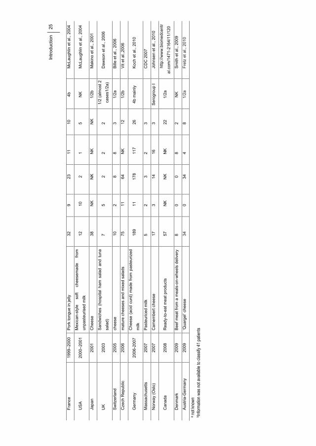
Intr
oduc
tion
25
Fra
nce
19
99–2
000
Por
k to
ngue
in je
lly
32
9 23
11
10
4b
M
cLau
ghlin
et a
l., 2
004
US
A
2000
–200
1 M
exic
an-s
tyle
so
ft ch
eese
mad
e fr
om
unpa
steu
rised
milk
12
10
2
1 5
NK
M
cLau
ghlin
et a
l., 2
004
Japa
n 20
01
Che
ese
38
NK
N
K
NK
N
K
1/2b
M
akin
o et
al.,
200
1
UK
20
03
San
dw
iche
s (h
ospi
tal
ham
sal
ad a
nd t
una
sala
d)
7 5
2 2
2 1/
2 (a
lmos
t 2
case
s1/2
a)
Da
wso
n et
al.,
200
6
Sw
itzer
land
20
05
chee
se
10
2 8
8 3
1/2a
B
ille
et a
l., 2
006
Cze
ch R
epub
lic
2006
m
atur
e ch
eese
s an
d m
ixed
sal
ads
75
11
64
NK
12
1/
2b
Vit
et a
l.,20
06
Ger
ma
ny
2006
-200
7 C
hees
e (a
cid
curd
) m
ade
from
pas
teur
ized
milk
18
9 11
17
8 11
7 26
4b
mai
nly
Koc
h et
al.,
201
0
Mas
sach
uset
ts
2007
P
aste
uric
ed m
ilk
5 2
3 2
3
CD
C 2
007
Nor
wa
y (O
slo)
20
07
Cam
embe
rt c
hee
se
17
3 14
16
3
Ser
ogro
up
I Jo
hnse
n et
al.,
201
0
Can
ada
2008
R
ead
y-to
-eat
me
at p
rodu
cts
57
NK
N
K
NK
22
1/
2a
http
://w
ww
.bio
me
dcen
tr
al.c
om/1
471-
216
4/11
/120
Den
mar
k 20
09
Bee
f mea
t fro
m a
mea
ls-o
n-w
heel
s de
liver
y 8
0 0
8 2
NK
S
mith
et a
l., 2
009
Aus
tria
-Ge
rman
y 20
09
‘Qua
rgel
’ che
ese
34
0
34
4 8
1/2a
F
retz
et a
l., 2
010
a not
know
n b In
forma
tion w
as no
t ava
ilable
to cl
assif
y 41 p
atien
ts

26 Introduction
Additionally, illness caused due to the consumption of contaminated foods has a wide
economic and public health impact worldwide. The Centers for Disease Control and
Prevention (CDC) estimate that foodborne diseases are responsible for about 76 million
illnesses, which result in 325,000 hospitalizations and 5000 deaths in the United States each
year (Mead et al., 1999). Also, the food industry can have major expenses due to product
contamination with L. monocytogenes as in cases with recall of products, factory closing,
extraordinary cleanings, and compensation to infected people. This was seen lately in a large
outbreak of L. monocytogenes in Canada in 2008 resulting in 57 cases with 22 deaths
(Anonymous, 2009a). The outbreak was caused by meat products from a company that itself
estimates their costs to be around $43 million for recalls, lost sales and compensations to
claimants (Anonymous, 2009b).
1.3.1.2. Incidence of L. monocytogenes in food processing plants
Several studies have confirmed the ability of L. monocytogenes to colonize food
processing factories (Harvey and Gilmour, 1992). Contamination of the final food products
occurs mainly during processing rather than from the raw material (Cox et al., 1989; Hu et al.,
2006; Samelis and Metaxopoulos, 1999, Autio et al., 1999, Miettinen et al., 1999; Norton et
al., 2001; Rørvik et al., 1995; Vogel et al., 2001, Wulff et al., 2006). Typing methods have
been used to point out equipment, floors and drains as important contamination sources in the
processing lines (Hoffmann et al., 2003; Miettinen et al., 1999; Norton et al., 2001; Keto-
Timonen et al., 2007) and to demonstrate that specific sub-types are able to persist in the food
processing environment for up to 10 years (Norton et al., 2001, Wulff et al., 2006). Moreover,
from recopilated data during surveys carried out in 2006 and 2007 it can be deduced that the
percentage of units in non-compliance with the EU Listeria criteria was considerably higher
in samples obtained from food processing than in those obtained from retails (Figure 1.3.1).
This pathogen represents a problem specifically in dairies, smoked fish houses and RTE
meat processing plants where, according to a research carried out by Kornacki and Gulter in
2007, a high prevalence has been detected within processing facilities in North America and
Europe. The DNA types found in the final products were also found in many samples from
the processing environment (Autio et al., 1999; Norton et al., 2001; Vogel et al., 2001; Wulff
et al., 2006). Also, the frequency of positive samples in a fish processing industry was lower
for fish before filleting compared to samples of fish after filleting and further in the
production process (Rørvik et al., 1995).

Introduction
27
Figure 1.3.1: Proportion of samples in non-compliance with the EU Listeria criteria, 2007.
Samples are based on single and batch data from retail, including sample units >=25.
Excluding HACPP and own check samples. RTE for infants and medical purposes also
include food for special nutritional uses. Taken from EFSA 2009.
1.3.1.3. Incidence of L. monocytogenes in foods
After several L. monocytogenes surveys, it is now generally accepted that the highest
incidence of L. monocytogenes ocurrs in RTE foods. In USA, the prevalence of L.
monocytogenes in RTE foods was generally determined to be 1.82% in 31,705 tested samples.
The highest rate of positive samples were from seafood salads (4.7%) and smoked seafood
(4.3%) (Gombas et al., 2003). In the EU, Listeria monocytogenes was seldom detected above
the legal (Gombas et al. 2003; Wallace et al. 2003) safety limit (contamination level of <10
cfu/g) from RTE foods but findings over this limit were most often found in smoked fish and
other ready-to-eat fishery products followed by ready to eat meat products and cheeses
(Anonymous, 2009a). In fact, fishery products had the highest proportion of samples
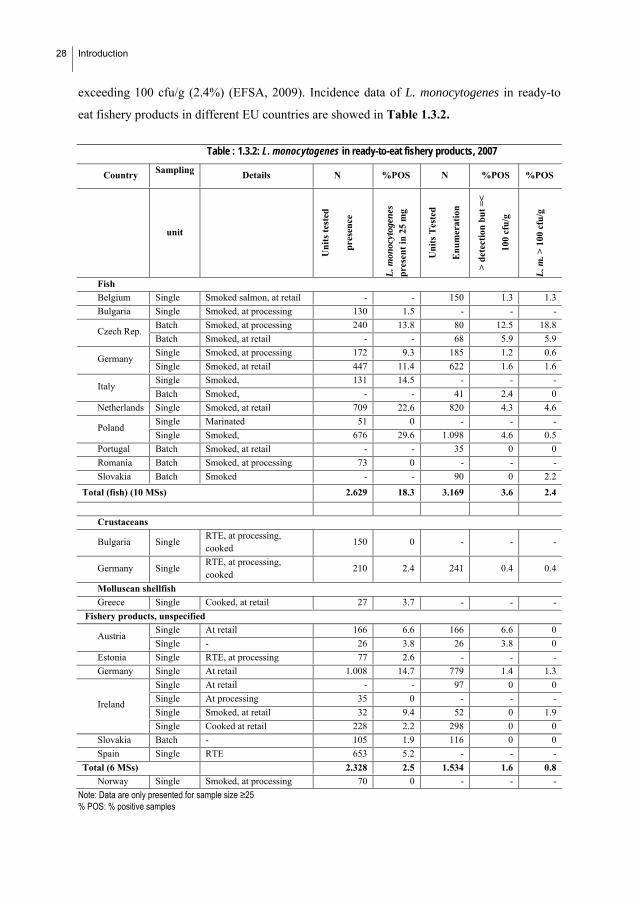
28 Introduction
exceeding 100 cfu/g (2.4%) (EFSA, 2009). Incidence data of L. monocytogenes in ready-to
eat fishery products in different EU countries are showed in Table 1.3.2.
Table : 1.3.2: L. monocytogenes in ready-to-eat fishery products, 2007
Country Sampling Details N %POS N %POS %POS
unit
Un
its
test
ed
pres
ence
L. m
onoc
ytog
enes
pr
esen
t in
25
mg
Un
its
Tes
ted
En
um
erat
ion
> d
etec
tion
bu
t =
<
100
cfu
/g
L. m
. > 1
00 c
fu/g
Fish
Belgium Single Smoked salmon, at retail - - 150 1.3 1.3
Bulgaria Single Smoked, at processing 130 1.5 - - -
Batch Smoked, at processing 240 13.8 80 12.5 18.8 Czech Rep.
Batch Smoked, at retail - - 68 5.9 5.9
Single Smoked, at processing 172 9.3 185 1.2 0.6 Germany
Single Smoked, at retail 447 11.4 622 1.6 1.6
Single Smoked, 131 14.5 - - - Italy
Batch Smoked, - - 41 2.4 0
Netherlands Single Smoked, at retail 709 22.6 820 4.3 4.6
Single Marinated 51 0 - - - Poland
Single Smoked, 676 29.6 1.098 4.6 0.5
Portugal Batch Smoked, at retail - - 35 0 0
Romania Batch Smoked, at processing 73 0 - - -
Slovakia Batch Smoked - - 90 0 2.2
Total (fish) (10 MSs) 2.629 18.3 3.169 3.6 2.4
Crustaceans
Bulgaria Single RTE, at processing, cooked
150 0 - - -
Germany Single RTE, at processing, cooked
210 2.4 241 0.4 0.4
Molluscan shellfish
Greece Single Cooked, at retail 27 3.7 - - -
Fishery products, unspecified
Single At retail 166 6.6 166 6.6 0 Austria
Single - 26 3.8 26 3.8 0
Estonia Single RTE, at processing 77 2.6 - - -
Germany Single At retail 1.008 14.7 779 1.4 1.3
Single At retail - - 97 0 0
Single At processing 35 0 - - -
Single Smoked, at retail 32 9.4 52 0 1.9 Ireland
Single Cooked at retail 228 2.2 298 0 0
Slovakia Batch - 105 1.9 116 0 0
Spain Single RTE 653 5.2 - - -
Total (6 MSs) 2.328 2.5 1.534 1.6 0.8
Norway Single Smoked, at processing 70 0 - - -
Note: Data are only presented for sample size ≥25 % POS: % positive samples
Introduction
29
In contrast with this high incidence, only a few cases of listeriosis were reported from
consumption of fish products and they involved a limited number of patients (Brett et al.
1998; Farber et al. 2000). Although L. monocytogenes was firstly isolated from imported
cooked crabmeat in 1987, the first of several cases of listeriosis positively linked to
consumption of fish or seafood was not reported until 1989. Further, some listeriosis
outbreaks have been related to the consumption of steamed mussels, shrimp, fish salads and
semicooked fish (Ben Embarek, 1994, Brett et al., 1998), cold-smoked rainbow trout
(Miettinen et al. 1999) and salmon (Tham et al., 2000).
The frequency with which L. monocytogenes can be found during environmental surveys
has raised doubts that the food processing industry can effectively eliminate L.
monocytogenes contamination. In this sense, some authors have hypothesized that three recent
anthropogenic practices increase the load within and transmission among reviewed habitats
and host populations: extended refrigerated storage of ready-to-eat foods allowing L.
monocytogenes growth in foods that are contaminated during production or subsequent
handling; feeding domestic ruminants with silage often contaminated with L. monocytogenes;
and dispersal of contaminated products of sewage treatment to agricultural fields and waters
(Ivanek et al., 2006). In this frame, prevention relies on progressively better defined tolerance
limits in foods and improved risk communication, focused on risk groups. Risk assessment is
increasingly used as a scientific process to assess the potencial for adverse health effects to
occur and as a basis for management of unacceptable risks (Notermans and Hoornstra, 2000).
1.3.2. Control of L. monocytogenes in food processing plants
1.3.2.1. Prerrequisites and Hazard Analysis Critical Control Point (HACCP):
importance for food safety
HACCP is an effective and rational means of assuring food safety from harvest to
consumption. Preventing problems from occurring is the paramount goal underlying any
HACCP system. To meet this goal, seven basic principles are used in developing HACCP
plans. These include hazard analysis, Critical Control Point identification, establishing critical
limits, monitoring procedures, corrective actions, verification procedures, and record keeping
and documentation. Under such systems, if a deviation occurs indicating that control has been
30 Introduction
lost, the deviation is detected and appropriate steps are taken to re-establish control in a timely
manner to assure that potentially hazardous products do not reach the consumer. But for a
HACCP to be succesful some prerequisites programs must be developed and implemented
These programs provide the basic environment and operating conditions that are necessary
for the production of safe, wholesome food. Many of the conditions and practices are
specified in federal, state and local regulations and guidelines. All prerequisite programs
should be documented and regularly audited, and are established and maintained separately
from the HACCP plan.
Prerequisites are aimed to prevent contamination of food-products with foodborne
pathogens such us L. monocytogenes during the manufacturing processing are importat
(Kornacki and Gulter, 2007). Thus:
i. The raw material must be of good microbiological quality.
ii. The employees must have a thorough understanding of food hygiene: Holah et al.
(2004) showed that although food factory designs, personnel hygiene and cleaning
and disinfection regimes are sufficient to control Listeria spp. to very low levels,
persistent strains can remain within factory high-risk production areas over
considerable time periods. Although hygienic conditions of food production in
industrially developed countries are continually improving, outbreaks of listeriosis
originating from consumption of contaminated food products still occur, ensuring
effective control of L. monocytogenes has proved to be challenging.
iii. The factory and equipment should be designed so they are easily cleaned to avoid
cross-contamination: biofilms may also cause technical problems such as total or
partial occlusion of pipes, outlets, valves, drains, filters, filtration membranes and
may coat the surfaces of heat exchangers and diverse sensors. Particularly
vulnerable are poorly accessible zones, pieces of equipment that are difficult to
dismantle, irregular or scratched surfaces and sites where routine cleaning fails to
prevent accumulation of organic material. Mechanical aids (high pressure jets,
brushing, scrubbing) may be used to supplement cleaning and disinfection chemical
treatments, but manual labour is costly and the hygienic results have a low
reproducibility. Identification and quantification of biofilm resident

Introduction
31
microorganisms and development of specific sanitation practices are increasing
concerns among food processors in the last few years (Belessi et al., 2010; Sofos
and Geornaras, 2010)
Once these and others prerrequisites are controlled, critical points in the process where
contamination can occur should be identified and controlled. Rocourt and Cossart (1997)
proposed that L. monocytogenes enters processing plants through soil on workers’ shoes,
clothing, and transport equipment; animals that excrete the bacterium or have contaminated
hides or surfaces; raw plant tissue; raw food of animal origin; and, possibly, healthy human
carriers. However, the widespread presence of L. monocytogenes in natural reservoirs and its
ability to grow at harsh conditions make its control in foods difficult (Bonnet and Montville
2005).
1.3.2.2. The problem of cross contamination
Cross-contamination can also occur after listeriocidal treatment (Reij and Aantrekker Den,
2004; Tompkin, 2002) and generally represents post-processing contamination from
environmental sources, including in food processing plants, retail operations, and household
kitchens (Soumet et al., 2005). Foodborne pathogens can remain viable on common food
contact surfaces for days or weeks and cross-contaminate other products (Lappi et al., 2004;
Lunden et al., 2000; Nesbakken et al., 1996).
There are numerous studies about the cross-contamination when food is sliced with blades.
Keskinen et al. (2008) study the stress and biofilm-forming ability on transfer of surface-dried
Listeria monocytogenes during slicing of delicatessen meat. They obtained significantly
greater transfer for blades used after 6 as opposed to 24 h of incubation. Also, Pappelbaum et
al. (2008) monitored the prevalence of Listeria spp. in frozen vegetables and environmental
samples (swabs) from a large produce processing factory. They conclude that the lack of
personnel hygiene was supporting the cross-contamination cycle in the plant. Another
possible source of cross-contamination could have been aerosol generation through hosing
under high pressure or in the cooling units. In fact protective barriers between the processing
lines were limited.

32 Introduction
In this frame, it is evident that cleaning and disinfection protocols must be effective: a key
point in preventing contamination is the control of hygiene at the food processing plant level,
and this is primarily obtained by efficient cleaning and especially disinfection, which is
regarded as a critical step in eliminating spoilage and pathogenic bacteria in the food
processing environment. The mechanisms by which L. monocytogenes survive under these
harsh conditions of physical and chemical stresses are not fully understood (Harvey et al.,
2007). It has been suggested that biofilm formation by L. monocytogenes have highlighted the
need for additional cleaning and disinfection strategies to control persistent L. monocytogenes
strains in the food processing environment (Holah et al. 2004).
1.3.2.3. Disinfectants
1.3.2.3.1. Classical disinfectants
There are a number of prerequisites for an efficient disinfection. Firstly, the disinfectant
must have the right spectrum of activity and be able to eliminate the relevant contaminants in
the production site. Generally, disinfectants have a very broad spectrum of targets, since they
are efficient against bacteria, viruses and fungi. However, Gram-negative bacteria tend to be
less susceptible than Gram-positive bacteria (McDonnell and Russell, 1999). Secondly, it is
important to use the right concentration, pH, temperature, and exposure time to obtain
sufficient elimination of bacteria. Finally, cleaning of the surface prior to disinfection is
necessary to remove organic compounds. Otherwise, the disinfection will be useless. A wide
range of chemical disinfectants are available for the food-industry, and they can be divided
into the following seven groups (Asselt and Giffel, 2005): alcohols, aldehydes, biguanides,
(bis)-phenols, halogen-releasing agents (HRA), peroxygens and quaternary ammonium
compounds (QACs). The most well-known disinfectant agents of each group, including their
applications and restrictions in use, are summarized in Table 1.3.3. Generally, disinfectants
act on multiple targets on a specific cell, which make these compounds highly active against
microorganisms, but also potentially harmful to humans. Bactericidal activity of a disinfectant
is well-known, but the mechanism of action is rarely fully understood (McDonnell and
Russell, 1999).

Introduction
33
Chlorine-based compounds, peroxygens, and compounds based on quaternary ammonium
compounds (QAC) are the most frequently applied disinfectants in the food industry. Each of
these three groups have some advantages and disadvantages (Table 1.3.4), and therefore the
choice of the disinfectant to be used depends on the production site.
Table 1.3.3: Biocide groups applications and restrictions in use.
Group Important types
Application Action on bacteria Comments
Ethanol Small spots.
Alcohols Isopropanol Quick wipe-downs.
Membrane damage and rapid denaturation of proteins. Interference with metabolism. Cell lysis
Not for large industrial application due to need of high concentrations (60-90%). Flammable.
Formaldehyde Decontaminate romos.
Extremely reactive. Reacts un-specifically with functional groups of proteins
Used less frequently in food production and processing due to toxicological considerations.
Aldehydes
Glutaraldehyde Instrument. Disinfectant.
Strong membrane association. Reacts unspecifically with functional groups of proteins. Inhibition of transport
Polymeric biguanides
Used in particular by the food industry
Alexidine Biguanides
Chlorhexidine
Most widely applied biocide in hand-washing and oral products.
Membrane active. Damage of intracellular membrane. Leakage of intracellular components.
Polyhexamethylene biguanides (PHMB) is a superior biocide due to lack of toxicology, colour, taste and surfactancy.
Bis-phenols Triclosan Soap, toothpaste, packing material, conveyer belts.
Membrane-active Inhibits a specific enzyme in lipid biosynthesis
The action at a specific target increases the risk of resistance.
Hypochlorite Frequently applied disinfectant in food industry.
Highly oxidizing agents Irreversible change and disruption of DNAprotein synthesis.
It is cheap. Halogen-releasing agents
Iodine Penetrates bacteria and attacks keygroups of proteins.
Expensive. Staining of skin and plastic parts.
Peracetic acid Frequently applied disinfectant in the food industry.
Mixture of water, hydrogen peroxide and acetic acid. Less sensitive to organic loads.
Peroxygens
Hydrogen peroxide
Oxidation of essential cell components as lipids, proteins and DNA.
Corrosive on some materials.
Quaternary ammonium compounds
Frequently applied disinfectant in the food industry.
Absorption to cell wall Perturbation of lipid bilayer. Cytoplasmic protein aggregation. Leakage of cytoplasmic materials.
More expensive than chlorine, but have residual action. Non-corrosive and non-tainting monocationic agents.

34 Introduction
Table 1.3.4: Advantages and disadvantages of some chemical disinfectants widely used in the food industry.
Disinfectant Advantages Disadvantages
Chlorine compounds
Cheap and effective. -Kills most types of microorganisms. -Less affected by hard water than some. -Does not form films. -Effective at low temperatures. -Relatively inexpensive. -Concentration easily determined by test strips.
-Inactivated in presence of organic material. -May corrode metals and weakenrubber. -Irritating to skin, eyes and throat. -Unstable, dissipates quickly. -Liquid chlorine loses strength in storage. -pH sensitive.
Iodophors
-Kills most types of microorganisms. -Less affected by organic matter than some. -Less pH sensitive than chlorine. -Concentration determined by test strips. -Solution colour indicates active sanitiser.
-May stain plastics and porous materials. -Inactivated above 50°C. -Reduced effectiveness at alkaline pH. -More expensive than hypochlorites. -May be unsuitable for CIP1 due to foaming.
Peroxy compounds
-Effectiveness. -Best against bacteria in biofilms. -Kills most types of microorganisms. -Relatively stable in use. -Effective at low temperatures. -Meets most discharge requirements. -Low foaming; suitable for CIP.
-Inactivated in presence of organic material. -More expensive than some. -Inactivated by some metals/organics. -May corrode some metals. -Not as effective as some against yeasts and moulds.
QAC
-Non corrosive -Less affected by organic matter than some -Residual antimicrobial activity if not rinsed -Can be applied as foam for visual control -Effective against Listeria monocytogenes -Effective for odour control -Concentration determined by test strips
-Expensive. -Inactivated by most detergents. -May be ineffective against certain organisms. -May be inactivated by hard water. -Effectiveness varies with formulation. -Not as effective at low temp. as some. -May be unsuitable for CIP due to foaming.
Acid-Anionic
- Sanitize and acid rinse in one step - Very stable - Less affected by organic matter than some - Can be applied at high temperature - Not affected by hard water
-Effectiveness varies with microorganism. -More expensive than some. -pH sensitive (use below pH 3.0). -Corrode some metals. -May be unsuitable for CIP due to foaming.
Carboxylic Acid
-Kills most types of bacteria -Sanitize and acid rinse in one step -Low foaming, suitable for CIP -Stable in presence of organic matter -Less affected by hard water than some
-Inactivated by some detergents. -pH sensitive (use below pH 3.5). -Less effective than chlorine at low temp. -May damage non-stainless steel materials. -Less effective against yeasts and moulds than some.
Chlorine Dioxide
-Kills most type of microorganisms -Stronger oxidiser (sanitizer) than chlorine -Less affected by organic matter than some -Less corrosive than chlorine -Less pH sensitive than some
-Unstable and cannot be stored. -Potentially explosive and toxic. -Relatively high initial equipment cost.
Ozone -Kills most type of microorganisms -Stronger oxidiser (sanitizer) than chlorine and chlorine dioxide
- Unstable and cannot be stored. -May corrode metals and weaken rubber. -Potentially toxic. -Inactivated by organic matter (similar to chlorine).
Hot Water / Heated Solutions
-Kills most types of microorganisms -Penetrates irregular surfaces -Suitable for CIP -Relatively inexpensive
-May form films or scale on equipment. -Burn hazard. -Contact time sensitive.

Introduction
35
1.3.2.3.2. Other compounds with potencial application in cleaning and disinfection
protocols
Enzymes
A promising strategy may be the use of enzymes that can dissolve the biofilm matrix (e.g.
DNase and alginate lyase) as well as quorum-sensing inhibitors that increase biofilm
susceptibility to antibiotics (Høiby et al. 2010). Several works have shown the potential
applicability of enzymes against biofilms: Gamble and Muriana (2007) tested three enzyme
preparations against single-species biofilms of 6 “strongly adherent” strains previously
isolated from meat and meat processing plants. They found that protease, lipase, and cellulase
were effective in releasing adherent cells from microplates. Longhi et al., (2008) examined the
effects of a protease treatment on the ability of L. monocytogenes to form biofilms and to
invade tissues. They have chosen serratiopeptidase (SPEP), an extracellular metalloprotease
produced by Serratia marcescens. Treatment of L. monocytogenes with sublethal
concentrations of SPEP reduced their ability to form biofilms and to invade host cells.
Zymograms of the treated cells revealed that Ami4b autolysin, internalin B, and ActA were
sharply reduced. These cell-surface proteins are known to function as ligands in the
interaction between these bacteria and the host cells, and their data suggest that treatment with
this natural enzyme may provide a useful tool in the prevention of the initial adhesion of L.
monocytogenes to the human gut. More recently, Harmsen et al. (2010) have shown that
DNase I treatment resulted in dispersal of biofilms, not only in microtiter tray assays but also
in flow cell biofilm assays.
Essential oils
In vitro and in vivo studies have demonstrated that essential oils act as antioxidants and
show antibacterial activities (Viuda-Martos 2010). Considering the large number of chemical
substances that make up essential oils (EOs), it is most probable that their antibacterial
activity cannot be explained by a single specific mechanism but rather by several different
mechanisms. Nevertheless, phenolic compounds are generally considered to be responsible
for the antibacterial properties of EOs. Several studies that have demonstrated the
effectiveness of EOs, alone or combined with other antimicrobials, as antilisterial agents in
foods are recopilated in Table 1.3.5.

36 Introduction
Table 1.3.5: Some examples of sucessful applications of essential oils (EOs) as antilisterial agents in foods
Essential oil Food Reference Thyme´s (0.08%) Minced pork Aureli et al., 1992 Rosemary (0.3%) and encapsulated rosemary oil (5%) Pork liver sausage Pandit and Shelef 1994 “DMC base natural” preservative comprising 50% Eos of rosemary, sage and citrus.
Soft cheese Mendoza-Yepes et al., 1997
Eugenol Cooked chicken breast, pieces Hao et al., 1998a Eugeniol Roast beef sirloin, sliced Hao et al., 1998b Pelargonium oil Quiche Lis-Balchin et al., 1998
Mint oil (Mentha piperita) Tzatziki (yogurt and cucumber salad pH 4.5), taramosalata (cod´s roe salad pH 5.0) and pâté (pH 6.8)
Tassou et al., 1995
Oregano Packed meat Tsigarida et al., 2000 Clove oil Mozzarella cheese Vrinda et al., 2001 Carvacrol Semi skimed milk Karatzas et al., 2001
Cilantro oil (6%) Vacuum packed ham Gill et al., 2002
Oils and vapours of lemon (Citrus limon), sweet orange (Citrus sinensis) and bergamot (Citrus bergamia)
Chicken skin Fisher and Philips 2006
Oregano or savory Pork meat Ghalfi et al., 2007
Spanish oregano (O; Corydothymus capitatus), Chinese cinnamon (C; Cinnamomum cassia), or winter savory (S; Satureja montana)
Bologna and ham Oussalah et al., 2007
Carvacrol Steak tartare Veldhuizen et al., 2007 Thyme Minced beef during refrigerated storage Solomako et al., 2008 Oregano Barbecued chicken Firouzi et al., 2007 Clove and cinnamon Pasteurized milk Cava et al., 2007
Cinnamon, bark Melon and waterlemon juices Mosqueda-Melgar et al., 2008
Basil, lemon balm, marjoram, oregano, rosemary, sage and thyme
Model media included potato starch (0, 1,5 or 10%), beef extract (1.5, 3, 6 or 12%), sunflower oil (0, 1, 5 or 10%).
Gutierrez et al., 2008
Satureja montana Minced pork Carramiñana et l., 2008 Basil, caraway, fennel, lemon balm, marjoram, nutmeg, oregano, parsley, rosemary, sage, and thyme
Ready-to-eat vegetables Gutierrez et al., 2008
Citron Fruit-based salads Belletti et al., 2008
Lemon balm, marjoram, oregano and thyme Model media based on lettuce, meat and milk
Gutierrez et al., 2009
Allspice, garlic, and oregano tomato films Du et al., 2009 Garlic, bay, black pepper, origanum, orange, thyme, tea tree, mint, clove, and cumin
Apple-carrot juice Irkin and Korukluoglu, 2009
Oregano and rosemary Fresh-cut vegetables Scollard et al., 2009 Thyme verbena, thyme red, Spanish oregano, ajowan, tea tree, clove, and sage oils tested at 1%, as well as with 2% rosemary oil
Ready-to-eat salad Cobo Molinos et al., 2009
Clove (Syzygium aromaticum L.), fennel (Foeniculum vulgare Miller), cypress (Cupressus sempervirens L.), lavender (Lavandula angustifolia), thyme (Thymus vulgaris L.), herb-of-the-cross (Verbena officinalis L.), pine (Pinus sylvestris) and rosemary (Rosmarinus officinalis)
Films for fish preservation Gómez-Estaca et al., 2010
There are few studies on the effects of essential oils in biofilms (Chorianopoulus et al.,
2008; Oliveira et al., 2010; Sandasi et al., 2008; Sandasi et al., 2010). Sandasi et al., 2010
studied the in vitro antibiofilm activity of selected culinary herbs and medicinal plants against
L. monocytogenes biofilms. The majority of extracts tested prevented cells adhesion to
polyvinyl chloride (PVC) surface. In contrast, inhibition of preformed biofilms (during 4

Introduction
37
hours) was more difficult to achieve, with only three extracts (Rosemarinus officialis, Mentha
piperita and Melaleuca alternifolia) inhibiting the growth of both strains by at least 50%.
Previously, Sandasi et al., 2008 showed that some essential oils at a concentration of 1 mg/ml
components promote the growth and development of a preformed L. monocytogenes biofilm
(6 h) in vitro. However, Chorianopoulus et al., 2008 observed that the essential oil of S.
thymbra (1%), as well as its hydrosol fraction (100%), presents sufficient bactericidal effect
on bacterial biofilms formed on stainless steel after 5 days. And also, Oliveira et al., 2010
studied the effect of disinfectant and essential oils (Cymbopogon citrtus Stapf. and
Cymbopogon nardus) in different phases of biofilm formation (3 hours of biofim formation)
of Listeria monocytogenes on stainless steel surface and found that essential oils have
effectively reduced the number of surface-adhered cells, especially after 60 min of contact.
Biosurfactants
A few studies have confirmed the effectiveness of biosurfactants produced by gram-
negative (Pseudomonas fluorescens) and Gram-positive (Lactobacillus helveticus) bacteria
against the adhesion of Listeria monocytogenes on stainless steel (Meylheuc et al., 2001 and
2006). Zeraik and Nitschke, 2010 studied the adhesion of different pathogenic bacteria
(Staphylococcus aureus, Listeria monocytogenes, and Micrococcus luteus) to polystyrene
surfaces previously condicioned with surfactin and rhamnolipid , whereas Nitschke et al.
(2009) to stainless steel and polypropylene previously adsorbed with surfactin.
Bacteriocins
The effectiveness of nisin and pediocin against planctonic L. monocytogenes cells has been
demonstrated in numerous studies (Nisin: Scannell et al., 2000; Szabo, and Cahill. 1999;
Harris et al., 1991; Coma et al., 2001; Nilsson et al., 2000; Benkerroum and Sandine. 1988;
Fang and Lin. 1994, Zhang and Mustapha. 1999. Pediocin: Degnan et al., 1993, Goff et al.,
1996; Liao et al., 1993; Scannell et al., 2000). In fact, biocontrol of L. monocytogenes in
refrigerated storage through co-culture with bacteriocin-producing lactic acid bacteria has
been proposed by several authors (Yousef et al., 1991.). However, both nisin-producing
lactococci and pediocin producing pediococci are hardly effective because they do not grow
well at refrigeration temperatures. The psychrotrophs C. piscicola L103 (Schobitz et al.,
1999), Leuconostoc carnosum 4010 (Budde et al., 2003), and several strains of Lb. bavaricus
(Winkowski et al., 1993) were proposed as alternative bacteriocin producers to prevent

38 Introduction
Listeria to grow at refrigeration temperature. Growth of the pathogen may also be retarded in
ready-to-eat, fresh-cut vegetables inoculated with Lb. delbrueckii (Harp and Gilliland. 2003).
On the contrary, studies about the effectiveness of bacteriocins against L. monocytogenes
biofilms are really scarce. Only three were found in the bibliography: Leriche et al., 1999;
Minei et al., 2008 and Guerrieri et al., 2008. The first one studied L. monocytogenes biofilm
growth in association with a nisin-producing strain of Lactococcus lactis. Both when L.
monocytogenes was combined with Lc. lactis and inoculated onto stainless steel coupons or
was inoculated onto a preexisting Lc. lactis biofilm, the antilisterial activity of the biocontrol
was effective. Minei et al., 2008 studied the influence of nisin (3 h) on L. monocytogenes
biofilms and observed that although treatment decreased the number of cells 4.6 log
CFU/cm2, a renewed biofilm was detected after 24 h of incubation. However, coculture with a
bacteriocin produced by E. faecium reduced efficiently the biofilm formation. Guerrieri et al.,
2008 studied the use of lactic acid bacteria (LAB) biofilms bacteriocin producers
(Lactobacillus plantarum 35d, Enterococcus casseliflavus IM 416K1) and by two non-
producers (L. plantarum 396/1, Enterococcus faecalis JH2-2) for the control of Listeria
monocytogenes in a small-scale model. They found the bacteriocin producers showed the best
antilisterial potentiality. Comparing the antilisterial activity of LAB biofilm against both
planktonic and adherent cells, the L. monocytogenes adherent cells showed a higher
resistance. And also in the studies performed with L. monocytogenes in co-culture with a
Pseudomonas putida strain it is produced a reduction of the antilisterial activity in the
lactobacilli biofilms added with P. putida, probably due to the pH increase and to a better
survival of Listeria in the presence of P. putida.
More recently, it has been showed that the presence of model resident Lactococcus lactis
biofilms reduced the initial adherence of L. monocytogenes. Moreover, significant differences
were seen in L. monocytogenes settlement as a function of the genetic background of resident
lactococcal biofilm cells (Habimana et al. 2009).
1.3.2.4. Resistance of L. monocytogenes biofilms to disinfectants
It is generally accepted that biofilm formation implies an increase in the resistante to
biocides. (Aarnisalo et al., 2000; Aarnisalo et al., 2007; Amalaradjou et al., 2009; Ammor et
al., 2004; Blackmann and Frank, 1996, Bremer et al., 2002, Chavant et al., 2003; Gram et al.,

Introduction
39
2007, Kastbjerg and Gram, 2009; Holah et al., 2002, Leriche et al., 1999; Minei et al., 2008,
Frank et al., 2003; 48 h: Pan et al., 2006, Yang et al., 2009).
According with Costerton et al. (1999) and Donlan and Costerton (2002), some
mechanisms responsible for that resistance may be one or more of the following:
i. A delayed penetration of the antimicrobial agent through the biofilm due to the
polymeric substances that make up the matrix of a biofilm (Ammor et al., 2004;
Bourion and Cerf 1996; Davies, 2003; Fatemi and Frank, 1999; Gilbert et al, 2002,;
Pan et al., 2006). Extracellular polymeric substances surrounding the cells may hinder
diffusion. Additionally, some authours have also pointed out that the abiotic part of the
biofilm plays a part in resistance because of its potent neutralization effect on many
compounds (Campanac et al. 2002).
ii. An altered growth rate of biofilms (Chavant et al., 2004, Fah and O´Toole).
iii. Other physiological changes due to the biofilm mode of growth. This suppose a more
speculative hypothese than the preceding one, in which at least some of the cells in a
biofilm adopt a distinct and protected biofilm phenotype (Chavant et al., 2004; Fah
and O´Toole; Davies, 2003).
iv. Formation of multispecies biofilms: L. monocytogenes is more resistant when grown
in multi-species biofilm than when grown as monospecies biofilm (Ammor et al.,
2004; Fatemi and Frank 1999; Saá Ibusquiza et al., 2010).
1.3.2.5. Cross-resistance responses of L. monocytogenes
1.3.2.5.1. Cross-resistance of acid-adapted L. monocytogenes.
Acid-adapted L. monocytogenes (pH 5.2, 2 h) had increased resistance to heat shock (52
°C), osmotic shock (25–30% NaCl) and alcohol stress, suggesting that acid adaptation also
provides cross-protection against other stress factors (Phan-Thanh et al., 2000). Cross-
resistance of acid-adapted cells to other stresses such as high temperatures, sodium chloride,
hydrogen peroxide, hypochlorite, crystal violet, ethanol, surfactants and3the bacteriocin nisin
(Begley et al., 2010; Bonnet and Montville, 2005) has important implications for the food
industry, particularly since foods commonly encounter sublethal acidic treatments during
processing (Phan-Thanh 4et al., 2000; van Schaik and Abee, 1999). Also, acid-tolerant strains
of L. monocytogenes are more resistant to CO2 (Francis et al. 2007; Jydegaard-Axelsen et al.,

40 Introduction
2004). Specifically with bacteriocins, acid tolerance response (ATR) confers cross-resistance
to nisin (Badaoui Najjar, et al., 2009; Begley et al., 2010; Bonnet and Montville, 2005; Ivanek
et al., 2006). So the cross-resistance induced by the ATR should be considered for the safety
of foods fermented with bacteriocin-producing cultures.
1.3.2.5.2. Cross-resistance between bacteriocins in L. monocytogenes
Song and Richard (1997) studied the antilisterial activity of three bacteriocins used at sub-
minimal inhibitory concentrations and cross-resistance of the survivors. They obtained that
the survivors displayed increased resistance not only toward the bacteriocin they were in
contact with, nisin, but toward the two other bacteriocins under study, pediocin AcH and
enterococcin EFS2. As the survivors to nisin are resistant to the other two bacteriocins, one
can expect that combined use of nisin and one of these bacteriocins would not result in better
inhibition. This conclusion is contradicted by the results of Hanlin et al. (1993) who showed
that nisin and pediocin AcH had greater antibacterial activity in combination than
individually.
1.3.3. Control of Listeria monocytogenes in foods
In general, there is not a unique strategy or technology to control L. monocytogenes in
foods. Preservation technologies were not designed exclusively to eliminate L.
monocytogenes so the application of multiple hurdles is necessary. However, in those foods
were L. monocytogenes has been identified as a frequent pathogenic bacterium, several
specific strategies have been developed:
1.3.3.1. Bacteriocins
Nisin and pediocin are the most investigated bacteriocins against L. monocytogenes.
Bacteriocins are suitable as processing aids, complementing other preservation methods.
Previous works have demonstrated that heat, freezing–thawing, acid (Muriana, 1996), high
hydrostatic pressure (Kalchayanand et al., 1994), and pulsed electric fields were more
listericidal in presence of bacteriocins than they were in their absence.
Introduction
41
1.3.3.2. Application of active packaging
Active packaging that contains antilisterial agents is increasingly investigated as a means
to improve food safety after processing. Listeriostatic or listericidal agents of active
packaging are applied as coatings to surfaces in contact with food (Cowan et al., 2003;
Hoffman et al., 2001) or incorporated into packaging materials (Brody et al., 2001). As an
example, we can cite: organic acids, bacteriocins, spice extracts, lysozyme, chitosan,
listeriophages, or EDTA.
1.3.3.3. Ozone (O3)
Ozone has been used in European countries for decades and has recently been approved in
the United States by the FDA for treatment, storage, and processing of foods, including meat
and poultry, unless useis precluded by a standard of identity (Food and Drug Administration.
2003). Although ozone treatment inactivates L. monocytogenes, listericidal activity of ozone
varies with temperature, medium composition, strain, and physiological status.
1.3.3.4. Electrolyzed water (EO).
The effectiveness of EO water against L. monocytogenes has been demonstrated in
suspension and in foods, like salmon (Ozer and Demirci 2005), eggs, fruits, and vegetables
(Sharma and Demirci 2003; Singh et al., 2003; Stan and Daeschel 2003; Bialka et al., 2004;
Koseki et al., 2004; Wang et al., 2004, Rahman et al., 2010; Park et al., 2004).
1.3.3.5 Phages
Bacteriophages have a potent antimicrobial activity by inducing the lysis of their host, are
ubiquitous in nature including food ecosystems, and are harmless to mammalian cells. Their
host specificity allows proper starter performance in fermented products and keeps the natural
microbiota undisturbed. Moreover, there are also several phage-encoded proteins with
antimicrobial activities, which may be useful for biological intervention approaches. Listeria
phages have already been succesfully used as processing aids to avoid Listeria contamination
during the manufacturing of soft cheeses (Carlton et al., 2005). However, limited work is
reported regarding phages to control foodborne pathogens. In this context, EFSA has
submitted a recommendation about more research on the use of phages in food
(http://www.efsa.europa.eu/en/scdocs/scdoc/1076.htm) and their possible use in RTE

42 Introduction
products products as it was recomended in the recent Scientific Opinion of the Panel on
Biological Hazards on “The use and mode of action of bacteriophages in food production”
(Anonymus 2009c).
1.3.3.6. Nanotechnology
The application of nanotechnology have been developed in the last few years. It is applied
to detect and combat Listeria in foods. With the first objective, a nanoparticle inmunoassay to
detect low concentrations of L. monocytogenes in food and environmental samples
(Jaakohuhta et al., 2007) and a highly stable biosensor material that mimics existing whole-
cell assays for detecting listeriolysin O (Zhao et al., 2006) have been developed, among
others. To combat L. monocytogenes in foods, Hong et al. (2008) studied the antimicrobial
activity of organically modified nano-clays and they observed that two of the three tested
caused rupture of cell membrane and inactivation of L. monocytogenes and Staphylococcus
aureus.
1.3.4. Listeria monocytogenes as a tool.
But Listeria has been also applied as a tool in medicine. Sawosz et al. (2010) tried to
construct bacteria-nanoparticle vehicles of gold and platinum with Listeria and Salmonella
enteritidis although in the case of Listeria monocytogenes they did not penetrate or were
removed. In the same line, Powell et al., 2010 used ovalbumin-derived designed peptides of
Listeria monocytogenes as a surrogate antigen to generate nanoparticle vaccines. All these
multiple applications constitute a promising field in biotechnology
Moreover, Tangney and Gahan (2010) described the potencial of Listeria monocytogenes
as a vector for anti-cancer therapies. Several recent studies about this medical application
were published: Paterson et al. (2010) designed Listeria and Salmonella bacterial vectors of
tumor-associated antigens for cancer immunotherapy. Shahabi et al., 2010 developed a live
and highly attenuated Listeria monocytogenes-based vaccine for the treatment of Her2/neu-
overexpressing cancers in human. Finally, Wood, et al. (2010) described Listeria-derived
ActA as an effective adjuvant for primary and metastatic tumor immunotherapy.
Introduction
43
References
Allerberger, F. and Wagner, M., 2010. Listeriosis: a resurgent foodborne infection.Clin Microbiol Infect., 16(1),
16-23. Review.
Allison, D.G., McBain, A.J. and Gilbert, P., 2000. Microbial biofilms: problems of control. in Community
Structure and Cooperation in Biofilms ed. Allison, D.G., Gilbert, P., Lappin-Scott, H. and Wilson, M.
pp. 309–327. Reading: Society for General Microbiology.
Anonymous, 2009a. Listeria monocytogenes outbreak. Final update: April 17, 2009. www.phac-aspc.gc.ca/alert-
alerte/listeria/listeria_2009-eng.php 24/04/2009.
Anonymous, 2009b. Listeria cost Maple Leaf Foods $43M. www.nowpublic.com/health/listeria-cost-maple-leaf-
foods-43m 19/02/2009
Anonymus, 2009c. The use and mode of action of bacteriophages in food production. The EFSA J. 1076, 1-26
Asselt, A. J. V. and Giffel, M. C. T., 2005. Pathogen resistance to sanitisers. In Handbook of hygiene control in
the food industry. Ed. Lelieveld, H.L.M., Mostert, M.A. and Holah, J. pp. 69-92. Cambridge:
Woodhead.
Aureli, P., Costantini, A. and Zolea, S. 1992. Antimicrobial activity of some plant essential oils againstListeria
monocytogenes. J. Food Prot. 55, 344–348.
Autio, T., Hielm, S., Miettinen, M., Sjo¨berg, A.M., Aarnisalo, K., Bjo¨rkroth, J., Mattila-Sandholm, T. and
Korkeala, H., 1999. Sources of Listeria monocytogenes contamination in a cold-smoked rainbow trout
processing plant detected by pulsed-field gel electrophoresis typing. Appl Environ Microbiol 65, 150–
155
Badaoui Najjar, M.Z., Chikindas, M.L., and Montville, T.J., 2009. The Acid Tolerance Response Alters
Membrane Fluidity and Induces Nisin Resistance in Listeria monocytogenes. Probiotics Antimicro.
Prot. 1,130–135
Barbuddhe, S.B., Chakraborty, T., 2009. Listeria as an enteroinvasive gastrointestinal pathogen. Curr. Top
Microbiol. Immunol. 337, 173-95. Review.
Begley, M., Cotter, P.D., Hill, C. and Ross, R.P., 2010. Glutamate Decarboxylase-Mediated Nisin Resistance in
Listeria monocytogenes. Appl Environ Microbiol. 76(19), 6541-6546
Belessi, C.E., Gounadaki, A.S., Psomas, A.N., Skandamis, P.N., 2010. Efficiency of different sanitation methods
on Listeria monocytogenes biofilms formed under various environmental conditions. Int. J. Food
Microbiol. Article in press.

44 Introduction
Belletti, N., Lanciotti, R., Patrignani, F. and Gardini, F., 2008. Antimicrobial Efficacy of Citron Essential Oil on
Spoilage and Pathogenic Microorganisms in Fruit-Based Salads. J. Food Sci. 73, M331-M338.
Belval, B.S., Gal, L., Margiewes, S., Garmyn D., Piveteau, P., Guzzo, J., 2006. Assessment of the roles of LuxS,
S-ribosyl homocysteine, and autoinducer 2 in cell attachment during biofilm formation by Listeria
monocytogenes EGD-e. Appl. Environ. Microbiol. 72(4), 2644-50.
Ben Embarek, P.K., 1994. Presence, detection and growth of Listeria monocytogenes in seafoods: a review. Int.
J. Food Microbiol. 23, 17–34.
Benkerroum, N., and Sandine, W. E., 1988. Inhibitory action of nisin against Listeria monocytogenes. J. Dairy
Sci. 71,3237–3245.
Bialka KL, Demirci A, Knabel SJ, Patterson PH, Puri VM. 2004. Efficacy of electrolyzed oxidizing water for the
microbial safety and quality of eggs. Poult Sci 83:2071–2078.
Bille, J., Blanc, D.S., Schmid, H., Boubaker, K., Baumgartner, A., Siegrist, H.H., Tritten, M.L., Lienhard, R.,
Berner, D., Anderau, R., Treboux, M., Ducommun, J.M., Malinverni, .R, Genné, D., Erard, P.H. and
Waespi, U., 2006. Outbreak of human listeriosis associated with tomme cheese in northwest
Switzerland, 2005. Euro Surveill. 11(6), 91-3.
Blackmann, I.C., and Frank, J.F., 1996. Growth of Listeria monocytogenes as a biofilm on various food
processing surfaces, J. Food Prot., 59, 827.
Blehert, D. S., R. J. Palmer, Jr., J. B. Xavier, J. S. Almeida, and P. E. Kolenbrander. 2003. Autoinducer 2
production by Streptococcus gordonii DL1 and the biofilm phenotype of a luxS mutant are influenced
by nutritional conditions. J. Bacteriol. 185, 4851–4860.
Boles, B.R., Thoendel, M. and Singh, P.K., 2005. Rhamnolipids mediate detachment of Pseudomonas
aeruginosa from biofilms. Mol. Microbiol. 57 (5), 1210–1223
Bonaventura Di, G., Piccolomini, R., Paludi, D., D'Orio, V., Vergara, A., Conter, M. and Ianieri, A., 2008.
Influence of temperature on biofilm formation by Listeria monocytogenes on various food-contact
surfaces: relationship with motility and cell surface hydrophobicity. J. Appl. Microbiol. 104, 1552-
1561.
Bonnet, M. and Montville T.J., 2005. Acid-tolerant Listeria monocytogenes persist in a model food system
fermented with nisin-producing bacteria. Lett. Appl. Microbiol. 40, 237.
Borucki, M.K., Peppin, J.D., White, D., Loge, F., Call, D.R., 2003. Variation in biofilm formation among strains
of Listeria monocytogenes. Appl. Environ. Microbiol. 69, 7336–7342.
Introduction
45
Bourion, F. Cerf, O., 1996. Disinfection efficacy against pure-culture and mixed-population biofilms of Listeria
innocua and Pseudomonas aeruginosa on stainless steel, Teflon(R) and rubber. Sci. Aliment. 16, 151-
166.
Bremer, P.J., Monk, I., and Butler, R., 2002. Inactivation of Listeria monocytogenes/Flaviobacterium spp.
Biofilms using chlorine: Impact of substrate, pH, time and concentration, Lett. Appl. Microbiol., 35,
321.
Brett, M.S.Y., Short, P. and McLauchlin, J., 1998. A small outbreak of listeriosis associated with smoked
mussels. Int. J. Food Microbiol. 43, 223–229.
Brody, A. L., Strupinski, E. R. and Kline, L. R., 2001. Antimicrobial packaging. In Active packaging for food
applications. Lancaster, Pa.: Technomic Pub. Co., Lancaster, pp. 131–194.
Brosch, R., Chen, J. and Luchansky, J.B., 1994. Pulsed-field fingerprinting of Listeriae: Identification of
genomic divisions for Listeria monocytogenes and their correlation with serovar. Appl. and Environ.
Microbiol. 60, 2584-2592.
Budde, B. B., Hornbaek, T., Jacobsen, T., Barkholt, V. and Koch. A. G., 2003. Leuconostoc carnosum 4010 has
the potential for use as a protective culture for vacuum-packed meats: Culture isolation, bacteriocin
identification, and meat application experiments. Int. J. Food Microbiol. 83, 171–184.
Campanac, C., Pineau, L., Payard, A., Baziard-Mouysset, G., Roques, C., 2002. Interactions between biocide
cationic agents and bacterial biofilms. Antimicrob Agents Chemother. 46(5), 1469-74.
Carlton, R.M., Noordman, W.H., Biswas, B., de Meester, E.D., Loessner, M.J., 2005. Bacteriophage P100 for
control of Listeria monocytogenes in foods: genome sequence, bioinformatic analyses, oral toxicity
study, and application. Regulatory Toxicology and Pharmacology, 43(3), 301-312.
Carramiñana, J.J., Rota, C., Burillo, J. and Herrera, A., 2008. Antibacterial efficiency of Spanish Satureja
montana essential oil against Listeria monocytogenes among natural flora in minced pork. J Food Prot.
71(3), 502-508.
Cava, R., Nowak, E., Taboada, A. and Marin-Iniesta, F., 2007. Antimicrobial activity of clove and cinnamon
essential oils against Listeria monocytogenes in pasteurized milk. J Food Prot. 70(12), 2757-2763.
Centers for Disease Control and Prevention (CDC) 2008. Outbreak of Listeria monocytogenes infections
associated with pasteurized milk from a local dairy-Massachusetts, 2007. MMWR Morb Mortal Wkly
Rep. 57(40), 1097-100.
Characklis, W. G., and Marshall, K. C., (ed.). 1990. Biofilms. John Wiley & Sons, Inc., New York, N.Y.

46 Introduction
Chasseignaux, E., Toquin, M.T., Ragimbeau, C., Salvat, G., Colin, P., Ermel, G., 2001. Molecular epidemiology
of Listeria monocytogenes isolates collected from the environment, raw meat and raw products in two
poultry- and pork-processing plants. J. Appl. Microbiol. 91(5), 888-899.
Chavant P, Gaillard-Martinie B, Hebraud M. 2004. Antimicrobial effects of sanitizers against planktonic and
sessile Listeria monocytogenes cells according to the growth phase. FEMS Microbiol Lett 236:241–248.
Chavant, P., Folio, P and Hebraud, M., 2003. Growth of Listeria monocytogenes in biofilm according to the
surface and the temperature and effects of different treatments on its survival. Sci. aliment. 23 (1), 116-
119.
Cole, S. P., J. Harwood, R. Lee, R. She, and D. G. Guiney. 2004. Characterization of monospecies biofilm
formation by Helicobacter pylori. J. Bacteriol. 186, 3124–3132.
Coma, V., Sebti, I., Pardon, P., Deschamps, A. and Pichavant. F. H., 2001. Antimicrobial edible packaging based
on cellulosic ethers, fatty acids, and nisin incorporation to inhibit Listeria innocua and Staphylococcus
aureus. J. Food Prot. 64, 470–475.
Costerton, J. W., and Lappin-Scott, H. M., 1995. Introduction to microbial biofilms, p. 1–11. In H. M. Lappin-
Scott and J. W. Costerton (ed.), Microbial biofilms. Cambridge University Press, Cambridge, United
Kingdom.
Costerton, J. W., Cheng, K.-J. , Geesey, G. G., Ladd, T. I., Nickel, J. C., Dasgupta, M. and Marrie, T. J., 1987.
Bacterial biofilms in nature and disease. Annu. Rev. Microbiol. 41, 435–464.
Costerton, J. W., Geesey, G. G. and Cheng, G. K., 1978. How bacteria stick. Sci. Am. 238, 86–95.
Costerton, J. W., Lewandowski, D. E. Caldwell, D. R. Korber, and Lappin-Scott, H. M., 1995. Microbial
biofilms. Annu. Rev. Microbiol. 49, 711–745.
Costerton, J.W., Stewart, P.S., Greenberg, E.P., 1999. Bacterial biofilms: a common cause of persistent
infections. Science. 21, 284(5418), 1318-22.
Cowan, M. M., Abshire, K. Z., Houk, S. L. and Evans, S. M., 2003. Antimicrobial efficacy of a silver–zeolite
matrix coating on stainless steel. J. Ind. Microbiol. Biotechnol. 30:102–106.
Cox, L. J., Kleiss, T., Cordier, J. L., Cordellana, C., Konkel, P., Pedrazzini, C., Beumer, R. and Siebenga, A.,
1989. Listeria spp. in food processing, nonfood and domestic environments. Food Microbiol. 6, 49–61.
Davey, M.E. and O'Toole, G.A., 2000. Microbial biofilms: from ecology to molecular genetics. Microbiol Mol
Biol Rev. 64(4), 847-67.

Introduction
47
Davies, D. G. and Geesey, G. G., 1995. Regulation of the alginate biosynthesis gene algC in Pseudomonas
aeruginosa during biofilm development in continuous culture. Appl. Environ. Microbiol. 61, 860–867.
Davies, D., 2003. Understanding biofilm resistance to antibacterial agents. Nature Rev. Drug Discov. 2(2), 114-
122.
Dawson, S.J., Evans, M.R., Willby, D., Bardwell, J., Chamberlain, N., Lewis, D.A., 2006. Listeria outbreak
associated with sandwich consumption from a hospital retail shop, United Kingdom. Euro Surveill.
11(6), 89-91.
Degnan, A. J., Buyong, N., and Luchansky, J. B., 1993. Antilisterial activity of pediocin AcH in model systems
in the presence of an emulsifier or encapsulated within liposomes. Int. J. Food Microbiol. 18, 127–138.
Donlan, R.M., Costerton, J.W., 2002. Biofilms: survival mechanisms of clinically relevant microorganisms.
Clin. Microbiol Rev. 15(2), 167-93.
Doumith, M., Buchreiser, C., Glaser, P., Jacquet, C. and Martin, P., 2004. Differentiation of the major Listeria
monocytogenes serovars by multiplex PCR. J. Clin. Microbiol. 42, 3819-3822.
Du, W.X., Olsen, C.W., Avena-Bustillos, R.J., 2009. Antibacterial effects of allspice, garlic, and oregano
essential oils in tomato films determined by overlay and vapor-phase methods. Journal of Food
Sicencie. 74(7), M390-M397.
Dunny, G.M., Leonard, B.A., 1997. Cell-cell communication in gram-positive bacteria. Annu. Rev. Microbiol.
51, 527-564.
EFSA, 2009. The community summary report on trends and sources of zoonoses and zoonotic agents in the
European Union in 2007. EFSA J. 223, 1-141.
El Marrackchi, A., Boum' handi, N. and Hamama, A., 2005. Performance of a new chromogenic plating medium
for the isolation of Listeria monocytognes from marine environments. Lett. Appl. Microbiol. 40, 87-91.
Embarek, P. K. B., 1994. Presence, detection and growth of Listeria monocytogenes in seafoods: a review. Int. J.
Food Microbiol. 23, 17-34.
Fang, T. J., and Lin, L.-W., 1994. Growth of Listeria monocytogenes and Pseudomonas fragi on cooked pork in
modified atmosphere packaging/nisin combination system. J. Food Prot. 57, 479–485.
Farber, J.M. and P.I. Peterkin., 1991. Listeria monocytogenes, a food-borne pathogen. Microbiol. Rev. 55, 476-
511.
Farber, J.M., E.M. Daley, M.T. MacKie, and B. Limerick. 2000. A small outbreak of listeriosis potentially
linked to the consumption of imitation crab meat. Lett. Appl. Microbiol. 31, 100–104.
48 Introduction
Fatemi, P., and Frank, J. F., 1999. Inactivation of Listeria monocytogenes/Pseudomonas biofilms by peracid
sanitizers. J. Food Prot. 62, 761–765.
Favero, M.S., Bond, W.W., Peterson, N.J. and Cook, E.H., 1983. Scanning electron microscopic observations of
bacteria resistant to iodophor solutions. In Proceedings of the International Symposium on Povidone pp.
158–166. University of Kentucky, Lexington. College of Pharmacy, University of Kentucky.
Firouzi, R., Shekarforoush, S.S., Nazer, A.H., Borumand, Z. and Jooyandeh, A.R., 2007. Effects of essential oils
of oregano and nutmeg on growth and survival of Yersinia enterocolitica and Listeria monocytogenes in
barbecued chicken. J. Food Prot. 70(11), 2626-2630.
Fisher, K. and Phillips, C.A., 2006. The effect of lemon, orange and bergamot essential oils and their
components on the survival of Campylobacter jejuni, Escherichia coli O157, Listeria monocytogenes,
Bacillus cereus and Staphylococcus aureus in vitro and in food systems. J Appl Microbiol. 101(6),
1232-40.
Fleming, A.D., Ehrlich, D.W., Miller, N.A., Monif, G.R., 1985. Successful treatment of maternal septicemia due
to Listeria monocytogenes at 26 weeks' gestation. Obstet. Gynecol. 66(3 Suppl), 52S-53S.
Folsom, J.P., Siragusa, G.R. and Frank, J.F., 2006. Formation of biofilm at different nutrient levels by various
genotypes of Listeria monocytogenes. J. Food Prot. 69 (4), 826-834.
Food and Drug Administration. 2003. Secondary direct food additives permitted in food for human consumption.
Code of Federal Regulations Title 21, Chapter I, Part 173, 144–145.
Fretz, R., Sagel, U., Ruppitsch, W., Pietzka, A. T., Stöger, A., Huhulescu, S., Heuberger, S., Pichler, J., Much,
P., Pfaff, G., Stark, K., Prager, R., Flieger, A., Feenstra, O., Allerberger, F., 2010. Austria Germany
cheese. Rapid communications www.eurosurveillance.org
Gamble, R. and Muriana, P. M., 2007. Microplate fluorescence assay for measurement of the ability of strains of
Listeria monocytogenes from meat and meat-processing plants to adhere to abiotic surfaces. Appl.
Environ. Microbiol. 73, 5235-5244.
Garmyn, D., Gal L., Lemaitre, J.P., Hartmann, A., Piveteau, P., 2009. Communication and autoinduction in the
species Listeria monocytogenes: A central role for the agr system. Commun Integr Biol., 2(4), 371-4.
Ghalfi, H., Benkerroum, N., Doguiet, D.D., Bensaid, M. and Thonart, P., 2007. Effectiveness of cell-adsorbed
bacteriocin produced by Lactobacillus curvatus CWBI-B28 and selected essential oils to control
Listeria monocytogenes in pork meat during cold storage. Lett Appl Microbiol. 44(3), 268-273.
Gilbert, P. and Allison, D.G., 1999. Biofilms and their resistance towards antimicrobial agents. In Dental Plaque
Revisited: Oral Biofilms in Nature and Disease ed. Newman, H.N. and Wilson, M.

Introduction
49
Gilbert, P., Allison, D.G. and Mcbain, A.J., 2002 Biofilms in vitro and in vivo: do singular mechanisms imply
cross-resistance? J. Appl. Microbiol. 92, 98S-110S.
Gill, A.O., Delaquis, P., Russo, P., Holley, R.A. 2002. Evaluation of antilisterial action of cilantro oil on vacuum
packed ham. Int J Food Microbiol., 73(1), 83-92.
Gilmour, M.W., Graham, M., Van Domselaar, G., Tyler, S., Kent, H., Trout-Yakel, K.M., Larios, O., Allen, V.,
Lee, B., Nadon, C.,2010. High-throughput genome sequencing of two Listeria monocytogenes clinical
isolates during a large foodborne outbreak. BMC Genomics 11, 120
http://www.biomedcentral.com/1471-2164/11/120.
Goff, J. F., Bhunia, A. K., and Johnson, M. G., 1996. Complete inhibition of low levels of Listeria
monocytogenes on refrigerated chicken meat with pediocin AcH bound to heat-killed Pediococcus
acidilactici cells. J. Food Prot. 59,1187–1192.
Gombas, D.E., Che,n Y., Clavero, R.S. and Scott, V. N., 2003. Survey of Listeria monocytogenes in ready-to-eat
foods. J. Food Prot. 66(4), 559–569.
Gómez-Estaca, J., López de Lacey, A., López-Caballero, M.E., Gómez-Guillén, M.C., Montero, P., 2010.
Biodegradable gelatin-chitosan films incorporated with essential oils as antimicrobial agents for fish
preservation. Food Microbiol. 27(7), 889-896.
Graves, L.M., Swaminathan, B., Reeves, M.W., Hunter, S.B., Weaver, R.E., Plikaytis, B.D. and Schuchat, A.,
1994. Comparison of ribotyping and multilocus enzyme electrophoresis for subtyping of Listeria
monocytogenes isolates. J. Clin. Microbiol. 32, 2936-2943.
Guerrieri, E., de Niederhäusern, S., Messi, P., Sabia, C., Iseppi, R., Anacarso, I. and Bondi, M., 2009. Use of
lactic acid bacteria (LAB) biofilms for the control of Listeria monocytogenes in a small-scale model.
Food Control 20, 861-865.
Gutierrez, J., Barry-Ryan, C., Bourke, P., 2009. Antimicrobial activity of plant essential oils using food model
media: efficacy, synergistic potential and interactions with food components. Food Microbiol. 26(2),
142-150.
Gutierrez, J., Rodriguez, G., Barry-Ryan, C. and Bourke P., 2008. Efficacy of plant essential oils against
foodborne pathogens and spoilage bacteria associated with ready-to-eat vegetables: antimicrobial and
sensory screening. J Food Prot. 71(9), 1846-1854.
Habimana, O., Meyrand, M., Meylheuc, T., Kulakauskas, S., Briandet, R., 2009. Genetic features of resident
biofilms determine attachment of Listeria monocytogenes. Appl. Environ. Microbiol. 75(24), 7814-
7821.

50 Introduction
Hansen, C.H., Vogel, B.F. and Gram, L., 2006. Prevalence and survival of Listeria monocytogenes in Danish
aquatic and fish-processing environments. J. Food Prot. 69, 2113-2122.
Hao, Y.Y., Brackett, R.E. and Doyle, M.P., 1998a. Efficacy of plant extracts in inhibiting Aeromonas hydrophila
and Listeria monocytogenes in refrigerated cooked poultry. Food Microbiol. 15, 367–378.
Hao, Y.Y., Brackett, R.E. and Doyle, M.P., 1998b. Inhibition of Listeria monocytogenes and Aeromonas
hydrophila by plant extracts in refrigerated cooked beef. J. Food Prot. 61 (3), 307–312.
Harmsen, M., Lappann, M., Knøchel, S. and Molin, S., 2010. Role of extracellular DNA during biofilm
formation by Listeria monocytogenes. Appl. Environ. Microbiol. 76(7), 2271-2279.
Harp, E., and Gilliland, S. E., 2003. Evaluation of a select strain of Lactobacillus delbrueckii subsp. lactis C. J.
Food Prot. 66, 1013–1018.
Harris, L. J., Fleming, H. P. and Klaenhammer. T. R., 1991. Sensitivity and resistance of Listeria monocytogenes
ATCC 19115, Scott A, and UAL500 to nisin. J. Food Prot. 54, 836–840.
Harvey, J. and Gilmour, A., 1992. Occurrence of Listeria species in raw milk and dairy products produced in
Northern Ireland. J. Appl. Bacteriol. 72, 119–125
Harvey, J., Keenan, K.P. and Gilmour, A., 2007. Assessing biofilm formation by Listeria monocytogenes strains.
Food Microbiol. 24, 380-392.
Hassan, A.N., Birt, D.M. and Frank , J.F., 2004. Behavior of Listeria monocytogenes in a Pseudomonas putida
biofilm on a condensate-forming surface. J Food Prot. 67(2), 322–327.
Helke, D.M. and Wong, A.C.L., 1994. Survival and growth characteristics of Listeria monocytogenes and
Salmonella typhimurium on stainless steel and buna-n rubber. J Food Prot. 57(11), 963–968.
Helke, D.M., Somers, E.B., Wong, A.C.L., 1993. Attachment of Listeria monocytogenes and Salmonella
typhimurium to stainless steel and buna-N in the presence of milk and milk components. J. Food Prot.
56, 479–484.
Helloin, E., Jänsch, L. and Phan-Thanh , L., 2003. Carbon starvation survival of Listeria monocytogenes in
planktonic state and in biofilm: a proteomic study. Proteomics. 3(10), 2052-64
Hellström, S., Kiviniemi, K., Autio, T. and Korkeala, H., 2008. Listeria monocytogenes is common in wild birds
in Helsinki region and genotypes are frequently similar with those found along the food chain. J. Appl.
Microbiol. 104, 883-888.
Herald, P.J. and Zottola, E.A., 1988. Attachment of Listeria monocytogenes to stainless steel surfaces at various
temperatures and pH values. J Food Sci 53(5), 1549–1552.

Introduction
51
Hoffman, A.D., Gall, K.L., Norton, D.M. and Weidmann, M., 2003. Listeria monocyotgoenes contamination
patterns for the smoked fish processing environment and for raw fish. J. Food Prot. 66, 52–60.
Hoffman, K. L., Han, I. Y. and Dawson, P. L., 2001. Antimicrobial effects of corn zein films impregnated with
nisin, lauric acid, and EDTA. J. Food Prot. 64, 885–889.
Høiby, N., Bjarnsholt, T., Givskov, M., Molin, S. and Ciofu, O., 2010. Antibiotic resistance of bacterial biofilms.
Int. J. Antimicrob. Agents 35, 322-332.
Holah, J.T., Bird, J. and Hall, K.E., 2004. The microbial ecology of high-risk, chilled food factories; evidence for
persistent Listeria spp. and Escherichia coli strains. J. Appl. Microbiol. 97(1), 68-77
Holah, J.T., Taylor, J.H., Dawson, D.J. and Hall, K.E., 2002. Biocide use in the food industry and the
disinfectant resistance of persistent strains of Listeria monocytogenes and Escherichia coli, J. Appl.
Microbiol., 92, 111.
Hon, S.I., Rhim, J.W., 2008. Antimicrobial activity of organically modified nano-clays. J. Nanosci. Nanotechnol.
8(11), 5818-5824.
Hood, S. K. and Zottola, E. A., 1997. Adherence to stainless steel by foodborne microorganisms during growth
in model food systems. Int. J. Food Microbiol. 37, 145-153.
Hoyle, B.D., Wong, C.K. and Costerton, J.W., 1992. Disparate efficacy of tobramycin on Ca(2+)-, Mg(2+)-, and
HEPES-treated Pseudomonas aeruginosa biofilms. Can. J. Microbiol. 38, 1214–1218.
Hu, Y., Gall, K., Ho, A., Ivanek, R., Gröhn Y.T. and Wiedmann, M., 2006. Daily variability of Listeria
contamination patterns in a cold-smoked salmon processing operation. J Food Prot., 69(9), 2123-33.
Huang, C.T., Yu, F.P., McFeters, G.A. and Stewart, P.S., 1995. Nonuniform spatial patterns of respiratory
activity within biofilms during disinfection. Appl. Environ. Microbiol. 61, 2252–2256.
Ichimiya, T., Yamaski, T. and Nasu, M., 1994. In-vitro effects of antimicrobial agents on Pseudomonas
aeruginosa biofilm formation. J. Antimicrob. Chemoth. 34, 331–341.
ILSI Research Foundation/Risk Science Institute Expert Panel on Listeria monocytogenes in Foods, 2005.
Achieving continuous improvement in reductions in foodborne listeriosis–a risk-based approach. J.
Food Prot. 68(9), 1932–1994.
International Commission on Microbiological Specifications for Foods, 1996. Microorganisms in Foods. 5,
Microbiological Specifications of Food Pathogens: a publication of the International Commission on
Microbiological Specifications for Foods (ICMSF) of the International Association of Microbiological
Societies. Blackie Academic & Professional, London.

52 Introduction
Irkin, R. and Korukluoglu, M., 2009. Growth inhibition of pathogenic bacteria and some yeasts by selected
essential oils and survival of L. monocytogenes and C. albicans in apple-carrot juice. Foodborne Pathog
Dis. 6(3), 387-94.
Ivanek, R., Gröhn Y.T. and Wiedmann M., 2006 Listeria monocytogenes in multiple habitats and host
populations: review of available data for mathematical modeling. Foodborne pathog. Dis. 3 (4), 319.
Ivanek, R., Grohn Y.T., Tauer, L.W. and Wiedmann, M., 2004. The cost and benefit of Listeria monocytogenes
food safety measures. Crit. Rev. Food Sci. 44(7-8), 513-523.
Jaakohuhta, S., Härmä, H., Tuomola, M., Lövgren, T., 2007. Sensitive Listeria spp. immunoassay based on
europium(III) nanoparticulate labels using time-resolved fluorescence. Int J Food Microbiol. 114(3),
288-294.
Jacquet, C, Catimel, B, Brosch, R, Buchrieser, C, Dehaumont, P, Goulet, V, Lepoutre, A, Veit, P, Rocourt, J.
1995. Investigations related to the epidemic strain involved in the French listeriosis outbreak in 1992.
Appl Environ Microbiol. 61(6), 2242-6.
Jacquet, C., Catimel, B., Goulet, V., Lepoutre, V., Veit, P., Dehaumont, P. and Rocourt, J., 1995. Typing of
Listeria monocytogenes during epidemiological investigations of the French listeriosis outbreaks in
1992, 1993 and 1995. In XII International Symposium on Problems of Listeriosis, Perth, Western
Australia, 2–6 October 1995. Canning Bridge, Western Australia: Promaco Conventions Pty Ltd.
Jacquet, C., Gouin, E., Jeannel, D., Cossart, P. and Rocourt, J., 2002. Expression of ActA, Ami, InlB, and
Listeriolysin O in Listeria monocytogenes of human and food origin. Appl. and Environ. Microbiol. 68,
616-622.
Jaradat, Z.W. and Bhunia, A.K., 2002. Glucose and nutrient concentrations affect the expression of a 104-
kilodalton Listeria adhesion protein in Listeria monocytogenes.Appl. Environ. Microbiol., 68(10),
4876-83.
Jay, J.M., 2003. Taxonomy, role and significance of microorganisms in food. In Modern Food Microbiology, 6th
Edition. Aspen Publishers, Gaithersburg, MD, p. 23.
Jensen, A., Larsen, M.H., Ingmer, H., Vogel, B.F. and Gram, L., 2007. Sodium chloride enhances adherence and
aggregation and strain variation influences invasiveness of Listeria monocytogenes strains. J. Food Prot.
70, 592-599.
Jeong, D.K. and Frank, J.F., 1994 Growth of Listeria monocytogenes at 21 ºC in biofilms with microorganisms
isolated from meat and diary environments. Lebensm-Wiss Technol 27, 415–424.

Introduction
53
Johansson, T., Rantala, L., Palmu, L. and Honkanen-Buzalski, T., 1999. Occurrence and typing of Listeria
monocytogenes strains in retail vacuum-packed fish products and in a production plant. Int. J. Food
Microbiol. 47, 111–119.
Johnsen, B.O., Lingaas, E., Torfoss, D., Strøm, E.G. and Nordøy, I., 2010 A large outbreak of Listeria
monocytogenes infection with short incubation period in a tertiary care hospital. J. Infect. article in
press.
Johnsen, B.O., Lingaas, E., Torfoss, D., Strøm, E.H., Nordøy, I., 2010. A large outbreak of Listeria
monocytogenes infection with short incubation period in a tertiary care hospital. J. Infect. article in
press.
Jordan, S.J., Perni, S., Glenn, S., Fernandes, I., Barbosa, M., Sol, M., et al., 2008. Listeria monocytogenes
biofilmassociated protein (BapL) may contribute to surface attachment of L. monocytogenes but is
absent from many field isolates. Appl Environ Microbiol 74, 5451–5456.
Kalchayanand, N., Sikes, T. and Dunne., C. P., 1994. Hydrostatic pressure and electroporation have increased
bactericidal efficiency in combination with bacteriocins. Appl. Environ. Microbiol. 60, 4174–4177.
Kalmokoff, M.L., Austin, J.W., Wan, X.-D., Sanders, G., Banerjee, S. and Farber, J.M., 2001. Adsorption,
attachment and biofilm formation among isolates of Listeria monocytogenes using model conditions. J.
Appl. Microbiol. 91, 725-734.
Karatzas, A.K., Kets, E.P.W., Smid, E.J. and Bennik, M.H.J., 2001. The combined action of carvacrol and high
hydrostatic pressure on Listeria monocytogenes Scott A. J. Appl. Microbiol. 90, 463– 469.
Kastbjerg, V.G. and Gram, L. 2009. Model systems allowing quantification of sensitivity to disinfectants and
comparison of disinfectant susceptibility of persistent and presumed nonpersistent Listeria
monocytogenes. J Appl Microbiol. 106(5), 1667-81.
Kathariou, S., 2002. Listeria monocytogenes virulence and pathogenicity, a food safety perspective. J. Food Prot.
65, 1811–1829.
Keskinen, L.A., Todd, E.C., Ryser, E.T., 2008. Impact of bacterial stress and biofilm-forming ability on transfer
of surface-dried Listeria monocytogenes during slicing of delicatessen meats. Int J Food Microbiol.
127(3), 298-304.
Keto-Timonen, R., Tolvanen, R., Lundén, J. and Korkeala, H., 2007. An 8-year surveillance of the diversity and
persistence of Listeria monocytogenes in a chilled food processing plant analyzed by amplified
fragment length polymorphism. J. Food Prot. 70, 1866-1873.
54 Introduction
Kim, K.Y. and Frank, J.F. 1995. Effect of growth nutrients on biofilm formation by Listeria monocytogenes on
stainless steel. J. Food Prot. 58(1), 24–28.
Klapper, P. Gilbert, B. P. Ayati, J. Dockery and P. S. Stewart, 2007. Senescence can explain microbial
persistence. Microbiol. 153, 3623–3630
Koch, J., Dworak, R., Prager, R., Becker, B., Brockmann, S., Wicke, A., Wichmann-Schauer, H., Hof, H.,
Werber, D., Stark, K., 2010. Large listeriosis outbreak linked to cheese made from pasteurized milk,
Germany, 2006-2007. Foodborne. Pathog. Dis. 7(12), 1581-1584.
Kolenbrander, P.E., 2000. Oral microbial communities: biofilms, interactions, and genetic systems. Annu. Rev.
Microbiol. 54, 413–437.
Kornacki, J.L. and Gulter, J., 2007. Incidence and control of Listeria in food processing facilities. In E.T. Ryser,
and E.H. Marth (Eds.), Listeria, listeriosis and food safety (pp.157-214). CRC Press.
Koseki S, Yoshida K, Isobe S, Itoh K. 2004. Efficacy of acidic electrolyzed water ice for pathogen control on
lettuce. J Food Prot 67:2544–2549.
Krysinski, E.P. and Brown, L.J., 1992. Effect of cleaners and sanitizers on Listeria monocytogenes attached to
product contact surfaces. J. Food Prot. 55 (4), 246 -251
Lado, B.H. and Yousef, A.E., 2007. Characteristics of Listeria monocytogenes important to food processors. In
E. T. Ryser, & E. H. Marth (Eds.), Listeria, listeriosis and food safety (pp. 157-214). CRC Press
Lappi, VR, Thimothe, J, Nightingale, KK, Gall, K, Scott, VN, Wiedmann, M., 2004. Longitudinal studies on
Listeria in smoked fish plants: impact of intervention strategies on contamination patterns.J Food Prot.
67(11), 2500-14
Laspidou, C.S. and Rittmann, B.E, 2002. A unified theory for extracellular polymeric substances, soluble
microbial products, and active and inert biomass. Water Res. 36, 2711–2720.
Lawrence, L.M., Gilmour, A., 1995. Characterization of Listeria monocytogenes isolated from poultry products
and from the poultry-processing environment by random amplification of polymorphic DNA and
multilocus enzyme electrophoresis. Appl. Environ. Microbiol. 61(6), 2139-2144.
Leistner, L., 2000. Basic aspects of food preservation by hurdle technology. Int. J. Food Microbiol. 55, 181–186.
Lemon, K. P., Higgins, D.E. and Kolter, R., 2007. Flagellar motility is critical for Listeria monocytogenes
biofilm formation. J. Bacteriol. 189: 4418-4424.
Leriche, V., Chassaing, D. and Carpentier, B., 1999. Behaviour of L. monocytogenes in an artificially made
biofilm of a nisin-producing strain of Lactococcus lactis. Int. J. Food Microbiol. 51, 169-182.
Introduction
55
Liao, C.-C., Yousef, A. E., Richter, E. R. and Chism, G. W., 1993. Pediococcus acidilactici PO2 bacteriocin
production in whey permeate and inhibition of Listeria monocytogenes in foods. J. Food Sci. 58, 430–
434.
Lis-Balchin, M., Buchbauer, G., Hirtenlehner, T., Resch, M., 1998. Antimicrobial activity of Pelargonium
essential oils added to a quiche filling as a model food system. Lett. Appl. Microbiol. 27(4), 207-210.
Liu D. (ed), 2008. Handbook of Listeria monocytogenes. Taylor & Francis group CRC press.
Liu, D., Lawrence, M.L., Ainsworth, A.J. and Austin, F.W., 2007. Toward an improved laboratory definition of
Listeria monocytogenes virulence. Int. J. Food Microbiol. 118, 101-115.
Longhi, C., Scoarughi, G.L., Poggiali, F.,, Cellini, A.,, Carpentieri, A.,, Seganti, L., Pucci, P., Amoresano, A.,
Cocconcelli, P.S., Artini, M., Costerton, J.W., Selan, L., 2008. Protease treatment affects both invasion
ability and biofilm formation in Listeria monocytogenes. Microb Pathog.45(1), 45-52.
Lundén, J., Autio, T., Markkula, A., Hellstrom, S. and Korkeala, H., 2003. Adaptive and cross-adaptive
responses of persistent and non-persistent Listeria monocytogenes strains to disinfectants. International
J. Food Microbiol. 82, 265-272.
Lundén, J., Tolvanen, R. and Korkeala, H., 2008. Acid and heat tolerance of persistent and nonpersistent Listeria
monocytogenes food plant strains. Lett. Appl. Microbiol. 46, 276-280.
Lundén, J.M., Miettinen, M.K., Autio, T.J. and Korkeala, H.J., 2000. Persistent Listeria monocytogenes strains
show enhanced adherence to food contact surface after short contact times. J. Food Prot.: 63 (9) 1204-
1207.
Lyautey, E., Hartman, A., Pagotto, F., Tyler, K., Lapen, D.R., Wilkes, G., Piveteau, P., Rieu, A., Robertson,
W.J., Medeiros, D.T., Edge, T.A., Gannon, V. and Topp, E., 2007. Characteristics and frequency of
detection of fecal Listeria monocytogenes shed by livestock, wildlife, and humans. Can. J. Microbiol.
53, 1158-1167.
MacGowan, A.P., Bowker, K., McLauchlin, J., Bennett, P.M. and Reeves, D.S., 1994. The occurrence and
seasonal changes in the isolation of Listeria spp. in shop bought food stuffs, human faeces, sewage and
soil from urban sources. I. J., Food Microbiol. 21, 325-334.
Mah, T.F., O'Toole, G.A., 2001. Mechanisms of biofilm resistance to antimicrobial agents. Trends Microbiol.
9(1), 34-39. Review
Mai, T.L. and Conner, D.E. , 2007. Effect of temperature and growth media on the attachment of Listeria
monocytogenes to stainless steel. Int. J. Food Microbiol. 120: 282-286.

56 Introduction
Makino, S.I., Kawamoto, K., Takeshi, K., Okada, Y., Yamasaki, M., Yamamoto, S. and Igimi, S., 2005. An
outbreak of food-borne listeriosis due to cheese in Japan, during 2001. Int. J Food Microbiol. 104(2),
189-96.
Makino, S.I., Kawamoto, K., Takeshi, K., Okada, Y., Yamasaki, M., Yamamoto, S., Igimi, S., 2005. An
outbreak of food-borne listeriosis due to cheese in Japan, during 2001. Int. J. Food Microbiol. 104(2),
189-96.
Marsh EJ, Luo H, Wang H. 2003. A three-tiered approach to differentiate Listeria monocytogenes biofilm-
forming abilities. FEMS Microbiol Lett 228(2):1969–1973.
Marshall, J. C., 1988. Adhesion and growth of bacteria at surfaces in oligotrophic habitats. Can. J. Microbiol. 34,
503–506.
Marshall, K. C. 1976. Interfaces in microbial ecology, p. 44–47. Harvard University Press, Cambridge, Mass.
Martin, S. E. and Fisher, C. W., 1999. Listeria monocytogenes. In Encyclopedia of Food Microbiology - volume
two. Ed. Robinson, R.K., Batt, C.A. and Patel, P.D. pp. 1228-1238. Academic press
McBain, A.J., Allison D. and Gilbert, P., 2000. Emerging strategies for the chemical treatment of microbial
biofilms. Biotechnol. Genet. Eng. Rev. 17, 267-79.
McDonnell, G. and Russell, A.D., 1999. Antiseptics and disinfectants: Activity, action, and resistance. Clin.
Microbiol. Rev. 12, 147-179.
McLauchlin, J., Greenwood, M.H. and Pini, P. N., 1990. The occurrence of Listeria monocytogenes in cheese
from a manufacturer associated with a case of listeriosis. Int. J. Food Microbiol. 10, 255–262.
Mead, P.S., Slutsker, L., Dietz, V., McCaig, L.F., Bresee, J.S., Shapiro, C., Griffin, P.M. and Tauxe, R.V., 1999.
Food-related illness and death in the United States. Emerg. Infect. Dis. 5 (5), 607–625.
Mendoza-Yepes, M.J., Sanchez-Hidalgo, L.E., Maertens, G. and Marin-Iniesta, F., 1997. Inhibition of Listeria
monocytogenes and other bacteria by a plant essential oil (DMC) en Spanish soft cheese. J. Food Safety
17, 47– 55.
Merritt, J., F. Qi, S. D. Goodman, M. H. Anderson, and W. Shi. 2003. Mutation of luxS affects biofilm formation
in Streptococcus mutans. Infect. Immun. 71:1972–1979. Prouty, A. M., W. H. Schwesinger, and J. S.
Gunn. 2002. Biofilm formation and interaction with the surfaces of gallstones by Salmonella spp.
Infect. Immun. 70, 2640–2649.

Introduction
57
Meylheuc, T. van Oss, C.J. and Bellon-Fontaine, M.N., 2001. Adsorption of biosurfactant on solid surfaces and
consequences regarding the bioadhesion of Listeria monocytogenes LO28. J Appl Microbiol. 91(5),
822-32.
Meylheuc, T., Renault, M. and Bellon-Fontaine, M.N., 2006. Adsorption of a biosurfactant on surfaces to
enhance the disinfection of surfaces contaminated with Listeria monocytogenes. Int J. Food Microbiol.
109, 71-78.
Miettinen, M.K., A. Siitonen, P. Heiskanen, H. Haajanen, K.J. Bjorkroth, and H.J. Korkeala. 1999. Molecular
epidemiology of an outbreak of febrile gastroenteritis caused by Listeria monocytogenes in cold-
smoked rainbow trout. J. Clin. Microbiol. 37, 2358–2360.
Miller, M.B., Bassler, B.L., 2001. Quorum sensing in bacteria. Annu. Rev. Microbiol. 55, 165-99.
Minei, C.C., Gomes, B.C., Regianne, P.R., D’ Angelis, C.E.M. and Martinis De, E.C.P., 2008. Influence of
peroxyacetic acid and nisin and coculture with Enterococcus faecium on Listeria monocytogenes
biofilm formation. J. Food Prot. 71, 634.
Mosqueda-Melgar, J., Raybaudi-Massilia, R.M., Martín-Belloso, O., 2008. Combination of high-intensity pulsed
electric fields with natural antimicrobials to inactivate pathogenic microorganisms and extend the shelf-
life of melon and watermelon juices. Food Microbiol. 25(3), 479-491.
Muriana, P. M. 1996. Bacteriocins for control of Listeria spp. in food. J. Food Prot. 59 (Suppl.):54–63.
Murray, E.G.D., Webb, R.A. and Swann, M.B.R., 1926. A disease of rabbits characterised by a large
mononuclear leucocytosis, caused by a hitherto undescribed bacillus Bacterium Monocytogenes (n.sp.).
J. Path. Bacteriol. 29, 407-409.
Nadon, C.A., Woodward, D.L., Young, C., Rodgers, F.G. and Wiedmann, M., 2001. Correlations between
molecular subtyping and serotyping of Listeria monocytogenes. J. Clin. Microbiol. 39, 2704-2707.
Nesbakken, T., Kapperud, G., Caugant, D.A., 1996. Pathways of Listeria monocytogenes contamination in the
meat processing industry. Int. J. Food Microbiol. 31(1-3), 161-71.
Nilsson, L., Chen, Y., Chikindas, M. L., Huss, H. H., Gram, L. and Montville. T. J., 2000. Carbon dioxide and
nisin act synergistically on Listeria monocytogenes. Appl. Environ. Microbiol. 66, 769–774.
Nitschke ,M., Araújo, L.V., Costa, S.G., Pires, R.C., Zeraik, A.E., Fernandes, A.C., Freire, D.M. and Contiero,
J., 2009. Surfactin reduces the adhesion of food-borne pathogenic bacteria to solid surfaces. Lett Appl
Microbiol. 49(2), 241-7.

58 Introduction
Norton, D.M., McCamey, M.A., Gall, K.L., Scarlett, J.M., Boor, K.J., Wiedmann, M., 2001. Molecular studies
on the ecology of Listeria monocytogenes in the smoked fish processing industry. Appl. Environ.
Microbiol., 67(1),198-205.
Norwood, D.E. and Gilmour, A., 1999. Adherence of Listeria monocytogenes strains to stainless steel coupons.
J. Appl. Microbiol. 86, 576–582.
Notermans, S. and E. Hoornstra., 2000. Risk assessment of Listeria monocytogenes in fish products: some
general principles, mechanism of infection and the use of performance standards to control human
exposure. Int. J. Food Microbiol. 62, 223–229.
Ojeniyi, B., Christensen, J. and Bisgaard, M., 2000. Comparative investigations of Listeria monocytogenes
isolated from a turkey processing plant, turkey products, and from human cases of listeriosis in
Denmark. Epidemiol. Infect. 125, 303–308.
O'Toole, G.A., 2004. Microbiology: Jekyll or hide?. Nature. 432(7018), 680-1.
Oussalah, M., Caillet, S., Salmiéri, S., Saucier, L. and Lacroix, M., 2007. Antimicrobial effects of alginate-based
films containing essential oils on Listeria monocytogenes and Salmonella typhimurium present in
bologna and ham. J Food Prot. 70(4), 901-908.
Ozer NP, Demirci A. 2005. Electrolyzed oxidizing water treatment for decontamination of raw salmon
inoculated with Escherichia coli O157:H7 and Listeria monocytogenes Scott A and response surface
modeling. J Food Eng 21:559–566.
Palmer, J., Flint, S. and Brooks, J., 2007. Bacterial cell attachment, the beginning of a biofilm. J. Ind. Microbiol.
Biotechnol. 34(9), 577–588.
Pandit, V. A., and Shelef L. A., 1994. Sensitivity of Listeria monocytogenes to rosemary (Rosmariusofficinalis
L.). Food Microbiol. 11, 57–63.
Pappelbaum, K., Grif, K., Heller, I., Wüirzne,r R., Hein, I., Ellerbroek, L., Wagner, M., 2008. Monitoring
hygiene on and at-line is critical for controlling Listeria monocytogenes during produce processing. J.
Food Prot. 71(4), 735-41.
Park H, Hung Y-C, Chung D. 2004. Effect of chlorine and pH on efficacy of electrolyzed water for inactivating
Escherichia coli O157:H7 and Listeria monocytogenes. Int J Food Microbiol 91:13–18.
Parker S.G., Flint S.H., Palmer J.S. and Brooks J.D., 2001. Factors influencing attachment of Thermophilic
Bacilli to stainless steel. J App Microbiol. 90, 901–908.

Introduction
59
Paterson, Y, Guirnalda, PD, Wood, LM., 2010. Listeria and Salmonella bacterial vectors of tumor-associated
antigens for cancer immunotherapy. Semin Immunol. 2010 Jun;22(3):183-9. Epub 2010 Mar 17.
Peel, M., Donachie, W. and Shaw, A., 1988. Temperature-dependent expression of flagella of Listeria
monocytogenes studied by electron microscopy, SDS-PAGE and Western blotting. J. Gen. Microbiol.
134(8) 2171-2178.
Phan-Thanh, L., Mahouin, F. and Alige, S. 2000. Acid responses of Listeria monocytogenes. Int. J. Food
Microbiol. 55, 121–126.
Piffaretti, J.C, Kressebuch, H., Aeschbacher, M., Bille, J., Bannerman, E., Musser, J.M., Selander, R.K.,
Rocourt, J., 1989. Genetic characterization of clones of the bacterium Listeria monocytogenes causing
epidemic disease. Proc. Natl. Acad. Sci. 86(10), 3818-3822.
Poimenidou, S., Belessi, C.A., Giaouris, E.D., Gounadaki, A.S., Nychas, G.J., Skandamis, P.N., 2009. Listeria
monocytogenes attachment to and detachment from stainless steel surfaces in a simulated dairy
processing environment. Appl Environ Microbiol. 75(22), 7182-8.
Porsby, C.H., Vogel, B.F., Mohr, M., Gram, L., 2008. Influence of processing steps in cold-smoked salmon
production on survival and growth of persistent and presumed non-persistent Listeria monocytogenes.
Int. J. Food Microbiol. 122, 287-295.
Prigent-Combaret, C. and Lejeune, P., 1999. Monitoring gene expression in biofilms. Methods Enzymol. 310,
56-79.
Rahman, S.M., Jin, Y.G., Oh, D.H., 2010. Combined effects of alkaline electrolyzed water and citric acid with
mild heat to control microorganisms on cabbage. J. Food Sci. 75(2), M111-5.
Rasmussen, O.F., Skouboe, P., Dons, L., Rossen, L. and Olsen, J.E., 1995. Listeria monocytogenes exists in at
least three evolutionary lines:evidence from flagellin, invasive associated protein and listeriolysin O
genes. Microbiol. 414, 2053-2061.
Reij, M. W. and Den Aantrekker, E. D., 2004. Recontamination as a source of pathogens in processed foods. Int.
J. Food Microbiol. 91, 1-11.
Renier, S., Hébraud, M. and Desvaux, M. 2010. Molecular biology of surface colonization by Listeria
monocytogenes: an additional facet of an opportunistic Gram-positive foodborne pathogen. Environ.
Microbiol. Article in press.
Richter, J.F., Gitter, A.H., Günzel, D., Weiss, S., Mohamed, W., Chakraborty, T., Fromm, M., Schulzke, J.D.,
2009. Listeriolysin O affects barrier function and induces chloride secretion in HT-29/B6 colon
epithelial cells. Am. J. Physiol Gastrointest Liver Physiol. 296(6), 1350-1359.

60 Introduction
Rickard, A.H., Gilbert, P., High, N.J., Kolenblander, P.E. and Handley, P.S., 2003. Bacterial coaggregation: An
integral process in the development of multi-species biofilms. Trends Microbiol. 11, 94–100.
Riedel, C.U., Monk, I.R., Casey, P.G., Waidmann, M.S., Gahan, C.G.M., Hill, C., 2009. AgrD-dependent
quorum sensing affects biofilm formation, invasion, virulence and global gene expression profiles in
Listeria monocytogenes. Mol. Microbiol., 71(5), 1177-1189.
Robbins, J.B., Fisher, C.W., Moltz, A.G. and Martin, S.E., 2005. Elimination of Listeria monocytogenes biofilms
by ozone, chlorine, and hydrogen peroxide. J. Food Prot. 68, 494-498.
Rocourt, J. and Hof, H., Schrettenbrunner, A., Malinverni, R. and Bille, J., 1986. Méningite purulente aiguë á
Listeria seeligeri chez un adulte immunocompétent. Schweizerische medizinische wochenschrift 116,
248-251.
Rocourt, J. and Cossart, P., 1997. Listeria monocytogenes. In: Doyle, M.P., Buechat, L.R., Montville, T.J. (Eds.),
Food Microbiology — Fundamentals and Frontiers. American Society for Microbiology (ASM) press,
Washington DC, pp. 337–352.
Rodrigues, D.A., Almeida, M.A., Teixeira, P.A., Oliveira, R.T., Azeredo, J.C., 2009. Effect of Batch and Fed-
Batch Growth Modes on Biofilm Formation by Listeria monocytogenes at Different Temperatures.
Curr. Microbiol. 59, 457–462.
Rodríguez, A., Autio, W.R. and McLandsborough, L.A., 2008. Effect of surface roughness and stainless steel
finish on Listeria monocytogenes attachment and biofilm formation. J. Food Prot. 71(1), 170-175.
Rørvik, L.M., Caugant, D.A. and Yndestad, M., 1995. Contamination pattern of Listeria monocytogenes and
other Listeria spp. in a salmon slaugterhouse and smoked salmon processing plant. Int. J. Food
Microbiol., 25, 19-25.
Ross, T., P. Dalgaard, and Tienungoon., S., 2000. Predictive modeling of the growth and survival of Listeria in
fishery products. Int. J. Food Microbiol. 62, 231–245.
Saá, P., Cabo, M.L. and Rodríguez, J.J., 2009. Effects of mussel processing soils on the adherence of Listeria
monocytogenes to polypropylene and stainless steel. J. Food Prot. 72, 1885-1890.
Sallen, B., Rajoharison, A., Desvarenne, S., Quinn, F. and Mabilat, C., 1996. Comparative analysis of 16S and
23S rRNA sequences of Listeria species. International Journal of Systematic Biology 46, 669-674.
Salvat, G., Toquin, M.T., Michel, Y. and Colin, P., 1995. Control of Listeria monocytogenes in the delicatessen
industries: the lessons of a listeriosis outbreak in France. Int. J. Food Microbiol. 25, 75–81.
Introduction
61
Samelis, J. and Metaxopoulos, J., 1999. Incidence and principal sources of Listeria spp. and Listeria
monocytogenes contamination in processed meats and a meat processing plant. Food Microbiol. 16,
465–477.
Sandasi, M., Leonard, C.M. and Viljoen, A.M., 2008. The effect of five common essential oil components on
Listeria monocytogenes biofilms. Food Control 19, 1070–1075.
Sasahara, K. and Zottola, E., 1993. Biofilm formation by Listeria monocytogenes utilizes a primary colonizing
microorganism in flowing systems. J. Food Prot. 56(12), 1022–1028.
Sauders, B.D., Pettit, D., Currie, B., Suits, P., Evans, A., Stellrecht, K., Dryja, D.M., Slate, D. and Wiedmann,
M., 2005. Low prevalence of Listeria monocytogenes in human stool. J. Food Prot. 68, 178-181.
Sauer, K., Cullen, M.C., Rickard, A.H., Zeef, L.A., Davies, D.G., Gilbert, P., 2004. Characterization of nutrient-
induced dispersion in Pseudomonas aeruginosa PAO1 biofilm. J. Bacteriol. 186(21), 7312-7326.
Scannell, A. G., Hill, C., Ross, R. P., Marx, S., Hartmeier, W., and Arendt. E. K., 2000. Development of
bioactive food packaging materials using immobilized bacteriocins lacticin 3147 and nisaplin. Int. J.
Food Microbiol. 60, 241–249.
Schlech, W.F., Lavigne, P.M., Bortolussi, R.A., Allen, A.C., Haldane, E.V., Wort, A.J., Hightower, A.W.,
Johnson, S.E., King, S.H., Nicholls, E.S. and Broome, C.V., 1983. Epidemic listeriosis evidence for
transmission by food. New Engl. J. Med. 308, 203-206.
Schobitz, R., Zaror, T., Leon, O. and Costa. M., 1999. A bacteriocin from Carnobacterium piscicola for the
control of Listeria monocytogenes in vacuum-packaged meat. Food Microbiol. 16:249–255.
Schwab, U., Hu, Y., Wiedmann, M. and Boor, K.J., 2005. Alternative sigma factor sigmaB is not essential for
Listeria monocytogenes surface attachment. J. Food Prot. 68 (2), 311–317.
Scollard, J., Francis, G.A. and O'Beirne, D., 2009. Effects of essential oil treatment, gas atmosphere, and storage
temperature on Listeria monocytogenes in a model vegetable system. J Food Prot. 72(6), 1209-1215.
Seeliger, H. P. R. and Jones, D., 1986 Listeria. In Bergey's manual of systematic bacteriology volume 2. Ed.
Sneath, P.H.A., Mair, N.S., Sharpe, M.E. and Holt, J.G. pp. 1235-1245. Baltimore: Williams & Wilkins.
Seeliger, H.P.R. and Höhne, K., 1979. Serotyping of Listeria monocytogenes and related species. Methods
Microbiol. 13, 31-49.
Shahabi V, Seavey MM, Maciag PC, Rivera S, Wallecha A. 2010. Development of a live and highly attenuated
Listeria monocytogenes-based vaccine for the treatment of Her2/neu-overexpressing cancers in human.
Cancer Gene Ther. Article in press.

62 Introduction
Sharma RR, Demirci A. 2003. Treatment of Escherichia coli O157:H7 inoculated alfalfa seeds and sprouts with
electrolyzed oxidizing water. Int J Food Microbiol 86:231–237.
Simões, M., Simões, L.C., Vieira, M.J., 2009. Species association increases biofilm resistance to chemical and
mechanical treatments. Water Res. 43, 229-237.
Simón de, M., Tarragó, C. and Ferrer, M. D., 1992. Incidence of Listeria monocytogenes in fresh foods in
Barcelona (Spain). Int. J. Food Microbiol. 16, 153-156.
Sinde, E. and Carballo J., 2000. Attachment of Salmonella spp. and Listeria monocytogenes to stainless steel,
rubber and polytetrafluorethylene: the influence of free energy and the effect of commercial sanitizers.
Food Microbiol. 17, 439-447.
Singh N, Singh RK, Bhunia AK. 2003. Sequential disinfection of Escherichia coli O157:H7 inoculated alfalfa
seeds before and during sprouting using aqueous chlorine dioxide, ozonated water, and thyme essential
oil. Lebensm Wiss U Technol 36:235–243.
Slack, M.P.E. and Nichols, W.W., 1981. The penetration of antibiotics through sodium alginate and through the
exopolysaccharide of a mucoid strain of Pseudomonas aeruginosa. Lancet 11, 502–503.
Smith, B., Larsson, J. T., Lisby, M., Mller, L., Madsen, S. B., Engberg, J., Bangsborg, J., Ethelberg S. and
Kemp. M., 2010. Outbreak of listeriosis caused by infected beef meat from a meals-on-wheels delivery
in Denmark 2009. Clin Microbiol Infect. Article in press.
Smoot, L.M. and Pierson, M.D., 1998. Effect of environmental stress on the ability of Listeria monocytogenes
Scott A to attach to food contact surfaces. J. Food Prot. 61 (10), 1293-1298.
Sofos, J.N. and Geornaras, I., 2010. Overview of current meat hygiene and safety risks and summary of recent
studies on biofilms, and control of Escherichia coli O157:H7 in nonintact, and Listeria monocytogenes
in ready-to-eat, meat products. Meat Sci., 86(1), 2-14.
Solomako, N., Govaris, A., Koidis, P. and Botsoglou, N., 2008. The antimicrobial effect of thyme essential oil,
nisin, and their combination against Listeria monocytogenes in minced beef during refrigerated storage.
Food Microbiol. 25(1),120-127.
Somers, E.B. and Wong, A.C., 2004. Efficacy of Two Cleaning and Sanitizing Combinations on Listeria
monocytogenes Biofilms Formed at Low Temperature on a Variety of Materials in the Presence of
Ready-to-Eat Meat Residue. J. Food Prot. 67(10), 2218-2229.
Song, H.J. and Richard, J., 1997. Antilisterial activity of three bacteriocins used at sub minimal. inhibitory
concentrations and cross-resistance of the survivors Int. J. Food Microbiol. 36 155-161.

Introduction
63
Soumet, C., Ragimbeau, C. and Maris, P., 2005. Screening of benzalkonium chloride resistance in Listeria
monocytogenes strains isolated during cold smoked fish production. Lett. Appl. Microbiol. 41, 291-296.
Speers, J.G.S. and Gilmour,A., 1985. The influence of milk and milk.components on the attachment of bacteria
to farm dairy equipmentsurfaces. J. Appl. Bacteriol. 59, 325–332.
Stan SD, Daeschel MA. 2003. Reduction of Salmonella enterica on alfalfa seeds with acidic electrolyzed
oxidizingwater and enhanced uptake of acidic electrolyzed oxidizingwater into seeds by gas exchange. J
Food Prot 66:2017–2022.
Stepanović, S., Cirković, I., Ranin, L. and Svabić-Vlahović, M., 2004. Biofilm formation by Salmonella spp. and
Listeria monocytogenes on plastic surface. Lett Appl Microbiol. 38(5), 428-32.
Swaminathan, B. and Gerner-Smidt, P., 2007. The epidemiology of human listeriosis. Microbes Infect. 9, 1236-
1243.
Szabo, E. A., and Cahill. M. E., 1999. Nisin and ALTA 2341 inhibit the growth of Listeria monocytogenes on
smoked salmon packaged under vacuum or 100% CO2. Lett. Appl. Microbiol. 28, 373–377.
Takhistov, P. and George, B., 2004. Linearized kinetic model of Listeria monocytogenes biofilm growth.
Bioproc. Biosyst. Eng. 26, 259–270
Tangney M, Gahan CG., 2010. Listeria monocytogenes as a vector for anti-cancer therapies. Curr Gene
Ther.;10(1):46-55.
Tassou, C.C., Drosinos, E.H., Nychas, G.J., 1995. Effects of essential oil from mint (Mentha piperita) on
Salmonella enteritidis and Listeria monocytogenes in model food systems at 4 degrees and 10 degrees
C. J Appl Bacteriol. 78(6), 593-600
Taylor, C.M., Beresford, M., Epton, H.A., Sigee, D.C., Shama, G., Andrew, P.W., and Roberts, I.S., 2002.
Listeria monocytogenes relA and hpt mutants are impaired in surface attached. growth and virulence. J
Bacteriol 184, 621–628.
Teixeira, P, J. Lima, J. Azeredo and R. Oliveira. 2008. Adhesion of Listeria monocytogenes to materials
commonly found in domestic kitchens. Int. J. Food Sci. Technol. 43: 1239-1244.
Tompkin, R.B., 2002. Control of Listeria in the food-processing environment. J. Food Prot. 65(4), 709–723.
Tresse, O., V. Lebret, T. Benezech and C. Faille., 2006. Comparative evaluation of adhesion, surface properties,
and surface protein composition of Listeria monocytogenes strains after cultivation at constant pH of 5
and 7. J. Appl. Microbiol. 101, 53-62.

64 Introduction
Tsigarida, E., Skandamis, P., Nycha, G.J., 2000. Behaviour of Listeria monocytogenes and autochthonous flora
on meat stored under aerobic, vacuum and modified atmosphere packaging conditions with or without
the presence of oregano essential oil at 5 ºC. J Appl Microbiol. 89(6), 901-909.
van Schaik, W., Gahan, C.G., Hill, C., 1999. Acid-adapted Listeria monocytogenes displays enhanced tolerance
against the lantibiotics nisin and lacticin 3147. J Food Prot. 62(5), 536-539
van der Veen, S., Abee T., 2010a. Importance of SigB for Listeria monocytogenes static and continuous-flow
biofilm formation and disinfectant resistance. Appl Environ Microbiol. 76(23):7854-60
van der Veen, S., Abee, T., 2010b. HrcA and DnaK are important for static and continuous-flow biofilm
formation and disinfectant resistance in Listeria monocytogenes. Microbiol. 156, 3782-3790.
Vatanyoopaisarn, S. A., Nazli, C. E. R., Dodd, C. E. D., Rees and Waites, W. M., 2000. Effect of flagella on
initial attachment of Listeria monocytogenes to stainless steel. Appl. Environ. Microbiol. 66, 860-863.
Vázquez-Boland, J. A., Kuhn, M., Berche, P., Chakraborty, T., Domínguez-Bernal, G., Goebel, W., González-
Zorn, B., Wehland, J. and Kreft, J., 2001. Listeria pathogenesis and molecular virulence determinants.
Clin. Microbiol. Rev. 14, 584-640.
Veldhuizen, E.J., Creutzberg, T.O., Burt, S.A. and Haagsman, H.P. 2007. Low temperature and binding to food
components inhibit the antibacterial activity of carvacrol against Listeria monocytogenes in steak
tartare. J Food Prot. 70(9), 2127-2132.
Verran, J., Rowe, D.L., Boyd, R.D., 2001. The effect of nanometer dimension topographical features on the
hygienic status of stainless steel. J Food Prot 64, 1183–118
Vit, M., Olejnik, R., Dlhý, J., Karpísková, R., Cástková, J., Príkazský, V., Príkazsk, M., Benes, C., Petrás, P.,
2007. Outbreak of listeriosis in the Czech Republic, late 2006-preliminary report. Euro Surveill. 12(2),
E070208.1.
Viuda-Martos, M., El Gendy, A.E., Sendra, E., Fernández-López, J., Abd El Razik, K.A., Omer, E.A., Pérez-
Alvarez, J.A., 2010. Chemical Composition and Antioxidant and Anti- Listeria Activities of Essential
Oils Obtained from Some Egyptian Plants. J. Agric. Food Chem.
Vogel, B.F., Huss, H.H., Ojeniyi, B., Ahrens, P. and Gram, L., 2001. Elucidation of Listeria monocytogenes
contamination routes in cold-smoked salmon processing plants detected by DNA-based typing methods.
Appl Environ Microbiol 67, 2586–2595.
Vrinda Menon, K., and Garg, S.R., 2001. Inhibitory effect of clove oil on Listeria monocytogenes in meat and
cheese. Food Microbiol. 18, 647– 650.

Introduction
65
Wallace, F.M., Call, J.E., Porto, A.C., Cocoma G.J. and Luchansky J.B., 2003. Recovery rate of Listeria
monocytogenes from commercially prepared frankfurters during extended refrigerated storage. J. Food
Prot. 66(4), 584–591.
Wang H, Feng H, Luo Y. 2004. Microbial reduction and storage quality of fresh-cut cilantro washed with acidic
electrolyzed water and aqueous ozone. Food Res Int 37:949–956.
Wang, X., Kim, Y., Wood, T.K., 2009. Control and benefits of CP4-57 prophage excision in Escherichia coli
biofilms. ISME J. 3(10), 1164-79
Waters, C.M. and Bassler, B.L., 2005. Quorum sensing: cell-to-cell communication in bacteria. Annu Rev Cell
Dev Biol. 21:319-46.
Watnick, P and Kolter, R., 2000. Biofilm, city of microbes. J Bacteriol 182(10),2675–2679.
Weis, J. and Seeliger, H.P.R., 1975. Incidence of Listeria monocytogenes in nature.Appl. Microbiol. 30, 29-32.
Wiedmann, M., Bruce, J.L., Keating, C., Johnson, A.E., McDonough, P.L. and Batt, C.A., 1997. Ribotypes and
virulence gene polymorphisms suggest three distinct Listeria monocytogenes lineages with differences
in pathogenic potential. Infect. Immun. 65, 2707-2716.
Williams A.J. and Nadel S., 2001. Bacterial meningitis: current controversies in approaches to treatment. CNS
Drugs, 15(12):909-919. http://europa.eu, visited the 12 of January 2010.
Wimpfheimer, L., Altman, N.S. and Hotchkiss, J.H., 1990. Growth of Listeria monocytogenes Scott A, serotype
4 and competitive spoilage organisms in raw chicken packaged under modified atmospheres and in air.
Int. J. Food Microbiol. 11, 205-214.
Winkowski, K., Crandall, A. D. and Montville T. J., 1993. Inhibition of Listeria monocytogenes byLactobacillus
bavaricus MN in beef systems at refrigeration temperatures. Appl. Environ. Microbiol. 59, 2552–2557.
Wood LM, Pan ZK, Shahabi V, Paterson Y., 2010. Listeria-derived ActA is an effective adjuvant for primary
and metastatic tumor immunotherapy. Cancer Immunol Immunother. 59(7):1049-58.
Wood, L.M., Pan, Z.K., Shahabi, V., Paterson, Y., 2010. Listeria-derived ActA is an effective adjuvant for
primary and metastatic tumor immunotherapy. Cancer Immunol. Immunother. 59(7), 1049-1058.
Wen, Z. T., and R. A. Burne. 2004. LuxS-mediated signaling in Streptococcus mutans is involved in regulation
of acid and oxidative stress tolerance and biofilm formation. J. Bacteriol. 186, 2682–2691.
Wulff, G., Gram, L., Ahrens, P. and Vogel, B.F., 2006. One group of genetically similar Listeria monocytogenes
strains frequently dominate and persist in several fish slaughter- and smokehouses. Appl Environ
Microbiol 72, 4313–4322.
66 Introduction
Xavier, K.B. and Bassler, B.L., 2003. LuxS quorum sensing: more than just a numbers game. Curr. Opin.
Microbiol. 6, 191–197.
Yousef, A. E., Luchansky, J. B., Degnan, A. J. and Doyle, M. P., 1991. Behavior of Listeria monocytogenes in
wiener exudates in the presence of Pediococcus acidilactici H or pediocin AcH during storage at 4 and
5º C. Appl. Environ. Microbiol. 57, 1461–1467.
Zeraik A.E. and Nitschke M., 2010. Biosurfactants as Agents to Reduce Adhesion of Pathogenic Bacteria to
Polystyrene Surfaces: Effect of Temperature and Hydrophobicity. Curr Microbiol. Apr 27. Article in
press.
Zhang, S., and Mustapha, A., 1999. Reduction of Listeria monocytogenes and Escherichia coli O157:H7
numbers on vacuum-packaged fresh beef treated with nisin or nisin combined with EDTA. J. Food Prot.
62, 1123–1127.
Zhao, J., Jedlicka, S.S., Lannu, J.D,, Bhunia, A.K., Rickus, J.L., 2006. Liposome-doped nanocomposites as
artificial-cell-based biosensors: detection of listeriolysin O. Biotechnol Prog. 22(1), 32-37.

22.. JJuussttiiffiiccaacciioonn aanndd oobbjjeeccttiivveess


Justification and objectives
69
2. JUSTIFICATION AND OBJECTIVES
Listeria monocytogenes is one of the most important food-borne pathogens, since it has a
high incidence in different kinds of food products, including fish products, and can cause
severe illness and even death in susceptible individuals. In general terms, this work is
therefore justified by the need of reducing the risk of the presence of L. monocytogenes in
food. Within this global objective three main reasons support the appropriateness of the
specific objectives raised in this Ph.D. thesis:
1. That the formation of biofilms is one of the principal strategy for L. monocytogenes to
persist in food processing plants. Persistence implies a high risk of cross-
contamination of food by biotransfer (contact, aerosols).
2. That the persistence of L. monocytogenes biofilms in the food industry may be related
to the resistance to external physical or chemical stimuli, including biocides.
3. That the transfer of biocide-resistant cells from biofilms to food can imply cross-
responses to preservation techniques.
The study of different aspects related to these three matters was considered to be useful to
prevent the persistence of biofilms formed by L. monocytogenes in the food environment and
thus improve the control of this bacterial pathogen.
Additionally, experimental systems were developed with the aim of simulating realistic
industrial conditions, which is not taken into consideration in many previous studies. Biofilms
were thus formed under experimental conditions simulating conditions found in mussel
processing plants. The use of mussels and mussel soils as experimental systems is justified by
the fact that Spain, particularly Galicia, is the number one mussel-producing European
country. In second term, single and dual species mature biofilms were used in this work to
assess the biocide activity of widely-used disinfectants as well as the potential of further
biocides as new disinfectants, whereas planktonic cells and young biofilms are commonly
used as experimental systems in previous works. Lastly, the structural complexity of biofilms
was measured in terms of the resistance to biocides instead of the number of cells.
Taking this into account, the main objectives of this work were:

70 Justification and objectives
1. To compare the adhesion kinetics of L. monocytogenes (strains CECT 5873, CECT 936,
CECT 911 and CECT 4032, representing serotypes 1/2a, 1/2b, 1/2c and 4b, respectively) in
polypropylene (PP) and stainless steel (SS) under two surface contamination conditions (with
and without conditioning film) simulating those found in mussels processing plants. This
permitted to select for subsequent studies those scenarios that give rise to the most risky
conditions.
2. To develop of a new method to obtain benzalkonium chloride adapted L. monocytogenes
by exposing exponential-phase cells once to a sub-lethal concentration of the biocide. This
permitted to obtain a BAC-adapted strain derived from L. monocytogenes CECT 5873, to
explore and discuss the effect of the inoculum size and the BAC concentration in the
exposition in the level of adaptation of L. monocytogenes to BAC and to compare the
proteomic pattern of BAC-adapted and non-adapted L. monocytogenes.
3. To compare the increase of resistance to biocides (BAC, peracetic acid and nisin) and
the changes in microscopic structure during biofilm formation at 25 ºC by three strains of L.
monocytogenes (CECT 911, CECT 4032, CECT 5873 and BAC-adapted CECT 5873) in
those scenarios selected in 1. This objective will permit to compare the effectiveness of the
checked biocides, to identify some cross-responses derived from BAC adaptation and to
highlight the importance of the structure in biofilm formation.
4. To compare the resistance to BAC and the microscopic structure of mixed-species
biofilms formed by different strains of L. monocytogenes and Pseudomonas putida CECT 845
under different scenarios and that obtained by the corresponding mono-species L.
monocytogenes biofilms. Obtained results in this section could give us an idea of how
important is to consider multispecies biofilm in food safety.
5. To compare the viability of BAC adapted and non-adapted L. monocytogenes CECT
5873 biofilm cells in modified atmospheres rich in CO2 and O2 during storage at 2.5 ºC once
they had been transferred by contact to cooked and live mussels, respectively. In addition, in
cooked mussels the combined effect of CO2 and nisin against the survival of L.
monocytogenes was also studied by using a first order factorial design.

33.. RReessuullttss aanndd ddiissccuussssiioonn


33..11.. Effects of mussel processing soils on
the adherence of Listeria monocytogenes to
polypropylene and stainless steel

Adherence
75
3.1. ADHERENCE OF LISTERIA MONOCYTOGENES: EFFECTS OF
MUSSEL PROCESSING SOILS ON THE ADHERENCE OF LISTERIA
MONOCYTOGENES TO POLYPROPYLENE AND STAINLESS
STEEL
A comparative study of adhesion kinetics of L. monocytogenes (strains CECT 5873, CECT
936, CECT 911 and CECT 4032, representing serotypes 1/2a, 1/2b, 1/2c and 4b, respectively)
to polypropylene (PP) and stainless steel (SS) under two surface contamination conditions in
plants processing cooked mussel was carried out. Either: i) contamination of clean surfaces
with mussel cooking juice carrying L. monocytogenes, or ii) contamination with L.
monocytogenes after soiling with mussel cooking juice, i.e. conditioning film (CF).
The kinetics of adhesion were successfully described by a modified logistic model.
Adhesion to polypropylene was higher than to stainless steel in all strains, except CECT 5873.
Adhesion was initially higher in the presence of CF, but numbers of adherent cells decreased
sharply in the late phase of study in 3 out of 8 cases as a result of cell detachment.
Combinations of strain, surface material and surface conditioning where adhesion was
most enhanced were defined as worst case scenarios (CECT 911-PP, 4032-PP-CF, 5873-SS,
and 4032-SS-CF). Subsequently, adhesion in worst case scenarios was compared with a
similar contamination event taking place in plants processing live mussels, using intervalvar
water of mussel as food residues.
Adhesion levels were higher in intervalvar water than in cooking juice, especially in both
cases with no conditioning film; this was attributed to more space available for adhesion or
physico-chemical conditions enhancing cells to adhere.

76 Adherence
3.1.1. Introduction
Listeria monocytogenes is a potencially pathogenic bacterium widely distributed in nature
and in food industries. Therefore it has been found in a wide range of sources, including
vegetables, processed foods, silage and soil. (Cox et al., 1989; Ivanek et al., 2006). L.
monocytogenes grows at temperatures ranging from 1 to 45 ºC, pH between 4 to 9, and high
salt concentrations (up to 10%) (Gandhi and Chikindas, 2007). Food are the primary route of
transmission of L. monocytogenes, which has been involved in numerous outbreaks and
sporadic cases of foodborne diseases in humans (Farber and Peterkin, 1991; McLauchlin et
al., 2004; Denny and McLauchlin, 2008).
L. monocytogenes comprises 13 distinct serological groups, but 95% of human isolates
belong to serotypes 1/2a, 1/2b, 1/2c and 4b (Kathariou, 2002). Although serotype 1/2a is more
frequently isolated from food and the environment, most outbreaks are caused by serotype 4b
(Borucki et al., 2003). In fact, serotype 4b strains were found in 59% of human cases of
listeriosis (Rocourt and Cossart, 1997).
L. monocytogenes can attach to food industry surfaces and evolve a biofilm (Kim and
Frank 1995). Biofilms can allow L. monocytogenes to persist in the food processing
environment (Frank and Koffi 1990; Pan et al., 2006). Although extensive research has been
conducted on L. monocytogenes biofilms, the control of biofilms is still a major problem for
the food industry (Chmielewski and Frank, 2003).
Bacterial adhesion to surfaces is the initial step in biofilm formation. The effects of
different environmental factors on the adherence of L. monocytogenes to different surfaces
have been the subject of several studies (Bonaventura et al., 2008, Chavant et al., 2002; Mai
and Conner, 2007; Moltz and Martin, 2005; Rodríguez et al., 2008; Smoot and Pierson,
1998). However, conflicting results have been obtained, and it has recently been stated that
biofilm formation is still poorly understood (Harvey et al., 2007). In fact, two main aspects
can be criticized in many studies. Firstly, adhesion was only determined during the first hours
of study, so the kinetics of adherence are not defined and it makes comparisons difficult.
Secondly, commercial or synthetic media were commonly used and these media do not
reproduce the actual conditions found in food processing plants.

Adherence
77
This study aimed to examine the adhesion kinetics of different L. monocytogenes strains on
stainless steel and polypropylene, which are widely used in the food industry. First, two
different surface contamination possibilities in plants processing cooked mussel were
compared: i) contamination of clean surfaces with food residues carrying L. monocytogenes
and ii) contamination of surfaces with L. monocytogenes after conditioning with food
residues. Following this, worst case scenarios, i.e. combinations of strain, surface material and
surface conditioning where adhesion was most enhanced, were further compared with a
similar contamination event which may occur in situ in plants processing live mussels i.e. use
of intervalvar water as a food residue matrix.
3.1.2. Materials and methods.
Bacterial strains
Listeria monocytogenes CECT 5873, CECT 911, CECT 4032 and CECT 936 were
provided by the Spanish Type Culture Collection (Valencia). Strains were stored at -80ºC in
nutrient broth containing 50% glycerol (v/v) until use.
Food processing surfaces
Stainless steel (AISI-304, 2B finish, 0.8 mm thickness) and polypropylene sheets (compact
00226) were cut into coupons (20 mm x 20 mm). Each polypropylene (PP) coupon was used
only once. Prior to being used, PP coupons were cleaned with alcohol and left overnight in
distilled water. In contrast, stainless steel coupons were used repeateadly. Prior to being used,
SS coupons were soaked in 2 M NaOH to remove any grease or food residues and then rinsed
several times with distilled water.
After cleaning, a circular shape (15 mm diam) was drawn on each coupon with a correction
pen (Tipp-Ex) to define the working area.
Mussel cooking juice (MCJ) and intervalvar water of mussel (IWM) were used as culture
media to simulate contamination of surfaces in plants processing cooked mussel and live
mussel, respectively.

78 Adherence
MCJ was prepared by cooking live mussels (ca. 1 kg) during 1 min at 100ºC in a pot with
no water added. Mussels prepared in this manner open and release liquid. About 400 ml MCJ
were obtained and stored in 50 ml aliquots at -20ºC (Table 3.1.1).
Table 3.1.1: Composition of mussel cooking juice (MCJ)
and intervalvar water of mussel (IWM) MCJ IWM
Carbohydrate (g/l) 9,19 2,7
Nitrogen (g/l) 1.70 1.85
pH 7.74 8,80
IWM was directly collected by opening live mussels (ca. 1 kg) with a knife. About 250 ml
IWM were obtained and similarly stored (Table 3.1.1). The pH of MCJ and IWM was
measured using a pHmeter.
Whenever required, aliquots of either MCJ or IWM were thawed and used either for
preparing the inocula or forming the conditioning film on coupons.
A conditioning film was formed on the working area of approximately half of the coupons
by drying 250 μl of mussel cooking juice (MCJ) or intervalvar water (IWM) under a stream of
air. Then coupons were sterilized by overnight exposure to ultraviolet light and subsequently
placed into sterile glass petri plates.
Preparation of inocula
Whenever required, stock strains were thawed and subcultured twice in Tryptone Soy
Broth (TSB; Difco, Spain) at 37 ºC prior to use.
Inocula were then prepared by adjusting the absorbance at 700 nm of each strain in TSB to
a value of 0.100, corresponding to a cell density of 108 CFU/ml previously, calibration curves.
Cells were then harvested by centrifugation (6000 rpm, 10 min, 25 ºC) and finally
resuspended in MCJ or in IWM. Two hundred and fifty μl-aliquots of each inoculum were
pipetted onto the working area of each coupon. In the case of the inocula dispensed on

Adherence
79
coupons with conditioning film, cells were resuspended in 1/2 x PBS instead. It was used 10
ml of MCJ and 12.5 ml of IMW in each experiment.
Two hundred μl of sterile distilled water were dispensed around the coupons placed in
Petri dishes to maintain humidity. Mass determinations initially and at each sampling time
indicated that this procedure was sufficient to prevent evaporation of inoculum.
Experimental design
During the first stage, the kinetics of adhesion of L. monocytogenes strains on
polypropylene and stainless steel were examined under two surface contamination
possibilities with different nutrient availability in plants processing cooked mussels:
Contamination of clean surfaces with food residues, i.e. mussel cooking juice,
carrying L. monocytogenes. In this case, nutrients were freely available.
Contamination of surfaces with L. monocytogenes after conditioning with dried
mussel cooking juice. In this case, inocula were prepared in 1/2 strength PBS and
nutrients were initially much less available.
During the second stage, the kinetics of adhesion of the worst case scenarios defined in the
first stage (combination of strain, surface material and surface conditioning showing most
adhesion), were examined in a similar contamination event which may occur in situ in plants
processing live mussel. Intervalvar water was used as matrix of food residues during these
experiments.
These studies were carried out during a period of 60-80 h at 25 ºC, which is a usual
temperature in the food processing environment. Two replicate coupons were examined at
each sampling time. Each experiment was carried out on 3 separate ocasions.
Determination of the number of adhered cells
The number of adhered cells was determined according to Herrera et al. (2007). Briefly,
coupons were drained and immediately immersed in 10 ml of PBS for 10 s to release non-
adhered cells. Adhered cells were collected by sequentially rubbing coupon surfaces with two
peptone water-moistened swabs for 30 s each. Both swabs were transferred to 10 ml peptone
water and subjected to vigorous vortexing for 1 min. The number of adhered cells was
80 Adherence
determined by plating appropriate serial dilutions on Triptone Soy Agar after incubation at 37
ºC for 24 h. Peptone water was always used as dilution medium. Results were expressed as
CFU per mm2.
Nutrient analysis
The liquid medium poured from coupons was centrifuged (2000 x g, 10 min) and the
supernatant was collected. In the case of coupons with conditioning film, the nutrients
remaining behind in the various food residues were analysed during biofilm growth. The
conditioning film was detached from the coupon surface by pipetting 10 ml of distilled water
up and down several times. Once detached, it was mixed with the liquid medium and the
whole volume was centrifuged. In both cases, supernatants were stored for subsequent
carbohydrate and nitrogen analysis.
The content of total carbohydrates was determined according with Dubois et al. (1956).
Total nitrogen was determined by the Kjeldahl method with a kjeltec 2300 analyser unit (Foss
Tecator, Barcelona, Spain).
Statistical analysis.
The fits of the models to the experimental data were performed according to a least-squares
method (quasi-Newton).
A Student´s test (α=0.05) was used to test the significance of the differences between
means of the total number of adhered cells.
3.3. Results and Discussion
Effects of mussel cooking juice on the kinetics of adhesion of L. monocytogenes on
polypropylene and stainless steel under different nutrient availability
A study on the effects of the presence of mussel cooking juice on the adhesion kinetics of
four L. monocytogenes strains to polypropylene and stainless steel surfaces, which are widely
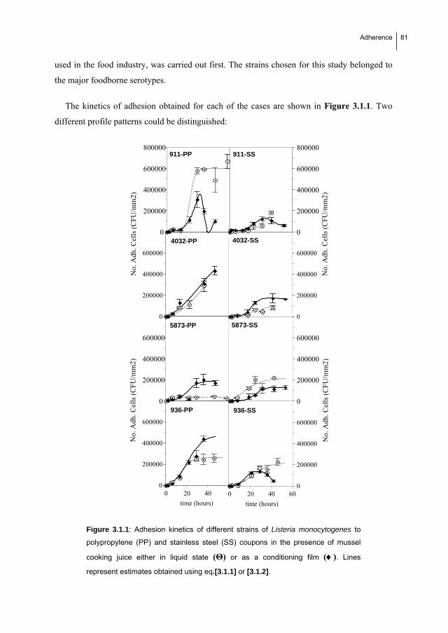
Adherence
81
used in the food industry, was carried out first. The strains chosen for this study belonged to
the major foodborne serotypes.
The kinetics of adhesion obtained for each of the cases are shown in Figure 3.1.1. Two
different profile patterns could be distinguished:
0
200000
400000
600000
800000
No.
Adh
. Cel
ls (
CF
U/m
m2)
911-PP 911-SS
0
200000
400000
600000
4032-PP
0
200000
400000
600000
4032-SS
0
200000
400000
600000
No.
Adh
. Cel
ls (
CF
U/m
m2)
5873-PP
0
200000
400000
600000
No.
Adh
. Cel
ls (
CF
U/m
m2)
5873-SS
0
200000
400000
600000
0 20 40
time (hours)
0
200000
400000
600000
0 20 40 60
time (hours)
936-PP 936-SS
0
200000
400000
600000
800000
No.
Adh
. Cel
ls (
CFU
/mm
2)
Figure 3.1.1: Adhesion kinetics of different strains of Listeria monocytogenes to
polypropylene (PP) and stainless steel (SS) coupons in the presence of mussel
cooking juice either in liquid state (Θ) or as a conditioning film (). Lines
represent estimates obtained using eq.[3.1.1] or [3.1.2].

82 Adherence
A sigmoidal type pattern, which is most frequent. This profile was described by a
logistic equation modified according to Cabo et al. (1999):
adadadad
admr)tm(r
e1
1
e1
aNAC
[3.1.1]
A pattern showing a sharp decrease after approximately 24 h. It appeared in 3 out
of the 16 experimental cases under study, namely, CECT 911 on polypropylene and
stainless steel and CECT 936 on polypropylene (Fig. 3.1.1), all of them in the
presence of conditioning film. An empirical additive model represented by two
logistic terms was used to describe this second pattern:
DDDDadadadad
Dadmr)tm(rmr)tm(r
e1
1
e1
a
e1
1
e1
aNAC
[3.1.2]
where,
NAC: number of adhered cells (CFU/mm2).
aad: maximum number of adhered cells (asymptote).
rad: specific adhesion coefficient (t-1).
mad: time at which the number of adhered cells is half of the maximum.
aD: total number of adhered cells which detach or die (CFU/mm2).
rD: specific detachment/death coefficient (t-1).
mD: time at which the number of detached/death cells is half of the maximum.
The values obtained for these parameters in each case are shown in Tables 3.1.2 and 3.1.3.
Fits of eq [3.1.1] and eq [3.1.2] to experimental data are shown by solid lines in Figure 3.1.1.
The values of parameters rad and mad have clearly shown that the strains CECT 911
(serotype 1/2c) and CECT 4032 (serotype 4b) had the highest adhesion ability (P < 0.05) to
both stainless steel and polypropylene, and that the adhesion of L. monocytogenes CECT 911
to polypropylene was the highest (as judged especially by the values of rad). On the contrary,
L. monocytogenes CECT 5873 (serotype 1/2a) showed the lowest adhesion (P < 0.05) in all

Adherence
83
the cases, except on stainless steel in the absence of conditioning film, a case in which the
adhesion of this strain was higher than all other strains.
Table 3.1.2: Values obtained for the parameters of eq [3.1.1] and eq [3.1.2] of adhesion to polypropylene in the
presence of mussel cooking juice
911 911-CF 4032 4032-CF 936 936-CF 5873 5873-CF
aad 603302 1134524 788373 650000 285000 500000 45000 190000
rad 0,420 0,171 0,068 0,075 0,198 0,140 0,550 0,211
mad 21,727 34,270 40,012 29,886 14,000 21,000 3,200 21,000
aD - 898804 - - - - - -
rD - 0,573 - - - - - -
mD - 35,67 - - - - - -
CF: presence of conditioning film.
Table 3.1.3: Values obtained for the parameters of eq [3.1.1] and eq [3.1.2] of adhesion to stainless steel in the
presence of mussel cooking juice
911 911-CF 4032 4032-CF 936 936-CF 5873 5873-CF
aad 250000 342779 1116597 204055 240993 169052 300000 131004
rad 0,122 0,109 0,017 0,252 0,122 0,253 0,102 0,242
mad 45,761 39,377 117,579 20,457 21,887 15,744 10,000 24,171
aD - 281047 - 198133 - 737975 - -
rD - 0,146 - 0,014 - 0,141 - -
mD - 50,27 - 43,302 - 53,76 - -
CF: presence of conditioning film
Although studies relating serotype to adhesion on polypropylene were not found, some
studies have shown that serotype 1/2c strains have higher adhesion ability on stainless steel
than serotype 1/2a and 4b strains (Norwood and Gilmour, 1999; Norwood and Gilmour
2001). This could be related to a different composition of flagella. The presence of flagella
and the consequent mobility have been shown to be determinant for initial adhesion
(Vatanyoopaisarn et al., 2000; Lemon et al., 2007). On the contrary, no significant differences
in initial adhesion were detected among different serotypes by Kalmokoff et al (2001). Even

84 Adherence
taking into account the variability in initial adhesion among different strains, discrepancies
between different studies are in some cases the result of differences in the experimental
design. It seems thus clear that sampling has to be carried out at several time points to define
adhesion kinetics and distinguish worst case scenarios, which would be defined by maximum
adhesion in minimum time, that is, by maximum values for aad and rad. (Herrera et al., 2007).
Otherwise, comparisons can be difficult to make. Additionally, it is disregarded that a time
limit between initial adhesion and mature biofilm formation cannot be fixed a priori.
In the present study, the adhesion to polypropylene was significantly higher (P < 0.05)
than to stainless steel in most of the scenarios and this is reflected in higher values of rad and
mad. The exception was L. monocytogenes CECT 5873, which adhered better to stainless steel
in the absence of conditioning film. This strain may exhibit a higher affinity towards
hydrophilic surfaces than the other strains tested.
Lower numbers (CFU/mm2) of adherenced CECT 911 and CECT 4032 on stainless steel in
the absence of conditioning film questioned the validity of the values for the asymptote of eq.
[3.1.1]. However, very similar values were obtained (aad=1116597 vs. aadreal=1115190; and
rad=0.018 vs. radreal=0.017 for CECT 4032) in later studies, in which the incubation period
was extended to 11 days (P < 0.05) (results not shown).
The present results agree with several studies showing that the physico-chemical properties
of surface materials (electric charge, hydrophobicity, roughness, etc.) are contributing
determinants of the initial adhesion of L. monocytogenes (Palmer et al., 2007; Rodríguez et
al., 2008; Sinde and Carballo, 2000). However, they may not be the only determinants. A
previous study by Sinde and Carballo (2000) had found that L. monocytogenes adhered in
higher numbers to polytetrafluoroethylene and rubber -hydrophobic substrates- than stainless
steel -a hydrophilic substrate-. Additionally, hydrophilic passivating groups were suggested to
be the best method for preventing cell attachment to synthetic substrates (Cunliffe et al.,
1999). On the contrary, other studies showed that L. monocytogenes formed biofilms faster on
stainless steel than polytetrafluoroethylene (strain LO28), (Chavant et al., 2002; Meylheuc et
al., 2001) or Buna-N rubber (strain Scott A), (Smoot and Pierson, 1998). In this latter work,
however, changes in cell hydrophobicity could not be correlated to differences in adhesion, or
to attachment of 21 L. monocytogenes strains to glass (Chae and Schraft, 2000). As Teixeira
et al. (2008) pointed out, a correlation between surface hydrophobicity and the extent of

Adherence
85
adhesion of L. monocytogenes is difficult to be established and other factors, such as surface
charge or roughness, should also be taken into account.
Adhesion was slightly higher (P < 0.05) under low nutrient availability conditions, i.e. in
the presence of conditioning film, particularly on polypropylene. However, it seems more
important that the number of cells adhered in the presence of conditioning film decreased in
the late phase of study in 3 out of 8 cases, that is, CECT 911 on both polypropylene and
stainless steel, and CECT 936 on stainless steel. Although the presence of conditioning film
enhanced initial adhesion, it hindered direct adhesion of cells to surface material. The
conditioning film plastified gradually over time. As a result, the system became more labile,
as the conditioning film tended to detach from the coupon surface, carrying the adhered cells.
This would be prevent a mature biofilm to be formed, but detachment involves a potential risk
of food contamination (Chavant et al., 2002; Norwood and Gilmour 1999).
With regard to the uptake of nutrients, it was approximately constant (1-2-g/l) in all the
cases, except for the strain CECT 5873 in the absence of conditioning film, in which a higher
carbohydrate content was consumed (ca. 4 g/l), despite lowers numbers of adhered cells was
low. Previous studies had shown that nutrient consumption was inversely proportional to
exopolysaccharide production, and therefore to adhesion (Laspidou et al., 2002).
Effects of intervalvar water of mussel on the kinetics of adhesion of L. monocytogenes
After this, the worst case scenarios were defined, i.e. combinations of strain, surface
material and surface conditioning showing most adhesion in plants processing cooked mussel.
These are:
CECT 911 on polypropylene in the absence of conditioning film (PP-ACF).
CECT 4032 on polypropylene in the presence of conditioning film (PP-CF).
CECT 5873 on stainless steel in the absence of conditioning film (SS-ACF).
CECT 4032 on stainless steel in the presence of conditioning film (SS-CF).
Subsequently, the kinetics of adhesion of these scenarios were compared with a similar
contamination event which may occur in situ in plants processing live mussels. Intervalvar
water was used as matrix of food residues in these subsequent comparative studies.

86 Adherence
As shown in Table 3.1.4 and Figure 3.1.2 adhesion was significantly higher (P < 0.05) in
the presence of intervalvar water, especially in the cases 1 and 3, both with no conditioning
film. However, only slight differences were observed in the presence of conditioning film. In
fact, practically no differences were found between IWM and MCJ in case CECT 4032 PP-
CF
Table 3.1.4: Values obtained for the parameters of eq [1] and eq [2] of adhesion in the
presence of intervalvar water of mussel
911-PP 4032-PP-CF 5873-SS 4032-SS-CF
aad 1444459.618 571898 809482 152976.856
rad 0.424 0.100 0.115 0.115
mad 25.517 23.087 41.286 33.36
aD - - - 1068974
rD - - - 35.63
mD - - - 0.650
PP: polypropylene SS: stainless steel CF: presence of conditioning film
Intervalvar water of mussel is nutritionally poorer than mussel cooking juice (see Table
3.1.1), but cells were not starved. In fact, the consumption of nutrients was low so nutrients
do not seem to play an important role.
At least three non-mutually exclusive factors could account for the higher adhesion found
in cases 3.1.2 A and 3.1.2.C (P < 0.05): i)
Higher probability of contact between cells and surface due to a lower nutrient
concentration,
Intervalvar water was not an adequate medium for cells to survive in the planktonic
state so they would tend to adhere,
pHs of intervalvar water and mussel cooking juice were different (7.74 in MCJ,
8.80 in IWM and about 7.50 in coupons with conditioning film). However, with
respect to the latter, it was shown that the lower the pH (in the range 6.0-9.0), the
higher the adhesion rate of L. monocytogenes Scott A to Buna N-rubber (Smoot and
Pierson, 1998).
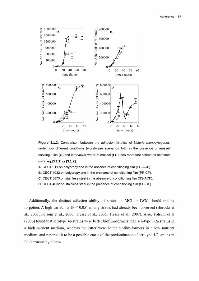
Adherence
87
0
200000
400000
600000
800000
0 20 40 60 80
No.
Adh
. Cel
ls (
CFU
/mm
2)
time (hours)
0
300000
600000
900000
1200000
1500000
1800000
0 20 40 60 80
No.
Adh
. Cel
ls (
CFU
/mm
2)
time (hours)
A.
0
200000
400000
600000
800000
0 20 40 60 80
No.
Adh
. Cel
ls (
CFU
/mm
2)
time (hours)
B.
0
200000
400000
600000
800000
0 20 40 60 80
No.
Adh
. Cel
ls (
CFU
/mm
2)
time (hours)
C. D.
Figure 3.1.2: Comparison between the adhesion kinetics of Listeria monocytogenes
under four different conditions (worst-case scenarios A-D) in the presence of mussel
cooking juice (Ο) and intervalvar water of mussel (). Lines represent estimates obtained
using eq.[3.1.1] or [3.1.2].
A.:CECT 911 on polypropylene in the absence of conditioning film (PP-ACF).
B.:CECT 4032 on polypropylene in the presence of conditioning film (PP-CF).
C.:CECT 5873 on stainless steel in the absence of conditioning film (SS-ACF).
D.:CECT 4032 on stainless steel in the presence of conditioning film (SS-CF).
Additionally, the distinct adhesion ability of strains in MCJ or IWM should not be
forgotten. A high variability (P < 0.05) among strains had already been observed (Borucki et
al., 2003; Folsom et al., 2006; Tresse et al., 2006; Tresse et al., 2007). Also, Folsom et al
(2006) found that serotype 4b strains were better biofilm-formers than serotype 1/2a strains in
a high nutrient medium, whereas the latter were better biofilm-formers in a low nutrient
medium, and reported it to be a possible cause of the predominance of serotype 1/2 strains in
food processing plants.
88 Adherence
Results from the present study showed a decrease in numbers of cells adhered to the
various surfaces after 40 h presumably because cell detachment became higher than adhesion.
This decrease was also observed by Herrera et al. (2007). This effect was observed in all cases
in the presence of conditioning film (CECT 911-PP, CECT 911-SS, CECT 936-PP, CECT
4032-SS) (Fig. 3.1.1, Tables 3.1.2, 3.1.3). However, in the cases of CECT 911-PP and CECT
4032-SS, the kinetics of adhesion showed a further increase subsequent to each decrease (P <
0.05) (Figure 3.1.1 and Figure 3.1.2, respectively). This may indicate a cyclic nature in L.
monocytogenes biofilm development. However, further studies would be needed to confirm
that a cyclical model is followed.
In conclusion, the initial adhesion of L. monocytogenes seems to depend mainly on the
interactions between cells and surfaces. The interactions are in turn mainly determined by
their physicochemical properties and possible space restrictions (hence the effects of the
conditioning film), along with environmental conditions. Adhesion is the initial step in
biofilm formation. However, subsequent biofilm growth was not dependent on initial
attachment (Chae and Schraft, 2000). Nonetheless, preventing the presence of pathogenic
bacteria on surfaces of food processing plants requires the control of environmental
conditions enhancing adhesion on a day to day basis. And this makes essential to carry out
studies allowing such conditions to be defined.
Biofilm formation: a cyclical dynamic?
Adherence
89
References
Bonaventura Di, G., R. Piccolomini, D. Paludi, V. D'Orio, A. Vergara, M. Conter, and A. Ianieri. 2008.
Influence of temperature on biofilm formation by Listeria monocytogenes on various food-contact
surfaces: relationship with motility and cell surface hydrophobicity. J. Appl. Microbiol. 104: 1552-1561.
Borucki, M.K., J.D. Peppin, D. White, F. Loge and D.R. Call. 2003. Variation in biofilm formation among
strains of Listeria monocytogenes. Appl. Environ. Microbiol. 69: 7339-7342.
Cabo. M.L., M.A. Murado, MªP. González and L. Pastoriza. 1999. A method for bacteriocin quantification. J.
Appl. Microbiol. 87: 907-914.
Chae, M. S. and H. Schraft. 2000. Comparative evaluation of adhesion and biofilm formation of different
Listeria monocytogenes strains. Int. J. Food Microbiol. 62: 103-111.
Chae, M. S., H. Schraft, L.T. Hansen and R. Mackereth. 2006. Effects of physicochemical surface characteristics
of Listeria monocytogenes strains on attachment to glass. Food Microbiol. 23: 250-259.
Chavant, P., B. Martinie, T. Meylheuc, M. Bellon-Fontaine, and M. Hebraud. 2002. Listeria monocytogenes
LO28: surface physicochemical properties and ability to form biofilms at different temperatures and
growth phases. Appl. Environ. Microbiol. 68: 728-737.
Chmielewski, R.A.N. and J.F. Frank. 2003. Biofilm formation and control in food processing facilities.
Comprehensive Rev Food Sci Food Safety. 2: 22-32.
Cox, L.J., T. Kleiss, J.L. Cordier. 1989. Listeria spp in food processing, non-food and domestic environments.
Food Microbiol. 6: 49-61.
Cunliffe, D., C.A. Smart, C. Alexander. 1999. Bacterial adhesion at synthetic surfaces. 1999. Appl. Environ.
Microbiol. 65: 4995-5002.
Denny, J. and J. McLauchlin. 2008. Human Listeria monocytogenes infections in Europe –an opportunity for
improved European surveillance. Eurosurveillance 13.
http://www.eurosurveillance.org/edition/v13n13/080327_5.asp. Accesed 27 March 2008.
Dubois, M., K.A. Gilles, J.K. Hamilton, P.A. Rebers and F. Smith. 1956. Colorimetric method for determination
of sugars and related substances. Anal. Chem. 28, 350–356.
Farber, J.M. and P.I. Peterkin. 1991. Listeria monocytogenes, a food-borne pathogen. Microbiol. Rev. 55:476-
511.

90 Adherence
Folsom, J. P., G. R. Siragusa and J. F. Frank. 2006. Formation of biofilm at different nutrient levels by various
genotypes of Listeria monocytogenes. J. Food Prot. 69: 826-834.
Frank, J.F., R.A. Koffi. 1990. Surface-adherent growth of Listeria monocytogenes in associated with increased
resistance to surfactant sanitizers and heat. J. Food Protect. 53, 550-554.
Gandhi, M and ML. Chikindas. 2007. Listeria: A foodborne pathogen that knows how to survive. Int. J. Food
Microbiol. 113: 1-15.
Harvey, J., K.P. Keenan, A. Gilmour. 2007. Assessing biofilm formation by Listeria monocytogenes strains.
Food Microbiol. 24: 380-392.
Herrera, J.J.R., M.L. Cabo, A.González, I. Pazos and L. Pastoriza. 2007. Adhesion and detachment kinetics of
several Strains of Staphylococcus aureus subsp.aureus under three different experimental conditions.
Food Microbiology 24: 585-591.
Ivanek, R., Y.T Grohn and M. Wiedmann. 2006. Listeria monocytogenes in multiple habitats and host
populations: review of available data for mathematical modelling. 2006. Foodborne Pathog. and Dis. 3:
319-336.
Kalmokoff, M. L., J. W. Austin, X. Wan, G. Sanders, S. Banerjee and J. M. Farber. 2001. Adsorption,
attachment and biofilm formation among isolates of Listeria monocytogenes using model conditions. J.
Appl. Microbiol. 91: 725-734.
Kathariou, S. 2002. Listeria monocytogenes virulence and pathogenicity, a food safety perspective. J. Food
Protect. 65: 1811-1829.
Kim, K.Y. and J.F. Frank. 1995. Effect of nutrients on biofilm formation by Listeria monocytogenes on stainless
steel. J. Food Protect. 58: 24-28.
Laspidou, C. S. and B. E. Rittmann. 2002. A unified theory for extracellular polymeric substances, soluble
microbial products, and active and inert biomass. Water Research, 36, 2711-2720.
Lemon, K. P., D. E. Higgins and R. Kolter. 2007. Flagellar motility is critical for Listeria monocytogenes biofilm
formation. J. Bacteriol. 189: 4418-4424.
Mai, T.L. and D.E. Conner. 2007. Effect of temperature and growth media on the attachment of Listeria
monocytogenes to stainless steel. Int. J. Food Microbiol. 120: 282-286.
McLauchlin, J., R.T. Mitchell, W.J. Smerdon and K. Jewell. 2004. Listeria monocytogenes and listeriosis: a
review of hazard characterisation for use in microbiological risk assessment of foods. Int. J. Food
Microbiol. 92: 15-33.
Adherence
91
Meylheuc, T., C. J. van Oss, and M. Bellon-Fontaine. 2001. Adsorption of biosurfactant on solid surfaces and
consequences regarding the bioadhesion of Listeria monocytogenes LO28. J. Appl. Microbiol. 91: 822-
832.
Moltz, A. G. and S. E. Martin. 2005. Formation of biofilms by Listeria monocytogenes under various growth
conditions. J. Food Prot. 68: 92-97.
Norwood, D. E. and A. Gilmour. 1999. Adherence of Listeria monocytogenes strains to stainless steel coupons.
J. Appl. Microbiol. 86: 576-582.
Norwood, D. E. and A. Gilmour. 2001. The differential adherence capabilities of two Listeria monocytogenes
strains in monoculture and multispecies biofilms as a function of temperature. Lett. Appl. Microbiol. 33:
320-324.
Palmer, J., S. Flint and J. Brooks. 2007. Bacterial cell attachment, the beginning of a biofilm. J. Ind. Microbiol.
Biotechnol. 34: 577-588.
Pan, Y., Jr. Breidt, S. Kathariou. 2006. Resistance of Listeria monocytogenes biofilms to sanitizing agents in a
simulated food processing environment. Appl. Environ. Microbiol. 72: 7711-7717.
Rocourt, J. and P. Cossart. 1997. Listeria monocytogenes. In: Doyle, M.P., Beuchat, L.R. and Montville T.
(Eds.), Food Microbiology: Fundamentals and Frontiers. American Society for Microbiology, ASM
Press, Washington DC., pp. 317-352.
Rodríguez, A., W.R. Autio, L.A. McLandsborough. 2008. Effect of surface roughness and stainless steel finish
on Listeria monocytogenes attachment and biofilm formation. J. Food Protect. 71: 170-175.
Sinde, E. and J. Carballo. 2000. Attachment of Salmonella spp. and Listeria monocytogenes to stainless steel,
rubber and polytetrafluorethylene: the influence of free energy and the effect of commercial sanitizers.
Food Microbiol. 17: 439-447.
Smoot, L. M. and M. D. Pierson. 1998. Effect of environmental stress on the ability of Listeria monocytogenes
Scott A to attach to food contact surfaces. J. Food Prot. 61: 1293-1298.
Teixeira, P, J. Lima, J. Azeredo and R. Oliveira. 2008. Adhesion of Listeria monocytogenes to materials
commonly found in domestic kitchens. Int. J. Food Sci. Technol. 43: 1239-1244 .
Tresse, O., V. Lebret, T. Benezech and C. Faille. 2006. Comparative evaluation of adhesion, surface properties,
and surface protein composition of Listeria monocytogenes strains after cultivation at constant pH of 5
and 7. J. Appl. Microbiol. 101: 53-62.

92 Adherence
Tresse, O., K. Shannon, A. Pinon, P. Malle, M.Vialette, G. Midelet-Bourdin. 2007. Variable adhesion of Listeria
monocytogenes isolates from food-processing facilities and clinical cases to inert surfaces. J. Food
Protect. 70: 1569-1578
Vatanyoopaisarn, S., A. Nazli, C. E. R. Dodd, C. E. D., Rees and W. M. Waites. 2000. Effect of flagella on
initial attachment of Listeria monocytogenes to stainless steel. Appl. Environ. Microbiol. 66: 860-863.

33..22 An efficient method to obtain
benzalkonium chloride-adapted cells of
Listeria monocytogenes CECT 5873.


Adaptation
95
3.2. AN EFFICIENT METHOD TO OBTAIN BENZALKONIUM
CHLORIDE-ADAPTED CELLS OF LISTERIA MONOCYTOGENES
CECT 5873.
This section presents a new method for obtaining BAC-adapted L. monocytogenes cells. A
factorial design was used to assess the effects of the inoculum size and BAC concentration on
the adaptation (measured in terms of lethal dose 50 -LD50-) of exponential L. monocytogenes
5873 cells after only one exposure. A significant empirical equation was obtained showing the
positive effect of the inoculum size and the positive interaction between the effects of BAC
and inoculum size on the level of adaptation achieved (LD50). However, a slight negative
effect of BAC, due to it being a biocide, was also significant. By applying the new procedure,
it was possible to increase BAC adaptation 4.17-fold in only 33 hours whereas 5 days were
necessary in the classical procedure based on successive stationary phase cultures in sublethal
BAC concentrations. Lastly, we present preliminary results of the comparison between the
proteomic patterns of non-BAC adapted and BAC-adapted cells.

96 Adaptation
3.2.1. Introduction
Listeria monocytogenes is a pathogenic bacteria widely found in nature and food industry
(Cox et al. 1989, Ivanek et al. 2006). The frequent use of disinfectants in food environments
can lead to the development of disinfectant resistant isolates due to inadequate cleaning and
disinfection protocols (Walton et al., 2008). Benzalkonium chloride (BAC) is a biocide
belonging to the group of quaternary ammonium compounds (QACs) that is commonly used
in the food industry. Previous works have found that persistent L. monocytogenes strains are
QAC resistant (Aase et al. 2000; Mullapudi et al., 2008). Changes in the cell membrane and
efflux pumps lead to this resistance (Mereghetti et al., 2000, Romanova et al., 2006). This
could be partly related to inadequate cleaning and disinfection protocols that result in long-
term exposure of microbial communities to subinhibitory concentrations of QACs, which
causes the emergence of more resistant clones with changes in their susceptibility to other
antimicrobials (Langsrud et al. 2003; Hegstad et al., 2010). Consequently, a lot of research in
applied microbiology is focused on studying the physiological consequences of BAC and
other biocides on bacterial resistance. To carry out this research it is necessary to generate
BAC-adapted bacterial cells in the experimental protocols.
Classical methods for obtaining bacterial cells adapted to antimicrobial agents are based on
successive exposures of stationary-phase cells (Aase et al. 2000; Soumet et al., 2005;
Taormina et al., 2001; To et al. 2006). Although this classical method is time-consuming and
does not ensure that the maximum level of adaptation is achieved, it is the procedure most
widely used in bacterial physiology experiments. However, some authors have found that L.
monocytogenes only begins to show gradual adaptation to BAC after several successive
subcultures in increasing BAC concentrations during 1 week (To et al. 2002), whereas others
have found it necessary to wait 5 (Aarnisalo et al. 2007) or 7 days (Aase et al. 2000) with no
growth to obtain BAC-adapted cells.
It is known that exponential-phase cells are metabolically more active than stationary-
phase cells. This implies that exponential cells are more able to activate a change in the
ongoing metabolic pathways in response to an external stimulus (Whistler et al., 1998; Zotta
et al., 2009). Stationary phase cells have already activated phase-associated resistance
mechanisms (Davies et al., 1996; Kang et al. 2004; Hengge-Aronis et al., 1991; Lange and
Hengge-Aronis, 1991; Hu et al., 1998; Munro et al., 1995). However, no previous studies in
Adaptation
97
which L. monocytogenes exponential-phase cells were exposed to biocides to generate
adaptation were found in the literature. Therefore, in the present work we studied the effects
of the concentration of bacterial cells (inoculum size) in exponential phase and the
concentration of BAC on the level of adaptation reached by L. monocytogenes after only one
exposure. Based on the results obtained we propose an alternative procedure for obtaining
cells with a higher level of adaptation in a shorter period of time.
3.2.2. Materials and methods
Bacterial strains, synthetic media and biocide
Listeria monocytogenes CECT 5873, CECT 911 and CECT 4032 were provided by the
Spanish Type Culture Collection (Valencia). Strains were frozen and stored at -80ºC in TSB
containing 50% glycerol (v/v) until use. Whenever required, one cryovial was thawed and
strains were subcultured twice in Triptone Soy Broth (TSB, Cultimed, S.L., Spain) at 37 ºC
before use. All subsequent cultures were grown in TSB at 25 ºC. In the adaptation
experiments, TSB with different concentrations of benzalkonium chloride was used.
Benzalkonium chloride was purchased from Sigma Chemical Co.
Experimental method
Inocula preparation
One ml aliquot of an overnight culture of L. monocytogenes CECT 5873 in stationary
phase was transferred to 49 ml of TSB (final volume: 50 ml) in a 250-ml erlenmeyer flask
(1:5) (v:v) and incubated at 25 ºC until the required growth phase according to the
experimental procedure applied (see below). Cultures were incubated until 1.0 optical density
(700nm) when inoculum in stationary phase was required and to 0.4-0.5 optical density (700
nm) when an exponential phase culture was needed, corresponding to cell densities of 2x10⁹
CFU/ml and 109 CFU/ml respectively according with previous calibration.
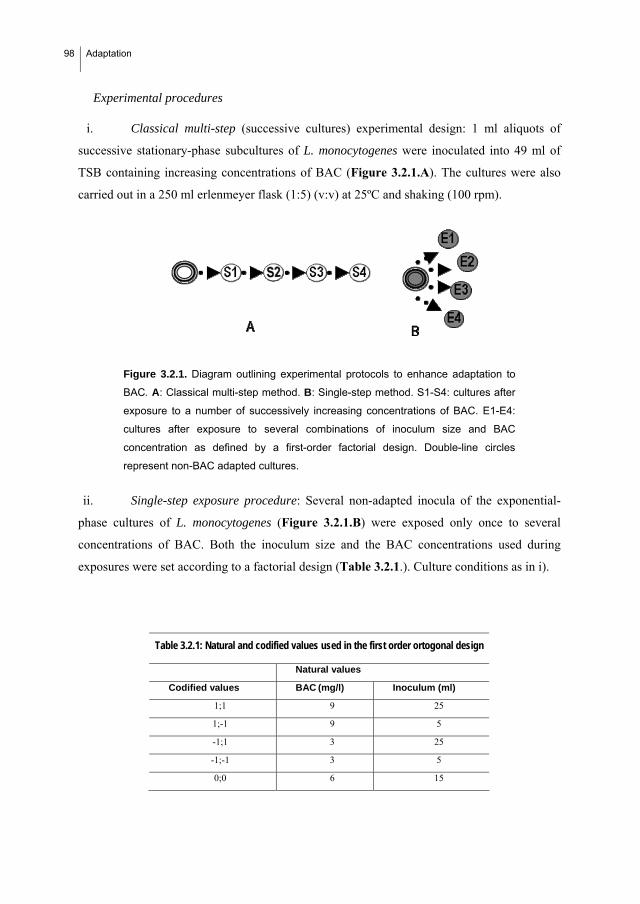
98 Adaptation
i. Classical multi-step (successive cultures) experimental design: 1 ml aliquots of
successive stationary-phase subcultures of L. monocytogenes were inoculated into 49 ml of
TSB containing increasing concentrations of BAC (Figure 3.2.1.A). The cultures were also
carried out in a 250 ml erlenmeyer flask (1:5) (v:v) at 25ºC and shaking (100 rpm).
Figure 3.2.1. Diagram outlining experimental protocols to enhance adaptation to
BAC. A: Classical multi-step method. B: Single-step method. S1-S4: cultures after
exposure to a number of successively increasing concentrations of BAC. E1-E4:
cultures after exposure to several combinations of inoculum size and BAC
concentration as defined by a first-order factorial design. Double-line circles
represent non-BAC adapted cultures.
ii. Single-step exposure procedure: Several non-adapted inocula of the exponential-
phase cultures of L. monocytogenes (Figure 3.2.1.B) were exposed only once to several
concentrations of BAC. Both the inoculum size and the BAC concentrations used during
exposures were set according to a factorial design (Table 3.2.1.). Culture conditions as in i).
Table 3.2.1: Natural and codified values used in the first order ortogonal design
Natural values
Codified values BAC (mg/l) Inoculum (ml)
1;1 9 25
1;-1 9 5
-1;1 3 25
-1;-1 3 5
0;0 6 15
Experimental procedures

Adaptation
99
Collection of BAC-adapted cells of L. monocytogenes
After each experimental assay described above, 0,5 ml of BAC-adapted cells were
collected by centrifugation at 2000 g during 5 minutes at 25 ºC. Obtained pelled pellet was
washed with PBS to eliminate residual BAC concentration by centrifuging and resuspending
twice in 0.05 M sterilized phosphate buffer. Cells were then transferred to a cryovial and
stored at -80ºC in TSB containing 50% glycerol (v/v) until their resistance to BAC was
determined.
Determination of the resistance to BAC in L. monocytogenes collected cells.
Dose-response assays in microtiter plates
One hundred and twenty five µl of the activated inoculum diluted (1:10) twice of Listeria
monocytogenes was added to microplate wells containing 125 µl solutions of different BAC
concentrations (0 to 15 mg /l, step: 1 mg/l). 3 replicates of each BAC concentration were
prepared and the microtiter plate was incubated at 37 ºC for 24 h. Absorbance at 700 nm was
measured using a spectrophotometer (iMarkTM, Biorad). Series with sterile water instead of
BAC was used as a control. The inhibition (I) of each BAC concentration was calculated from
the absorbance values obtained in the dose-response assay according to the following
equation [3.2.1]:
I (%)= [1-(AX/A0)]*100 [3.2.1]
I: Inhibition (%)
Ax: average of the absorbance values obtained for each concentration of BAC
A0: average of the absorbance values obtained in absence of BAC.
Determination of resistance
Two different methods were used to quantify the bacterial resistance to benzalkonium
chloride:

100 Adaptation
i) In terms of the minimum inhibitory concentration (MIC), defined as the minimum
value of dose were the absence of growth in detected. Used in the screening of the
mutants carried out in the genetic studies.
ii) In terms of LD50, that was obtained by adjusting the inhibition values calculated
according with Eq. [3.2.1] to the modified logistic equation [3.2.2.] (Cabo et al.
1999) and the Solver application of Excel. Used in the rest of experiments.
I (%) =
1
1
1
150)50( rLDDLDr ee
k [3.2.2]
LD50: Dose that kills 50% of initial population
k: Maximum inhibition BAC value (asymptote)
r: Specific inhibition coefficient (dimensions: inverse of the dose)
D: Dose
Proteomic assays
Cytosolic proteins were extracted from controlled exponential-phase cultures (OD. O.4-
0.5) and then purified using the ReadyPrep™ 2-D Cleanup Kit (Biorad, S.L.). First- and
second-dimension electrophoresis were carried out according to Sánchez et al. 2010. In the
proteomic experiments, BAC-adapted cells (LD50=7.22 mg/l) obtained after one exposure of
7.5 ml of exponential cells of L. monocytogenes to 5.25 mg/l of BAC were used.
Statistics
Factorial designs were carried out according to Box and Hunter (1989). A Student’s t test
(α=0.05) was used to test for significant differences between means of the adaptation level
and between the coefficients of the equations obtained in the factorial design. A Fisher test
(α=0.05) was used to test the consistency of the models.
Genetic procedures
Information about bacterial strains, plasmids, culture conditions, nucleic acid manipulations,
mutagenesis methods and cloning methods employed can be checked in Sleator et al., 2000
and Stack et al., 2005.

Adaptation
101
3.2.3 Results
Adaptation of L. monocytogenes CECT 5873, 911 and 4032 to benzalkonium chloride by
the classical multi-step procedure after the exposure to different BAC concentrations
0 0,9 1,8 2,25 3 4,50
2
4
6
8
10
Re
sist
an
ce L
D5
0 (
mg
/l)
BAC (mg/l) concentration
CECT 5873
5 days
0 1,1 2,2 2,75 3,6 5,50
2
4
6
8
Re
sist
an
ce L
D5
0 (
mg
/l)
BAC (mg/l)] concentration
CECT 911
5 days
BAC (mg/l) concentration
CECT 4032
0 1,5 3 3,75 502468
10
Re
sist
an
ce L
D5
0 (
mg
/l)
4 days
Figure 3.2.2: Increase in BAC-resistance of L. monocytogenes CECT 5873, 911
and 4032 according to a classical multi-step procedure

102 Adaptation
Stationary phase cells of each strain were tested for growth under a sequence of
successively increasing sublethal concentrations of BAC and their adaptation capacities were
compared (Figure 3.2.2).
The adaptation level of each strain was found to be related to its susceptibility to BAC.
Following the classical procedure, the LD50 of CECT 5873 – the most susceptible strain –
increased from 2.71 mg/l to 7.1 mg/l (2.61-fold) after five successive cultures in 5 days.
However, the maximum adaptation of CECT 911 and CECT 4032 – both more resistant to
BAC – was achieved after only one culture, with LD50 values of 5.06 mg/l (from 3.80,
increasing 1.33-fold) and 9.19 (from 6.96, increasing 1.32-fold), respectively.
It was thus shown that, using stationary phase cells and the classical experimental protocol,
some L. monocytogenes strains can reach the maximum level of adaptation to BAC in only
one culture (Figure 3.2.1). This means that it is possible to reduce the experimental time
needed to achieve BAC-adapted cells, which in previous studies were obtained after days or
even weeks of culture (Aase et al. 2000; To et al. 2002; Romanova et al. 2006).
Although this experimental protocol is probably the most frequently used system for
obtaining BAC-adapted-cells, the present results indicate that it is not optimum for all strains;
therefore, each strain should be considered independently. In fact, the maximum adaptation
capacity of L. monocytogenes CECT 5873 was not reached even after 5 successive cultures,
whereas CECT 4032 and CECT 911 seemed to reach it after only one culture. This also
indicates a higher adaptation potential of the strain CECT 5873. This strain was therefore
chosen for subsequent studies in which the possibility of increasing the adaptation level was
explored with exponential phase cells submitted to a single-step experimental design (see
Figure 3.2.1 B).
An alternative strategy for obtaining BAC-adapted L. monocytogenes cells
Supported by the theory that exponential cells are metabolically more active than
stationary-phase cells, in the present work we propose a new method for obtaining L.
monocytogenes cells with a high degree of BAC adaptation in a short period of time based on
only one exposure of exponential phase cells to sublethal concentrations of BAC. For this, the
effects of the inoculum size and the concentration of BAC in the exposure on the level of
adaptation achieved by L. monocytogenes CECT 5873 were explored by using a first-order

Adaptation
103
factorial design that allows both the individual and combined effects of these two variables to
be determined.
The experimental results obtained were adequately described by the following empirical
equation (r2= 0.925), graphically shown in Figure 3.2.3 (data are showed in the supporting
information chapter):
LD50=7,96-0,91B+1,84I+2,46BI [3.2.3]
In which a significant positive individual effect of the L. monocytogenes inoculum size and
a significant effect of the interaction between it and the concentration of BAC in the exposure
wasfound. However, a negative individual effect of the benzalkonium chloride used in the
exposure was also significant in the equation.
LD
50 (
mg
/l)
Figure 3.2.3. Effects of the Inoculum size and BAC concentration during the
exposures on the resistance (LD50) of non-adapted inocula of L. monocytogenes
CECT 5873. Independent variables are expressed in coded values.
The significant first order negative term describing the individual effect of BAC reflects
the biocide effect of BAC. This which exposing to low inoculum concentrations, could imply
the absence of adaptation. Therefore, over [BAC: inoculum] ratios, L. monocytogenes does
not grow. This is the case of the combination [1,-1], in which the basal resistance level (LD50
value of 2.71 mg/l) was used for calculation purposes. Therefore, it is recommended to
determine the growth limits of the strain before setting the BAC concentrations to be used.
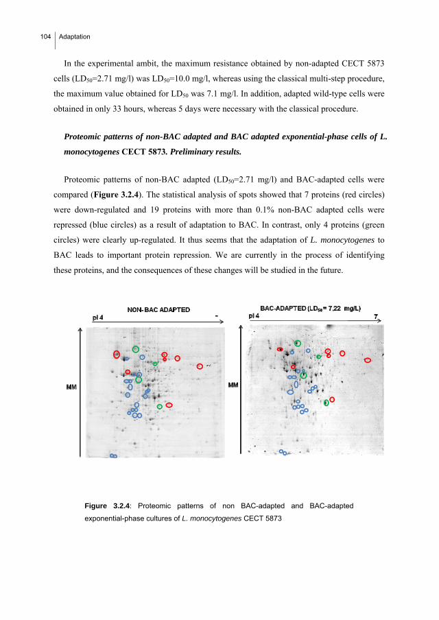
104 Adaptation
In the experimental ambit, the maximum resistance obtained by non-adapted CECT 5873
cells (LD50=2.71 mg/l) was LD50=10.0 mg/l, whereas using the classical multi-step procedure,
the maximum value obtained for LD50 was 7.1 mg/l. In addition, adapted wild-type cells were
obtained in only 33 hours, whereas 5 days were necessary with the classical procedure.
Proteomic patterns of non-BAC adapted and BAC adapted exponential-phase cells of L.
monocytogenes CECT 5873. Preliminary results.
Proteomic patterns of non-BAC adapted (LD50=2.71 mg/l) and BAC-adapted cells were
compared (Figure 3.2.4). The statistical analysis of spots showed that 7 proteins (red circles)
were down-regulated and 19 proteins with more than 0.1% non-BAC adapted cells were
repressed (blue circles) as a result of adaptation to BAC. In contrast, only 4 proteins (green
circles) were clearly up-regulated. It thus seems that the adaptation of L. monocytogenes to
BAC leads to important protein repression. We are currently in the process of identifying
these proteins, and the consequences of these changes will be studied in the future.
Figure 3.2.4: Proteomic patterns of non BAC-adapted and BAC-adapted
exponential-phase cultures of L. monocytogenes CECT 5873

Adaptation
105
Isolation of BAC mutants of Listeria monocytogenes. Identification of genes and
proteins associated with BAC resistance in L. monocytogenes
A bank of transposon mutants of Listeria monocytogenes EGDe and LO28 (Cao et al.
2007) was screened for mutants with respect to their BAC sensitivity or resistance, Several
BAC mutants were isolated and confirmed by their MIC values. The region of transposon
insertion of the resistant mutants was determined by sequencing the PCR products obtained
using the appropriate Marq primers (Cao et al. 2007). The proteins predicted to be encoded by
the disrupted genes were deduced from the ListiList website
(http:/genolist.pasteur.fr/ListiList/). The MIC’s of the selected mutants and the proteins
encoded by the disrupted genes are shown in Table 3.2.2.
Tabla 3.2.2: MIC values (mg/l) of BAC in the wild type and isolated mutants of L. monocytogenes. Strain MIC of BAC (mg/l) Identified protein
L. monocytogenes EGDe 2.5
1 sensitive mutant 2.0
1 resistant mutant 6.5 lmo 2277: unknown protein
1 resistant mutant 6 stress response protein encoding DNA
L. monocytogenes LO28 2.5
1 resistant mutant 7.0 lmo 601: cell surface protein
The role of the protein encoded by lmo2277 in the BAC resistance of L. monocytogenes
EDGe is currently under investigation using further mutagenesis and cloning strategies.
4.2.4. Discussion
In this work it has been shown that the exposure of high concentrations of exponential cells
of L. monocytogenes to sublethal BAC concentrations is a more efficient protocol than the
classical method used in applied microbiology for obtaining cells with a high level of BAC
adaptation. To achieve this, the individual and combined effects of both variables, the
inoculum size and the concentration of BAC during exposure were studied using a first order
factorial design.
106 Adaptation
The equation [3.2.3] highlights the relevant positive effect of the inoculum size, with two
significant terms (individual and combined with BAC) in the empirical equation. There are
some previous studies that have considered the inoculum effect to initiate growth in bacteria
(Bidlas et al., 2008; Skandamis et al. 2007; Koutsoumanis and Sofos, 2005; Pascual et al.
2001; Robinson et al. 2001). Most of these works agree that increasing the inoculum levels
allows Listeria monocytogenes to grow under more stressful environmental (pH, temperature,
salt, temperature, aw) or chemical and antimicrobial conditions (bacteriocins, sodium lactate,
sodium diacetate). Two possible explanations for the inoculum effect are considered: 1) that it
is due to the heterogeneity of the bacterial population, which means that the larger the
inoculum size the longer the resistant fraction of the population (Skandamis et al, 2007;
Koutsoumanis and Sofos, 2005; Pascual et al. 2001); and 2) that a co-operative effect due to
cell-cell signalling (quorum sensing) occurs at high cell densities. However, this last
explanation was only partially demonstrated by Robinson et al. (2001), who found that using
spent medium caused a decrease in the lag phase of L. monocytogenes. However, these
authors did not observe any significant changes in the probability of growth. Moreover,
Bidlas et al. (2008) proposed a “composite model” that demonstrated in five different
pathogenic bacteria (one of which was Listeria innocua) According to this model, the effect
of the inoculum size is only the consequence of the time required for a specific inoculum size
to start growing (TTD, time to detection of growth) and it is directly related to microbial
number. This is in agreement with the present study, the cooperative effect associated with the
inoculum size was checked by exposing L. monocytogenes adapted cells (LD50=6.62) to
different inoculum sizes and a concentration of BAC that allowed the BAC:inoculum size
ratio to be constant. No significant differences were obtained in the resistance obtained after
exposure (Table 3.2.2). The equation 3.2.3 ecuation reflects the larger the inoculums size the
higher BAC concentration needed to kill the cells.
So, one evident consequence of the described inoculum effect is that increasing the
microbial inoculum exposed to a set biocide concentration (BAC in our case) will decrease
the time to growth of the exposed strain, thus facilitating adaptation and increasing cell
resistance (measured as LD50). This is precisely reflected by the significant I-containing terms
of the equation [3.2.3].
The proposed procedure significantly reduces the time needed to obtain BAC-adapted L.
monocytogenes cells in batch cultures. This is especially important considering that all the

Adaptation
107
methods in the literature are based on successive exposures to increasing BAC concentrations
without increasing the inocula, which makes them to be tedious and time consuming.
Table 3.2.3: Effects of the inoculum size of the level of adaptation achieved by L. monocytogenes exposed to the
same ratio BAC:cell.
Inoculum (ml) [BAC] (mg/l) µg[BAC]/cell. LD50
1 0.88 0.000045 5.58
5 4.09 0.000045 5.00
5 4.55 0.00005 6.62
10 7.5 0.000045 5.72
10 8.33 0.00005 6.39
15 11.54 0.00005 No growth
The results obtained are in agreement with two previous works which demonstrated that
the metabolic activity of exponential cells allows them to develop adaptation mechanisms
quicker than stationary phase cells exposed to sublethal concentrations of a biocide: Davies et
al. 1996 studied the acid tolerance response (ATR) of L. monocytogenes and demonstrated
that this response is growth-phase dependent and that the low metabolic activity of stationary
phase cells implies a phase-specific resistance mechanism; and McMahon et al. (2000) found
that L. monocytogenes has a higher adaptive response to heat-shock when working cultures
were obtained after two successive cultures until the exponential phase instead of culturing
twice untill stationary phase or one till stationary and the second one untill exponential phase.
However, exponential-phase cells are more susceptible to biocides than stationary-phase cells
(Luppens et al., 2001; Hayman et al. 2007; Weeks et al. 2006; Pascual et al. 2001). This could
be a disadvantage of the proposed method, as biocide concentration during the exposure could
not increase over the growth limit unless the inoculum concentration is increased sufficiently.
But this fact will be also present in the classical method. This susceptibility of the cells to
BAC is reflected in the negative first order term for the benzalkonium chloride, which
indicates that it is important to control the concentration of the disinfectant carefully during
the exposures.
In addition, we found significant differences in the expression of proteins between BAC
non-adapted and semi-adapted (LD50= 7.22 mg/l) L. monocytogenes CECT 5873 cells.
Although such proteins should be identified further, the balance of protein expressed proteins
showed that adaptation to BAC seems to imply a repression. There is little previous

108 Adaptation
information on this subject for L. monocytogenes. In fact, only two proteomic works were
found with proteomics in Listeria monocytogenes (Mbandi et al., 2007; Folsom et al, 2007);
however, only in the first changes in the protein profiles associated with the exposure to
biocides were studied. They studied protein variations when the bacteria were exposed to the
organic acids. Sodium diacetate treatment produced a higher number of unmatched proteins
than sodium lactate (124 versus 53 in lactate), and a increase in protein expression (20 versus
5 in lactate) and in the number of novel proteins (90 versus 45 in lactate). The combination of
the treatments involved a higher number of repressed proteins (41 versus ~30 in the single salt
treatment). Six proteins were further investigated.
There have been few proteomic studies carried out on the resistance of bacteria to
chemicals. Bore et al. (2007) also studied adapted tolerance to benzalkonium chloride
Escherichia coli K-12 using transcriptome and proteome analyses. Changes in the expression
levels of adapted cells were shown for porins, drug transporters, glycolytic enzymes,
ribosomal subunits and several genes and proteins involved in protection against oxidative
stress and antibiotics. However, they found that genes related to these functions were mainly
up-regulated, which is in contrast with the present results.
In summary, the results obtained permitted us to propose a new method for obtaining
Listeria monocytogenes cells with a higher level of BAC adaptation in a shorter period of
time than the classical method based on successive cultures of stationary phase cells in
sublethal BAC concentrations. This method consists in only one exposure of a high
concentration of exponential phase cells to sublethal BAC concentrations, always under the
growth limit of the strain for BAC.

Adaptation
109
References.
Aarnisalo, K., Lundén, J., Korkeala, H. and Wirtanen, G., 2007. Susceptibility of Listeria monocytogenes strains
to disinfectants and chlorinated alkaline cleaners at cold temperatures. LWT – Food Science and
Technology, 40, 1041-1048.
Aase, B., Sundheim, G., Langsrud, S. and Rørvik, L.M., 2000. Occurrence of and a possible mechanism for
resistance to a quaternary ammonium compound in Listeria monocytogenes. Int. J. Food Microbiol. 62,
57-63.
Bidlas, E., Du, T. and Lambert, R.J.W., 2008. An explanation for the effect of inoculum size on MIC and the
growth/no growth interface. Int. J. Food Microbiol. 126, 140-152.
Bore, E., Hébraud, M., Chafsey, I., Chambon, C., Skjaeret, C., Moen, B., Møretrø, T., Langsrud, Ø., Rudi, K.,
Langsrud, S., 2007. Adapted tolerance to benzalkonium chloride in Escherichia coli K-12 studied by
transcriptome and proteome analyses. Microbiol. 153(Pt 4):935-46.
Box, G.E.P., Hunter, W.G., and Hunter, J.S., 1989. En “Estadística para investigadores”. Ed. Reverté
(Barcelona).
Cao, M., Bitar, A.P., Marquis, H., 2007. A mariner-based transposition system for Listeria monocytogenes.
Appl. Environ. Microbiol. 73(8), 2758-2761
Cox, L.J., Kleiss, T. and Cordier, J.L., 1989. Listeria spp in food processing, non-food and domestic
environments. Food Microbiol. 6, 49-61.
Davies, E.A., Falahee, M.B., Adams, M.R., 1996. Involvement of the cell envelope of Listeria monocytogenes in
the acquisition of nisin resistance. J. Appl. Bacteriol. 81(2), 139-146.
Ferreira A., Sue D., O'Byrne C.P., Boor K.J., 2003.Role of Listeria monocytogenes sigma(B) in survival of lethal
acidic conditions and in the acquired acid tolerance response. Appl Environ Microbiol. 69(5), 2692-8.
Folsom, J.P., Frank, J.F., 2007. Proteomic analysis of a hypochlorous acid-tolerant Listeria monocytogenes
cultural variant exhibiting enhanced biofilm production. J. Food Prot. 70(5), 1129-36.
Hayman, M. M., Anantheswaran, R. C. and Knabel, S. J. 2007. The effects of growth temperature and growth
phase on the inactivation of Listeria monocytogenes in whole milk subject to high pressure processing.
Int. J. Food Microbiol. 115, 220-226.
Hegstad, K., Langsrud, S., Lunestad, B.T., Scheie, A.A., Sunde, M., Yazdankhah, S.P., 2010. Does the wide use
of quaternary ammonium compounds enhance the selection and spread of antimicrobial resistance and
thus threaten our health?. Microb. Drug Resist. 16(2), 91-104.

110 Adaptation
Hengge-Aronis, R., W. Klein, R. Lange, M. Rimmele, and W. Boos. 1991. Trehalose synthesis genes are
controlled by the putative sigma factor encoded by rpoS and are involved in stationary phase
thermotolerance in Escherichia coli. J. Bacteriol. 173, 7918–7924.
Ivanek, R., Grohn, Y.T. and Wiedmann, M., 2006. Listeria monocytogenes in multiple habitats and host
populations: review of available data for mathematical modelling. 2006. Foodborne Path Dis. 3, 319-
336.
Kang B.R., Cho, B.H., Anderson A.J., Kim Y. C., 2004. The global regulator GacS of a biocontrol bacterium
Pseudomonas chlororaphis O6 regulates transcription from the rpoS gene encoding a stationary-phase
sigma factor and affects survival in oxidative stress. Gene 325 (2004) 137–143
Koutsoumanis, K.P. and Sofos, J.N., 2005. Effect of inoculum size on the combined temperature, pH and aw
limits for growth of Listeria monocytogenes. Int. J. Food Microbiol. 104, 83-91.
Lange, R., and Hengge-Aronis, R., 1991. Identification of a central regulator of stationary phase gene expression
in Escherichia coli. Mol. Microbiol. 5:49–59.
Langsrud, S., Sidhu, M.S., Heir, E. and Holck, A.L., 2003. Bacterial disinfectant resistance - a challenge for the
food industry. Int. Bio. Biod.51, 283-290.
Luppens, S.B., Abee, T., Oosterom, J., 2001. Effect of benzalkonium chloride on viability and energy
metabolism in exponential- and stationary-growth-phase cells of Listeria monocytogenes. Journal of
Food Protection 64, 476.
Mbandi, E., Phinney, B.S., Whitten, D., Shelef, L.A., 2007. Protein variations in Listeria monocytogenes
exposed to sodium lactate, sodium diacetate, and their combination. J. Food Prot. 70(1), 58-64.
McMahon, C.M., Byrne, C.M., Sheridan, J.J., McDowell, D.A., Blair, I.S., Hegarty, T., 2000. The effect of
culture growth phase on induction of the heat shock response in Yersinia enterocolitica and Listeria
monocytogenes. J. Appl. Microbiol. 89(2), 198-206.
Mereghetti, L., Quentin, R., Marquet-Van Der Mee, N. and Audurier, A. 2000. Low Sensitivity of Listeria
monocytogenes to Quaternary Ammonium Compounds. Appl. Environ. Microbiol. 66, 5083-5086.
Mullapudi, S., Siletzky, R.M. and Kathariou, S., 2008. Heavy-metal and benzalkonium chloride resistance of
Listeria monocytogenes isolates from the environment of turkey-processing plants. Appl. Environ.
Microbiol. 1464-1468.
Munro PM, Flatau GN, Clément RL, Gauthier MJ., 1995. Influence of the RpoS (KatF) sigma factor on
maintenance of viability and culturability of Escherichia coli and Salmonella typhimurium in seawater.
Adaptation
111
Appl Environ Microbiol. 61(5):1853-8. The sigma factor RpoS is essential for stationary-phase-specific,
multiple-stress resistance.
Pascual, C., Robinson, T.P., Ocio, M.J., Aboaba, O.O. and Mackey, B.M., 2001. - The effect of inoculum size
and sublethal injury on the ability of Listeria monocytogenes to initiate growth under suboptimal
conditions. Lett. Appl. Microbiol. 33, 357-361.
Robinson, T.P., Aboaba, O.O., Kaloti, A., Ocio, M.J., Baranyi, J. and Mackey, B.M., 2001. The effect of
inoculum size on the lag phase of Listeria monocytogenes. Int. J. Food Microbiol. 70, 163-173.
Romanova, N. A., Wolffs, P. F. G., Brovko, L. Y. and Griffiths, M. W. 2006. Role of Efflux Pumps in
Adaptation and Resistance of Listeria monocytogenes to Benzalkonium Chloride. Appl. Environ.
Microbiol. 72, 3498-3503
Sánchez, B., Cabo, M.L., Margolles, A. and Herrera, J.J., 2010. A proteomic approach to cold acclimation of
Staphylococcus aureus CECT 976 grown at room and human body temperatures. Int. J. Food Microbiol.
144, 160-168.
Skandamis, P.N, Stopforth, J.D., Yoon, Y., Kendall, P.A., Sofos, J.N. 2007. Modeling the effect of storage
atmosphere on growth-no growth interface of Listeria monocytogenes as a function of temperature,
sodium lactate, sodium diacetate, and NaCl. J Food Prot. 70(10), 2329-38.
Sleator, R.D., Gahan, C.G.M., O’Driscoll, B., Hill, C., 2000. Analysis of the role of betL in contributing to the
growth and survival of Listeria monocytogenes LO28. Int. J. Food Microbiol. 60 261–268
Soumet, C., Ragimbeau, C. and Maris, P. 2005. Screening of benzalkonium chloride resistance in Listeria
monocytogenes strains isolated during cold smoked fish production. Lett. Appl. Microbiol. 41, 291-296.
Stack, H.M:, Sleator, R.D., Bowers, M., Hill, C. 2005. Role for HtrA in Stress Induction and Virulence Potential
in Listeria monocytogenes. Appl. Environ. Microb. 71, 4241-4247.
Taormina, P. J. and Beuchat, L. R. 2002. Survival of Listeria monocytogenes in commercial food-processing
equipment cleaning solutions and subsequent sensitivity to sanitizers and heat. J. Appl. Microbiol. 92,
71-80.
To, M.S., Favrin, S., Romanova, N. and Griffiths, M.W., 2002. Postadaptational resistance to benzalkonium
chloride and subsequent physicochemical modifications of L. monocytogenes. Appl. Environ. Microbiol.
5258-5264.
Walton, J.T., Hill, D.J., Protheroe, R.G., Nevill, A., Gibson, H., 2008. Investigation into the effect of detergents
on disinfectant susceptibility of attached Escherichia coli and Listeria monocytogenes. J. Appl.
Microbiol. 105(1), 309-315.

112 Adaptation
Weeks, M. E., Nebe von Caron, G., James, D. C., Smales, C. M. and Robinson, G. K. 2006. Monitoring changes
in nisin susceptibility of Listeria monocytogenes Scott A as an indicator of growth phase using FACS.
J. Microbiol. Methods 66, 43-55.
Whistler, C.A., Corbell, N.A., Sarniguet, A., Ream, W., Loper, J.E., 1998. The two-component regulators GacS
and GacA influence accumulation of the stationary-phase sigma factor rs and the stress response in
Pseudomonas fluorescens Pf-5. J. Bacteriol. 180, 6635–6641.
Zotta, T., Ricciardi, A., Ciocia, F., Rossano, R., Parente, E., 2008. Diversity of stress responses in dairy
thermophilic streptococci. International J. Food Microbiol. 124, 34–42.
33..33.. Resistance to benzalkonium
chloride, peracetic acid and nisin
during formation of mature biofilms by
Listeria monocytogenes.


Mature biofilms
115
3.3. RESISTANCE TO BENZALKONIUM CHLORIDE, PERACETIC
ACID AND NISIN DURING FORMATION OF MATURE BIOFILMS
BY LISTERIA MONOCYTOGENES
Increase of resistance to the application of benzalkonium chloride (BAC), peracetic acid
(PA) and nisin during biofilm formation at 25 ºC by three strains of L. monocytogenes
(CECT 911, CECT 4032, CECT 5873 and BAC-adapted CECT 5873) in different scenarios
was compared. For this purpose, resistance after 4 and 11 days of biofilm formation was
quantified in terms of lethal dose 90% values (LD90), determined according with a dose-
response logistic mathematical model. Microscopic analyses after 4 and 11 days of L.
monocytogenes biofilm formation were also carried out. Results demonstrated a relation
between the microscopic structure and the resistance to the assayed biocides in matured
biofilms. The worst cases being biofilms formed by the strain 4032 (in both stainless steel
and polypropylene), which showed a complex “cloud type” structure that correlates with the
highest resistance of this strain against the three biocides during biofilm maturation.
However, that increase in resistance and complexity appeared not to be dependent on initial
bacterial adherence, thus indicating mature biofilms rather than planctonic cells or early-
stage biofilms must be considered when disinfection protocols have to be optimized. PA
seemed to be the most effective of the three disinfectants used for biofilms. We
hypothesized both its high oxidising capacity and low molecular size could suppose an
advantage for its penetration inside the biofilm. We also demonstrated that organic material
counteract with the biocides, thus indicating the importance of improving cleaning
protocols. Finally, by comparing strains 5873 and 5873 adapted to BAC, several adaptative
cross-responses between BAC and nisin or peracetic acid were identified.

116 Mature biofilms
3.3.1. Introduction
Listeria monocytogenes is a psychotrophic pathogenic bacterium (O’Driscoll et al., 1996)
that is found throughout the food industry (Kastbjerg and Gram, 2009). The ability of L.
monocytogenes to adhere to surfaces and form biofilms has been demonstrated in numerous
studies (Adrião et al., 2008; Beresford et al., 2001; Blackman and Frank, 1996; Palmer et al.,
2007; Smoot and Pierson, 1998; Rieu et al., 2008; Somers and Wong, 2004, Takahashi et al.,
2010). The formation of L. monocytogenes biofilms is frequently evaluated by quantifying
the biomass (Bonaventura Di, 2008; Djordjevic et al., 2002; Harvey et al., 2007; Folsom et
al., 2006), using sometimes in situ microscopic analyses (Borucky et al., 2003; Chae and
Scraft, 2000; Chavant et al., 2004; Kalmokoff et al., 2001; Rodríguez et al., 2008; Moltz and
Martin, 2005). However, quantifying the total biomass does not permit to determine the
consequences of different biofilm structure and composition.
One consequence of biofilm formation is the acquisition of (adaptive) resistance to
cleaning and disinfection agents, which can lead to serious economic and health problems
(Aarnisalo et al., 2007; Gram et al., 2007; Leriche et al., 1999; Minei et al., 2008).
Therefore, the efficacy of different biocides against L. monocytogenes biofilms has been
examined in various studies (Aarnisalo et al., 2000; Amalaradjou et al., 2009; Ammor et al.,
2004; Chavant et al., 2002; Rieu et al., 2010; Tyh-JenK and Frank, 1993; Yang et al., 2009).
Industrial disinfectants including quaternary ammonium compounds, alcohols, chlorinated
compounds, and other oxidizing agents such as peracetic acid, ozone and peroxide
derivatives among others, have been tested in most of these studies (Aarnisalo et al., 2007;
Frank et al., 2003; Pan et al., 2006; Krysinski and Brown, 1992; González-Fandos et al.,
2005). In contrast, there are few studies on natural antimicrobials, and only some essential
oils (Chorianopoulus et al., 2007; Oliveira et al., 2010; Sandasi et al., 2008; Sandasi et al.,
2010; Tajkarimi et al., 2010) and lactic acid bacterial biofilms (Leriche et al., 1999; Minei et
al., 2008; Guerrieri et al., 2009) have been tested for controlling L. monocytogenes biofilms.
However, immature biofilms were used in most studies, which greatly affects the efficacy
of disinfectants (72 h: Aarnisalo et al., 2007; 24 h: Bonaventura Di et al., 2008; 40 h:
Borucki et al., 2003; 28 h: Frank et al., 2003; 48 h: Pan et al., 2006). Only Gram et al.
(2007) and Kastbjerg and Gram, (2009) simulated real conditions in their studies by
allowing the biofilms to mature for up to seven days. In addition, the experimental methods
Mature biofilms
117
used to quantify the sensitivity of biofilms to disinfectants involve determining the
minimum inhibitory concentration (MIC), which can lead to underestimating the efficacy of
the disinfectant under study, as noted by other authors (Kastbjerg and Gram, 2009).
Therefore, this study aimed to compare the mature biofilms formed by three strains of L.
monocytogenes in different scenarios by determining the increase in resistance to
benzalkonium chloride (BAC), peracetic acid (PA) and nisin. This will allow us to elucidate
some of the real repercussions of L. monocytogenes biofilm formation in different scenarios.
BAC belongs to the quaternary ammonium chloride (QUACs) family and was chosen due to
its widespread use as an industrial disinfectant. PA was chosen due to its efficacy and
degradability (Briñez et al 2006), and nisin due to the lack of studies on it and because it is a
natural antimicrobial agent widely used in foods (Cabo et al., 2009; Zhao et al., 2004). In
addition, microscopic analysis was used to determine whether the increase in resistance to
disinfectants is related to any significant changes in the structure of the biofilms formed by
L. monocytogenes.
3.3.2. Material and methods
Bacterial strains and synthetic media
Listeria monocytogenes CECT 5873, CECT 911, CECT 4032 were provided by the
Spanish Type Culture Collection (Valencia). Strain 5873 adapted to BAC (5873A) was
obtained in a previous experiment though sequential exposure of the strain 5873 to sub-
lethal concentrations of BAC (LD50: 2.71 mg/l; BAC-adapted strain: 7.22 mg/l) (see section
3.2). Strains were frozen and stored at -80º C in TSB containing 50% glycerol (v/v) until
use. Whenever required, one cryovial was thawed and subcultured twice in TSB (Cultimed,
S.L., Spain) at 37 ºC before use for activation.
Food soils media
Mussel cooking juice (MCJ) and intervalvar water of mussel (IWM), used as culture
media to simulate contamination of surfaces in plants processing cooked mussel and live
mussel, were prepared as it was previously described (Saá et al. 2009). The composition of
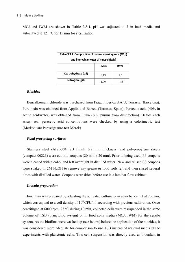
118 Mature biofilms
MCJ and IWM are shown in Table 3.3.1. pH was adjusted to 7 in both media and
autoclaved to 121 ºC for 15 min for sterilization.
Table 3.3.1: Composition of mussel cooking juice (MCJ)
and intervalvar water of mussel (IWM)
MCJ IMW
Carbohydrate (g/l) 9,19 2,7
Nitrogen (g/l) 1.70 1.85
Biocides
Benzalkonium chloride was purchased from Fragon Iberica S.A.U. Terrassa (Barcelona).
Pure nisin was obtained from Applin and Barrett (Terrassa, Spain). Peracetic acid (40% in
acetic acid/water) was obtained from Fluka (S.L. purum from disinfection). Before each
assay, real peracetic acid concentrations were checked by using a colorimetric test
(Merkoquant Peressigsäure-test Merck).
Food processing surfaces
Stainless steel (AISI-304, 2B finish, 0.8 mm thickness) and polypropylene sheets
(compact 00226) were cut into coupons (20 mm x 20 mm). Prior to being used, PP coupons
were cleaned with alcohol and left overnight in distilled water. New and reused SS coupons
were soaked in 2M NaOH to remove any grease or food soils left and then rinsed several
times with distilled water. Coupons were dried before use in a laminar flow cabinet.
Inocula preparation
Inoculum was prepared by adjusting the activated culture to an absorbance 0.1 at 700 nm,
which correspond to a cell density of 108 CFU/ml according with previous calibration. Once
centrifuged at 6000 rpm, 25 ºC during 10 min, collected cells were resuspended in the same
volume of TSB (planctonic system) or in food soils media (MCJ, IWM) for the sessile
system. As the biofilms were washed up (see below) before the application of the biocides, it
was considered more adequate for comparison to use TSB instead of residual media in the
experiments with planctonic cells. This cell suspension was directly used as inoculum in
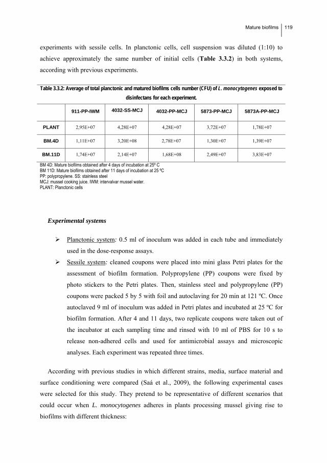
Mature biofilms
119
experiments with sessile cells. In planctonic cells, cell suspension was diluted (1:10) to
achieve approximately the same number of initial cells (Table 3.3.2) in both systems,
according with previous experiments.
Table 3.3.2: Average of total planctonic and matured biofilms cells number (CFU) of L. monocytogenes exposed to
disinfectans for each experiment.
911-PP-IWM 4032-SS-MCJ 4032-PP-MCJ 5873-PP-MCJ 5873A-PP-MCJ
PLANT 2,95E+07 4,28E+07 4,28E+07 3,72E+07 1,78E+07
BM.4D 1,11E+07 3,20E+08 2,78E+07 1,30E+07 1,39E+07
BM.11D 1,74E+07 2,14E+07 1,68E+08 2,49E+07 3,83E+07
BM 4D: Mature biofilms obtained after 4 days of incubation at 25º C BM 11D: Mature biofilms obtained after 11 days of incubation at 25 ºC PP: polypropylene. SS: stainless steel MCJ: mussel cooking juice. IWM: intervalvar mussel water. PLANT: Planctonic cells
Experimental systems
Planctonic system: 0.5 ml of inoculum was added in each tube and immediately
used in the dose-response assays.
Sessile system: cleaned coupons were placed into mini glass Petri plates for the
assessment of biofilm formation. Polypropylene (PP) coupons were fixed by
photo stickers to the Petri plates. Then, stainless steel and polypropylene (PP)
coupons were packed 5 by 5 with foil and autoclaving for 20 min at 121 ºC. Once
autoclaved 9 ml of inoculum was added in Petri plates and incubated at 25 ºC for
biofilm formation. After 4 and 11 days, two replicate coupons were taken out of
the incubator at each sampling time and rinsed with 10 ml of PBS for 10 s to
release non-adhered cells and used for antimicrobial assays and microscopic
analyses. Each experiment was repeated three times.
According with previous studies in which different strains, media, surface material and
surface conditioning were compared (Saá et al., 2009), the following experimental cases
were selected for this study. They pretend to be representative of different scenarios that
could occur when L. monocytogenes adheres in plants processing mussel giving rise to
biofilms with different thickness:
120 Mature biofilms
CECT 911 on polypropylene and intervalvar mussel water (PP-IMW). That
corresponded with the conditions that gave rise to the highest level of adherence.
Strain 4032 in polypropylene and stainless steel using MCJ as medium. This
strain showed the highest adhesion ability in both materials.
Strain 5873 in polypropylene in MCJ. It showed the lowest level of adherence out
from 16 experimental cases assayed. In this work we use this scenario to compare
the biofilms formed by this strain (5873 wild type) and the physiologically-BAC-
adapted (5873A).
Dose-response assays
In the planctonic system: 0.5ml aliquots of the biocides (benzalkonium chloride, peracetic
acid and nisin) at various doses were added to 0.5 ml of the culture.
In the case of sessile cells: after 4 and 11 days of incubation, coupons were drained and
washed as previously described before the biocide dosification. 0.5ml aliquots of the
biocides were added to the formed biofilms at various doses covering the total coupon
surface. Doses were as follows:
Benzalkonium chloride (mg/l): 2.5, 5,10, 17, 25, 50, 100, 250, 500, 1000
Peracetic acid (mg/l): 1, 2.5, 5, 10, 17, 25, 50, 100, 250, 500
Nisin (UI/ml): 5, 10, 15, 20, 50, 75, 100, 125, 150, 165, 185, 200, 400, 1500.
Several blanks with distilled water instead of antimicrobial were included in each
experimental series. All experiments were carried out at 25 ºC.
After dosage and a time of exposition of 10 minutes, the biocides had to be neutralised.
BAC and peracetic acid were neutralized by adding to a ratio 9:1 (v/v), a solution
comprising 10 ml of a buffer containing 34 g/l KH2PO4 adjusted to pH 7.2 with NaOH, 3 g/l
soybean lecithin, 30 ml/l Tween 80, 5 g/l Na2S2O3 and 1 g/l L-histidine. This solution was
applied during 1 min at room temperature, which was considered sufficient according with
preliminary experiments. Nisin was neutralized by culturing surviving cells in Nutrient Agar
previously adjusted to pH=9. The lack of effect of this pH on the growth of all the strains of
L. monocytogenes was checked in preliminary experiments.

Mature biofilms
121
After neutralization, in the sessile system, adhered cells were collected by thoroughly
rubbing with two moistened swabs and resuspended by vigorously vortexing swabs for 50 s
in 9 ml of peptone water according to Herrera et al., 2007. Then adhered cells were
determined by plating appropriate serial dilutions of this suspension on Triptone Soy Agar
and subsequent incubation at 37 ºC for 24 h. Peptone water (Cultimed, S.A.) was always
used as dilution medium.
Percentage mortality was calculated according to equation [3.3.1]:
M (%) =100*
NTVC
NVCNTVC, where: [3.3.1]
M: Percentage of death (%)
NTVC: number of total viable cells (CFU/ml) in the planctonic system or total
adhered cells (CFU/mm2) in the sessile system.
NVC: number of surviving planctonic or adhered cells after treatment with the
corresponding doses of biocides (in CFU/ml or CFU/mm2).
Determination of the resistance: calculation of Lethal Dose 90 (LD90 )
LD90 values were used as a direct measurement of the biofilm resistance to the biocides.
Values for this parameter were obtained by adjusting experimental data of mortality
obtained in the dose-response assays to the following modified logistic equation obtained
according with previous works (Cabo et al., 1999; Cabo et al., 2009):
M (%) =
111.0
1
11.01
190)90( rLDDLDr ee
k [3.3.2]
LD90: Dose that kills 90% of initial population
k: Maximum inhibition value
r: Specific inhibition coefficient (dimensions: inverse of the dose)
D: Dose
122 Mature biofilms
Microscopic analyses
Sessile cells were fixed in 2% glutaraldehyde with 0.1 M cacodylate buffer (pH 7.4) for
at least 2 h at 4º C. The fixing solution was washed out with PBS, and samples were
dehydrated with a series of increasing concentrations of ethanol: 30, 50, 70, 80 and 95% for
30 min each, and pure ethanol for 1 h. Ethanol was progressively replaced with
amyloacetate and then samples were subjected to critical point drying with CO2 (73 atm;
31.3º C). Afterwards, samples were coated with gold (10-20 nm), and later observed by
using a Philips XL30 Scanning Electronic Microscope.
Statistical analysis
The fits of the models to the experimental data were performed according to a least-
squares method (quasi-Newton). Significant differences between the obtained LD90 values
were identified by a one-way analysis of variance (Anova).
3.3.3. Results
Resistance to benzalkonium chloride (BAC) of planctonic cells and mature L.
monocytogenes biofilms
Values of the parameters (r and k) obtained after the mortality values were adjusted to
equation [3.3.2] are shown in Table 3.3.3.
We compared the LD90 values obtained for planctonic cells and maturebiofilms (4 and 11
days old) formed by different strains of L.monocytogenes (911-PP-IWM, 4032-SS-MCJ and
5873-PP-MCJ) after the exposition to BAC (Figure 3.3.1). To study whether the
physiological adaptation of L. monocytogenes to BAC results in the formation of more-
resistant mature biofilms, the strain CECT 5873A was included in this and subsequent
studies.
LD90 obtained for the planctonic cells in TSB (Table 3.3.2) were significantly (p < 0.05)
higher for the CECT 4032 strain, which was the most resistant to the disinfectants tested,
followed by the wild 5873 and 911 strains.

Mature biofilms
123
Table 3.3.3:Values of the parameters (k and r) obtained after fitting of experimental data to eq [3.3.2]:
911-PP-IWM 4032-SS-MCJ 4032-PP-MCJ 5873-PP-MCJ 5873A-PP-MCJ
BM. 4D BM.11D BM. 4D BM.11D BM. 4D BM.11D BM. 4D BM.11D BM. 4D BM.11D
KBAC 97.363 95.506 99.65 99.450 99.060 93.188 99.477 99.097 99.748 91.275
rBAC 0.308 0.1746 0.053 0.033 0.235 0.007 0.256 0.231 0.138 0.404
Knisin 99.595 99.056 86.413 91.913 84.872 87.189 69.412 83.274 76.843 89.048
rnisin 0.013 0.007 0.215 0.006 0.255 0.023 0.028 0.327 0.0730 0.029
KPA 99.342 99.810 99.704 99.736 99.998 97.457 98.976 98.560 94.353 99.883
rPA 0.330 0.419 0.169 0.069 0.480 0.097 0.439 0.263 2.816 2.255
911-PP 4032-SS 4032-PP 5873-PP 5873A-PP0
50
100
150
200600900
1200
LD
90
(m
g/l)
of
be
nza
lko
niu
m c
hlo
rid
e Planctonic cells MB-4 Days MB-11 Days
Figure 3.3.1: Values of LD90 (mg/l) obtained after the exposure of planctonic cells
and mature biofilms of L. monocytogenes to benzalkonium chloride (BAC).
A significant increase (p<0.05) in LD values (i.e. resistance) between planctonic cells and
biofilms was observed. However, whereas no significant difference was observed between
the resistance to BAC of biofilms formed by 5873 on polypropylene (PP) after 4 and 11
days, the resistance of biofilms formed by the strains 911 and 4032 increased significantly
(p<0.05) as they matured, and was quantitatively higher in the case of 4032 on both stainless
steel and polypropylene. On stainless steel (4032-SS-MCJ), the resistance (LD90 values) in

124 Mature biofilms
biofilms incubated for 4 and 11 days was multiplied by factors of 3.7 and 6 respectively with
respect to the planctonic system. On polypropylene (4032-PP-MCJ), the resistance increased
by a factor of 36 after 11 days with respect to the planctonic system.
Resistance to nisin of planctonic cells and mature L. monocytogenes biofilms
The results obtained for planctonic cells showed that strain 911 were the most sensitive to
nisin, followed by the strain 5873 adapted to BAC. Statistically similar results were obtained
for the other two strains (p < 0.05).
As for BAC, a significant increase (p<0.05) in resistance to nisin was found at both 4 and
11 days of biofilm maturation for the planctonic cells in all the assayed scenarios (Figure
3.3.2). No significant differences in LD90 values obtained for the biofilms formed in the
different scenarios after 4 days were observed. However, after 11 days of incubation,
biofilms formed by strain 4032 on stainless steel were significantly more resistant than those
formed by the same strain on polypropylene and by those formed by strain 911.
911-PP 4032-SS 4032-PP 5873-PP 5873A-PP0
100
200
300
400
500
600
LD
90
(U
I/m
l) o
f n
isin
.
Planctonic cells MB-4days MB-11days
Figure 3.3.2: Values of LD90 (mg/l) obtained after the exposition of planctonic
cells and matured biofilms of L. monocytogenes to nisin.
Biofilms formed by the strain 5873 were the most sensitive and did not show significant
differences during biofilm maturation. However, a significant increase in resistance (p <
0.05) in the BAC-adapted strain with respect to biofilms formed by the non-adapted strain
was observed after 11 days of incubation.
Beside LD90, k and r parameter values obtained from the model (Table 3.3.3) could also
be helpful for comparative purposes. In the particular case of nisin, two important
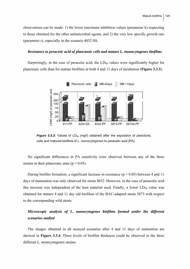
Mature biofilms
125
observations can be made: 1) the lower maximum inhibition values (parameter k) respecting
to those obtained for the other antimicrobial agents, and 2) the very low specific growth rate
(parameter r), especially in the scenario 4032-SS.
Resistance to peracetic acid of planctonic cells and mature L. monocytogenes biofilms
Surprisingly, in the case of peracetic acid, the LD90 values were significantly higher for
planctonic cells than for mature biofilms at both 4 and 11 days of incubation (Figure 3.3.3).
911-PP 4032-SS 4032-PP 5873-PP 5873A-PP0
25
50
75
100300
400
LD
90
(m
g/l)
of
pe
race
tic a
cid
Planctonic cells MB-4days MB-11days
Figure 3.3.3: Values of LD90 (mg/l) obtained after the exposition of planctonic
cells and matured biofilms of L. monocytogenes to peracetic acid (PA).
No significant differences in PA sensitivity were observed between any of the three
strains in their planctonic state (p > 0.05).
During biofilm formation, a significant increase in resistance (p < 0.05) between 4 and 11
days of maturation was only observed for strain 4032. However, in the case of peracetic acid
this increase was independent of the host material used. Finally, a lower LD90 value was
obtained for mature 4 and 11 day old biofilms of the BAC-adapted strain 5873 with respect
to the corresponding wild strain
Microscopic analysis of L. monocytogenes biofilms formed under the different
scenarios studied
The images obtained in all assayed scenarios after 4 and 11 days of maturation are
showed in Figure 3.3.4. Three levels of biofilm thickness could be observed in the three
different L. monocytogenes strains:
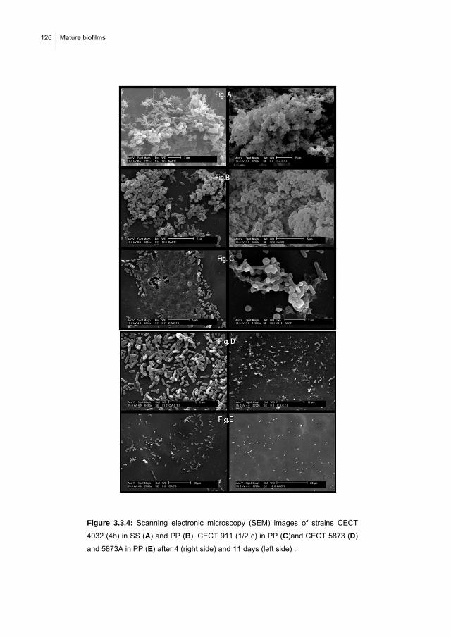
126 Mature biofilms
Figure 3.3.4: Scanning electronic microscopy (SEM) images of strains CECT
4032 (4b) in SS (A) and PP (B), CECT 911 (1/2 c) in PP (C)and CECT 5873 (D)
and 5873A in PP (E) after 4 (right side) and 11 days (left side) .
FFiigg.. CC
FFiigg.. AA
FFiigg..BB

Mature biofilms
127
Strain CECT 4032 (SS and PP) has the most complex structure, with a cloud-type
cell distribution that makes contact between the cells and the biocide more
difficult.
Strain CECT 911 shows a structure of intermediate complexity. The cells form a
monolayer at 4 days and some isolated microcolony-type formations are visible
after 11 days.
Strain CECT 5873, both the wild type and BAC-adapted (5873A), forms the
simplest structure, namely a monolayer, and shows the least resistance to the
biocides tested.
Study of the relationship between the adherence kinetics of L. monocytogenes and
mature biofilm formation
The adherence kinetics of the different L. monocytogenes strains in all assayed scenarios
are represented in Figure 3.3.5. Results showed that the strain 4032, which forms the
thickest biofilms according to the microscopic images, has a slightly (but not significant)
higher maximum level of adherence than the other two strains, with no significant
differences between polypropylene and stainless steel. No significant differences were found
between the maximum number of adhered Listeria cells in the rest of the scenarios.
A. B.
0 2 4 6 8 10 12 14
time (days)
C.
0
2
4
6
8
10
0 2 4 6 8 10 12 14
log
adh
cells
(C
FU
/mm
2).
time (days)
B
0 2 4 6 8 10 12 14
time (days)
Figure 3.3.5: Adherence kinetics of Listeria monocytogenes in different
experimental cases.
A.: CECT 911 on polypropylene (E) in the presence of intervalvar water (IWM).
B.:CECT 4032 on stainles steel (V) in the presence of cooked mussel water
(MCW) and on polypropylene (B) in MCW.
C.: CECT 5873 (C) and CECT 5873A (BAC-adapted) (H) on polypropylene in
MCW.

128 Mature biofilms
3.3.4. Discussion
The mature biofilms formed by the three strains of L. monocytogenes in different
scenarios were compared by studying the increase in resistance to three biocides
(benzalkonium chloride, peracetic acid and nisin) with SEM analysis of their
microstructures after incubation for 4 and 11 days at 25 °C.
The results showed a clear relation between the structure of the biofilms formed and the
acquisition of resistance to BAC and nisin from the planctonic state, with strain 4032
(serotype 4b) developing the thickest (“cloud shape”) and most biocide-resistant structure,
presumably as a result of reduced access to the bacterial cells. In contrast, the increase in
resistance does not appear to be correlated with the adherence levels during biofilm
formation or with each strain’s sensitivity in the planctonic state to the biocides tested.
Concerning the first, when comparing obtained results with the adherence levels previously
obtained during the initial stages of biofilm development (72 hours) (Chae et al., 2006; Saá
et al., 2009), it can be seen that the highest adherence levels were obtained by strain CECT
911 after 40 hours of incubation, whereas the initial adherence of strain CECT 4032 to
stainless steel was surprisingly low. On the contrary, the material has a specific effect on the
biofilm formed for BAC with higher resistance of biofilms formed on stainless steel,
although the opposite effect (higher resistance of biofilms formed on polypropylene) was
obtained for nisin.
Furthermore, this structure-resistance relation supports the theory that the increased
resistance associated with the formation of biofilms is due more to the population structure
in the sessile state than to the associated metabolic and physiological changes (including
quorum sensing) that occur at cellular level (Folsom et al., 2006; Kalmokoff et al., 2001).
Indeed, this hypothesis could explain the results reported by other groups (Kastbjerg and
Gram, 2009; Stopforth et al., 2002), who found no significant differences in disinfectant
sensitivity between the planctonic and sessile stages of L. monocytogenes, as is the case with
strain 5873 and BAC here.
Biofilm maturation does not, however, have the same effect on the action of peracetic
acid, with the lethal doses obtained being higher for planctonic cells in the presence of
organic matter (present in TSB). This is likely due to interference by the organic material
found in TSB, which reduces the effective concentration of this compound due to its high

Mature biofilms
129
reactivity and non-specificity. Indeed, it was found that using PBS as the culture medium led
to LD values similar to those obtained for biofilms at 4 days (data not shown).
These counterproductive effects of the organic matter highlight the importance of
effective cleaning protocols prior to disinfection to optimize the action of disinfectants, as
has been previously reported in other experimental systems (Briñez et al., 2006; Fatemi and
Frank, 1999; Gram et al., 2007; González-Fandos et al., 2005).
Moreover, the results obtained suggest that PA is more effective against biofilms than
BAC and nisin, as reported previously (Fatemi and Frank, 1999; Stopforth et al., 2002). In
fact, no significant increase in resistance (LD90 values) was observed in the 911 and 5873
scenarios during biofilm maturation, and the increase in resistance during biofilm formation
of strain 4032 was not significantly different on either of the materials. This could be due to
the physicochemical properties of PA: 1) its high reactivity and oxidizing ability, together
with its higher decomposition rate, facilitate its entry into the biofilm matrix and greatly
reduce its effective concentration in systems containing large amounts of organic matter
(such as that containing planctonic cells); and 2) its small size, which also greatly increases
its ability to penetrate the biofilm network. In fact, previous studies performed with ozone (a
molecule that also has high reactivity and a small size) in our laboratory showed that it is
more effective on Staphylococcus aureus biofilms than on planctonic cells (Cabo et al.,
2009). In contrast, the structure of the biofilm presents a significant physical barrier to the
entry of BAC and nisin, thereby reducing their ability to diffuse into the matrix and reach
the cells, although with some differences. Indeed, the results obtained highlight two reasons
why nisin is less effective than BAC for disinfecting surfaces potentially contaminated with
L. monocytogenes: 1) it acts at a slower rate, as shown by the lower r values (Table 3.3.3)
and has lower efficacy which indicates that nisin has less capacity to eliminate the entire
bacterial population, as shown by the maximum inhibition values (k parameter, Table
3.3.3), which in most cases are lower than 90%.
The different mechanisms of action of BAC and nisin (destabilization of the cytoplasmic
membrane) and PA (it is intracellular and much more non-specific) probably explain why
mature biofilms formed by the BAC-adapted strain 5873 are more resistant to BAC and
nisin than the wild strain (particularly after 11 days of incubation) and more sensitive to PA
at both 4 and 11 days of incubation. Furthermore, these results also highlight why the cross-

130 Mature biofilms
responses to adaptations acquired upon exposure of a strain to sub-lethal concentrations of
industrial disinfectants should be studied: First, studies would allow risk situations that
could arise from the use of a particular disinfectant, and which may generate positive cross-
responses (as is the case with BAC and nisin), to be identified. Second, effective treatments,
which generate a negative cross-response (as is the case with BAC and PA) could be
developed that could be used if resistance to a disinfectant used in a processing plant is
detected. Only one similar study (Stopforth et al., 2002), in which the authors found no
cross-response between the adaptation of L. monocytogenes biofilms to acid and subsequent
exposure to PA, BAC and sodium hypochlorite, has been reported in the literature.
Assuming that the increased biocide resistance is related to the ability of the adhered cells
to form three-dimensional cloud-type (strain 4032-SS-11D) or flat structures (911-4D), or
microcolonies (5873) consisting of a complicated network of bacterial cells and secreted
extracellular polysaccharides (EPSs), it becomes clear that an understanding of the
composition and structure of the extracellular matrix is essential in order to improve the
efficacy of the disinfection strategies currently used. Despite this, and although several
studies appear to indicate that the structure of L. monocytogenes biofilms differs from the
classical fungus-type model (Dubravka et al., 2009; Blaschecket al., 2007) the chemical
composition of these EPSs and their spatial distribution is unknown. Most of the studies
reported are focused on the factors that affect the cell’s ability to form biofilms, such as
variations in the fatty acid composition (Gianotti et al., 2008) or the role of N-acylated
homoserine lactones (Belval et al., 2006). Such an understanding, and the application of
treatments prior to disinfection that ensure disintegration of the extracellular matrix, would
allow current cleaning protocols to be improved, thereby increasing the efficacy of those
disinfectants that are unable to penetrate the extracellular matrix of biofilms.
The experimental procedures considered here are also an improvement on those
frequently used as they use pure active ingredients that avoid multi-resistance responses.
Moreover, setting out the results in terms of microbial kinetics equations allows the bacterial
resistance (LD90) to be quantified appropriately and an objective comparison can be made
between different test cases. The majority of studies reported in the literature use semi-
quantitative methods (MIC) and compare commercial disinfectants with unknown
compositions or mixtures of different active ingredients, which makes determining a cause-
effect relationship more difficult (Frank et al., 2003; González-Fandos et al., 2005; Jacquet
Mature biofilms
131
and Reynaud, 1994). Other studies only determine the response of the population on a short
timescale and with two or three biocide concentrations (Aarnisalo et al., 2000; Thy-Jenq et
al., 1993).
In conclusion, we have demonstrated that 1) the acquired resistance to disinfectants
during maduration of L. monocytogenes biofilms reflects the possible consequences of
biofilms rather than adherence, 2) the microscopic structure of mature biofilms correlates
with resistance to disinfectants, and 3) biofilms formed by BAC-resistant L. monocytogenes
CECT 5873 are more resistant to BAC and nisin but more sensitive to PA.

132 Mature biofilms
References
Aarnisalo, K., Lundén, J., Korkeala, H. and Wirtanen, G., 2007. Susceptibility of Listeria monocytogenes
strains to disinfectants and chlorinated alkaline cleaners at cold temperatures. Food Sci. Technol. 40,
1041-1048.
Aarnisalo, K., Salo, S., Miettinen, H., Suihko, M., Wirtanen, G., Autio, T., Lunden, J., Korkeala, H. and
Sjorberg, A., 2000. Bactericidal eficiencies of commercial disinfectants against Listeria
monocytogenes on surfaces. J. Food Saf. 20, 237-250.
Adrião, A., Vieira, M., Fernandes, I., Barbosa, M., Sol, M., Tenreiro, R.P., Chambel, L., Barata, B., Zilhao, I.,
Shama, G., Perni, S., Jordan, S.J., Andrew, P.W. and Faleiro, M.L., 2008. Marked intra-strain
variation in response of Listeria monocytogenes dairy isolates to acid or salt stress and the effect of
acid or salt adaptation on adherence to abiotic surfaces. Int. J. Food Microbiol. 123, 142-150.
Amalaradjou, M.A. and Norris, C.E. and Venkitanarayanan, K., 2009. Effect of octenidine hydrochloride on
planktonic cells and biofilms of Listeria monocytogenes. Appl. Environ. Microbiol. 75 (12), 4089-
4092.
Ammor, S., Chevallier, I., Laguet, A., Labadie, J., Talon, R. and Dufour, E., 2004. Investigation of the
selective bactericidal effect of several decontaminating solutions on bacterial biofilms including
useful, spoilage and/or pathogenic bacteria. Food Microbiol. 21, 11-17.
Belval, S.C., Gal, L., Margiewes, S., Garmyn, D., Piveteau, P. and Guzzo, J., 2006. Assessment of the roles of
LuxS, S-ribosyl homocysteine, and autoinducer 2 in cell attachment during biofilm formation by
Listeria monocytogenes EGD-e. Appl. and Environ. Microbiol. 72 (4), 2644–2650.
Beresford, M.R., Andrew, P.W. and Shama, G., 2001. Listeria monocytogenes adheres to many materials
found in food-processing environments. J. Appl. Microbiol. 90, 1000-1005.
Blackman, I. and Frank, J.F., 1996. Growth of Listeria monocytogenes as a biofilm on various food-processing
surfaces. J. Food Prot. 59 (8), 827 -831
Blascheck, H.P., Wang, H.H. and Meredith E.A (Ed.), 2007. Biofilms in the Food Environment.
Bonaventura Di, G., Piccolomini, R., Paludi, D., D'Orio, V., Vergara, A., Conter, M. and Ianieri, A., 2008.
Influence of temperature on biofilm formation by Listeria monocytogenes on various food-contact
surfaces: relationship with motility and cell surface hydrophobicity. J. Appl. Microbiol. 104, 1552-
1561.
Borucki, M.K., Peppin, J.D., White, D., Loge, F. and Call, D.R., 2003. Variation in biofilm formation among
strains of Listeria monocytogenes. Appl. Environ. Microbiol. 69, 7336-7342.

Mature biofilms
133
Briñez, W.J., Roig-Sagués, A.X., Hernández Herrero, M.M., López-Pedemonte, T. and Guamis, B., 2006.
Bactericidal efficacy of peracetic acid in combination with hydrogen peroxide against pathogenic and
non pathogenic strains of Staphylococcus spp., Listeria spp. and Escherichia coli. Food Control 17,
516-521.
Cabo, M.L., Murado, M.A., González, MªP. and Pastoriza, L., 1999. A method for quantification. J. Appl.
Microbiol. 87, 907-914.
Cabo, M.L., Torres, B., Herrera, J.J., Bernárdez, M. and Pastoriza, L., 2009. Application of nisin and pediocin
against resistance and germination of Bacillus spores in sous vide products. J. Food Prot. 73 (3), 515-
523.
Chae, M. S. and Schraft, H., 2000. Comparative evaluation of adhesion and biofilm formation of different
Listeria monocytogenes strains. Int. J. Food Microbiol. 62, 103-111.
Chae, M. S., Schraft, H., Truelstrup Hansen, L. and Mackereth, R., 2006. Effects of physicochemical surface
characteristics of Listeria monocytogenes strains on attachment to glass. Food Microbiol. 23, 250-259.
Chavant, P., Martinie, B., Meylheuc, T., Bellon-Fontaine, M. and Hebraud, M., 2002. Listeria monocytogenes
LO28: Surface physicochemical properties and ability to form biofilms at different temperatures and
growth phases. Appl. Environ. Microbiol. 68, 728-737.
Chavant, P., Folio, P and Hebraud, M., 2003. Growth of Listeria monocytogenes in biofilm according to the
surface and the temperature and effects of different treatments on its survival. Sci. aliment. 23 (1),
116-119
Chorianopoulos, N.G., Giaouris, E.D., Skandamis, P.N., Haroutounian, S.A. and Nychas, G.J., 2007.
Disinfectant test against monoculture and mixed-culture biofilms composed of technological, spoilage
and pathogenic bacteria: bactericidal effect of essential oil and hydrosol of Satureja thymbra and
comparison with standard acid–base sanitizers. J. Appl. Microbiol. 104, 1586–1596.
Djordjevic, D., Wiedmann, M. and McLandsborough, L.A., 2002. Microtiter plate assay for assessment of
Listeria monocytogenes biofilm formation. Appl. and Environ. Microbiol. 68 (6), 2950–2958.
Dubravka, M., Ružica, A., Branka, V., Krnjaić D., Jelena, P. and Sara, S., 2009.Scanning electron microscopy
of Listeria monocytogenes biofilms on stainless steel surfaces. Acta Vet., 59 (4), 423-435
Fatemi, P. and Frank, F.J., 1999. Inactivation of Listeria monocytogenes/Pseudomonas biofilms by peracid
sanitizers. J. Food Prot. 62 (7), 761-765.
Folsom, J.P., Siragusa, G.R. and Frank, J.F., 2006. Formation of biofilm at different nutrient levels by various
genotypes of Listeria monocytogenes. J. Food Prot. 69 (4), 826-834.
134 Mature biofilms
Frank, J.F., Ehlers, J. and Wicker, L., 2003. Removal of Listeria monocytogenes and poultry soil-containing
biofilms using chemical cleaning and sanitizing agents under static conditions. Food Prot. Trends 23
(8), 654-663.
Gianotti, A., Serrazanetti, D., Sado Kamdem, S. and Guerzoni, M.E., 2008. Involvement of cell fatty acid
composition and lipid metabolism in adhesion mechanism of Listeria monocytogenes. Int. J. Food
Microbiol. 123, 9-17.
González-Fandos, E., Sanz, J., García-Fernández, M.C. and García-Arias, M.T., 2005. Effectiveness of
disinfectants used in the food industry on microorganisms of sanitary interest. Acta Aliment. 34 (3),
253-258.
Gram, L., Bagge-Ravn, D., Ng, Y.Y., Gymoese, P. and Vogel, B.F., 2007. Influence of food soiling matrix on
cleaning and disinfection efficiency on surface attached Listeria monocytogenes. Food Control 18,
1165-1171.
Guerrieri, E., de Niederhäusern, S., Messi, P., Sabia, C., Iseppi, R., Anacarso, I. and Bondi, M., 2009. Use of
lactic acid bacteria (LAB) biofilms for the control of Listeria monocytogenes in a small-scale model.
Food Control 20, 861-865.
Harvey, J., Keenan, K.P., Gilmour, A., 2007. Assessing biofilm formation by Listeria monocytogenes strains.
Food Microbiol. 24, 380-392.
Herrera, J.J.R., Cabo, M.L., González, A., Pazos, I. and Pastoriza, L., 2007. Adhesion and detachment kinetics
of several strains of Staphylococcus aureus subsp. aureus under three different experimental
conditions. Food Microbiol. 24, 585-591.
Jacquet, C. and Reynaud, A., 1994. Differences in the sensitivity to eight disinfectants of Listeria
monocytogenes strains as related to their origin (short note). Int. J. Food Microbiol. 22, 79-83.
Kalmokoff, M.L., Austin, J.W., Wan, X., Sanders, G., Banerjee, S. and Farber, J.M., 2001. Adsorption,
attachment and biofilm formation among isolates of Listeria monocytogenes using model conditions.
J. Appl. Microbiol. 91, 725-734.
Kastbjerg, V.G. and Gram, L., 2009. Model systems allowing quantification of sensitivity to disinfectants and
comparison of disinfectant susceptibility of persistent and presumed nonpersistent Listeria
monocytogenes. J. Appl. Microbiol. 106, 1667-1681.
Krysinski, E.P. and Brown, L.J., 1992. Effect of cleaners and sanitizers on Listeria monocytogenes attached to
product contact surfaces. J. Food Prot. 55 (4), 246 -251

Mature biofilms
135
Leriche, V., Chassaing, D. and Carpentier, B., 1999. Behaviour of L. monocytogenes in an artificially made
biofilm of a nisin-producing strain of Lactococcus lactis. Int. J. Food Microbiol. 51, 169-182.
Minei, C.C., Gomes, B.C., Regianne, P.R., D’ Angelis, C.E.M. and Martinis De, E.C.P., 2008. Influence of
peroxyacetic acid and nisin and coculture with Enterococcus faecium on Listeria monocytogenes
biofilm formation. J. Food Prot. 71, 634.
Moltz, A.G. and Martin, S.E., 2005. Formation of biofilms by Listeria monocytogenes under various growth
conditions. J. Food Prot. 68 (1), 92-97.
O’Driscoll, B., Gaham, C.G.M. and Hill, C., 1996. Adaptative acid tolerance response in Listeria
monocytogenes: isolation of an acid-tolerant mutant which demonstrated increased virulence. Appl.
Environ. Microbiol. 62, 1693-1698.
Oliveira, M.M.M.d., Brugnera, D.F., Cardoso, M.d.G., Alves, E. and Piccoli, R.H., 2010. Disinfectant action of
Cymbopogon sp. essential oils in different phases of biofilm formation by Listeria monocytogenes on
stainless steel surface. Food Control 21, 549-553.
Palmer J., Flint, S. and Brooks J., 2007. Bacterial cell attachment, the beginning of a biofilm. J. Ind. Microbiol.
Biotechnol. 34(9), 577–588.
Pan, Y., Breidt, F., Jr. and Kathariou, S., 2006. Resistance of Listeria monocytogenes biofilms to sanitizing
agents in a simulated food processing environment. Appl. Environ. Microbiol. 72, 7711-7717.
Rieu, A., Briandet, R., Habimana, O., Garmyn, D., Guzzo, J. and Piveteau, P., 2008. Listeria monocytogenes
EGD-e biofilms: no mushrooms but a network of knitted chains. Appl. Environ. Microbiol. 74 (14),
4491-4497.
Rieu A., Guzzo J.and Piveteau P., 2010. Sensitivity to acetic acid, ability to colonize abiotic surfaces and
virulence potential of Listeria monocytogenes EGD-e after incubation on parsley leaves. J. Appl.
Microbiol. 108 (2), 560-570.
Rodríguez, A., Autio, W.R. and McLandsborough, L.A., 2008. Effect of surface roughness and stainless steel
finish on Listeria monocytogenes attachment and biofilm formation. J. Food Prot. 71 (1), 170-175.
Saá, P., Cabo, M.L. and Rodríguez, J.J., 2009. Effects of mussel processing soils on the adherence of Listeria
monocytogenes to polypropylene and stainless steel. J. Food Prot. 72, 1885-1890.
Sandasi, M., Leonard, C.M. and Viljoen, A.M., 2008. The effect of five common essential oil components on
Listeria monocytogenes biofilms. Food Control 19, 1070-1075.

136 Mature biofilms
Sandasi, M., Leonard, C.M. and Viljoen, A.M., 2010. The in vitro antibiofilm activity of selected culinary
herbs and medicinal plants against Listeria monocytogenes. Lett. Appl. Microbiol. 50 (1), 30-35.
Somers, E.B. and Wong, A.C., 2004. Efficacy of Two Cleaning and Sanitizing Combinations on Listeria
monocytogenes Biofilms Formed at Low Temperature on a Variety of Materials in the Presence of
Ready-to-Eat Meat Residue. J. Food Prot. 67 (10), 2218-2229.
Smoot, L.M. and Pierson, M.D., 1998. Effect of environmental stress on the ability of Listeria monocytogenes
Scott A to attach to food contact surfaces. J. Food Prot. 61 (10), 1293-1298.
Stoporth, J.D., Samelis, J., Sofos, J.N., Kendall, P.A. and Smith, G.C., 2002. Biofilm formation by acid-
adapted and nonadapted Listeria monocytogenes in fresh beef decontamination washings and its
subsequent inactivation with sanitizers. J. Food Prot. 65 (11), 1717-1727.
Tajkarimi, M.M., Ibrahim, S.A. and Cliver, D.O., 2010. Antimicrobial herb and spice compounds in food.
Food Control 21, 1199-1218.
Takahashi, H, Suda, T, Tanaka, Y. and Kimura, B., 2010. Cellular hydrophobicity of Listeria monocytogenes
involves initial attachment and biofilm formation on the surface of polyvinyl chloride. Lett. Appl.
Microbiol. 50, 618-625.
Tyh-Jenq Ren and Frank, J. H., 1993. Susceptibility of starved planktonic and biofilm Listeria monocytogenes
to quaternary ammonium sanitizer as determined by direct viable and agar plate counts. J. Food Prot.
56 (7), 573-576.
Yang, H., Kendall, P.A., Medeiros, L.C. and Sofos, J.N., 2009. Efficacy of sanitizing agents against Listeria
monocytogenes biofilms on high-density polyethylene cutting board surfaces. J Food Prot. 72 (5),
990-998
Zhao, T., Doyle, M.P. and Zhao, P., 2004. Control of Listeria monocytogenes in a biofilm by competitive-
exclusion microorganism. Appl. and Environ. Microbiol. 70, 3996–4003.

33..44.. Adherence kinetics, resistance to
benzalkonium chloride and microscopic
analysis of mixed biofilms formed by L.
monocytogenes and Pseudomonas putida

Mixed biofilms
139
3.4 ADHERENCE KINETICS, RESISTANCE TO BENZALKONIUM
CHLORIDE AND MICROSCOPIC ANALYSIS OF MIXED BIOFILMS
FORMED BY LISTERIA MONOCYTOGENES AND PSEUDOMONAS
PUTIDA.
Comparison between the resistance to BAC and the microscopic structure of mixed-species
biofilms formed by different strains of L. monocytogenes and Pseudomonas putida CECT 845
under different scenarios and these obtained by the corresponding mono-species L.
monocytogenes biofilm was carried out. The association of P. putida with L. monocytogenes
quickens biofilm formation and increases significantly (p<0.05) the BAC-resistance of the
biofilm after 4 days of incubation at 25 ºC respect to monospecies biofilms. According with
the adherence profiles of P. putida, two different patterns of association (A and B) were
observed, being type A pattern much more resistant to BAC. After 11 days of incubation, in 2
out of 5 experimental cases (4032 and BAC-adapted 5873 on polypropylene) a destructuration
of mixed biofilms occurred, being accompanied by a sharp decrease in the number of adhered
cells. Microscopic analyses demonstrated that the most complex three-dimensional structure
showed the highest resistance to BAC (4032-SS). Results clearly highlight that it is necessary
to consider natural bacterial association for improving the safety effectiveness of disinfection
protocols.

140 Mixed biofilms
3.4.1. Introduction
Listeria monocytogenes is a pathogenic bacterium implicated in numerous foodborne
outbreaks (Denny et al., 2008; Gilmour et al., 2010). It is widely recognized that one of the
most frequent contamination routes of L. monocytogenes in foods is through cross
contamination from surfaces in food processing plants (Ammor et al., 2004; Norwood and
Gilmour, 1999; Porsby et al., 2008), where it is able to attach and form resistant biofilms
(Aase et al., 2000; To et al., 2002; Takahashi et al., 2009). This resistance is related to the
three-dimensional structure of the biofilm, which defines the interactions between the
bacterial species (Wuertz et al., 2004) and determines the degree to which chemicals can
access the cells (Bourion and Cerf, 1996, Qu et al., 2010). Moreover, it has been
demonstrated that biofilm cells are phenotypically different from the corresponding
planktonic cells (Nadell et al., 2008).
But the situation is even more complicated, as it is known that more than two species
coexist in real biofilms and it has been demonstrated that this could increase their resistance
to biocides (Sharma and Anand, 2002; Kastbjerg and Gram, 2009). Some studies on L.
monocytogenes have analyzed the attachment of multispecies biofilms involving this
bacterium, with different results. It has been found that in some cases the association with
other bacteria increases the number of adhered cells in the mixed biofilm: L. monocytogenes
with Pseudomonas (Sasahara and Zottola, 1993; Marshall and Smimidt, 1988; Hassan et al.,
2003) or with Flavobacterium (Bremer et al., 2001). However, other studies have found that
the number of adhered cells in the mixed-biofilm decreased: L. monocytogenes with
Pseudomonas fragi and Staphylococcus xylosus (Norwood and Gilmour, 2001); with
Staphyloccocus sciuri (Leriche et al., 2000); and with E. faecium (Minei et al., 2008). Lastly,
some studies found that the number of adhered cells did not vary due to the presence of other
species in the biofilm: L. monocytogenes with Staphyloccocus aureus (Rieu et al., 2008),
except for one strain; and with Salmonella enterica and Ps. putida (Chorianopulos et al.,
2007).
Although it is recognized that bacteria in nature form complex mixed-species communities,
the European standard EN 13697 points out that adhered cells should be obtained by
inoculation of the target microorganism and subsequent drying for testing the bactericidal
activity of chemical disinfectants. However, following this procedure, the effectiveness of

Mixed biofilms
141
disinfectants will be overestimated. Even though there are few studies on disinfection against
mixed biofilms (Bourion and Cerf, 1996; Bremer et al., 2002; Chlorianopoulous et al., 2007;
Fatemi and Frank, 1999; Lebert et al., 2009; Minei et al., 2007), thus, to improve the efficacy
of disinfection systems commonly used it is necessary to continue studying the repercussions
of the increased resistance to disinfectants related to the association between L.
monocytogenes and other bacterial genera that are normally present in the environment, such
as Pseudomonas species.
Thus, the main aim of this work was to study the association between L. monocytogenes
and Pseudomonas putida by comparing the resistance to BAC and the microscopic structure
of the dual-species biofilm with that of L. monocytogenes mono-species biofilms.
3.4.2. Material and methods
Bacterial strains
Listeria monocytogenes, CECT 911, CECT 4032, CECT 5873 and Ps. putida 845 were
provided by the Spanish Type Culture Collection (Valencia). Strain 5873 was adapted to BAC
in previous experiments after two exposures of the wild type strain 5873 to sub-lethal
concentrations of BAC (wild strain LD50: 2.71 mg/l; BAC-adapted strain LD50: 7.22 mg/l, see
section 3.2). Strains were frozen and stored at -80 ºC in TSB containing 50% glycerol (v/v)
until use. Whenever required, one cryovial was thawed and subcultured twice in TSB
(Cultimed, S.L., Spain) at 37 ºC or at 31º C for activation of Listeria monocytogenes and
Pseudomonas putida, respectively.
Food soils media
In order to simulate contamination of surfaces in plants processing cooked mussels and
live mussel, mussel cooking juice (MCJ) and intervalvar water of mussel (IWM) were used as
culture media (Saá Ibusquiza et al. 2010, see section 3.2). The composition of MCJ and IWM
is shown in Table 3.3.1.

142 Mixed biofilms
Disinfectant
Benzalkonium chloride was purchased from Fragon Iberica S.A.U. Terrassa (Barcelona).
Food processing surfaces
Stainless steel (SS, AISI-304, 2B finish, 0.8 mm thickness) and polypropylene sheets (PP,
compact 00226) were cut into coupons (20 mm x 20 mm). PP coupons were cleaned with
alcohol and left overnight in distilled water. New and reused SS coupons were soaked in 2M
NaOH to remove any grease or food soils left and then rinsed several times with distilled
water. Coupons were dried in a laminar flow cabinet.
Inocula preparation
Inoculum of each strain (Listeria and Pseudomonas) was prepared by adjusting the
activated culture to an absorbance 0.1 at 700 nm, centrifuged (6000 rpm, 10 min, 25 ºC) and
the pellet was re-suspended in the same volume of the corresponding residual media (MCJ or
IWM). To form mixed-species biofilms, obtained pellets of each strain were resuspended in
half of the total centrifuged volume, thus achieving the same final concentration of P. putida
and L. monocytogenes cells, which correspond to a cell density of 108 CFU/ml of each species
according to previous calibrations.
Experimental systems
Cleaned coupons were placed into mini glass Petri plates for the assessment of biofilm
formation. Polypropylene (PP) coupons were fixed by photo stickers to the Petri plates. Then,
cleaned stainless steel or polypropylene (PP) coupons were packed 5 by 5 with foil and
autoclaved for 20 min at 121 ºC. Once autoclaved, 9 ml of cell suspension was added to Petri
plates and incubated at 25 ºC for biofilm formation. After 4 and 11 days, two replicate
coupons were taken out of the incubator at each sampling time and rinsed with 10 ml of PBS
for 30s to release non-adhered cells and used for antimicrobial assays and microscopic
analyses. Each experiment was repeated three times.
According with previous studies in which different strains, media, surface material and
surface conditioning were compared (Saá et al., 2009, Saá Ibusquiza et al., 2010, see sections
3.1 and 3.2 respectively of this thesis) the following experimental cases were assayed. They

Mixed biofilms
143
pretend to be representative of different scenarios that can occur when L. monocytogenes
adheres in plants processing mussel giving rise to biofilms with different thicknes:
CECT 911 on polypropylene and intervalvar mussel water (PP-IMW). It
corresponds with the conditions that gave rise to the highest level of adherence.
Strain 4032 on polypropylene and stainless steel using MCJ as medium. This strain
showed the highest adhesion ability on both materials.
Strain 5873 on polypropylene in MCJ. It showed the lowest level of adherence out
of the 16 experimental cases assayed. In this work we use this scenario to compare
the biofilms formed by this strain (5873 wild type) and the physiologically-BAC-
adapted (5873A).
Determination of the number of adhered cells
First of all, coupons were drained and immediately immersed in 10 ml of PBS during 10s
to release non-adhered cells. The number of adhered cells was determined according to
Herrera et al. 2007. They were collected by thoroughly rubbing with two moistened swabs
and resuspended by vigorously vortexing swabs for 50 s in 9 ml of peptone water (Cultimed,
S.A.) and subsequent plating. The number of adhered cells of P. putida was determined by
plating the prepared dilutions in Violet red bile agar with glucose (VRBG, Cultimed S.L.) and
incubation at 37 ºC during 24 hours. Number of adhered cells of L. monocytogenes was
determined by plating in Palcam Agar (Liofilchem, S.L.R., Italia) and incubation at 37 ºC
during 48 hours before counting.
Inactivation assays
After 4 and 11 days of incubation, coupons were drained and inmediately inmersed in 10
ml of PBS for 10 s to release non-adhered cells before the addition of biocide.
After PBS was drained, 0.5 ml aliquots of benzalkonium chloride were added to the
formed biofilms at various doses (mg/l): 2.5, 5, 10, 17, 25, 50, 100, 250, 500, 1000.
Several blanks with distilled water instead of BAC were included in each experimental
series. All experiments were carried out at 25 ºC.
144 Mixed biofilms
After the addition of BAC and a time of exposure of 10 minutes, the effect of BAC was
neutralized by adding a solution comprising 10 ml of a buffer containing 34 g/l KH2PO4
adjusted to pH 7.2 with NaOH, 3 g/l soybean lecithin, 30 ml/l Tween 80, 5 g/l Na2S2O3 and 1
g/l L-histidine , to a ratio 9:1 (v/v). Neutralizing solution was applied during 1 min at room
temperature according with preliminary experiments. After neutralization, adhered cells of P.
putida and L. monocytogenes were collected and determined as previously described.
Finally, percentage of mortality was calculated according to equation [3.3.1]:
M (%) =100*
NTAC
NACNTAC, where: [3.3.1]
M: Mortality (%)
NTAC: number adhered cells (CFU/mm2) in biofilms without treatment.
NAC: number of adhered cells after treatment with the corresponding doses of BAC
(CFU/mm2).
Determination of the Lethal Dose 90 (LD90).
LD90 values were obtained by fitting experimental data of mortality obtained in the dose-
response assays to the following modified logistic equation obtained according with previous
works (Cabo et al., 1999; Cabo et al., 2009):
M (%) =
111.0
1
11.01
190)90( rLDDLDr ee
k [3.3.2]
LD90: Dose that kills 90% of initial population.
k: Maximum BAC inhibition value (asymptote).
r: Specific inhibition coefficient (dimensions: inverse of the dose).
D: dose.

Mixed biofilms
145
Microscopic analyses
Cells were fixed in 2% glutaraldehyde with 0.1 M cacodylate buffer (pH 7.4) for at least 2
h at 4 ºC. The fixing solution was washed out with PBS, and samples were dehydrated with a
series of increasing concentrations of ethanol: 30, 50, 70, 80 and 95% for 30 min each, and
pure ethanol for 1 h. Ethanol was progressively replaced with amyloacetate and then samples
were subjected to critical point drying with CO2 (73 atm; 31.3 ºC). Afterwards, samples were
coated with gold (10-20 nm), and finally observed by using a Philips XL30 Scanning
Electronic Microscope.
Statistical analysis
Fits of the model to the experimental data were performed according to a least-squares
method (quasi-Newton). A Student´s test (α=0.05) was used to test the significance of the
differences between means of the total number of adhered cells and between the lethal doses
values obtained in each experimental case.
3.4.3. Results
Comparison between the adherence of L. monocytogenes in monoculture and in the
presence of Pseudomonas putida CECT 845
Adherence kinetics of 3 strains of L. monocytogenes (CECT 911, 4032 and 5873) in
monoculture and co-cultured with P. putida were compared in different scenarios, which were
selected according to previous studies (Saá et al. 2009; Saá Ibusquiza et al. 2010 see sections
3.1 and 3.3 respectively of this thesis).
Slightly, higher adherence levels were reached by L. monocytogenes in a monoculture
compared with a mixed culture of L. monocytogenes and P. putida at the beginning of
incubation (compare open and closed symbols in Figure 3.4.1). Whereas adherence of L.
monocytogenes in monoculture remained constant or even increased (as in 4032) after the
plateau phase was reached, a sharp decrease in the number of adhered cells was detected for
strains 4032 and BAC-adapted 5873 on polypropylene in the presence of P. putida. In the
latter strain, adherence decreased to almost undetectable values after 14 days of incubation.

146 Mixed biofilms
A. B.
0 2 4 6 8 10 12 14
time (days)
C.
0
1
2
3
4
5
6
7
0 2 4 6 8 10 12 14
log
adh
cell
s (C
FU/m
m2)
.
time (days)
0 2 4 6 8 10 12 14
time (days)
D. E.
0 2 4 6 8 10 12 14
time (days)
0 2 4 6 8 10 12 14
time (days)
Figure 3.4.1: Adhesion kinetics of different Listeria monocytogenes in monoculture (O)
and in the presence of Pseudomonas putida 845 (●).
A.: CECT 911 on polypropylene in the presence of intervalvar water (IWM),
B.: CECT 4032 on stainless steel in the presence of cooked mussel water (MCW),
C.: CECT 4032 on polypropylene in MCW, D: CECT 5873 on polypropylene in MCW and
E.: CECT 5873A (BAC-adapted) on polypropylene in MCW.
Observed (*) and expected (continuous line) adhesion kinetics of Pseudomonas putida
in mixed biofilms are also included in these graphs.
Although modelling the experimental results could be helpful for comparing the adherence
of L. monocytogenes in the different scenarios it was not feasible, because there were no
experimental points before 4 days of incubation. However, in the case of P. putida, a
satisfactory fit (r2=0,995) to a two-term additive logistic-type model [3.4.3] that adequately
describes the increase and decrease of biofilm population (Herrera et al. 2007; Saá et al. 2009,
see section 3.1) was obtained (Figure 3.4.1, Table 3.4.1).
Table 3.4.1: Values for the parameters obtained after fitting experimental results of adhesion of Ps. putida in
presence of different L. monocytogenes strains to equation [3.4.3]
911-PP-IWM 4032-SS-MCJ 4032-PP-MCJ 5873-PP-MCJ 5873-PP-MCJ
aad 30499 1800000 370723 199237 45000
rad 5,014 2,974 1,101 3,204 5,014
mad 0,700 1,843 0,998 1,881 0,700
aD 26244 1497876 268936 184311 26244
rD 10,425 5,500 4,173 4,965 8,500
mD 1,676 4,500 1,820 51,632 8,000
PP: polypropylene. SS: stainless steel MCJ: mussel cooking juice. IMW: intervalvar water.

Mixed biofilms
147
DDDDadadadad
Dadmr)tm(rmr)tm(r
e1
1
e1
a
e1
1
e1
aNAC
[3.3.3],
where,
NAC: number of adhered cells, log (CFU/mm2).
aad: maximum number of adhered cells (asymptote).
rad: specific adhesion coefficient (t-1).
mad: time at which the number of adhered cells is half of the maximum.
aD: total number of adhered cells which detach or die, log (CFU/mm2).
rD: specific detachment/death coefficient (t-1).
mD: time at which the number of detached/death cells is half of the maximum.
Analysis of the adjusted experimental data permitted to distinguish two different adherence
patterns for P. putida that gave rise to two different patterns of association with L.
monocytogenes:
Type A.: This pattern was observed in the associations between P. putida and
strains 4032 (in both materials) and the wild type strain 5873 (Fig. 3.4.1.B., C., D.
respectively) This type has a sharp initial increase in the adherence level that
permits higher maximum adherence levels (compare the parameters aad and rad
shown in Table 3.4.2) to be obtained compared to type B association. The increase
in adherence of P. putida was especially clear with strain 4032 on SS, in which a 2-
log increase in the maximum level with respect to that obtained for the type B
pattern was observed.
Type B: This type has a flatter adherence profile that implies a 1-log decrease in the
maximum adherence levels of P. putida with respect to those obtained in pattern
type A. This profile occurs between P. putida and strains 911 and BAC-adapted
5873 (Fig. 3.4.1.A., E. respectively). Conversely, in these experimental cases a
slightly higher adherence level of L. monocytogenes was observed.
As we will see further, both types of association patterns give rise to mature mixed-species
biofilms (after 4 days) with different levels of resistance to benzalkonium chloride.

148 Mixed biofilms
Comparison between the resistance to BAC of monospecies L. monocytogenes biofilms
and dual-species biofilms formed from the association between L. monocytogenes and P.
putida
The comparison of the LD90 of BAC obtained in all assayed scenarios demonstrated a
significant variation in the resistance to BAC of the dual-species biofilms after 4 and 11 days
of incubation with respect to the single-species biofilms. This indicates that the presence of P.
putida modifies the L. monocytogenes biofilm structure, but in different ways after 4 and 11
days of incubation:
After 4 days a significant increase (p<0.05) in the resistance to BAC was observed in the
dual-species biofilms formed by L. monocytogenes and P. putida in all experimental cases
(Figure 3.4.2). However, quantitative differences were observed depending on the type of
association pattern between the strains. Thus, in the biofilm formed by P. putida and L.
monocytogenes 4032-SS (type A), the LD90 value (i.e. resistance to BAC) increased until a
factor of around 6; however, in the cases that showed a type B pattern, the resistance was
multiplied by a maximum factor of 2. Specially significant is the case of L. monocytogenes
5873 (WT), whose association with P. putida permits the formation of resistant biofilms at 25
ºC.
After 11 days of incubation the situation was completely different: a significant reduction
in BAC resistance was shown in all mixed biofilms formed by L. monocytogenes and P.
putida. Moreover, a significant decrease in the resistance of mixed-species biofilms compared
with that obtained for mono-species biofilms was observed in all cases except for 4032-SS
and 5873 (wt).
It seems, then, that destabilization of mixed-species biofilms occurred in all experimental
assays. However, only for 4032 and BAC-adapted 5873 strain on polypropylene this
destructuration is accompanied by a sharp decrease in the number of adhered cells (Figure
3.4.1.C. and E. respectively). Furthermore, for 5873, the results obtained demonstrated that
BAC-adaptation hindered its association capacity with P. putida, giving rise to a mixed-
species biofilm that was significantly (p<0.05) less resistant to BAC and which had a higher
biofilm destabilization rate than the corresponding wild type. Lastly, in the case of 911-IWM,
a 1-log decrease in the number of adhered cells between 4 and 11 days of incubation was
observed (Figure 3.4.1).
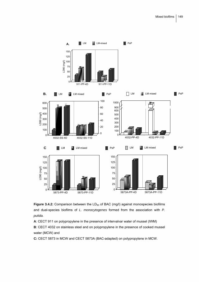
Mixed biofilms
149
911-PP-4D 911-PP-11D0
25
50
75
100
125
150
LD
90 (
mg/
l)
LM LM-mixed PsPA.
4032-PP-4D 4032-PP-11D0
100
200
300
400
500
600900
1000
LM LM mixed PsP
4032-SS-4D 4032-SS-11D0
100
200
300
400
500
600
0
20
40
60
80
100
LD
90
(m
g/l)
LM LM mixed PsP
*
B.
5873-PP-4D 5873-PP-11D0
25
50
75
100
125
150
LD
90
(m
g/l)
LM LM mixed PsP
5873A-PP-4D 5873A-PP-11D0
25
50
75
100
125
150
LM LM mixed PsPC .
Figure 3.4.2: Comparison between the LD90 of BAC (mg/l) against monospecies biofilms
and dual-species biofilms of L. monocytogenes formed from the association with P.
putida.
A: CECT 911 on polypropylene in the presence of intervalvar water of mussel (IWM)
B: CECT 4032 on stainless steel and on polypropylene in the presence of cooked mussel
water (MCW) and
C: CECT 5873 in MCW and CECT 5873A (BAC-adapted) on polypropylene in MCW.
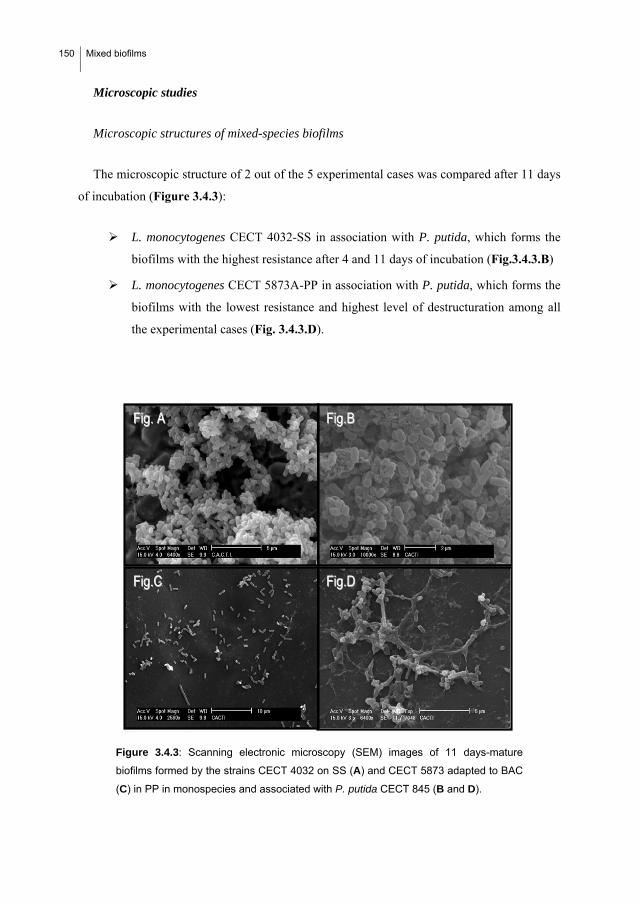
150 Mixed biofilms
Microscopic studies
Microscopic structures of mixed-species biofilms
The microscopic structure of 2 out of the 5 experimental cases was compared after 11 days
of incubation (Figure 3.4.3):
L. monocytogenes CECT 4032-SS in association with P. putida, which forms the
biofilms with the highest resistance after 4 and 11 days of incubation (Fig.3.4.3.B)
L. monocytogenes CECT 5873A-PP in association with P. putida, which forms the
biofilms with the lowest resistance and highest level of destructuration among all
the experimental cases (Fig. 3.4.3.D).
Figure 3.4.3: Scanning electronic microscopy (SEM) images of 11 days-mature
biofilms formed by the strains CECT 4032 on SS (A) and CECT 5873 adapted to BAC
(C) in PP in monospecies and associated with P. putida CECT 845 (B and D).
FFiigg..DD FFiigg..CC
FFiigg..BB FFiigg.. AA

Mixed biofilms
151
The SEM images obtained of the biofilm formed by L. monocytogenes CECT 4032-SS
showed that it had a similar spatial cell arrangement in both the absence (Figure 3.4.3.A) and
in the presence (Figure 3.4.3.B) (Saá Ibusquiza et al., 2010, see section 3.3) of P. putida. In
fact, a cloud-type structure can be observed in both cases.
As a consequence, the LD90 values were similar (155.64 mg/l of BAC in the monospecies
biofilms and 166.51 mg/l of BAC in the mixed-species biofilm). However, a monolayer-
biofilm without a three-dimensional cell arrangement was formed by the BAC-adapted 5873
strain both alone (Fig. 3.4.3.C) and associated with P. putida (Figure 3.4.3D), and low values
of LD90 were obtained (32.77 and 20.69 mg/l) in monospecies and mixed-species biofilms
respectively). By the way, cells with a rough plasmatic membrane were observed in the mixed
biofilms formed by BAC-adapted cells of L. monocytogenes CECT 5873.
Figure3.4.4: Scanning electronic microscopy (SEM) images of 11 days-mature mixed
species biofilms formed by CECT 4032 on SS (A) and in PP (B) with P. putida.
FFiigg..AA
FFiigg..BB
152 Mixed biofilms
On the other hand, Figure 3.4.4 showed a clear effect of material on the association
between P. putida and L. monocytogenes CECT 4032, being the destructuration of the mixed
biofilm after 11 days of maturation significantly higher in PP (Figure 3.4.4B) than on
stainless steel (Figure 3.4.4A)
Microscopic analysis of the biofilm after disinfection
A picture of the mixed 11-day matured biofilm formed by strain 4032 was taken after it
was exposed to 100 mg/l of BAC for 10 minutes (Figure 3.4.5). Curiously, whereas a low
number of L. monocytogenes cells were observed after the treatment, exopolysaccharide
matrix remained attached to the stainless steel
Figure 3.4.5: Scanning electronic microscopy (SEM) images of residues left after
treatment of biofilms formed by the strain CECT 4032 and P. putida on SS with 100
mg/l of BAC.
3.4.4. Discussion
The results obtained demonstrate that the presence of Pseudomonas putida in L.
monocytogenes biofilms significantly increases their resistance to BAC with respect to the
resistance of monospecies L. monocytogenes biofilms after 4 days of incubation.

Mixed biofilms
153
These results are in agreement with those obtained by previous authors, who found a
smaller log decrease for mixed L. monocytogenes biofilms exposed to disinfectants compared
to the corresponding monospecies biofilm (Ammor et al., 2004).
The presence of Pseudomonas putida quickens the biofilm maturation process compared to
the corresponding monospecies L. monocytogenes biofilm. A possible explanation for this
could be that the exopolysaccharide matrix secreted by Pseudomonas could facilitate the
adhesion of L. monocytogenes, as has been previously stated by other authors (Nadell et al.,
2009). However, lower adherence levels of L. monocytogenes in the mixed biofilm were
observed in this study, which could be due to competitive effects (specific or non-specific,
Jameson effects) between the two strains during biofilm formation (Mellefont et al., 2008).
This has been previously demonstrated in other bacterial species (Chorianopoulos et al.,
2007).
Nonetheless, although after 4 days of incubation a significant increase in the resistance to
BAC is observed when P. putida is present in the biofilm, in those cases in which an initial
sharp increase in the adherence of P. putida was observed (type A), 4032-SS-PP and 5873-
PP, the increase in resistance was quantitatively higher than in the other two cases. It seems
that when experimental conditions imply that P. putida has a higher affinity for the surface
compared to L. monocytogenes, a favourable association between the two species occurs, at
least with respect to resistance to BAC. Specially in the case of the wild type 5873 strain, in
which the resistance to BAC increased from 20.51 mg/l to 130 mg/l in the absence and
presence of P. putida respectively. However, the modifications derived from BAC adaptation
in the same strain gave rise to a mixed biofilm with P. putida that is considerably less
resistant and has a higher detachment rate.
After 11 days of incubation, a significant decrease in the resistance to BAC with respect to
that obtained after 4 days was observed. This was probably related to the destructuration of
the mixed-species biofilm observed in all experimental cases. However, only in 2 (4032-PP
and 5873A) out of the 5 experimental cases assayed was this destructuration accompanied by
a drastic decrease in the number of adhered cells. In the case of 4032-PP, the decrease in the
adhered L. monocytogenes cells is already observed from day 4 of incubation (Figure 3.4.1).
This contrasts with its good capacity to form biofilms and its BAC resistance value. This

154 Mixed biofilms
could be explained by the detachment of the cells in clusters and the high resistance of the
remaining adhered cells (Stoodley et al., 2001).
Although detachment of cells from monospecies biofilm has been previously described by
several authors (S.aureus: Herrera et al., 2007, P. aeruginosa: Boles et al., 2005, B. cereus:
Wijman et al., 2007), the reasons are still unclear. Some authors related it to nutritional
(Marshall, 1988) and gas transference restrictions (Applegate and Bryers, 1991). This does
not agree with our results, in which the low level of destructuration observed in 4032-SS and
5873-PP contrast with the high biomass concentration and the consequent nutritional and
respiratory restrictions. Others authors believed that EPSs can be used as nutrients by the
cells, making it easier for starving cells to detach from the biofilm (Takhistov and George,
2004). However, it could also be the result of competitive interactions between L.
monocytogenes and P. putida during biofilm formation (Simões et al., 2007). Also, Rodríguez
et al., 2007 believe that after drying, Listeria biofilms weakens adhesion forces and it causes
cell dettachment. In fact, in our experiments with mixed L. monocytogenes-P.putida biofilms,
progressive water lost of the media that initially covered the coupons was also observed.
Additionally, different detachment regulating systems have been demonstrated in various
bacterial species that could specifically disrupt the external matrix (EPS) of the biofilms
(Sauer et al., 2004; Liu et al., 2007; Boles et al., 2005).
In the other three cases, the decrease in BAC resistance (which probably reflects biofilm
destructuration) was not accompanied by a drastic decrease in the number of adhered cells,
which was only slight during the incubation time. One possible explanation for this could be
that even a low detachment level during biofilm formation is enough to increase the
accessibility of BAC to L. monocytogenes cells, but insufficient to be reflected in a decrease
in the total number of adherent cells.
Another interesting observation is that LD90 values of BAC obtained for dual-species
biofilms were close to those obtained for P. putida biofilms in the association type A. This
suggests that when two or more strains are integrated in a single biofilm, the effectiveness of
the disinfectant seems to depend mainly on the cell arrangement in the final structure, being
less relevant the particular sensitivity of the individual species involved.
The microscopic analysis demonstrated the risk associated to the formation of a three-
dimensional structure by adhered pathogenic bacteria. The best example is that biofilms

Mixed biofilms
155
formed by the strain 4032 on SS, which showed similar cell arrangement (Figure 3.4.3A and
3.4.3B) and similar values of BAC resistance, 155.64 mg/l and 166.51 mg/l, in monospecies
and mixed-species biofilm, respectively (Figure 3.4.2).
Additionally, results clearly demonstrated an effect of the surface on biofilm formation of
L. monocytogenes CECT 4032. If we compare the LDs (Figure 3.4.2) and the microscopic
structure of strain 4032 cells attached in the presence of P. putida on SS and PP (Figures
3.4.4A and 3.4.4B), it can be observed that “cloud” shaped structures formed by the mixed
biofilm on stainless steel (LD90 =166.51 mg/l of BAC) appeared as microcolonies in the case
of polypropylene (LD90= 49.97 mg/l of BAC), which demonstrates that the two strains have a
high affinity for stainless steel. In fact, P. putida reached a 1-log higher maximum adherence
level on stainless steel than on a polypropylene surface (aAD in SS: 1800000; aAD in PP:
370323). Lastly for this strain, the microscopic analysis of the biofilm after disinfection
showed that the exopolysaccharide matrix remained attached to the stainless steel whereas the
number of remaining cells were low. This matrix could function as a trap for new colonizing
cells that would recontaminate the surface and form another mature biofilm (Pan et al. 2006).
Furthemore, rough plasmatic membrane observed in the mixed biofilms formed by BAC-
adapted cells of L. monocytogenes CECT 5873 (See Figure 4.4.3D) could be due to the
inactive cell metabolism as a consequence of nutrient starvation, membrane modifications
associated with BAC-adaptation or both of these.
Although when the LD90 values of monospecies and mixed-species biofilms are compared
it seems to be clear that each strain has an inherent capacity to form biofilms, the results
obtained demonstrate that the association with P. putida is relevant. Similar conclusions were
found by Simões et al., 2009 and Davies et al., 2003 that observed the importance of the
biofilm architecture for metabolic cooperation where antimicrobial-resistant phenotypes are
formed. Moreover Simões et al., 2009 stated that biofilm species association promotes
community stability and functional resilience even after chemical and mechanical treatment.
Two facts show this clearly:
The wild type 5873 in the presence of Ps. Putida forms a biofilm with a higher
BAC resistance than when the biofilm is formed by a monoculture.
The differences between the biofilms formed by 4032 on polypropylene and on
stainless steel are related to the higher adherence capacity of P. putida on the latter

156 Mixed biofilms
surface, on which the maximum adherence levels increased more than 1-log. As a
consequence, biofilms with higher BAC-resistance were formed on SS, after 4 and
11 days of incubation. However, another explanation for these differences could be
related to a decrease in the metabolic activity derived from the formation of a more
dense and complex biofilm on SS (Rodrigues et al. 2009), which implies more
stability and probably a delay in the beginning of the destructuration phase.
Moreover, the presence of intercellular connections and the proximity between
cells of different species in the biofilm could facilitate the transfer of genetic
material, including genes associated with resistance to external stimuli (Ammor et
al., 2004, Wuertz et al., 2004, Nadell et al., 2009).
Finally, the following conclusions and perspectives can be drawn:
i. The presence of P. putida increases the resistance to BAC of the biofilms formed
by L. monocytogenes, giving rise to situations of higher microbiological risk. This
highlights the need to consider microbial communities and not individual genera in
food safety studies.
ii. Biofilm formation between P. putida and L. monocytogenes depends on the
bacterial strains involved, their interactions and the interactions between them and
the environmental conditions.
iii. The resistance of mixed-species biofilms of L. monocytogenes and P. putida to
BAC seems to be related to their microscopic structure and to the association
between the involved strains.
iv. This structure did not correlate with the number of adhered cells, which implies that
indirect variables, such as the resistance to disinfectants, should be considered for
biofilm evaluation.
v. The application of doses of industrial disinfectants, such as a benzalkonium
chloride that kill practically 100% of cells in the mixed P. putida-L. monocytogenes
biofilm does not ensure the absence of organic residues (probably from the external
EPS matrix). Therefore, new disinfection methods or improvements of the current
disinfection strategies should be developed to ensure that there are not biofilm
residues left which could act as a trap for new cells that could recolonize the
surface.
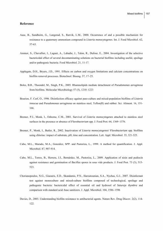
Mixed biofilms
157
Reference
Aase, B., Sundheim, G., Langsrud, S., Rørvik, L.M., 2000. Occurrence of and a possible mechanism for
resistance to a quaternary ammonium compound in Listeria monocytogenes. Int. J. Food Microbiol. 62,
57-63.
Ammor, S., Chevallier, I., Laguet, A., Labadie, J., Talon, R., Dufour, E., 2004. Investigation of the selective
bactericidal effect of several decontaminating solutions on bacterial biofilms including useful, spoilage
and/or pathogenic bacteria. Food Microbiol. 21, 11-17.
Applegate, D.H., Bryers, J.D., 1991. Effects on carbon and oxygen limitations and calcium concentrations on
biofilm removal processes. Biotechnol. Bioeng. 37, 17–25.
Boles, B.R., Thoendel, M., Singh, P.K., 2005. Rhamnolipids mediate detachment of Pseudomonas aeruginosa
from biofilms. Molecular Microbiology 57 (5), 1210–1223
Bourion, F. Cerf, O., 1996. Disinfection efficacy against pure-culture and mixed-population biofilms of Listeria
innocua and Pseudomonas aeruginosa on stainless steel, Teflon(R) and rubber. Sci. Aliment. 16, 151-
166.
Bremer, P.J., Monk, I., Osborne, C.M., 2001. Survival of Listeria monocytogenes attached to stainless steel
surfaces in the presence or absence of Flavobacterium spp. J. Food Prot. 64, 1369–1376.
Bremer, P., Monk, I., Butler, R., 2002. Inactivation of Listeria monocytogenes/ Flavobacterium spp. biofilms
using chlorine: impact of substrate, pH, time and concentration. Lett. Appl. Microbiol. 35, 321-325.
Cabo, M.L., Murado, M.A., González, MªP. and Pastoriza, L., 1999. A method for quantification. J. Appl.
Microbiol. 87, 907-914.
Cabo, M.L., Torres, B., Herrera, J.J., Bernárdez, M., Pastoriza, L., 2009. Application of nisin and pediocin
against resistance and germination of Bacillus spores in sous vide products. J. Food Prot. 73 (3), 515-
523.
Chorianopoulos, N.G., Giaouris, E.D., Skandamis, P.N., Haroutounian, S.A., Nychas, G.J., 2007. Disinfectant
test against monoculture and mixed-culture biofilms composed of technological, spoilage and
pathogenic bacteria: bactericidal effect of essential oil and hydrosol of Satureja thymbra and
comparison with standard acid–base sanitizers. J. Appl. Microbiol. 104, 1586–1596
Davies, D., 2003. Understanding biofilm resistance to antibacterial agents. Nature Rev. Drug Discov. 2(2), 114-
122.

158 Mixed biofilms
Denny, J., McLauchlin, J., 2008. Human Listeria monocytogenes infections in Europe –an opportunity for
improved European surveillance. Eurosurveillance 13,
http://www.eurosurveillance.org/edition/v13n13/080327_5.asp.
Fatemi, P., Frank, F.J., 1999. Inactivation of Listeria monocytogenes/Pseudomonas biofilms by peracid
sanitizers. J. Food Prot. 62 (7), 761-765.
Gilmour, M.W., Graham, M., Van Domselaar, G., Tyler, S., Kent, H., Trout-Yakel, K.M., Larios, O., Allen, V.,
Lee, B., Nadon, C., 2010. High-throughput genome sequencing of two Listeria monocytogenes clinical
isolates during a large foodborne outbreak. BMC Genomics, 11, 120-135
Hassan, A.N., Birt, D.M., Frank, J.F., 2004. Behavior of Listeria monocytogenes in a Pseudomonas putida
biofilm on a condensate-forming surface. J. Food Prot. 67, 322-327.
Herrera, J.J.R., Cabo, M.L., González, A., Pazos, I., Pastoriza, L., 2007. Adhesion and detachment kinetics of
several strains of Staphylococcus aureus subsp. aureus under three different experimental conditions. F.
Microbiol. 24, 585-591.
Jeong, D.K., Frank, J.F., 1994. Growth of Listeria monocytogenes at 10 °C in biofilms with microorganisms
isolated from meat and dairy processing environments. J. Food Prot. 57, 576–586.
Kastbjerg, V.G., Gram, L., 2009. Model systems allowing quantification of sensitivity to disinfectants and
comparison of disinfectant susceptibility of persistent and presumed nonpersistent Listeria
monocytogenes. J. Appl. Microbiol. 106, 1667-1681.
Lebert, I., Leroy, S., Talon, R., 2007. Effect of industrial and natural biocides on spoilage, pathogenic and
technological strains grown in biofilm. Food Microbiol. 24, 281-287.
Leriche, V., Carpentier, B., 2000. Limitation of adhesion and growth of Listeria monocytogenes on stainless
steel surfaces by Staphylococcus sciuri biofilms J. Appl. Microbiol. 88, 594–605.
Liu, D., Lawrence, M.L., Ainsworth, A.J., Austin, F.W., 2007. Toward an improved laboratory definition of
Listeria monocytogenes virulence. Int. J. of Food Microbiol. 118, 101-115.
Marshall, J. C. 1988. Adhesion and growth of bacteria at surfaces in oligotrophic habitats. Can. J. Microbiol. 34,
503–506
Mellefont, L.A., McMeekin, T.A., Ross, T., 2008. Effect of relative inoculum concentration on Listeria
monocytogenes growth in co-culture. Int. J. Food Microbiol. 121, 157-168.

Mixed biofilms
159
Minei, C.C., Gomes, B.C., Regianne, P.R., D’ Angelis, C.E.M., De Martinis, E.C.P., 2008. Influence of
peroxyacetic acid and nisin and coculture with Enterococcus faecium on Listeria monocytogenes
biofilm formation. J. Food Prot. 71, 634.
Nadell, C.D., Xavier, J.B., Foster, K.R., 2008. The sociobiology of biofilms. FEMS Microbiol. 33 (1) 206
Norwood, D.E., Gilmour, A., 1999. Adherence of Listeria monocytogenes strains to stainless steel coupons. J.
Appl. Microbiol. 86, 576-582.
Norwood, D.E., Gilmour, A., 2001. The differential adherence capabilities of two Listeria monocytogenes strains
in monoculture and multispecies biofilms as a function of temperature. Lett. Appl. Microbiol. 33, 320-
324.
Pan, Y., Breidt, F.Jr., Kathariou, S., 2006. Resistance of Listeria monocytogenes Biofilms to Sanitizing Agents
in a Simulated Food Processing Environment. Appl. Environ. Microbiol. 72, 7711-7717.
Porsby, C.H., Vogel, B.F., Mohr, M., Gram, L., 2008. Influence of processing steps in cold-smoked salmon
production on survival and growth of persistent and presumed non-persistent Listeria monocytogenes.
Int. J. Food Microbiol. 122, 287-295.
Rodrigues, D.A., Almeida, M.A., Teixeira, P.A., Oliveira, R.T., Azeredo, J.C., 2009. Effect of Batch and Fed-
Batch Growth Modes on Biofilm Formation by Listeria monocytogenes at Different Temperatures.
Curr. Microbiol. 59, 457–462.
Rodríguez, A., Autio, W.R., McLandsborough, L.A., 2008. Effect of surface roughness and stainless steel finish
on Listeria monocytogenes attachment and biofilm formation. J. Food Prot. 71, 170-175.
Rieu, A., Lemaître, J., Guzzo, J., Piveteau, P., 2008. Interactions in dual species biofilms between Listeria
monocytogenes EGD-e and several strains of Staphylococcus aureus. Int. J. Food Microbiol. 126, 76-82.
Rodríguez A., Autio W.R., McLandsborough, L.A., 2007. Effect of biofilm dryness on the transfer of Listeria
monocytogenes biofilms grown on stainless steel to bologna and hard salami. J. Food Prot. 70(11),
2480-2484.
Saá, P., Cabo, M.L., Rodríguez, J.J., 2009. Effects of mussel processing soils on the adherence of Listeria
monocytogenes to polypropylene and stainless steel. J. Food Prot. 72, 1885-1890.
Saá Ibusquiza, P., Rodríguez, J.J, Cabo, M.L., 2010. Resistance to benzalkonium chloride, peracetic acid and
nisin during formation of mature biofilms by Listeria monocytogenes. Food Microbiol.
doi:10.1016/j.fm.2010.09.014.
160 Mixed biofilms
Sasahara, K.C., Zottola, E.A., 1993. Biofilm formation by Listeria monocytogenes utilizes a primary colonizing
microorganism in flowing systems. J. Food Prot. 56 (12), 1022-1028.
Sauer, K., Cullen, M.C., Rickard, A.H., Zeef, L.A., Davies, D.G., Gilbert, P., 2004. Characterization of nutrient-
induced dispersion in Pseudomonas aeruginosa PAO1 biofilm. J. Bacteriol. 186(21), 7312-7326.
Sharma, M., Anand, S.K., 2002. Characterization of constitutive microflora of biofilms in dairy processing lines.
Food Microbiol. 19, 627-636.
Simões, L.C., Simões, M., Vieira, M.J., 2007. Biofilm interactions between distinct bacterial genera isolated
from drinking water. Appl. Environ. Microbiol. 73, 6192-6200.
Simões, M., Simões, L.C., Vieira, M.J., 2009. Species association increases biofilm resistance to chemical and
mechanical treatments. Water Res. 43, 229-237.
Stoodley, P., Wilson, S., Hall-Stoodley, L., Boyle J.D., Lappin-Scott H.M., Costerton, J.W., 2001. Growth and
Detachment of Cell Clusters from Mature Mixed-Species Biofilms. Appl. Environ. Microbiol. 67 (12)
5608-5613.
Takahashi, H., Miya, S., Igarashi, K., Suda, T., Kuramoto, S., Kimura, B., 2009. Biofilm formation ability of
Listeria monocytogenes isolates from raw ready-to-eat seafood. J. Food Prot. 72(7), 1476-1480.
Takhistov, P., George, B., 2004. Linearized kinetic model of Listeria monocytogenes biofilm growth. Bioproc.
Biosyst. Eng. 26, 259–270
To, M.S., Favrin, S., Romanova, N., Griffiths, M.W., 2002. Postadaptational Resistance to Benzalkonium
Chloride and Subsequent Physicochemical Modifications of Listeria monocytogenes. Appl. Environ.
Microbiol. 68, 5258-5264.
Wijman, J.G., de Leeuw, P.P., Moezelaar, R., Zwietering, M.H., Abee, T., 2007. Air-liquid interface biofilms of
Bacillus cereus: formation, sporulation, and dispersion. Appl. Environ. Microbiol. 73(5), 1481
Wuertz, S, Okabe, S; Hausner, M., 2004. Microbial communities and their interactions in biofilm systems: an
overview. Water Sci. Technol. 49(11-12) 327-336.
33..55.. Comparison between the resistance of
benzalkonium chloride-adapted and non-
adapted biofilms of Listeria monocytogenes
to modified atmosphere packaging (MAP)
and nisin once transferred to mussels


MAP
163
3.5. COMPARISON BETWEEN THE RESISTANCE OF
BENZALKONIUM CHLORIDE-ADAPTED AND NON-ADAPTED
BIOFILMS OF LISTERIA MONOCYTOGENES TO MODIFIED
ATMOSPHERE PACKAGING (MAP) AND NISIN ONCE
TRANSFERRED TO MUSSELS.
Benzalkonium chloride (BAC) adapted and non-adapted CECT 5873 L. monocytogenes
biofilm cells were transferred by contact to cooked or live mussels and packed under CO2 and
O2 rich atmospheres, respectively. Viability of transferred cells during storage of these packed
samples at 2.5 ºC was compared. In addition, in cooked mussels the combined effect of CO2
and nisin against the survival of L. monocytogenes was also studied by using a first order
factorial design. Obtained results demonstrated that biofilms formed by BAC-adapted L.
monocytogenes cells could be more resistant to the application of modified atmospheres rich
in CO2 and nisin once they have been transferred to cooked mussels by contact (simulating
cross-contamination). This implies an increase in the risk associated with the presence of
these cells in food processing plants. Significant empirical equations obtained after 7, 11 and
20 days showed an inhibitory effect of CO2 and nisin against L. monocytogenes. However, a
significant positive interaction between both variables highlights an incompatibility between
CO2 and nisin at high concentrations. Results also demonstrated that L. monocytogenes could
persist after cross-contamination during the processing of live mussels, so L. monocytogenes
is of concern as a contaminant in live mussels packaged in high O2 atmospheres.

164 MAP
3.5.1. Introduction
The aim to maintain a high level of mussel (Mytilus galloprovinciales) commercialization
in Galicia and the increase in international competitiveness have led to the development of
alternatives of preservation that allow long distance markets to be reached such as modified
atmosphere packaging (MAP). It has been applied extensively to fish products (Cabo et al.,
2003; Sivertsvik et al., 2002; Ohlsson and Bengtsson, 2002; Skara et al., 2003). MAP is used
for preservation of mussels with two objectives: 1) to increase the shelf life of cooked mussels
in a CO2-rich atmospheres; and 2) to increase the viability of live mussels in an O2-rich
atmosphere (Pastoriza et al., 2004).
According to Rutherford et al. (2007), Listeria monocytogenes is often found in processing
environments as a post-processing contaminant. It is transferred to food through contact at
food processing plants, where L. monocytogenes forms biofilms (Porsby et al., 2008,
Rodríguez and McLandsborough 2007). Also it has been previously shown that persistent
Listeria monocytogenes cells are resistant to Quaternary Ammonium compounds, such as
benzalconium chloride (BAC) (Aase et al., 2000; Mullapudi et al., 2008), probably due to
inadequate disinfection protocols that lead to residual sublethal concentrations of disinfectants
(Langsrud et al., 2003).
It is possible for L. monocytogenes to grow in modified atmosphere packed mussels. In
fact, thermal soft treatments (like those applied in boiled mussels) may not eliminate this
bacterium (Doyle et al., 2001, Huang, 2004), it can resist high CO2 atmospheres (Fang and
Lin 1994; Jenssen and Lammert, 2002; Szabo and Cahill, 1999; Olarte et al., 2002) and the
aerobic conditions of high O2 atmospheres are not expected to damage L. monocytogenes
cells. In this context, the application of antimicrobials, such as bacteriocins, with a specific
effect against these opportunistic bacteria can be useful for ensuring food safety (Cabo et al.,
2001; Cabo et al., 2005; Cabo et al., 2009¸ Cleveland et al., 2001, Delves-Broughton et al.,
1996).
It is therefore possible that in a mussel processing plant, BAC-adapted biofilms of L.
monocytogenes can be transferred by contact to the products during processing and it could
survive refrigerated under MAP during storage. Nonetheless, no previous studies on the
repercussions of BAC-adaptation of L. monocytogenes to the resistance to MAP were found.
Therefore, the main objective of this work was to compare the viability of BAC adapted and

MAP
165
non-adapted L. monocytogenes biofilm cells in modified atmospheres rich in CO2 and O2 once
they had been transferred by contact to cooked and live mussels, respectively. In addition, in
cooked mussels an experimental factorial design allowed us to study the combined effects of
CO2 and nisin against the survival of L. monocytogenes.
3.5.2. Material and methods
Bacterial strains and synthetic media
Listeria monocytogenes CECT 5873 was provided by the Spanish Type Culture Collection
(Valencia). Strain CECT 5873 adapted to BAC was obtained in previous experiments after
two exposures of the wild type strain 5873 to sub-lethal concentrations of BAC (wild strain
LD50: 2.71 mg/l; BAC-adapted strain LD50: 7.22 mg/l, see section 3.2). Strains were frozen
and stored at -80 ºC in TSB containing 50% glycerol (v/v) until use. Whenever required, one
cryovial was thawed and this strain was subcultured twice in TSB (Cultimed, S.L., Spain) at
37 ºC before use.
Method for biofilm formation
Food processing surfaces
Stainless steel sheets (AISI-304, 2B finish, 0.8 mm thickness) were cut into coupons (10
mm x 10 mm). New and reused SS coupons were soaked in 2M NaOH to remove any grease
or food soils left and then rinsed several times with distilled water. Coupons were dried before
use in the laminar flow cabinet.
Food soils media
Mussel cooking juice (MCJ, Carbohydrate 9,19 g/l, Nitrogen 1.70 g/l), used as culture
media to simulate contamination of surfaces in plants processing cooked mussel, was
prepared as it was previously described (Saá et al., 2009).
166 MAP
Inocula preparation
Working cultures of BAC-adapted and non-adapted strains of L. monocytogenes CECT
5873 were adjusted to an absorbance 0.1 at 700 nm, which correspond to a cell density of 108
CFU/ml according with previous calibration. Adjusted cultures were centrifuged at 6000 rpm
during 10 min at 25 ºC and collected cells were resuspended in MCJ. This suspension was
used as inoculum.
Biofilm formation
Mini glass Petri plates (5 cm diameter) containing cleaned coupons were packed 5 by 5
with foil and autoclaved for 20 min at 121 ºC. Once autoclaved, 9 ml of inoculum was added
per mini Petri plate and incubated at 25 ºC during 7 days for biofilm formation.
Transference assays
Preparation of mussels
Fresh mussels from Galicia (Mytilus galloprovincialis) were always acquired in the same
market. For cooking, fresh mussels were cooked during 1 minute at 100 ºC without adding
water. After cooking, 10 g weight mussels were selected for the experiments.
Transference of biofilms from coupons to mussels
After 7 days of biofilm maturing, coupons were rinsed with 10 ml of PBS for 30 s to
release non-adhered cells before transferring. Total number of adhered cells in the coupons
after 7 days were 7,88 log CFU. The surface was rubbing with two moistened swabs which
were resuspended by vigorously vortexing for 50 s in 9 ml of peptone water according to
Herrera et al. 2007 and plating on Triptone Soy Agar and incubated at 37 ºC for 24 h. The
cells of the upper superface were taking account in the counting (7.88 log CFU)the other
superface was in touch with the glass of the petri plates. Biofilm cells were the transferred as
follows:
to cooked mussel: by contact between the coupon and the meat without additional
pressure during 120 seconds according with previous kinetic experiments that
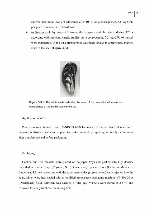
MAP
167
showed maximum levels of adherence after 100 s. As a consequence, 5.6 log CFU
per gram of mussel were transferred.
to live mussel: by contact between the coupons and the shells during 120 s
according with previous kinetic studies. As a consequence, 7.2 log CFU of mussel
were transferred. In this case transference was made always in a previously marked
zone of the shell (Figure 3.5.1).
Figure 3.5.1. The white circle indicates the area of the mussel-shell where the
transference of the biofilm was carried out.
Application of nisin
Pure nisin was obtained from DANISCO (A/S Denmark). Different doses of nisin were
prepared in distilled water and applied to cooked mussel by pipetting uniformly on the meat
after transference and before packaging.
Packaging
Cooked and live mussels were placed on polyspan trays and packed into high-density
polyethylene barrier bags (Cryobac, S.L.). Once ready, gas mixtures (Carburos Metálicos,
Barcelona, S.L.) set according with the experimental design (see below) were injected into the
bags, which were heat-sealed with a modified atmosphere packaging machine VP-430-20-A
(Dordalpack, S.L.). Nitrogen was used as a filler gas. Mussels were stored at 2.5 ºC and
removed for analysis at each sampling time.

168 MAP
Experimental design
In cooked mussels: a first order factorial design (Box et al., 1989) with 4
combinations of variables and 5 replicates in the center of the domain was carried
out. The independent variables were the proportion of CO2 (C) the gas mixture and
the concentration of nisin applied on the meat of the mussel (N). Natural and coded
levels for each variable are showed in Table 3.5.1.
In live mussels: the effect of increasing concentrations of O2 (30, 60 and 90%) on
the survival of L. monocytogenes were assayed.
Table 3.5.1: Natural and codified values used in the
first order ortogonal design
Natural values
Codified values CO2 (%) Nisin (UI/g)
1;1 90 210
1;-1 90 21
-1;1 20 210
-1;-1 20 21
0;0 55 115
Microbiological analyses
Determination of the Number of Viable Cells of L. monocytogenes (VLM)
In cooked mussels, determination of VLM was carried out after 7, 11 and 20 days of
storage. At each day of sampling, 5 mussels were minced with sterilized scissors and 10 g of
this mixture was homogenized in 90 ml of sterile peptone water (Cultimed, S.A.) in a Lab-
Blender-400 Stomacher. Number of VLM were determined by serially ten-fold diluting it in
peptone water (Cultimed, S.A.), subsequent spread out in Palcam (Liofilchem, S.L.R., Italia)
and Triptone Soy Agar (Cultimed, S.A.) and finally incubated at 37 ºC during 48h. TSA was
used to assure sublethal damaged cells, that may have problems to growth in a selective media
as PALCAM, were computed.
MAP
169
In live mussels, determination of VLM was carried out after 5, 8 and 12 days of storage.
Adhered cells were collected by thoroughly rubbing with two moistened swabs in the marked
area and resuspended by vigorously vortexing the swabs for 50 s in 9 ml of peptone water
according to Herrera et al. 2007. Proper serial ten-fold dilutions were then plated on Triptone
Soy Agar and Palcam and incubated at 37 ºC for 24 h and 48 h respectively. Peptone water
was always used as dilution medium.
Biofilm contaminated mussels were packed in increasing concentrations of O2 and stored
at 2.5 ºC. The viability of the 5873 BAC adapted and non-adapted biofilm cells to the
application of modified O2-rich atmospheres was compared after 5, 8 and 12 days of storage.
Mortality test in mussels packaged in O2-rich atmospheres
Mortality test were carried out according with previous studies (Pastoriza et al., 2004).
Briefly, it was carried out after 2, 6, 8, 10, 12, 13, 14 days of storage at 2.5 ºC by tapping on
gaping bivalve shelves and observing mussel closure. Any shells gaping after tapping were
considered dead.
Statistics
A Student t-test (α=0.05) was used to test the significance of the differences between
means of the total number of viable cells (log CFU), between the mortality (%) rate in live
mussels and between the coefficients of the equations obtained in the factorial design. A
Fisher Test (α=0.05) was used to test the consistence of the models.
3.5.3. Results
Studies on cooked mussels
Effects of CO2 and nisin on the viability of BAC-adapted and non-adapted biofilms cells
once transferred to cooked mussels.
A first order factorial design (Box et al., 1988) was used to compare the sensitivity of
biofilms cells of 5873 BAC adapted and non-adapted strains to the combined application of
CO2 and nisin during storage at 2.5 ºC. Both CO2 percentage and nisin concentration used

170 MAP
during the expositions were set following the factorial design (Table 3.5.1). Viable cells of L.
monocytogenes (VLM, in log CFU/g) in cooked mussels after 7, 11 and 20 days of storage
could be predicted satisfactory according to the following empirical equations [3.5.1-3.5.6]:
1) BAC non-adapted L. monocytogenes CECT 5873:
After 7 days: VLM=4.25-1.18C-1.48N-1.18CN [3.5.1]
After 11 days: VLM=5.25-0.45C-0.74N+0.46CN [3.5.2]
After 20 days: VLM=5.72-1.43C-0.86N [3.5.3]
2) BAC-adapted 5873 strain:
After 7 days: VLM=3.65-0.57C-0.87N [3.5.4]
After 11 days: VLM=5.24-0.45C-0.94N [3.5.5]
After 20 days: VLM=5.47-0.37C-0.79N+0.54CN [3.5.6]
Graphycal representations of all the empirical equations are shown in Figure 3.5.2 and
3.5.3 (data are showed in the supporting information chapter). The comparison of the effects
of the two variables on the wild type strain and the BAC adapted strains showed that the
biggest differences occured after 7 and 20 days of storage. At these times of storage, it can be
clearly observed that BAC adaptation gave L. monocytogenes a higher resistance to CO2 and
nisin. In fact, the CO2 and nisin coefficients obtained for the non-adapted strain after 7 (eq.
[3.5.1]) and 20 (eq. [3.5.3]) days were significantly higher (p<0.05) than those obtained at the
same times of storage for the non-adapted strain (eq. [3.5.4] and eq. [3.5.6], respectively). In
Table 3.5.2, log reductions (LR) obtained at different combinations of CO2 and nisin in
adapted and non-adapted biofilms of L. monocytoenes are showed. At high concentrations of
both variables, 90% CO2 and 210 UI/ml of nisin, the model predicts log reductions (LR=log
UFC/g transferred-log UFC/g after storage) of 3.39 and 5.12 in the number of viable L.
monocytogenes cells per gram of the 5873 BAC adapted and non-adapted strains respectively
after 7 days of storage of the contaminated cooked mussels. However, after 11 days of storage
the differences between the effects of CO2 and nisin decreased and similar polynomial
equations were obtained to predict the VLM. There was one difference: there was a

MAP
171
significant positive interaction between CO2 and nisin at this time of storage for the non-
adapted strain, which is reflected in a decrease in the effect of CO2. This positive interaction,
also significant in equation [3.5.6], contrasts with the beneficial individual effects of the CO2
and the nisin and can be interpreted as an increase in the viable Listeria monocytogenes
biofilm cells in mussels preserved under high concentrations of both CO2 and nisin.
CECT 5873 Adapted-20DAYSCECT 5873 Adapted-11 DAYSCECT 5873 Adapted-7 DAYS
VLM
(lo
g U
FC
/g)
VLM
(lo
g U
FC
/g)
VLM
(lo
g U
FC
/g)
Figure 3.5.2. Response surfaces corresponding to the combined effects of CO2
and nisin against viable L. monocytogenes CECT 5873 BAC-adapted biofilm cells
(VLM, in log CFU/g) after 11 and 20 days of storage at 2.5 ºC. Variables are
expressed as codified values (see Table 3.5.1).
CECT 5873-20 DAYSCECT 5873-11 DAYSCECT 5873-7 DAYS
VLM
(lo
g U
FC
/g)
VLM
(lo
g U
FC
/g)
VLM
(lo
g U
FC
/g)
Figure 3.5.3. Response surfaces corresponding to the combined effects of CO2
and nisin against viable L. monocytogenes CECT 5873 BAC non adapted biofilm
cells (VLM, in log CFU/g) after 11 and 20 days of storage at 2.5 ºC. Variables are
expressed as codified values (see Table 3.5.1).

172 MAP
Those results have also demonstrated that the effects of the variables change during the
time of storage. In fact, if we calculate the log reductions in L. monocytogenes at different
combinations of CO2-nisin in the experimental design (see Table 3.5.2), it can be clearly
observed that the effectiveness of the variables decrease with the time of storage, especially
from 7 to 11 days.
Table 3.5.2: Log reduction (LR) in L. monocytogenes biofilm cells transferred to cooked mussels and
stored under different combinations of CO2 and nisin.
Vc (CO2, nis) (1,1) Vc (CO2, nis) (0,0) Vc (CO2, nis) (-1,-1)
Non adapted 7D 5.12 1.32 -0.18a
11D 1.08 0.35 -1.3
20D 2.17 -0.12 -2.41
BAC-adapted 7D 3.39 1.95 0.51
11D 1.75 0.36 -1.03
20D 0.75 0.13 -1.57 a Negative values indicate the model predicts the growth of L. monocytogenes
Studies on live mussels
Preliminary experiments: viability of live mussels packaged under O2-rich atmospheres
In order to assure that mussels were viable during the set time of study, preliminary
experiments to study the effect of the different O2-rich atmospheres on the viability of live
mussels without inoculation with L. monocytogenes were carried out.
Before 13 days of storage at 2.5 ºC, no significant differences were obtained between the
mortality of mussels packed under different O2 concentrations, being in all cases lower than
20%. At 14 days mortality decreased from 45% in air (21%O2) till 10% in mussels packed
under 90% O2 (Figure 3.5.4). This is in accordance with previous results that demonstrated
mortality of mussels decreased when packaging at high O2 concentration (Pastoriza et al.
2004). According with those results and to assure mussels were alive during the experiments,
sampling times were set at 5, 8 and 12 days of storage.

MAP
173
Air 30% O2 60% O2 90% O20
20
40
60
80
100
Mo
rta
lity
(%)
Figure 3.5.4. Mortality of live mussels packaged under different O2-rich
atmospheres (30%, 60% and 90%) and air after 14 days of storage at 2.5 ºC.
Comparison of the resistance to O2 of BAC adapted and non-adapted, transferred to live
mussels
The results did not show significant differences between the number of viable BAC
adapted L. monocytogenes cells compared to the non-adapted cells during storage (Figure
3.5.5). In fact, the sensitivity of the BAC-adapted cells was only slightly higher in live
mussels packed at 60% O2 (0.25 log/g).
day0 day5 day8 day12Air 30%O2 60%O2 90%O2
5
5,5
6
6,5
7
7,5
8
CF
U
day0 day5 day8 day12 day0 day5 day8 day12 day0 day5 day8 day12
Figure 3.5.5. Viability of L. monocytogenes cells transferred to live mussels
packaged under different O2-rich atmospheres (30%, 60% and 90%) and air at 0,
5, 8 and 12 days of storage at 2.5 ºC. BAC non-adpated biofilm cells are showed in
black columns and BAC adapted CECT 5873 in grey columns.

174 MAP
3.5.4. Discussion
Cooked mussels
The results obtained demonstrate that biofilms cells formed by BAC-adapted CECT 5873
L. monocytogenes cells were more resistant to the application of modified atmospheres rich in
CO2 and nisin once they have been transferred to cooked mussels by contact (simulating
cross-contamination). This implies an increase in the risk associated with the presence of
these cells in food processing plants. This was particularly significant after 7 days of storage,
when the coefficients that quantitatively described the effects of CO2 and nisin were
significantly lower in the equation that describes the number of non-adapted L.
monocytogenes cells (compare Eq. [3.5.1] and Eq. [3.5.4]). This is in agreement with other
results recently published that demonstrated mature biofilms formed by BAC-adapted cells
were more resistant to nisin compared to those formed by the non-adapted cells (Saá
Ibusquiza et al., 2010).
However, those effects are not constant over the storage time. In fact, after 11 days a
decrease in the effect of the two variables was observed both in the BAC adapted and non-
adapted cells, which is reflected in the similar 3D surfaces shown in Figure 3.5.2 and 3.5.3
respectively. Moreover, at this storage time nisin is more effective against the viability of
BAC adapted L. monocytogenes biofilm cells than against the non-adapted cells. The
variations observed in the effects of the variables over time reflect the kinetic conditions of
the microbial system. And they are in agreement with those obtained by Minei et al., (2008),
who found a significant decrease in the rate of L. monocytogenes biofilm formation after nisin
had been added in the early stages of incubation, followed by the recommencement of biofilm
formation after 12 hours.
Although it is very difficult to draw conclusions at this point without carrying out
additional mechanistic experiments, several facts taken from previously published results
could help us to explain the appearance of the identified adaptive responses:
A confluence in the cross-responses of L. monocytogenes to acid (ATR) and BAC.
It has been previously demonstrated that acid-tolerant strains of L. monocytogenes
are more resistant to CO2 (Francis et al., 2007, Jydegaard-Axelsen et al., 2004) and
nisin (Badaoui Najjar et al., 2009; Begley et al., 2010; Bonnet and Montville 2005).

MAP
175
The confluence of the two cross-responses suggests that adaptation to BAC and
acid could imply similar modifications in L. monocytogenes. However, we could
not find any previous studies on the cross-resistance response between disinfectants
and CO2.
A confluence of the membrane as a target for the CO2, nisin and BAC action. It has
been demonstrated that CO2 modifies bacterial membrane permeability by
changing the lipid composition (Jydegaard-Axelson et al. 2004, Lungu et al., 2009;
Tassou et al., 2004; Nilsson et al., 2000). Nisin forms pores in the bacterial
membrane of its targets by interacting with lipid II, a cell wall precursor (Christ et
al., 2007; Dalmau et al., 2002; Wiedemann et al., 2007). It has been recently
demonstrated that BAC induces membrane damage in Bacillus cereus (Ceragioli et
al., 2010). In addition, a direct relation between the sensitivity to BAC and the
increase in membrane permeability has been observed in E. coli and L.
monocytogenes (Walton et al., 2008). Changes in the components in the outer
membrane (QAC-resistance genes such as qacA, qacB, smr, qacG and qacH code
for efflux membrane proteins) could also participate in the resistance of L.
monocytogenes to BAC (Soumet et al., 2005).
A relation between the resistance to nisin and the resistance to BAC could be
related to the proton motive force (PMF)-efflux pump regulation system. In fact, it
has been demonstrated that adding nisin can dissipate the basal PMF across the
membrane (Bruno et al., 1992). However, Romanova et al., (2006) demonstrated
that adding reserpine (an inhibitor of the efflux pump mdrL and lde caused by
BAC) to physiologically adapted and BAC-sensitive L. monocytogenes implied a
decrease in the MIC value of BAC, absent in those strains that are naturally
resistant. Finally, alterations in the PMF caused by nisin and BAC could help CO2
to act by preventing protons being expelled from the cytoplasm, as well as
acidification (Garcia-Gonzalez et al. 2007).
Therefore, it seems that BAC, nisin and CO2 have common points of action in the cell
membrane, and it is biologically reasonable that some cross-resistances between them could
appear after successive exposures.

176 MAP
Although quantitative differences between the effects of the variables against the BAC
adapted and non-adapted L. monocytogenes transferred biofilms were identified, the signs of
the individual and combined effects of CO2 and nisin were very similar. CO2 and nisin
showed an inhibitor effect against both types of biofilms. This is in agreement with previous
works with planktonic cells (effect of CO2: Sheridan et al 1995, Olarte et al 2002; effect of
nisin: Nisson et al. 1997, López- Mendoza et al. 2007). However, no previous studies on the
effectiveness of CO2 against L. monocytogenes biofilms were found. In the case of nisin, two
types of studies were found: those that study controlling Listeria monocytogenes with nisin-
producing biofilms (Guerrieri et al., 2009, Leriche et al., 1999) and only one article that
assayed controlling Listeria monocytogenes biofilms with nisin (Minei et al., 2008). In this
last, although bacterial growth was initially reduced up to 4.6 log CFU/cm2 when compared
with L. monocytogenes cultures on untreated coupons, after 24 h of incubation, a renewed
biofilm was detected.
To elucidate the real repercussions of the identified cross-responses additional experiments
with more strains should be carried out. However, the positive interaction between CO2 and
nisin, which reflects incompatibility at high concentrations of the two variables, contrasts with
previous results in which a synergic effect between nisin and CO2 against the survival of L.
monocytogenes has been demonstrated (López-Mendoza et al. 2007; Nilsson et al. 2000;
Szabo and Cahill, 1998). This discrepancy could be explained by an increase in
incompatibility between the effects of CO2 and nisin associated with the presence of an
external polysaccharide matrix in the mature biofilm transferred to cooked mussels. In fact, a
significant decrease in nisin effectiveness against mature L. monocytogenes biofilms
compared with that observed against planktonic cells has been previously observed (Saá
Ibusquiza et al., 2010). Moreover, the ineffectiveness of nisin in the presence of CO2 against
an exopolysaccharide producer, Pseudomonas fragi, has been demonstrated (Fang and Lin,
1994). Variations in the EPS composition during biofilm formation could explain the
variations in the effect of nisin over the storage time.
However, the results of the individual and combined effects of CO2 and nisin is that LR of
L. monocytogenes decreased during the time of storage, thus demonstrating that L.
monocytogenes biofilm cells (both adapted and non-adapted) can resist the preservation
hurdles and growth in cooked mussels at this temperature.

MAP
177
Live mussels
No significant differences were found between the number of viable L. monocytogenes
cells transferred to live mussels from BAC adapted and non-adapted biofilms over 12 days of
storage at 2.5 ºC. This indicates that cellular modifications in L. monocytogenes related to
BAC adaptation do not have any significant effects on its survival under high O2
concentrations. Moreover, no significant differences were found in the number of viable L.
monocytogenes cells recovered from the live mussels during storage, which indicates that L.
monocytogenes could persist after cross-contamination during the processing of live mussels.
This is in agreement with previous results obtained by Allende et al., (2002), who found no
significant differences in the number of viable L. monocytogenes cells inoculated into a mixed
vegetable salad during storage at 4 and 7 ºC under different O2-rich atmospheres.
Overall, the results allow us to conclude that BAC adapted L. monocytogenes biofilms
transferred to cooked mussels are more resistant to CO2 and nisin than non-adapted biofilms
but that this resistance can vary over the storage time. In addition, nisin and CO2 rich
atmospheres are not effective enough to control a hypothetical cross-contamination of L.
monocytogenes biofilm cells transferred from industrial surfaces to ready-to-eat fish products,
such as cooked mussels. Finally, it must be pointed out that L. monocytogenes is a pathogen
of concern in live mussels packaged in high O2 atmospheres.

178 MAP
References
Aase, B., Sundheim, G., Langsrud, S., and Rørvik, L. M., 2000. Occurrence of and a possible mechanism for
resistance to a quaternary ammonium compound in Listeria monocytogenes. Int. J. Food Microbiol. 62(1-
2), 57-63.
Allende, A., Martínez, B., Selm,a V., Gil, M.I., Suárez, J.E. and Rodríguez, A., 2007. Growth and bacteriocin
production by lactic acid bacteria in vegetable broth and their effectiveness at reducing Listeria
monocytogenes in vitro and in fresh-cut lettuce. Food Microbiol. 24(7-8), 759-66.
Badaoui, Najjar, M.Z., Chikindas, M.L., and Montville, T.J., 2009. The Acid Tolerance Response Alters
Membrane Fluidity and Induces Nisin Resistance in Listeria monocytogenes. Probiotics Antimicro.
Prot. 1,130–135
Begley, M., Cotter, P.D., Hill, C. and Ross, R.P., 2010. Glutamate Decarboxylase-Mediated Nisin Resistance in
Listeria monocytogenes. Appl Environ Microbiol. 76(19), 6541-6546
Bonnet, M. and Montville, T.J., 2005. Acid-tolerant Listeria monocytogenes persist in a model food system
fermented with nisin-producing bacteria. Lett. Appl. Microbiol.40(4), 237.
Box, G.E.P., Hunter, W.G. and Hunter, J.S., 1989. En “Estadística para investigadores”. Ed. Reverté
(Barcelona).
Bruno, M. E., Kaiser, A. and Montville, T. J., 1992. Depletion of proton motive force by nisin in Listeria
monocytogenes cells. Appl. Environ. Microbiol. 58(7), 2255–2259.
Cabo, M. L., Pastoriza, L., Bernárdez, M., and Herrera, J. J. R., 2001. Effectiveness of CO2 and nisaplin on
increasing shelf-life of fresh pizza. Food Microbiol. 18(5), 489-498.
Cabo M.L., Herrera J.R., Sampedro G. and Pastoriza, L., 2005. Application of nisin, CO2 and a permeabilizing
agent in the preservation of refrigerated blue whiting (Micromesistius poutassou). J. Sci. Food Agr. 85,
1733
Cabo, M. L., Herrera, J. J., Crespo, M. D., and Pastoriza, L., 2009. Comparison among the effectiveness of
ozone, nisin and benzalkonium chloride for the elimination of planktonic cells and biofilms of
Staphylococcus aureus CECT4459 on polypropylene. Food Control. 20(5), 521-525.
Ceragioli M., Mols M., Moezelaar R., Ghelardi E., Senesi S. and Abee T., 2010. Comparative transcriptomic and
phenotypic analysis of the responses of Bacillus cereus to various disinfectant treatments. Appl. Environ.
Microbiol. 76(10), 3352-3360.

MAP
179
Christ, K., Wiedemann, I., Bakowsky, U., Sahl, H., and Bendas, G., 2007. The role of lipid II in membrane
binding of and pore formation by nisin analyzed by two combined biosensor techniques. BBA
Biomembranes. 1768(3), 694-704.
Cleveland, J., Montville, T. J., Nes, I. F., and Chikindas, M. L., 2001. Bacteriocins: Safe, natural antimicrobials
for food preservation. Int. J. Food Microbiol. 71(1), 1-20.
Dalmau M., Maier, E., Mulet, N., Viñas, M. and Benz., R., 2002. Bacterial membrane injuries induced by
lactacin F and nisin. Int. microbial. 5, 73-80
Delves-Broughton J., Blackburn P., Evans R.J. and Hugenholtz J., 1996. Applications of the bacteriocin, nisin.
Anton. Leeuw.69 (2), 193.
Doyle, M.E., Mazzotta, A.S., Wang, T., Wiseman, D.W. and Scott, V.N., 2001. Heat resistance of Listeria
monocytogenes. J. Food Prot. 64(3), 410-429.
Fang, T.J. and Lin., L-W, 1994. Growth of Listeria monocytogenes and Pseudomonas fragi on cooked pork in a
modified atmosphere packaging/nisin combination system. J. Food Prot. 57, 479-485
Francis, G. A., Scollard, J., Meally, A., Bolton, D. J., Gahan, C. G. M., Cotter, P. D., et al., 2007. The glutamate
decarboxylase acid resistance mechanism affects survival of Listeria monocytogenes LO28 in modified
atmosphere-packaged foods. J. Appl. Microbiol. 103(6) 2316-2324.
Garcia-Gonzalez, L., Geeraerd, A. H., Spilimbergo, S., Elst, K., Van Ginneken, L., Debevere, J. et al., 2007.
High pressure carbon dioxide inactivation of microorganisms in foods: The past, the present and the future.
Int. J. Food Microbiol. 117(1), 1-28
Guerrieri, E., de Niederhäusern, S., Messi, P., Sabia, C., Iseppi, R., Anacarso, I., et al., 2009. Use of lactic acid
bacteria (LAB) biofilms for the control of Listeria monocytogenes in a small-scale model. Food Control.
20(9), 861-865.
Herrera, J.J.R., Cabo, M.L., González, A., Pazos, I. and Pastoriza, L., 2007. Adhesion and detachment kinetics of
several Strains of Staphylococcus aureus subsp.aureus under three different experimental conditions.
Food Microbiol. 24, 585-591.
Huang L. (2004). Thermal resistance of Listeria monocytogenes, Salmonella Heidelberg, and Escherichia coli
O157:H7 at elevated temperatures. J. Food Prot 67(8), 1666-70
Jydegaard-Axelsen, A.-M., Høiby, P.E., Holmstrøm, K., Russell, N. and Knøchel, S. (2004). CO2- and
anaerobiosis-induced changes in physiology and gene expression of different Listeria monocytogenes
strains. Appl. Environ. Microbiol. 70(1), 4111

180 MAP
Langsrud, S., Sidhu M.S., Heir, E. and Holck A.L. (2003). Bacterial disinfectant resistance - a challenge for the
food industry. Int. Biodeter. Biodegr. 51, 283-290.
Leriche, V., Chassaing, D., and Carpentier, B. (1999). Behaviour of L. monocytogenes in an artificially made
biofilm of a nisin-producing strain of lactococcus lactis. Int. J. Food Microbiol. 51(2-3), 169-182.
Lopez-Mendoza, M. C., Ruiz, P., and Mata, C. M. (2007). Combined effects of nisin, lactic acid and modified
atmosphere packaging on the survival of Listeria monocytogenes in raw ground pork. Int. J. Food Sci.
Tech., 42(5), 562-566.
Lungu, B., Ricke, S.C. and Johnson, M.G., 2009 Growth, survival, proliferation and pathogenesis of Listeria
monocytogenes,under low oxygen or anaerobic conditions: A review. Anaerobe. 15, 7-17
Minei, C.C., Gomes, B.C., Regianne, P.R., D’ Angelis, C.E.M. and Martinis De, E.C.P. (2008). Influence of
peroxyacetic acid and nisin and coculture with Enterococcus faecium on Listeria monocytogenes biofilm
formation. J. Food Prot. 71(3), 634-638.
Mullapudi S., Siletzky R.M. and Kathariou S. (2008). Heavy-metal and benzalkonium chloride resistance of
Listeria monocytogenes isolates from the environment of turkey-processing plants. Appl. Environ.
Microbiol. 74(5), 1464-1468
Nilsson, L., Chen, Y., Chikindas, M.L., Huss, HH, Gram, L. and Montville, T.J. (2000). Carbon Dioxide and
Nisin Act Synergistically on Listeria monocytogenes. Appl. Environ. Microbiol. 66(2):769-74.
Ohlsson T. and Bengtsson, N., 2002. Minimal processing technologies in the food industry. Woodhead
Publishing. Cambridge.
Olarte, C., González-Fandos, E., Giménez, M., Sanz, S., and Portu, J. (2002). The growth of Listeria
monocytogenes in fresh goat cheese (cameros cheese) packaged under modified atmospheres. Food
Microbiol. 19(1), 75-82.
Pastoriza, L., Sampedro, G., Cabo, M.L. & Herrera, J.R. (2004). Elevated concentrations of oxygen on the
stability of live mussel stored refrigerated. Eur. Food Technol. 218, 415-419.
Porsby, C. H., Vogel, B. F., Mohr, M., and Gram, L. (2008). Influence of processing steps in cold-smoked
salmon production on survival and growth of persistent and presumed non-persistent Listeria
monocytogenes. . Int. J. Food Microbiol. 122(3), 287-295.
Rodríguez, A. and McLandsborough, L.A. (2007). Evaluation of the transfer of Listeria monocytogenes from
stainless steel and high-density polyethylene to bologna and american cheese. J. Food Prot.70 (3), 600.

MAP
181
Romanova, N. A., Wolffs, P. F. G., Brovko, L. Y., and Griffiths, M. W. (2006). Role of efflux pumps in
adaptation and resistance of Listeria monocytogenes to benzalkonium chloride. Appl. Environ. Microbiol.
72(5), 3498-3503.
Rutherford, T. J., Marshall, D. L., Andrews, L. S., Coggins, P. C., Wes Schilling, M., anmd Gerard, P. (2007).
Combined effect of packaging atmosphere and storage temperature on growth of Listeria monocytogenes
on ready-to-eat shrimp. Food Microbiol.24(7-8), 703-710.
Saá, P., Cabo, M.L. and Rodríguez, J.J. (2009). Effects of mussel processing soils on the adherence of Listeria
monocytogenes to polypropylene and stainless steel J.Food Prot. 72, 1885-1890.
Saá Ibusquiza, P., Rodríguez, J.J. & Cabo, M.L. (2010). Resistance to benzalkonium chloride, peracetic acid and
nisin during formation of mature biofilms by Listeria monocytogenes. Food microbiology
doi:10.1016/j.fm.2010.09.014.
Sheridan, J.J., Doherty, A., Allen, P., McDowell, D.A., Blair, I.S. and Harrington, D., 1995. Investigations on
the growth of Listeria monocytogenes on lamb packaged under modified atmospheres. Food Microbiol.
12, 259-266.
Skara, T., Rosnes, JT and Sivertsvik, M., 2003. Safe and sound: minimally processed fish products. Food
Technol. Int.
Sivertsvik, M., Jeksrud, W.K. and Rosnes, J.T., 2002. A review of modified atmosphere packaging of fish and
fishery products - significance of microbial growth, activities and safety. Int. J. Food Sci. Technol. 37,
107-127
Soumet, C., Ragimbeau, C., and Maris, P. (2005). Screening of benzalkonium chloride resistance in Listeria
monocytogenes strains isolated during cold smoked fish production. Lett. Appl. Microbiol. 41(3), 291-296.
Szabo, E.A. and Cahill, M.E. (1998). The combined affects of modified atmosphere, temperature, nisin and
ALTA 2341 on the growth of Listeria monocytogenes. Int. J. Food Microbiol.18, 43(1-2), 21-31.
Szabo, E.A. and Cahill, M.E. (1999). Nisin and ALTATM 2341 inhibit the growth of Listeria monocytogenes on
smoked salmon packaged under vacuum or 100% CO2. Lett. Appl. Microbiol.28, 373–377
Tassou, C.C., Lamibropoulou, K. and Nychas, G.J. (2004). Effect of prestorage treatmlents and storage
conditions on the survival of Salmonella enteritidis PT4 and Listeria monocytogenes on fresh marine and
freshwater aquaaculture fish. J. Food Prot. 67(1), 193-198.Wiedemann, I., Böttiger, T., Bonelli, R.R.,
Schneider, T., Sahl, H.G. & Martínez B. (2006). Lipid II-based antimicrobial activity of the lantibiotic
plantaricin C. Appl. Environ. Microbiol. 72(4), 2809-14.


55.. CCoonncclluussiioonnss


Conclusions
185
CONCLUSIONS:
A review of the results presented in this Ph.D. thesis has permitted to draw the following
conclusions:
1. Initial adhesion kinetics of L. monocytogenes depends on environmental and
nutritional conditions, bacterial strain, physico-chemical properties of the surface and the
interactions between any of these factors. Initial adhesion was thus shown to be higher on
polypropylene than on stainless steel. The presence of mussel soils as a conditioning film
enhanced adhesion, the number of cells decreased sharply after 24 hours, likely due to a
gradual plastification of the conditioning film leading to partial detachment. Additionally,
reducing nutrient concentration increased significantly the maximum level of adhesion.
Experimental data were successfully fitted to a logistic model which was shown to be a useful
tool for comparative purposes, permitting the effect of independent variables to be
differentiated and thus obtain more accurate conclusions.
2. Individual and combined effects of inoculum size and benzalkonium chloride
concentration on adaptation of L. monocytogenes CECT 5873 to benzalkonium chloride were
determined by means of an orthogonal factorial design which showed that adapted cells could
be achieved in a short period by exposing a large inoculum of exponential-phase cells to sub-
lethal concentrations only once. An alternative procedure to generate benzalkonium chloride-
adapted cells is thus proposed. This procedure is more efficient and less time-consuming than
classical methods based on successive exposure of stationary-phase cells to sub-lethal
concentrations of biocide. Lastly, a significant protein repression resulting from adaptation to
benzalkonium chloride was detected in proteomic studies.
3. From studies carried out on mature biofilms several conclusions are drawn:
3.1. Resistance of mature biofilms to disinfectants (measured in terms of LD90)
increased with time in most of the cases, and it was correlated with biofilm
thickness as observed by scanning electron microscopy. However, no correlation
was found with cell adhesion (measured in terms of the number of adhered
cells). These results underline the importance of the extracellular matrix, rather
than the number of cells, to evaluate the repercussions of biofilm formation.

186 Conclusions
3.2. Mature biofilms formed by benzalkonium chloride-adapted L. monocytogenes
CECT 5873 were more resistant to benzalkonium chloride and nisin (positive
cross-adaptive response) than biofilms by the wild-type strain, which suggests
some overlap between the effects of the two biocides. In contrast, biofilms by
adapted cells were more sensitive to peracetic acid (negative cross-adaptive
response). It seems therefore convenient to use peracetic acid for removal of
persistent strains in processing plants where benzalkonium chloride has been
routinely applied for disinfection.
3.3. Peracetic acid was significantly more effective than benzalkonium chloride or
nisin against L. monocytogenes biofilms formed under different conditions. On
the basis of the mechanism of action, it is hypothesized that a high reactivity and
small size facilitate peracetic acid to enter and disrupt biofilms.
4. Dual-species biofilms formed by L. monocytogenes and Pseudomonas putida were
more resistant to benzalkonium chloride than single-species biofilms by L. monocytogenes.
This higher resistance can give rise to situations of higher microbiological risk and
consequently highlights the need to consider microbial consortia and not individual strains in
food safety studies. Also, it was demonstrated that the increase in benzalkonium chloride
resistance depends on co-existing bacteria and biofilm structure, rather than on cell adhesion,
which can not be used for assessment of biofilm structure.
5. Benzalkonium chloride-adapted L. monocytogenes biofilms transferred to cooked
mussels are more resistant to CO2 and nisin than non-adapted biofilms, and this resistance
changed over the time of storage. Nisin and CO2 were found not to be effective enough to
control L. monocytogenes cross-contamination from biofilms on industrial surfaces in cooked
mussels packed under CO2-enriched atmospheres. L. monocytogenes was also found to be of
concern as a microbial contaminant in live mussels packed under oxygen-enriched
atmospheres.

66.. SSuuppppoorrttiinngg iinnffoorrmmaattiioonn


Supporting information
189
SUPLEMENTARY MATERIAL section 3.2
Tables related to the equation [3.2.3]. Results of factorial designs and test of significance
for models in equations [3.2.3], that describe the effects of BAC (B) and inocula (I) on the
Viable Cells of non-adapted L. monocytogenes (VLM) CECT 5873 against the level of
adaptation achieved.
Table related to equation 3.2.3: Effects of BAC (B) and inocula (I) on the Viable Cells of L. monocytogenes (VLM) CECT 5873 semiadapted cells against the level of adaptation achieved
B I VLM VLMe Coefficients t Model
1 1 10.00 10.49 9.00 57.94 7.82
1 -1 2.71 3.19 3.00 4.68 -0.99 B
-1 1 7.5 7.99 -2.00 9.44 1.42 I
-1 -1 9.14 9.63 3.00 12.66 2.24 BI
0 0 9.00 7.82 Average value = 8.31
0 0 8.24 7.82 Expected average value = 7.82
0 0 8.00 7.82 Var (Ee) = 0.2244
0 0 8.00 7.82 t(<0,05; =3) = 3.182
SS MS MSM / MSE = 16.506 VLMe (=0,05) = 6.590
Model 31.871 3 10.624 MSMLF / MSM = 0.795 VLMe (=0,05) = 9.120
Error 2.574 4 0.644 MSE / MSEe = 2.868 VLMe (=0,05) = 9.010
Experim. error
0.673 3 0.224 MSLF / MSEe = 8.473 VLMe (=0,05) = 9.550
Lack of fitting
1.901 1 1.901 r2 = 0.925
Total 34.445 7 4.921 corrected r2 = 0.869
VLM ( in log CFU/g); VLMe: expected response; NS: not significant coefficients; SS: sum of squares; : degrees of freedom; MSE, MSEe and MSLF: mean squares for total error experimental error and lack of fitting, respectively.
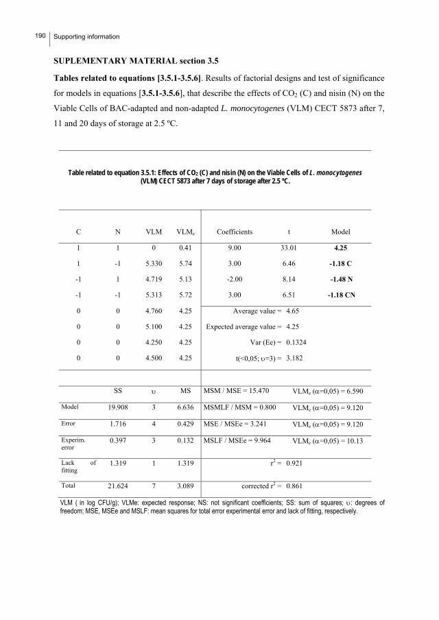
190 Supporting information
SUPLEMENTARY MATERIAL section 3.5
Tables related to equations [3.5.1-3.5.6]. Results of factorial designs and test of significance
for models in equations [3.5.1-3.5.6], that describe the effects of CO2 (C) and nisin (N) on the
Viable Cells of BAC-adapted and non-adapted L. monocytogenes (VLM) CECT 5873 after 7,
11 and 20 days of storage at 2.5 ºC.
Table related to equation 3.5.1: Effects of CO2 (C) and nisin (N) on the Viable Cells of L. monocytogenes (VLM) CECT 5873 after 7 days of storage after 2.5 ºC.
C N VLM VLMe Coefficients t Model
1 1 0 0.41 9.00 33.01 4.25
1 -1 5.330 5.74 3.00 6.46 -1.18 C
-1 1 4.719 5.13 -2.00 8.14 -1.48 N
-1 -1 5.313 5.72 3.00 6.51 -1.18 CN
0 0 4.760 4.25 Average value = 4.65
0 0 5.100 4.25 Expected average value = 4.25
0 0 4.250 4.25 Var (Ee) = 0.1324
0 0 4.500 4.25 t(<0,05; =3) = 3.182
SS MS MSM / MSE = 15.470 VLMe (=0,05) = 6.590
Model 19.908 3 6.636 MSMLF / MSM = 0.800 VLMe (=0,05) = 9.120
Error 1.716 4 0.429 MSE / MSEe = 3.241 VLMe (=0,05) = 9.120
Experim. error
0.397 3 0.132 MSLF / MSEe = 9.964 VLMe (=0,05) = 10.13
Lack of fitting
1.319 1 1.319 r2 = 0.921
Total 21.624 7 3.089 corrected r2 = 0.861
VLM ( in log CFU/g); VLMe: expected response; NS: not significant coefficients; SS: sum of squares; : degrees of freedom; MSE, MSEe and MSLF: mean squares for total error experimental error and lack of fitting, respectively.

Supporting information
191
Table related to equation 3.5.2: Effects of CO2 (C) and nisin (N) on the Viable Cells of L. monocytogenes (VLM) CECT 5873 after 11 days of storage after 2.5 ºC.
C N VLM VLMe Coefficients t Model
1 1 4.48 4.52 9.00 52.83 5.25
1 -1 5.04 5.08 3.00 3.22 -0.45 C
-1 1 4.46 4.50 -2.00 5.27 -0.74 N
-1 -1 6.87 6.91 3.00 3.28 0.46 CN
0 0 4.95 5.25 Average value = 5.30
0 0 5.36 5.25 Expected average value = 5.25
0 0 5.63 5.25 Var (Ee) = 0.0792
0 0 5.25 5.25 t(<0,05; =3) = 3.182
SS MS MSM / MSE = 20.454 VLMe (=0,05) = 6.590
MSMLF / MSM = 0.753 VLMe (=0,05) = 9.12 Model
Error
3.867
0.252
3
4
1.289
0.063 MSE / MSEe = 0.796 VLMe (=0,05) = 9.12
Experim. error
0.237 3 0.079 MSLF / MSEe = 0.185 VLMe (=0,05) = 10.13
Lack of fitting
0.015 1 0.015 r2 = 0.939
Total 4.119 7 0.588 corrected r2 = 0.893
Notations in previous table.

192 Supporting information
Table related to equation 3.5.3: Effects of CO2 (C) and nisin (N) on the Viable Cells of L. monocytogenes (VLM) CECT 5873 after 20 days of storage after 2.5 ºC.
C N VLM VLMe Coefficients t Model
1 1 3.76 3.43 9.00 130.74 5.72
1 -1 5.35 5.15 3.00 23.15 -1.43C
-1 1 6.50 6.29 -2.00 13.90 -0.86N
-1 -1 8.34 8.02 3.00 1.03 0CN
0 0 5.55 5.72 Average value = 5.46
0 0 5.56 5.72 Expected average value = 5.72
0 0 5.30 5.72 Var (Ee) = 0.0153
t(<0,05; =3) = 3.182 0 0 5.43 5.72
SS MS MSM / MSE = 44.749 VLMe (=0,05) = 6.590
Model 11.171 2 5.585 MSMLF / MSM = 0.526 VLMe (=0,05) = 9.120
Error 0.624 5 0.125 MSE / MSEe = 8.145 VLMe (=0,05) = 9.010
Experim. error
0.046 3 0.015 MSLF / MSEe = 9.550 VLMe (=0,05) = 10.13
Lack of fitting
0.578 2 0.289 r2 = 0.947
Total 11.795 7 1.685 corrected r2 = 0.926
Notations in previous table.

Supporting information
193
Table related to equation 3.5.4: Effects of CO2 (C) and nisin (N) on the Viable Cells of L. monocytogenes (VLM) CECT 5873 BAC-adapted after 7 days of storage after 2.5 ºC.
C N VLM VLMe Coefficients t Model
1 1 2.04 2.21 9.00 36.69 3.65
1 -1 3.65 3.94 3.00 4.05 -0.57 C
-1 1 3.04 3.35 -2.00 6.15 -0.87 N
-1 -1 4.91 5.08 3.00 0.50 0 CN
0 0 4.16 3.65 Average value = 3.89
0 0 3.90 3.65 Expected average value = 3.65
0 0 4.00 3.65 Var (Ee) = 0.0791
t(<0,05; =3) = 3.182 0 0 3.50 3.65
SS MS MSM / MSE = 14.756 VLMe =0,05) = 6.590
Model 4.293 2 2.146 MSMLF / MSM = 0.557 VLMe (=0,05) = 9.120
Error 0.727 5 0.145 MSE / MSEe = 1.840 VLMe (=0,05) = 9.010
Experim. error
0.237 3 0.079 MSLF / MSEe = 3.099 VLMe (=0,05) = 9.550
Lack of fitting
0.490 2 0.245 r2 = 0.855
Total 5.020 7 0.717 corrected r2 = 0.797
Notations in previous table.

194 Supporting information
Table related to equation 3.5.5: Effects of CO2 (C) and nisin (N) on the Viable Cells of L. monocytogenes (VLM) CECT 5873 BAC-adapted after 11 days of storage after 2.5 ºC.
C N VLM VLMe Coefficients t Model
1 1 3.78 3.85 9.00 105.19 5.24
1 -1 5.54 5.73 3.00 6.32 -0.45 C
-1 1 4.55 5.74 -2.00 13.35 -0.94 N
-1 -1 6.55 6.62 3.00 0.85 0.0CN
0 0 5.44 5.24 Average value = 5.37
0 0 5.26 5.24 Expected average value = 5.24
0 0 5.24 5.24 Var (Ee) = 0.0198
0 0 5.53 5.24 t(<0,05; =3) = 3.182
SS MS MSM / MSE = 51.095 VLMe (=0,05) = 5.790
Model 4.327 2 2.163 MSMLF / MSM = 0.518 VLMe (=0,05) = 19.250
Error 0.212 5 0.042 MSE / MSEe = 2.136 VLMe (=0,05) = 9.010
Experim. error
0.059 3 0.020 MSLF / MSEe = 3.839 VLMe (=0,05) = 9.550
Lack of fitting
0.152 2 0.076 r2 = 0.953
Total 4.538 7 0.648 corrected r2 = 0.935
Notations in previous table.

Supporting information
195
Table related to equation 3.5.6: Effects of CO2 (C) and nisin (N) on the Viable Cells of L. monocytogenes (VLM) CECT 5873 BAC-adapted after 20 days of storage after 2.5 ºC..
C N VLM VLMe Coefficients t Model
1 1 4.90 4.85 9.00 76.06 5.47
1 -1 5.40 5.35 3.00 3.66 -0.37 C
-1 1 4.57 4.51 -2.00 7.79 -0.79 N
-1 -1 7.23 7.18 3.00 5.31 0.54 CN
0 0 5.63 5.47 Average value = 5.42
0 0 5.14 5.47 Expected average value = 5.47
0 0 5.44 5.47 Var (Ee) = 0.0414
0 0 5.45 5.47 t(<0,05; =3) = 3.182
SS MS MSM / MSE = 38.310 VLMe (=0,05) =6.590
Model 4.233 3 1.411 MSMLF / MSM = 0.754 VLMe (=0,05) = 9.120
Error 0.147 4 0.037 MSE / MSEe = 0.890 VLMe (=0,05) = 9.120
Experim. error
0.124 3 0.041 MSLF / MSEe = 0.560 VLMe (=0,05) = 10.13
Lack of fitting
0.023 1 0.023 r2 = 0.966
Total 4.380 7 0.626 corrected r2 = 0.941
Notations in previous table.


77.. PPeerrssppeeccttiivveess


Perspectives
199
PERSPECTIVES
The results achieved throughout this Ph.D. thesis and the consequent conclusions drawn from
those results allows a number of future research lines to be enunciated:
1. Search for genes and proteins associated with biofilm formation in L.
monocytogenes, and quantitative analysis of such genes and proteins during biofilm formation
in different strains. A high variability was observed when initial adhesion and biofilm
formation of several strains were compared, and this seems to indicate that there might be
biological determinants associated with the type of biofilm formed.
2. Examine the potential of the method newly proposed for attaining benzalkonium
chloride-adapted L. monocytogenes. This should comprise a screening of a high number of
strains, firstly. Subsequently it should be studied if it can be extended to other biocides and
even to further bacterial species with the aim of establishing this method as a general protocol
in microbial physiology.
3. Design of disinfection strategies aimed at removing mature L. monocytogenes
biofilms. This also comprises the search of agents (of chemical, physical or biological origin)
that can diffuse into and disturb such biofilms and have or enhance bactericidal effects. This
search needs, in turn, to identify the chemical composition of the extracellular matrix, which
is unknown, and determine the microstructure of those biofilms. Furthermore, bacterial
species commonly associated with L. monocytogenes in the food environment must be
identified and physiological characteristics of mixed biofilms formed by these consortia must
be determined in order to design disinfection strategies correctly. Also, the study of the role of
each microorganism could give new interesting possibilities (based in microbial ecology) to
control high persistant biofilms.
4. Identify cross-responses to environmental and technological factors resulting from the
adaptation of L. monocytogenes to industrial biocides. For instance, it would be highly
interesting to find out if the cross-adaptive response between benzalkonium chloride and CO2
shown by L. monocytogenes CECT 5873 is also developed by other strains. Additionally, the
mechanisms of adaptation should be clarified by using molecular biology tools. These studies

200 Perspectives
would permit the potential risk associated with the routine use of each disinfectant in each
food processing plant to be better known.

RReessuummeenn


Resumen
203
RESUMEN
Introducción
Características biológicas: L. monocytogenes es un bacilo Gram positivo, anaerobio
facultativo, móvil a temperaturas inferiores a 25 ºC (Seeliger and Jones, 1986) y
altamente resistente en condiciones de estrés: pHs ácidos, baja aw, bajas
concentraciones de O2 y baja temperatura (Ross et al., 2000, Kathariou, 2002). Todo
ello contribuye a su ubicuidad (Cox et al. 1989, Ivanek et al. 2006) y a su condición de
bacteria patógena, causante de listeriosis.
Patogeneidad: está asociada a un grupo de riesgo constituido por mujeres
embarazadas, individuos de avanzada edad e inmunodeprimidos. A pesar de su
ubicuidad, la incidencia anual de listeriosis es de 0.3 casos al año por cada 100.000
habitantes, baja si la comparamos con otras infecciones trasmitidas por alimentos
(EFSA 2006). Lo que contrasta con su elevada tasa de mortandad (20-30%), haciéndola
especialmente relevante. Existen 13 serotipos: 1/2a, 1/2b, 1/2c, 3a, 3b, 3c, 4a, 4ab, 4b,
4c, 4d, 4e, 7 (Seeliger and Höhne, 1979; Seeliger and Jones, 1986), siendo 1/2a, 1/2b,
1/2c and 4b los responsables del 95% de los casos de listeriosis en humanos (Farber and
Peterkin, 1991; Doumith et al., 2004; Swaminathan and Gerner-Smidt, 2007).
Incidencia en alimentos: Según los datos epidemiológicos, el 99% de los casos de
listeriosis en humanos se deben al consumo de productos alimenticios contaminados. En
Europa rara vez se detectan productos contaminados por encima del límite de seguridad
(contaminación <10 UFC/g) pero los productos listos para el consumo en los que ha
sido detectada son productos de la pesca, cárnicos y quesos. De entre alimentos
anteriores, las últimas inspecciones realizadas en Europa mostraron una mayor
incidencia en productos de la pesca listos para el consumo, en los cuales se detectaron
una mayor proporción de muestras contaminadas por encima de 100 UFC/g (2.4%)
(EFSA, 2009), sobre todo durante y después del procesado (Cox et al., 1989; Hu et al.,
2006; Samelis and Metaxopoulos, 1999, Autio et al., 1999, Miettinen et al., 1999;
Norton et al., 2001; Rørvik et al., 1995; Vogel et al., 2001a, Wulff et al., 2006).).
204 Resumen
Control: Dada la importancia del este patógeno en el ámbito alimentario, se han
desarrollado diferentes estrategias para el control de Listeria monocytogenes a
diferentes niveles, desde la consideración de medidas para evitar su aparición mediante
la implementación del sistema de prerrequisitos y análisis de riesgo de los puntos
críticos de control (HACCP), hasta el diseño de estrategias de conservación que
aseguren el control de L. monocytogenes, pasando además por los esfuerzos realizados
en la aplicación de protocolos de limpieza y desinfección efectivos.
Justificación y objetivos
Con el objetivo final de incrementar la seguridad alimentaria respecto a L.
monocytogenes, tres razones justifican la realización del presente trabajo:
1. Que L. monocytogenes puede persistir en plantas de procesado formando
biopelículas, definidas como comunidades microbianas sésiles caracterizadas por
células irreversiblemente adheridas al sustrato o interfase y embebidas en una matriz de
sustancias poliméricas extracelulares (Donlan and Costerton 2002). En el desarrollo de
estas biopelículas desde el estado planctónico se pueden diferenciar tres fases: 1) Fase
de adherencia: reversible y dependiente de los nutrientes del medio (Kim and Frank
1994; Mai and Corner 2007; Stepanovic et al., 2004; Hood et al., 1997), del movimiento
flagelar (Lemon et al. 2007; Vatanyoopaisarn et al. 2000; Tresse et al., 2006 and Geeriri
et al., 2008), del tipo de superficie (Balckman and Frank, 1996; Di Bonaventura et al.,
2008; Krysinski and Brown 1992; Meylheuc et al., 2001; Rodríguez et al. 2008; Saá et
al., 2009; Safyan et al. 2006; Sinde and Carballo, 2000; Somers and Wong, 2004;
Smoot and Pierson, 1998; Teixeira et al., 2008) y de variables de estado como pH
(Herald and Zottola 1988; Tresse et al., 2006; Poimenidou et al., 2009) 2) Maduración:
durante la que las células bacterianas se adhieren ya de manera irreversible para formar
estructuras tipo microcolonias que segregan una matriz polimérica (EPSs) (Høiby et. al.
2010) y forman biopelículas que van incrementando su densidad a lo largo del tiempo
(Takhistov and George 2004, Marsh and Wang, 2003) dependiendo del medio externo
(Folsom et al 2006; Rodrigues et al. 2009) y 3) Desprendimiento: en la que el biofilm se

Resumen
205
desprende del sustrato por diversas razones tales como falta de nutrientes (Marshall,
1988) o restricciones en la transferencia de gases (Applegate and Bryers, 1991)
pudiendo ser fuente de contaminaciones cruzadas. En la actualidad se conocen dos
sistemas de comunicación célula-célula que regulan la formación de biopelículas en L.
monocytogenes: los sistemas LuxS y Agr (Belval et al. 2006; Garmyn et al., 2009).
Pero la importancia de la formación de biofilms en bacterias en general y en L.
monocytogenes en particular es todavía mayor si tenemos en cuenta que las biopelículas
de una sola especie raramente se encuentran en el ambiente y que, al menos en algunos
casos, la asociación entre diferentes especies para formar una biopelícula mixta
incrementa su resistencia a antimicrobianos (Simões et al., 2007a).
2: Que la persistencia de L. monocytogenes en plantas de procesado pudiera
relacionarse con su resistencia a aquellos biocidas frecuentemente utilizados en los
procesos de limpieza y desinfección (Gilbert et al. 2002; Lundén et al. 2000, 2003a). Y
de hecho, en numerosos estudios se ha demostrado que los biofilms de Listeria
monocytogenes son más resistentes que las células plantónicas a los desinfectantes
comúnmente empleados en la industria (Aarnisalo et al., 2000; Aarnisalo et al., 2007;
Amalaradjou et al., 2009; Ammor et al., 2004; Blackmann et al., 1996, Bremer et al.,
2002, Chavant et al., 2003; Gram et al., 2007, Kastberg 2009; Holah et al., 2002,
Leriche et al., 1999; Minei et al., 2008, Frank et al., 2003; 48 h: Pan et al., 2006, Yang
et al., 2009) tales como cloruro de benzalconio (perteneciente a la familia de QACs) y
ácido peracético.
3: Que las biopelículas de L. monocytogenes que persisten en las plantas de
procesado (con resistencias adquiridas) pueden transferirse fácilmente por contacto al
alimento, causando problemas graves de contaminación cruzada.
De acuerdo con lo expuesto, los objetivos del presente trabajo fueron:
1. Comparar las cinéticas de adhesión de diferentes cepas de L. monocytogenes (CECT
5873, CECT 936, CECT 911 y CECT 4032) en polipropileno (PP) y acero inoxidable
(SS) con y sin superficie de acondicionamiento, simulando residuos presentes en plantas
de procesado de mejillón. A partir de los resultados obtenidos en este primer objetivo se

206 Resumen
seleccionaron aquellos escenarios de mayor riesgo, que constituyeron los casos
experimentales para el desarrollo de los objetivos subsiguientes.
2. Desarrollar un procedimiento eficiente para la obtención de células de L.
monocytogenes adaptadas al cloruro de benzalconio (BAC). Para ello, se estudiaron, se
discutieron y se formalizaron los efectos del tamaño de inóculo y concentración de BAC
durante la exposición sobre el nivel final de adaptación. Además, se compararon los
perfiles proteicos de células adaptadas y no adaptadas al BAC.
3. Estudiar la formación de biopelículas maduras por tres cepas de L. monocytogenes
(CECT 4032, CECT 911, CECT 5873 y CECT 5873 adaptada al BAC) en aquellos
escenarios seleccionados en 1. como de mayor riesgo mediante el estudio del aumento
de resistencia a biocidas (BAC, ácido peracético y nisina) y el análisis microscópico de
la estructura biopelicular formada en diferentes etapas de la maduración a 25 ºC. Ello
permitió además comparar la efectividad de los biocidas e identificar algunas respuestas
cruzadas derivadas de la adaptación al cloruro de benzalconio.
4. Estudiar la influencia de la asociación entre Pseudomonas putida y L. monocytogenes
sobre la resistencia al BAC y la estructura microscópica de la biopelícula mixta formada
en diferentes escenarios respecto a la correspondiente biopelícula monoespecie de L.
monocytogenes.
5. Estudiar el efecto de la aplicación de atmósferas ricas CO2 y O2 sobre la viabilidad de
células procedentes de biopelículas de L. monocytogenes no adaptadas y adaptadas al
cloruro de benzalconio una vez transferidas a mejillón cocido y vivo, respectivamente.
En el caso del mejillón cocido, se estudió además la aplicación combinada de CO2 y
nisina mediante un diseño factorial de primer orden.
Resultados y Discusión
1. Efectos de la presencia de residuos de plantas de procesado de mejillón
sobre la adherencia de Listeria monocytogenes en polipropileno y acero
inoxidable. J. Food Protect. 72(9):1885-1890 (2009).

Resumen
207
1.1. Introducción
L. monocytogenes puede adherirse a las superficies en las superficies en la industria
alimentaria (Kim and Frank 1995) y desarrollar un biofilm, lo cual puede contribuir a su
persistencia (Frank and Koffi 1990; Pan et al., 2006). La adherencia bacteriana a
superficies es la fase inicial del desarrollo de biofilms. Los estudios recogidos en
bibliografía hasta el momento presentaban controversias entre sí (Harvey et al., 2007).
Por tanto, el estudio llevado a cabo, trató de esclarecerlas determinando la adherencia
no solo durante las primeras horas, y evitandom la utilización de medios comerciales
puesto que no reproducen las condiciones reales de las plantas de procesado.
Para ello, se compararon las cinéticas de adherencia de L. monocytogenes (cepas
CECT 5873, CECT 936, CECT 911 y CECT 4032) en polipropileno (PP) y acero
inoxidable (INOX). Además, se simularon dos situaciones que reproducen a escala
experimental dos casuísticas diferentes respecto a la acumulación de residuos resultado
de una limpieza deficitaria en una planta de procesado:
Contaminación con L. monocytogenes de superficies con residuos secos,
pegados al material, formando una película de acondicionamiento: PA.
Contaminación con L. monocytogenes de superficies limpias con residuos de
mejillón en disolución.
1.2. Materiales y Métodos
Cepas: las cepas se obtuvieron en la Colección Española de Cultivos Tipo (CECT)
Medios residuales: en estos experimentos se utilizaron el agua de cocción de
mejillón (ACM) y el agua intervalvar (AIV). La primera, obtenida a partir de la cocción
de los mejillones durante 1 min., resultó tener la siguiente composición: 9,19 g/l de
carbohidratos totales y 1698,7 mg/l de Nitrógeno; pH=7.74. El agua intervalvar,
obtenida a partir del escurrido de mejillón vivo, resultó tener la siguiente composición:
2,7g/l de carbohidratos totales, 185 mg/l de Nitrógeno; pH=8.80.
El inóculo se preparó ajustando el cultivo activado a una absorbancia de 0.1 a 700
nm correspondiente a una densidad celular de 108 CFU/ml de acuerdo con calibraciones
previas. Una vez se centrifuga a 6000 rpm, 25 ºC durante 10 min, las células

208 Resumen
recolectadas se resuspenden en el mismo volumen del medio residual objeto de estudio
en cada caso (tampón fosfato 0.05M diluido en agua destilada (1:1) (v:v) para aquellos
casos con película de acondicionamiento, ACM o bien AIV). En cada cupón se
dispensaron alícuotas de 250 μl de inóculo dentro del área circular (15 mm de diámetro)
previamente delimitada en los cupones (20 mm x 20 mm) mediante typpex. A
continuación se incubaron a 25 ºC y el número de células adheridas se cuantificó a
diferentes tiempos durante un período en torno a 60-80 h mediante el método puesto a
punto previamente en nuestro laboratorio (Herrera et al., 2007).
1.3. Resultados y discusión
Los valores de adherencia obtenidos en las diferentes condiciones ensayadas se
describieron satisfactoriamente mediante un modelo logístico modificado de acuerdo
con Cabo et al. (1999).
adadadad
admr)tm(r
e1
1
e1
aNCA
[1.1]
No obstante, en 3 de los 8 casos objeto de estudio en presencia de película de
acondicionamiento, se produce un descenso pronunciado del número de células
adheridas después de aproximadamente 24 h (CECT 911-PP y en INOX y la CECT
936-PP (Fig.1.1)), que resultó describible mediante un modelo empírico aditivo
representado por 2 términos logísticos:
DDDDadadadad
Dadmr)tm(rmr)tm(r
e1
1
e1
a
e1
1
e1
aNCA
[1.2]
NCA: número de células adheridas (UFC/mm2).
aad: máximo número de células adheridas (asíntota)
rad: coeficiente de adherencia específico (t-1).
mad: tiempo al cual el número de células adheridas es la mitad del máximo.
aD: número total de células adheridas que mueren o bien se desprenden
(UFC/mm2).

Resumen
209
rD: coeficiente de desprendimiento/muerte (t-1).
mD: tiempo al cual el número de células desprendidas/muertas es la mitad del
máximo.
0
200000
400000
600000
800000
No.
Adh
. Cel
ls (
CFU
/mm
2)911-PP 911-INOX
0
200000
400000
600000
4032-PP
0
200000
400000
600000
4032-INOX
0
200000
400000
600000
No.
Cél
. Adh
. (U
FC/m
m2)
5873-PP
0
200000
400000
600000
No.
Cél
. Adh
. (U
FC/m
m2)
5873-INOX
0
200000
400000
600000
0 20 40
tiempo (horas)
0
200000
400000
600000
0 20 40 60
tiempo (horas)
936-PP 936-INOX
0
200000
400000
600000
800000
No.
Cél
. Adh
. (U
FC/m
m2)
Figura 1.1 Cinéticas de adherencia de diferentes cepas de Listeria
monocytogenes en cupones de polipropileno (PP) y en acero inoxidable
(INOX) en presencia de agua de cocción de mejillón (ACM) sin (O) y con (F)
película de acondicionamiento. Las líneas representan las estimaciones
obtenidas empleando las ecuaciones [1.1] y [1.2].

210 Resumen
En la Figura 1.1. se representaron los ajustes de los resultados experimentales
obtenidos a las ecuaciones [1.1] y [1.2]. Tal y como se observa, los resultados
mostraron una mayor adherencia en polipropileno que en acero inoxidable en todas las
cepas de L. monocytogenes excepto para la CECT 5873. Se demostró además un efecto
positivo de la presencia de película de acondicionamiento sobre la adherencia inicial de
L. monocytogenes, aunque en la fase más tardía del estudio se produce desprendimiento
celular (Figura 1.1).
Además, a partir de estos primeros resultados se seleccionaron aquellos casos
experimentales (escenarios) que podrían dar lugar a unos mayores niveles de
adherencia: CECT 911-PP-SPA, 4032-PP-PA, 5873-INOX-SPA, y 4032-INOX-PA.
Precisamente en estos casos concretos, se comparó la adherencia con la obtenida en el
supuesto de una contaminación producida en plantas de procesado de mejillón vivo,
para lo que se utilizó agua intervalvar (AIV) de mejillón como medio de cultivo
(Figura 1.2).
Los resultados demostraron claramente mayores niveles de adherencia en presencia
de agua intervalvar, especialmente en los dos casos experimentales sin película de
acondicionamiento. Aunque para conocer la razón exacta de estos resultados sería
necesario recurrir a experimentación adicional, se postulan como posibilidades el que la
nueva composición del medio implique condiciones físico-químicas que incrementen la
adherencia celular o que dejen mayor espacio disponible.
Para los casos seleccionados a partir de los experimentos realizados como más
representativos de los resultados obtenidos se estudió la formación de biopelículas
maduras después de 4 y 11 días de incubación a 25 ºC (subapartado 3 de resultados y
discusión). Asimismo, con la finalidad de simular la formación de biopelículas
adaptadas al BAC en condiciones reales y de comparar la capacidad adaptativa a este
desinfectante de las tres cepas objeto de estudio, a continuación se desarrolló el segundo
de los objetivos expuestos, que permitió proponer un procedimiento para la obtención
de células de L. monocytogenes adaptadas al cloruro de benzalconio y mejorar
notablemente el método clásico basado en la exposición sucesiva de células en fase
estacionaria a concentraciones subletales de BAC.

Resumen
211
0
200000
400000
600000
800000
0 20 40 60 80
No.
Cél
. Adh
. (U
FC/m
m2)
tiempo (horas)
0
300000
600000
900000
1200000
1500000
1800000
0 20 40 60 80
No.
Cél
. Adh
. (U
FC/m
m2)
tiempo (horas)
A.
0
200000
400000
600000
800000
0 20 40 60 80
No.
Cél
. Adh
. (U
FC/m
m2)
tiempo (horas)
B.
0
200000
400000
600000
800000
0 20 40 60 80
No.
Cél
. Adh
. (U
FC/m
m2)
tiempo (horas)
C. D.
Figura 1.2 Comparación de las cinéticas de adherencia de Listeria
monocytogenes bajo las condiciones de mayor riesgo en presencia de agua
de cocción de mejillón (○) y de agua intervalvar () en diferentes casos
experimentales. A. CECT 911 en polipropileno en ausencia de película de
acondicionamiento (PP-SPA). B. CECT 4032 en polipropileno en presencia
de película de acondicionamiento (PP-PA). C. CECT 5873 en acero
inoxidable en ausencia de película de acondicionamiento (INOX-SPA). D.
CECT 4032 en acero inoxidable en presencia de película de
acondicionamiento (INOX-PA). Las líneas representan las estimaciones
obtenidas empleando las ec. .[1.1] o [1.2]
2. Desarrollo de un procedimiento eficiente para la obtención de células de
Listeria monocytogenes CECT 5873 adaptadas al cloruro de benzalconio.
2.1. Introducción
Numerosos estudios han demostrado que las cepas persistentes de L. monocytogenes
son resistentes a los compuestos de amonio cuaternario (QACs) (Aase et al. 2000;

212 Resumen
Mullapudi et al., 2008), entre ellos el cloruro de benzalconio (BAC), biocida
perteneciente a la familia QACs que se emplea comúnmente en la industria alimentaria.
Sin embargo, no existen trabajos previos centrados en el estudio de la adaptación de L.
monocytogenes al BAC.
En general, la obtención de bacterias resistentes adaptadas a biocidas se lleva a cabo
experimentalmente mediante subcultivos sucesivos de células en fase estacionaria en
medio de cultivo con concentraciones crecientes de BAC hasta la ausencia de
crecimiento, considerándose éste como el punto máximo de adaptación. Sin embargo,
este método clásico resulta tedioso, con fases experimentales demasiado largas y no
asegura alcanzar el máximo nivel de adaptación (To et al. 2002, Aarnisalo et al. 2007,
Aase et al. 2000). En este sentido, y partiendo de que las células en fase exponencial son
metabólicamente más activas (Whistler et al., 1998; Zotta et al., 2009), una alternativa
posible podría ser diseñar un procedimiento experimental basado en la exposición de
células en fase exponencial.
De acuerdo con ello, en el presente trabajo se planteó en dos etapas: 1) comparación
entre el nivel de adaptación alcanzado por 3 cepas de L. monocytogenes (CECT 4032,
CECT 911 Y CECT 5873) una vez aplicado el protocolo experimental clásico y
selección de aquella cepa con mayor capacidad de adaptación y 2) estudio del efecto del
tamaño del inóculo y de la concentración de BAC sobre el nivel de adaptación
alcanzado tras una sola exposición de células de L. monocytogenes CECT 5873 en fase
exponencial. Para ello, se utilizó un diseño factorial de primer orden que permitió
describir adecuadamente los resultados en el ámbito experimental ensayado. A partir de
los resultados obtenidos, se propuso un método que mejora notablemente el actualmente
utilizado, permitiendo alcanzar mayores niveles de adaptación en menos tiempo.
Además, se realizaron estudios proteómicos preeliminares para identificar cambios en
los perfiles proteicos consecuencia de la adaptación al BAC y se identificaron posibles
genes (así como sus proteínas asociadas) relacionados con dicha adaptación.
2.2. Materiales y Métodos
Cepas: Las cepas utilizadas se obtuvieron en la CECT.

Resumen
213
Procedimientos experimentales:
i. Cultivos sucesivos en fase estacionaria (método clásico): consistente en
siembras sucesivas de 1 ml de inóculo en fase estacionaria en 49 ml de TSB
(Caldo de Soja y Triptona, Cultimed, S.L.) con concentraciones crecientes de
BAC (Sigma Chemical Co) hasta la ausencia de crecimiento. Estos primeros
experimentos se realizaron con las cepas CECT 911, CECT 4032 y CECT
5873.
ii. Cultivos en fase exponencial: consistente una sola exposición de
combinaciones [inóculo de L. monocytogenes CECT 5873 (I): BAC (B)] de
acuerdo a un plan factorial ortogonal de primer orden (Tabla 2.1).
Recolección de células: después de cada exposición, las células se recolectaron
mediante centrifugación (2000 g durante 5 minutos a 25 ºC) y se almacenaron
congeladas en glicerol al 50% (v/v) hasta los ensayos para determinar su resistencia.
Determinación de la resistencia de las células de L. monocytogenes CECT 5873
recolectadas al cloruro de benzalconio. Mediante el cálculo de la dosis letal 50 (DL50) a
partir de un ensayo tipo dosis-respuesta, de acuerdo con lo descrito por Cabo et al.
1999.
Tabla 2.1: Valores naturales y codificados de las variables en el diseño factorial
Valores naturales
Valores codificados BAC (mg/l) Inóculo (ml)
1;1 9 25
1;-1 9 5
-1;1 3 25
-1;-1 3 5
0;0 6 15
Ensayos de proteómica: la obtención y comparación de los perfiles proteicos de las
células adaptadas y no adaptadas al BAC se realizó de acuerdo a los procedimientos de
Sánchez et al. 2010.

214 Resumen
2.3. Resultados y discusión
Adaptación de células de L. monocytogenes CECT 5873, 911 y 4032 al BAC
aplicando el método clásico.
Los resultados obtenidos mostraron que en las cepas 911 y 4032 el nivel de
adaptación máxima se alcanzó después de tan solo una exposición (desde DL50 de 3.80
mg/l en la salvaje hasta 5.06 mg/l en el caso de la 911 y desde 6.96 mg/l hasta DL50 9.19
mg/l en el caso de la 4032), lo cual demuestra la falta de optimización del método
clásico e indica que el control de la adaptación de la cepa objeto de estudio después de
cada exposición al BAC permitiría, al menos en algunos casos, reducir el tiempo de
experimentación empleado en estudios previos (Aase et al. 2000; To et al. 2002;
Romanova et al. 2006).
Por el contrario, la cepa 5873 no alcanzó su nivel máximo de adaptación ni después
de 5 exposiciones sucesivas al BAC, indicando una mayor capacidad adaptativa de esta
cepa respecto a las anteriores. Dicha capacidad adaptativa se refleja en un mayor
incremento entre la resistencia de la cepa 5873 salvaje (DL50=2.71 mg/l) y la cepa 5873
adaptada después de 5 exposiciones (DL50=7.1 mg/l). Capacidad que justifica la
selección de esta cepa para los estudios subsiguientes, en los que se ensayaron los
efectos del tamaño del inóculo y de la concentración de BAC sobre el nivel de
adaptación tras una sola exposición de células de L. monocytogenes 5873 en fase
exponencial.
Una estrategia alternativa para la obtención de células adaptadas de L.
monocytogenes CECT 5873 al cloruro de benzalconio.
Los resultados experimentales resultaron describibles mediante la siguiente ecuación
empírica (poner el valor de r=0.925), cuya representación se observa en la Figura 2.1:
DL50=7,82-0,99B+1,42I+2,24BI [2.1]
A partir de la ecuacion [2.1] se deduce claramente la importancia del tamaño del
inóculo durante las exposiciones en la resistencia final adquirida así como una

Resumen
215
interacción positiva entre aquél y la concentración de BAC. Sin embargo, el efecto
individual negativo del cloruro de benzalconio refleja la necesidad de mantener la
relación [nºde células: BAC] en un valor tal que se encuentre por encima de los límites
de crecimiento de la cepa. En definitiva, como consecuencia de la aplicación de este
protocolo, y dentro del ámbito experimental, se consiguió incrementar la resistencia al
BAC aproximadamente por un factor de 3.69 (DL50=10 mg/l) con respecto a la cepa
salvaje (células no adaptadas DL50=2.71 mg/l) después de 33 horas de exposición. Ello
supuso una mejora considerable respecto al método tradicional, con el que la resistencia
al BAC de L. monocytogenes CECT 5873 se incrementó por un factor de factor: 2.61
después de después de 5 días de exposiciones sucesivas a concentraciones subletales de
BAC.
DL
50 (
mg
/l)
Figura 2.1. Efectos del tamaño de inóculo y de la concentración de BAC
durante la exposición sobre la resistencia (DL50) de células de L.
monocytogenes CECT 5873. Las variables independientes se expresan en
valores codificados (Tabla 2.1).
Además, un análisis comparativo inicial entre los perfiles proteicos de células de L.
monocytogenes CECT 5873 no adaptada y adaptada al BAC demostró que la adaptación
al biocida parece acompañarse de una represión proteica.
Por último, con el objetivo final de identificar aquellos genes responsables de la
resistencia al BAC en L. monocytogenes, se llevó a cabo un estudio de sensibilidad al
BAC de un banco de mutantes (alrededor de 10000 para ambas cepas) de Listeria
monocytogenes EGDe y LO28, identificándose los genes y proteínas asociadas con la
resistencia al biocida en aquellos casos que obtuvimos valores de MIC superiores a la
cepa salvaje. La implicación exacta de la proteína codificada lmo2277 en la resistencia

216 Resumen
de L. monocytogenes EDGe al BAC se está llevando a cabo mediante mutagénesis y
estrategias de clonación.
3. Resistencia al cloruro de benzalconio, ácido peracético y nisina durante la
formación de biofilms maduros de Listeria monocytogenes. Food Microbiology.
doi:10.1016/j.fm.2010.09.014.
3.1. Introducción
Una de las consecuencias de la formación de biofilms que puede provocar serios
problemas económicos y de salud es la adquisición de resistencia (adaptativa) a biocidas
(Aarnisalo et al., 2007; Gram et al., 2007; Leriche et al., 1999; Minei et al., 2008). De
hecho, la eficacia de diferentes biocidas para erradicar biofilms de L. monocytogenes ha
sido evaluada en varios estudios (Aarnisalo et al., 2000; Ammor et al., 2004; Chavant et
al., 2003; Tyh-JenK and Frank, 2003). La mayoría se han centrado en desinfectantes
industriales como compuestos de cloruro cuaternario, alcoholes, compuestos clorados y
otros agentes oxidantes tales como el ácido peracético, ozono y derivados de peróxidos
(Aarnisalo et al., 2007; Frank et al., 2003; Pan et al., 2006; Krysinski and Brown, 1992;
González-Fandos et al., 2005). Por el contrario, existen pocos estudios de
antimicrobianos naturales y sólo algunos de aceites esenciales (Chorianopoulus et al.,
2008; Sandasi et al., 2008; Sandasi et al., 2009) y ácido láctico y bacteriocinas
producidos por biofilms de bacterias lácticas (Leriche et al., 1999; Minei et al., 2008;
Guerrieri et al., 2009).
La mayoría de los trabajos existentes en bibliografía emplean biofilms inmaduros (72
h: Aarnisalo et al., 2007; 24 h: Bonaventura et al., 2008; 40 h: Borucki et al., 2003; 28
h: Frank et al., 2003; 48 h: Pan et al., 2006). La excepción es el grupo de Gram et al.,
(2007, 2008), en cuyos estudios simulan condiciones reales con biopelículas de más de
7 días. Sin embargo, de acuerdo con resultados previos obtenidos en esta tesis, 60 horas
de incubación a 25ºC resultaba insuficiente para la formación de una biopelícula madura
resistente. Por ello, en el presente capítulo se estudió la formación de las biopelículas
maduras durante 11 días a 25 ºC en aquellos casos seleccionados como de mayor riesgo
a partir del desarrollo del objetivo 1. y de la CECT 5873 adaptada al BAC obtenida tras

Resumen
217
el desarrollo del objetivo 2. Para la evaluación del estado de maduración de las
biopelículas se utilizaron dos criterios complementarios: la adquisición de resistencia a
tres biocidas (cloruro de benzalconio (BAC), ácido peracético (PA) y nisina (NIS) y el
análisis microscópico de las estructuras biopeliculares formadas. Ello supone una
mejora importante del planteamiento experimental habitual en la bibliografía publicada,
en donde la formación de biopelículas es evaluada frecuentemente cuantificando
simplemente biomasa (Di Bonaventura, 2008; Djordjevic et al., 2002; Harvey et al.,
2006; Folsom et al., 2006). Y sólo en algunos casos se realizaron análisis microscópicos
in situ (Borucky et al., 2003; Chae and Scraft, 2000; Chavant et al., 2003; Kalmokoff et
al., 2001; Rodríguez et al., 2008; Moltz and Martin, 2005).
3.2. Materiales y Métodos
Casos experimentales estudiados:
911-PP-AIV (Biopelículas maduras de L. monocytogenes CECT 911
formadas sobre polipropileno y con agua intervalvar de mejillón)
4032-PP-ACM (Biopelículas maduras de L. monocytogenes CECT 4032
formadas sobre polipropileno (PP) y con agua de cocción de mejillón)
5873-INOX-ACM (Biopelículas maduras de L. monocytogenes CECT 5873
formadas sobre acero inoxidable y con agua de cocción de mejillón).
5873A-INOX-ACM (Biopelículas maduras de L. monocytogenes CECT 5873
adaptada al BAC (LD50: 2.71 mg/l; Cepa adaptada al BAC: 7.2 mg/l)
formadas sobre acero inoxidable (INOX) y con agua de cocción de mejillón.
Esta cepa se obtuvo en experimentos previos mediante 2 exposiciones
sucesivas a concentraciones crecientes de inóculo y subletales de BAC de la
cepa salvaje correspondiente.
Medios residuales y biocidas: el agua de cocción de mejillón e intervalvar se preparó
tal y como se describe en el primer apartado (Saá et al., 2009). Posteriormente el pH se
ajustó a 7 y se autoclavó a 121 ºC durante 15 minutos para su esterilización.
218 Resumen
Biopelículas: la formación de las biopelículas se llevó a cabo en cupones (20 mm x
20 mm) de acero inoxidable o polipropileno previamente esterilizados y situados
individualmente en mini placas Petri. En todos los casos, se añadieron 9 ml de inóculo
para el desarrollo del biofilm. Y a cada tiempo de ensayo (4 y 11 días), los cupones se
lavan mediante inmersión en PBS durante 10 s para eliminar las células no adheridas.
Ensayos dosis-respuesta y determinación del nº de células viables
Se añaden 0.5 ml de cada concentración de biocida (BAC (mg/l): 2.5, 5,10, 17, 25,
50, 100, 250, 500, 1000; PA (mg/l): 1, 2.5, 5, 10, 17, 25, 50, 100, 250, 500; Nisina
(UI/ml): 5, 10, 15, 20, 50, 75, 100, 125, 150, 165, 185, 200, 400, 1500) sobre toda la
superficie del cupón previamente lavado. Se espera el tiempo de exposición
predeterminado (10 minutos) y se neutraliza añadiendo 10 ml de una disolución
neutralizante (34 g/l KH2PO4 ajustado a pH 7.2 con NaOH, 3 g/l soybean lecithin, 30
ml/l Tween 80, 5 g/l Na2S2O3 y 1 g/l L-histidine). El número de células viables se
determina siguiendo el método ya descrito. En los ensayos realizados en células
plantónicas se utilizó el mismo procedimiento, pero en tubos y utilizando TSB como
medio de ensayo. Experimentos preeliminares permitieron conseguir aproximadamente
la misma concentración de inóculo en ambos sistemas. Todos los experimentos dosis-
respuesta se realizaron por triplicado
3.3. Resultados
Resistencia al cloruro de benzalconio de células plantónicas y biopelículas de L. monocytogenes.
Se compararon los valores de resistencia (LD90) al BAC obtenidos en células
plantónicas con los obtenidos en el caso de biopelículas maduras (tras 4 y 11 días).
Además, y con el objetivo de investigar si la adaptación fisiológica después de la
exposición al BAC desencadenaba resistencias cruzadas en las biopelículas maduras que
pudieran formar, la cepa CECT 5873 adaptada al BAC (LD50= 7.22) se incluyó en este
y en los siguientes estudios.
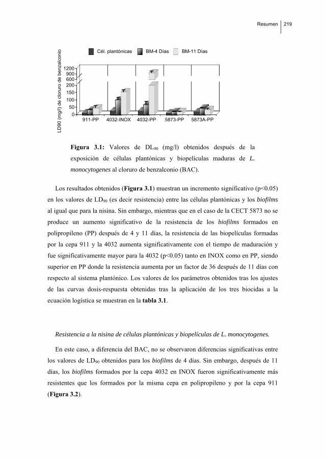
Resumen
219
911-PP 4032-INOX 4032-PP 5873-PP 5873A-PP0
50
100
150
200600900
1200
LD
90
(m
g/l)
de
clo
ruro
de
be
nza
lco
nio Cél. plantónicas BM-4 Días BM-11 Días
Figura 3.1: Valores de DL90 (mg/l) obtenidos después de la
exposición de células plantónicas y biopelículas maduras de L.
monocytogenes al cloruro de benzalconio (BAC).
Los resultados obtenidos (Figura 3.1) muestran un incremento significativo (p<0.05)
en los valores de LD90 (es decir resistencia) entre las células plantónicas y los biofilms
al igual que para la nisina. Sin embargo, mientras que en el caso de la CECT 5873 no se
produce un aumento significativo de la resistencia de los biofilms formados en
polipropileno (PP) después de 4 y 11 días, la resistencia de las biopelículas formadas
por la cepa 911 y la 4032 aumenta significativamente con el tiempo de maduración y
fue significativamente mayor para la 4032 (p<0.05) tanto en INOX como en PP, siendo
superior en PP donde la resistencia aumenta por un factor de 36 después de 11 días con
respecto al sistema plantónico. Los valores de los parámetros obtenidos tras los ajustes
de las curvas dosis-respuesta obtenidas tras la aplicación de los tres biocidas a la
ecuación logística se muestran en la tabla 3.1.
Resistencia a la nisina de células plantónicas y biopelículas de L. monocytogenes.
En este caso, a diferencia del BAC, no se observaron diferencias significativas entre
los valores de LD90 obtenidos para los biofilms de 4 días. Sin embargo, después de 11
días, los biofilms formados por la cepa 4032 en INOX fueron significativamente más
resistentes que los formados por la misma cepa en polipropileno y por la cepa 911
(Figura 3.2).

220 Resumen
911-PP 4032-INOX 4032-PP 5873-PP 5873A-PP0
100
200
300
400
500
600
DL
90
(U
I/m
l) d
e n
isin
a.
Cél. plantónicas BM-4días BM-11días
Figura 3.2: Valores de DL90 (mg/l) obtenidos después de la exposición de
células plantónicas y biopelículas maduras de L. monocytogenes a nisina.
De nuevo, los biofilms formados por la cepa 5873 resultaron ser los más sensibles, no
observándose diferencias significativas entre los valores de su resistencia obtenidos
durante la maduración. Sin embargo, se produjo un incremento significativo en la
resistencia (p < 0.05) al BAC de la cepa adaptada con respecto a los biofilms formados
por la no adaptada después de 11 días de incubación.
Table 3.1: Valores de los parámetros (k y r) obtenidos del ajuste de los datos experimentales a un modelo
logístico (Cabo et al., 1999; Cabo et al., 2009)
911-PP-IWM 4032-SS-MCJ 4032-PP-MCJ 5873-PP-MCJ 5873A-PP-MCJ
BM. 4D BM.11D BM. 4D BM.11D BM. 4D BM.11D BM. 4D BM.11D BM. 4D BM.11D
KBAC 97.363 95.506 99.65 99.450 99.060 93.188 99.477 99.097 99.748 91.275
rBAC 0.308 0.1746 0.053 0.033 0.235 0.007 0.256 0.231 0.138 0.404
Knisin 99.595 99.056 86.413 91.913 84.872 87.189 69.412 83.274 76.843 89.048
rnisin 0.013 0.007 0.215 0.006 0.255 0.023 0.028 0.327 0.0730 0.029
KPA 99.342 99.810 99.704 99.736 99.998 97.457 98.976 98.560 94.353 99.883
rPA 0.330 0.419 0.169 0.069 0.480 0.097 0.439 0.263 2.816 2.255
Por último, en el caso de la nisina, y tal y como se muestra en la tabla 3.1., se
obtienen menores valores de k y de r que en el caso del BAC y el ácido peracético

Resumen
221
Resistencia a ácido peracético (AP) de células plantónicas y biopelículas de L. monocytogenes.
En el caso del ácido peracético, y a diferencia de los anteriores, se obtuvieron valores
de LD90 significativamente más altos para las células plantónicas cuando se utilizaba
TSB como medio de cultivo, que para las biopelículas maduras después de 4 y 11 días
de incubación (Figura 3.3). Sin embargo, cuando en experimentos posteriores (data not
shown) se sustituyó el TSB por tampón fosfato (PBS) se obtenían los mismos valores de
resistencia que los obtenidos en los biofilms de 4 días de maduración, indicando un
efecto de la materia orgánica presente en el medio (data not shown).
Únicamente en el caso de la cepa 4032 se observó un incremento significativo en la
resistencia (p < 0.05) entre 4 y 11 días de maduración en ambos materiales. Finalmente,
en el caso de la cepa 5873 adaptada al BAC, los valores de resistencia de las
biopelículas maduras formadas tras 4 y 11 días de incubación fueron significativamente
menores que en el caso de las formadas por la cepa salvaje correspondiente.
911-PP 4032-INOX 4032-PP 5873-PP 5873A-PP0
20406080
100300350400
DL
90
(m
g/l)
de
áci
do
pe
racé
tico
Cél. plantónicas BM-4días BM-11días
Figura 3.3: Valores de DL90 (mg/l) obtenidos después de la exposición de
células plantónicas y biopelículas maduras de L. monocytogenes al ácido
peracético (AP)
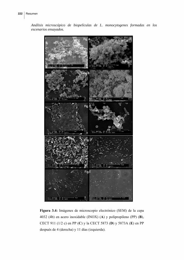
222 Resumen
Análisis microscópico de biopelículas de L. monocytogenes formadas en los escenarios ensayados.
Figura 3.4: Imágenes de microscopio electrónico (SEM) de la cepa
4032 (4b) en acero inoxidable (INOX) (A) y polipropileno (PP) (B),
CECT 911 (1/2 c) en PP (C) y la CECT 5873 (D) y 5873A (E) en PP
después de 4 (derecha) y 11 días (izquierda).
FFiigg.. CC
FFiigg.. AA
FFiigg..BB

Resumen
223
Las imágenes obtenidas después de 4 y 11 días de maduración mostradas en la
Figura 3.4 permitieron diferenciar tres niveles de densidad celular, siendo claramente la
CECT 4032 la que muestra una estructura tridimensional más compleja.
Estudio de la relación de las cinéticas de adherencia y la formación de biopelículas
de L. monocytogenes.
Las cinéticas de adherencia obtenidas (Figura 3.5) muestran la ausencia de
diferencias significativas entre el número máximo de células adheridas de Listeria en
los diferentes escenarios.
A. B.
0 2 4 6 8 10 12 14
tiempo (dias)
C.
0
2
4
6
8
10
0 2 4 6 8 10 12 14
log
cel.
adh
(UF
C/m
m2)
.
tiempo (dias)
B
0 2 4 6 8 10 12 14
tiempo (dias)
Figura 3.5: Cinéticas de adherencia de Listeria monocytogenes en
diferentes casos experimentales. CECT 911 en polipropileno (E) en
presencia de agua intervalvar (AI). CECT 4032 en acero inoxidable (V) en
presencia de agua de cocción de mejillón (ACM) y en polipropileno (B) en
ACM C: CECT 5873 (C) y la CECT 5873A (adaptada al BAC) (H) en
polipropileno en ACM.
3.4. Discusión
Los resultados obtenidos demostraron que la cepa 4032 es la que forma biopelículas
maduras más resistentes después de 11 días de incubación a 25 ºC (Figuras 3.1, 3.2,
3.3) con respecto a los 4 días y al estado plantónico. Además, las imágenes obtenidas
tras el análisis microscópico mostraron que es también esta cepa la que forma
estructuras tridimensionales más complejas (Figura 3.4). Los resultados demostraron,
por tanto, una relación entre la estructura tridimensional de las biopelículas maduras de
L. monocytogenes y su resistencia a los biocidas ensayados. Esta aparente relación entre
estructura-resistencia apoya la teoría de que el aumento de resistencia observado en
biopelículas bacterianas tiene más que ver con la disposición de las células que con
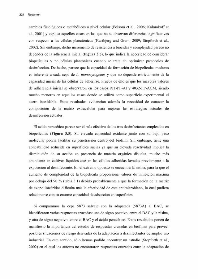
224 Resumen
cambios fisiológicos o metabólicos a nivel celular (Folsom et al., 2006; Kalmokoff et
al., 2001) y explica aquellos casos en los que no se observan diferencias significativas
con respecto a las células planctónicas (Kastbjerg and Gram, 2009; Stopforth et al.,
2002). Sin embargo, dicho incremento de resistencia a biocidas y complejidad parece no
depender de la adherencia inicial (Figura 3.5), lo que indica la necesidad de considerar
biopelículas y no células plantónicas cuando se trata de optimizar protocolos de
desinfección. De hecho, parece que la capacidad de formación de biopelículas maduras
es inherente a cada cepa de L. monocytogenes y que no depende estrictamente de la
capacidad inicial de las células de adherirse. Prueba de ello es que los mayores valores
de adherencia inicial se observaron en los casos 911-PP-AI y 4032-PP-ACM, siendo
mucho menores en aquellos casos donde se utilizó como superficie experimental el
acero inoxidable. Estos resultados evidencian además la necesidad de conocer la
composición de la matriz extracelular para mejorar las estrategias actuales de
desinfección actuales.
El ácido peracético parece ser el más efectivo de los tres desinfectantes empleados en
biopelículas (Figura 3.3). Su elevada capacidad oxidante junto con su bajo peso
molecular podría facilitar su penetración dentro del biofilm. Sin embargo, tiene una
aplicabilidad reducida en superficies sucias ya que su elevada reactividad implica la
disminución de su acción en presencia de materia orgánica disuelta, mucho más
abundante en cultivos líquidos que en las células adheridas lavadas previamente a la
exposición al desinfectante. En el extremo opuesto se encuentra la nisina, para la que el
aumento de complejidad de la biopelícula proporciona valores de inhibición máxima
por debajo del 90 % (tabla 3.1) debido probablemente a que la formación de la matriz
de exopolisacáridos dificulta más la efectividad de este antimicrobiano, lo cual pudiera
relacionarse con su enorme capacidad de adsorción en superficies.
Si comparamos la cepa 5873 salvaje con la adapatada (5873A) al BAC, se
identificaron varias respuestas cruzadas: una de signo positivo, entre el BAC y la nisina,
y otra de signo negativo, entre el BAC y el ácido peracético. Estos resultados ponen de
manifiesto la importancia del estudio de respuestas cruzadas en biofilms para preveer
posibles situaciones de riesgo derivadas de la adaptación a desinfectantes de amplio uso
industrial. En este sentido, sólo hemos podido encontrar un estudio (Stopforth et al.,
2002) en el cual los autores no encontraron respuestas cruzadas entre la adaptación de

Resumen
225
biofilms de L. monocytogenes al ácido y la exposición subsiguiente al AP, al BAC y al
hipoclorito sódico.
Finalmente, los resultados obtenidos muestran que la utilización de ingredientes
puros activos mejora los empleados habitualmente, productos comerciales mezclas de
varios compuestos, ya que evitan posibles respuestas de resistencia múltiples (Frank et
al., 2003; González-Fandos et al., 2005; Jacquet and Reynaud, 1994) Paula, yo este
párrafo lo sacaría, realmente no hacemos ninguna comparación entre los desinfectantes
puros y los comerciales.
La determinación de la resistencia del biofilm (DL90) a partir de principios básicos de
la cinética microbiana permite su adecuada cuantificación y posibilita la comparación
objetiva entre los distintos casos estudiados. Ello supone además una alternativa útil a la
utilizada en la mayoría de los trabajos previos, en donde sólo se estudian 2 o tres
concentraciones de biocidas en los ensayos dosis-respuesta (Aarnisalo et al., 2000; Thy-
Jenq et al., 1993), se dosifican mezclas de desinfectantes (en algunos casos de
composición desconocida) y se cuantifica la resistencia del biofilm mediante métodos
semicuantitativos (MIC) y durante un periodo demasiado corto de incubación.
4. Cinéticas de adherencia, resistencia al cloruro de benzalconio y análisis
microscópico de biopelículas mixtas formadas por Listeria monocytogenes y
Pseudomonas putida.
4.1. Introducción
Una de las vías frecuentes de contaminación alimentaria es mediante contaminación
cruzada a través de superficies de plantas de procesado de alimentos (Ammor et al.,
2004; Norwood and Gilmour, 1999; Porsby et al., 2008) donde L. monocytogenes puede
adherirse y formar biofilms (Aase et al., 2000; To et al., 2002; Takahashi et al., 2009).
Esta resistencia se relaciona con la estructura tridimensional de las células que
constituyen el biofilm, la cual es el resultado de interacciones entre las especies
bacterianas presentes (Wuertz et al., 2004) y determina el grado de acceso de los
biocidas a las células (Bourion and Cerf, 1996, Qu et al., 2010). Además, se ha
demostrado que las células de los biofilms son fenotípica y genotípicamente diferentes a

226 Resumen
las células plantónicas (Nadell et al., 2008). Esta situación se complica aún más debido
a que en situaciones reales más de dos especies bacterianas coexisten formando biofilms
y se ha demostrado que esto podría incrementar su resistencia a los biocidas (Sharma
and Anand, 2002; Kastbjerg and Gram, 2009).
4.2. Matreriales y métodos
En el presente trabajo se estudió el efecto de la presencia de P. putida CECT 845
sobre la resistencia al cloruro de benzalconio de biopelículas maduras formadas por L.
monocytogenes en los 5 escenarios considerados en el apartado 3. Se utilizó el mismo
diseño experimental que el descrito en dicho apartado, salvo la inoculación simultánea
de P. putida y L. monocytogenes a la misma concentración (108 UFC/ml en ambos
casos).
4.3. Resultados y discusión
Comparación entre la adherencia de L. monocytogenes en monocultivo y en presencia
de Pseudomonas putida CECT 845
Se compararon las cinéticas de adherencia de 3 cepas de L. monocytogenes (CECT
911, 4032 and 5873) en monocultivo y co-cultivo con P. putida en diferentes escenarios
seleccionados de acuerdo a los estudios previos (Saá et al. 2009; Saá Ibusquiza et al.
2010).
A. B.
0 2 4 6 8 10 12 14
tiempo (dias)
C.
0 2 4 6 8 10 12 14
tiempo (dias)
D. E.
0 2 4 6 8 10 12 14
tiempo (dias)
0 2 4 6 8 10 12 14
tiempo (dias)
0
1
2
3
4
5
6
7
0 2 4 6 8 10 12 14
log
cel.
adh.
(U
FC/m
m2)
.
tiempo (dias)
Figura 4.1: Cinéticas de adherencia de Listeria monocytogenes en monocultivo (O)
y en presencia de Pseudomonas putida 845 (●). A: CECT 911 en polipropileno y
agua intervalvar (AIV); B: CECT 4032 en acero inoxidable y agua de cocción de
mejillón (ACM); C: CECT 4032 en polipropileno en ACM; D: CECT 5873 en
polipropileno en ACM and E: CECT 5873A (adaptada al BAC) en polipropileno en
ACM. Además se incluyen en esta gráfica las cinéticas de adherencia observadas de
P. putida (*) y esperadas (línea continua) en las biopelículas mixtas.

Resumen
227
Tal y como muestra la figura 4.1, el resultado más significativo es el descenso
pronunciado del número de células adheridas en presencia de P. putida observado en el
caso de la cepa 4032 y la 5873 adaptada al BAC en polipropileno (Figura 4.1.B. y
4.1.E.).
Aunque el modelado de los datos experimentales facilita su comparación, en el caso
de L. monocytogenes la ausencia de puntos antes en los 4 primeros días imposibilitó el
ajuste matemático. No así en el caso de P. putida, donde los resultados de adherencia
resultaron describibles (r2=0.995) mediante un modelo aditivo de los ecuaciones
logísticas que describen adecuadamente el incremento y descenso de la población de la
biopelícula (ecuación 1.2) (Herrera et al. 2007; Saá et al. 2009). Y este ajuste de los
datos experimentales permitió identificar 2 patrones de adherencia de este
microorganismo en presencia de L. monocytogenes, que, como veremos, van a dar lugar
a dos patrones de asociación del inóculo mixto:
Patrón tipo A: que tiene lugar cuando Pseudomonas se asocia con las cepas
de Listeria 4032, especialmente en INOX, y con la 5873 salvaje (Fig. 4.1.B.
C. y D. respectivamente). Se caracteriza por mayores niveles de adherencia
máxima (veánse valores de parámetro aad y rad en la tabla 4.1) que los
obtenidos en el patrón B.
Tabla 4.1.: Valores de los parámetros obtenidos tras ajuste de los resultados experimentales de adherencia
de Ps. putida en presencia de diferentes cepas de L. monocytogenes a la ecuación [1.2]
911-PP-IWM 4032-SS-MCJ 4032-PP-MCJ 5873-PP-MCJ 5873-PP-MCJ
aad 30499 1800000 370723 199237 45000
rad 5,014 2,974 1,101 3,204 5,014
mad 0,700 1,843 0,998 1,881 0,700
aD 26244 1497876 268936 184311 26244
rD 10,425 5,500 4,173 4,965 8,500
mD 1,676 4,500 1,820 51,632 8,000
Patrón tipo B: que tiene lugar cuando Pseudomonas se asocia con las cepas
de Listeria 911 y la 5873 adaptada al BAC (Fig. 4.1.A. y E.

228 Resumen
respectivamente). En este caso, los valores máximos de adherencia de P.
putida descienden 1-log con respecto al patrón de tipo A.
Estos dos patrones identificados van a dar lugar a biopelículas maduras de 4 días de
maduración con diferente resistencia al cloruro de benzalconio.
Comparación entre la resistencia al BAC de biopelículas monoespecie de L.
monocytogenes y biopelículas mixtas formadas tras la asociación de L. monocytogenes
y P. putida
Los resultados obtenidos mostraron que P. putida tiene un papel determinante en la
formación de las estructuras biopeliculares mixtas, observándose que su asociación con
L. monocytogenes modifica significativamente (p<0.05) la resistencia al BAC después
de 4 y 11 días de incubación a 25 ºC respecto a los biofilms monoespecie (Figura 4.2).
No obstante, los efectos de la presencia de P. putida fueron diferentes tras 4 y 11 días de
incubación.
Después de 4 días de incubación, los resultados obtenidos demostraron que la
presencia de P. putida en la biopelícula supone un incremento significativo de la
resistencia al cloruro de benzalconio en todos los casos experimentales ensayados
(Figura 4.2.). Estos resultados están de acuerdo con los obtenidos previamente por
otros autores (Ammor et al., 2004; Bourion and Cerf 1996). Sin embargo, en todos los
escenarios ensayados los niveles de adherencia de L. monocytogenes disminuyen
significativamente en presencia de Pseudomonas putida respecto a los observados en
monocultivo (Figura 4.1).
A este tiempo de maduración, y tal y como se anunció previamente, los resultados de
resistencia al BAC permitieron diferenciar los casos experimentales en dos grupos de
acuerdo con las cinéticas de adherencia de P. putida desde el inóculo mixto: i) patrón
favorable para la formación de biopelículas, que ocurre en 3 de los 5 escenarios
estudiados (cepas 4032 y 5873 salvaje, Fig. 4.1.B. C. y D. respectivamente) y se
caracteriza por una mayor velocidad de adherencia de P. putida respecto a L.
monocytogenes. Y que después de 4 días de incubación da lugar a las biopelículas más
resistentes al BAC, llegándose a multiplicar los valores de la resistencia (medida en
términos de la DL90) por un factor de 6 y ii) patrón desfavorable para la formación de

Resumen
229
biopelículas, que ocurre en los escenarios de las cepas 911 y 5873 adaptada al BAC,
(Fig. 4.1.A. y E. respectivamente) en los que la velocidad de adherencia de P. putida es
menor que la de L. monocytogenes. En estos casos la resistencia aumenta por un factor
de 2. Especialmente significativo es el aumento de resistencia observado en la CECT
5873, cuya asociación con L. monocytogenes le permite formar biopelículas resistentes.
911-PP-4D 911-PP-11D0
25
50
75
100
125
150
DL9
0 (m
g/l)
LM LM-mixtas PsPA.
4032-PP-4D 4032-PP-11D0
100
200
300
400
500
600900
1000
LM LM mixtas PsP
4032-INOX-4D 4032-INOX-11D0
100
200
300
400
500
600
DL9
0 (
mg
/l)
LM LM mixtas PsP
*
B.
5873-PP-4D 5873-PP-11D0
25
50
75
100
125
150
DL
90
(mg
/l)
LM LM mixtas PsP
5873A-PP-4D 5873A-PP-11D0
25
50
75
100
125
150
LM LM mixtas PsPC.
Figura 4.2: Comparación entre la DL90 de BAC (mg/l) frente a biofilms
monoespecie y biofilms mixtos de L. monocytogenes en presencia de P.
putida empleando diferentes cepas de L. monocytogenes. A.: con la CECT
911-PP-AI; B.: CECT 4032-INOX-ACM, 4032-P-ACM; C.: CECT 5873-PP-
ACM y CECT 5873A (adaptada al BAC)-PP-ACM

230 Resumen
Por el contrario, después de 11 días de incubación se observa una disminución
significativa (p<0.05) de la resistencia al cloruro de benzalconio de la biopelícula
constituida por L. monocytogenes y P. putida respecto a los valores obtenidos a los 4
días de incubación que refleja una desestructuración de la biopelícula mixta ocurrida en
todos los casos experimentales ensayados, estando acompañada además en 2 de los 5
casos ensayados (4032 y 5873A en polipropileno) por un descenso pronunciado del
número de células adheridas (Figura 4.1.C. y E.). Mientras que el desprendimiento de
células de biofilms de monoespecie ha sido descrito previamente por varios autores
(S.aureus: Herrera et al., 2007, P. aeruginosa: Boles et al., 2005, B. cereus: Wijman et
al., 2007), las razones por las cuales se produce son todavía inciertas (Marshall, 1988,
Takhistov and George, 2004, Rodríguez et al., 2007, Sauer et al., 2004; Liu et al., 2007;
Boles et al., 2005). Además, los resultados obtenidos demuestran que la adaptación de
la cepa 5873 al BAC disminuye su capacidad de asociación con P. putida,
proporcionando biopelículas mixtas significativamente menos resistentes (p<0.05) al
BAC y con una mayor tendencia a desestabilizarse que la cepa salvaje.
Análisis de microscopio
El análisis microscópico de las estructuras formadas demostraron una mayor
complejidad tridimensional para el biofilm formado por la cepa CECT 4032-INOX
(Figuras 4.3 A y B) tanto en presencia como en ausencia de P. putida tras 11 días de
incubación. En el extremo opuesto se encuentra la CECT 5873A, cepa cuya asociación
al BAC proporciona biopelículas de menor resistencia y con el mayor nivel de
desestructuración de todos los casos ensayados (Figura 3.3.C y D)
Además, la distribución de las cepas de acuerdo a su resistencia al BAC se mantiene
en presencia de P. putida respecto a la obtenida en las correspondientes biopelículas
maduras monoespecie, siendo la 4032 (serotipo 4b) la más resistente y la cepa 911 la
que presenta valores de DLs iguales en ambos casos a 4 y 11 días de incubación. La
excepción es la cepa 5873 salvaje, que en presencia de P. putida forma una biopelícula
mucho más resistente al BAC que en el monocultivo, donde no se produce un aumento
de la resistencia con el tiempo de incubación. En cualquier caso, y salvo este caso
concreto, los resultados obtenidos apoyan la teoría de que la capacidad de L.
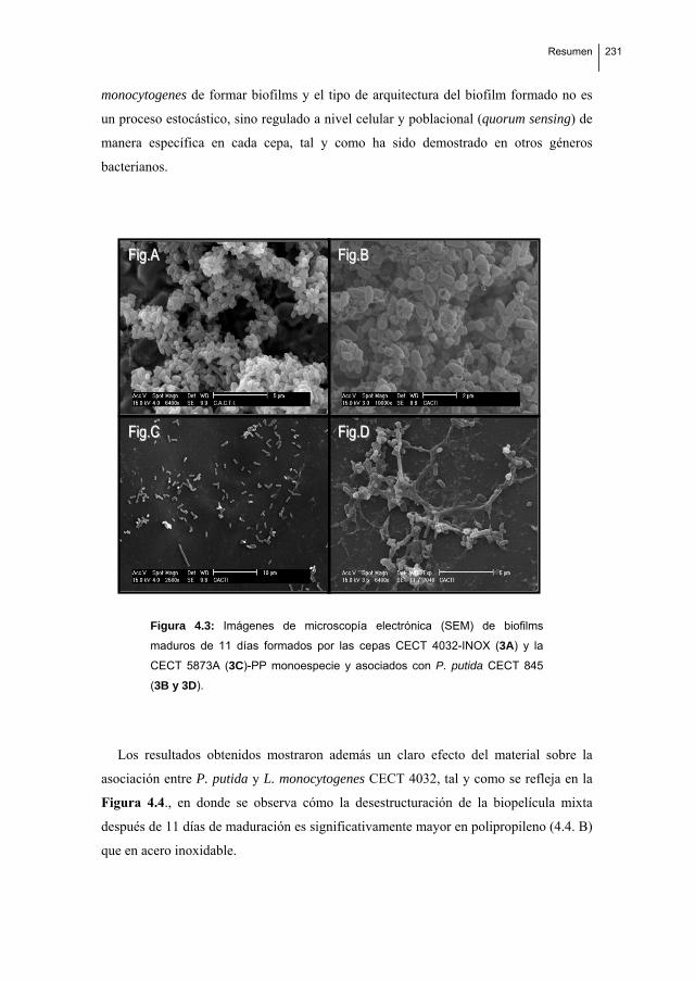
Resumen
231
monocytogenes de formar biofilms y el tipo de arquitectura del biofilm formado no es
un proceso estocástico, sino regulado a nivel celular y poblacional (quorum sensing) de
manera específica en cada cepa, tal y como ha sido demostrado en otros géneros
bacterianos.
Figura 4.3: Imágenes de microscopía electrónica (SEM) de biofilms
maduros de 11 días formados por las cepas CECT 4032-INOX (3A) y la
CECT 5873A (3C)-PP monoespecie y asociados con P. putida CECT 845
(3B y 3D).
Los resultados obtenidos mostraron además un claro efecto del material sobre la
asociación entre P. putida y L. monocytogenes CECT 4032, tal y como se refleja en la
Figura 4.4., en donde se observa cómo la desestructuración de la biopelícula mixta
después de 11 días de maduración es significativamente mayor en polipropileno (4.4. B)
que en acero inoxidable.
FFiigg..DD FFiigg..CC
FFiigg..BB FFiigg..AA
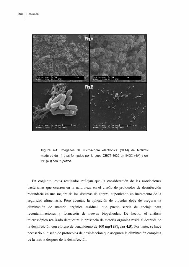
232 Resumen
Figura 4.4: Imágenes de microscopía electrónica (SEM) de biofilms
maduros de 11 días formados por la cepa CECT 4032 en INOX (4A) y en
PP (4B) con P. putida.
En conjunto, estos resultados reflejan que la consideración de las asociaciones
bacterianas que ocurren en la naturaleza en el diseño de protocolos de desinfección
redundaría en una mejora de los sistemas de control suponiendo un incremento de la
seguridad alimentaria. Pero además, la aplicación de biocidas debe de asegurar la
eliminación de materia orgánica residual, que puede servir de anclaje para
recontaminaciones y formación de nuevas biopelículas. De hecho, el análisis
microscópico realizado demuestra la presencia de materia orgánica residual después de
la desinfección con cloruro de benzalconio de 100 mg/l (Figura 4.5). Por tanto, se hace
necesario el diseño de protocolos de desinfección que aseguren la eliminación completa
de la matriz después de la desinfección.
FFiigg..AA
FFiigg..BB

Resumen
233
Figura 4.5: Imágenes de microscopía electrónica (SEM) de los residuos de
los biofilms formados por la cepa CECT 4032 en INOX y P. putida tras la
acción del BAC empleando una concentración de 100 mg/l.
Aunque tanto los resultados obtenidos en el capítulo previo como en este dejan claro
que la mayor o menor capacidad de formar biopelículas es inherente a cada cepa
bacteriana, también ponen en evidencia que la presencia de P. putida en el biofilm no es
irrelevante. Dos hechos principales observados apoyan esta afirmación:
1) Que la cepa salvaje 5873 en presencia de Ps. putida forma biofilms con una
mayor resistencia al BAC que los correspondientes biofilms monoespecie
(Figura 4.2 C).
2) Que las diferencias entre los biofilms formados por la cepa 4032 en polipropileno
y en acero inoxidable se deben a una mayor capacidad de adherencia de P. putida
esta última, con máximos de adherencia incrementados en más de 1-log. Como
consecuencia, se forma un biofilm con una mayor resistencia al BAC en INOX,
después de 4 y 11 días de incubación (Figura 4.2. B). Sin embargo, otra explicación
para estas diferencias podría deberse a un descenso en la actividad metabólica
derivada de la formación de un biofilm más denso y complejo sobre acero inoxidable
(Rodrigues et al. 2009), lo cual implica una mayor estabilidad y probablemente un
retraso en el comienzo de la fase de desestructuración del biofilm. Además, la
presencia de conexiones intercelulares y la proximidad entre las células de diferentes
especies en el biofilm podría facilitar la transferencia de material genético, incluídos
los genes asociados a la resistencia a estímulos externos (Ammor et al., 2004, Wuertz
et al., 2004, Nadell et al., 2009).

234 Resumen
El desarrollo de los objetivos anteriores, permitió concluir que la cepa 4032 (4b),
sola o asociada con P. putida, es la que forma estructuras biopeliculares más resistentes
a desinfectantes y que el incremento de dicha resistencia se relaciona sin duda con su
capacidad de formar estructuras tridimensionales complejas. Sin embargo, para los
estudios de adaptación al BAC se eligió la cepa CECT 5873 por ser la que mostraba la
mayor capacidad adaptativa al cloruro de benzalconio, demostrándose a posteriori su
condición de mala formadora de biopelículas y como consecuencia, su mayor facilidad
para su transferencia a alimentos.
5. Efectos combinado de CO2 y nisina sobre células de Listeria monocytogenes
transferidas a mejillón desde biopelículas adaptadas y no adaptadas al cloruro de
benzalconio. Accepted in Journal of Food Protection
5.1. Introducción
De acuerdo con las últimas inspecciones realizadas por la EFSA (2009, 2010), uno
de los tipos de alimentos con mayor incidencia en L. monocytogenes son los productos
de la pesca listos para el consumo. Tal y como se ha descrito, su presencia en alimentos
se asocia a su transferencia por contacto desde las plantas de procesado, donde L.
monocytogenes persiste formando biopelículas (Porsby et al. 2008, Rodríguez and
McLandsborough 2007 dicho en el apartado anterior). Y también, en algunos casos se
ha demostrado que las cepas de L. monocytogenes persistentes son resistentes a QACs
como el cloruro de benzalconio (BAC) (Aase et al 2000). Por otro lado, el envasado en
atmósferas modificadas (EAM) es una técnica de conservación aplicada con éxito en
alimentos de origen marino (Pastoriza et al. 1996a, 1996b, 1998, 2002, 2004; Cabo et
al. 2003), pudiendo, sin embargo, favorecer el crecimiento de microorganismos
anaerobios facultativos como L. monocytogenes. En estas situaciones, la aplicación de
bacteriocinas constituye una solución útil para incrementar la calidad y seguridad (Cabo
et al. 2001, 2005, 2009). De este modo, en una planta de procesado de mejillón puede
existir el riesgo de que células de Listeria monocytogenes adaptadas al BAC persistan
formando biopelículas y se transfieran por contacto al mejillón antes del envasado. Sin
embargo, no existen estudios previos acerca de los efectos de la adaptación de L.

Resumen
235
monocytogenes a desinfectantes de amplio uso industrial sobre su resistencia a la
aplicación de atmósferas modificadas.
El principal objetivo de este trabajo es comparar la viabilidad de células procedentes
de una biopelícula adaptada y no adaptada al BAC en atmósferas ricas en CO2 y O2 una
vez han sido transferidas por contacto a mejillón cocido y vivo, respectivamente, y
almacenadas a 2.5ºC. Además, en el caso concreto del mejillón cocido, se recurrió a un
diseño factorial de primer orden (Box & Hunter, 1988) para el estudio adicional del
efecto combinado del CO2 y la nisina.
5.2.Materiales y métodos
Cepas: Listeria monocytogenes CECT 5873 adaptada al BAC (DL50=7.2 mg/l) se
obtuvo en experimentos previos a partir de dos cultivos sucesivos de la cepa salvaje
(DL50=2.7 mg/l) en concentraciones subletales de BAC.
Medios residuales, inóculo y formación e biopelículas: La preparación de los medios
residuales y el procedimiento para la formación de biopelículas se realizó como se
describe en los apartados previos. Se utilizaron biofilms de L. monocytogenes CECT
5873 adaptada y no adaptada al BAC formados en cupones de INOX (10 mm x 10 mm)
después de 7 días de incubación a 25 ºC. Tras el lavado con PBS durante 30 segundo, el
número
Ensayos de transferencia: la transferencia de las células adheridas a los mejillones se
realizó mediante el contacto del cupón y la vianda (mejillón cocido) o concha (mejillón
vivo) durante 2 minutos. El tiempo de contacto (10 min) se determinó a partir de
estudios preliminares, alcanzándose 5.6 log UFC de L. monocytogenes por de mejillón
cocido y 7.2 log UFC por gramo de mejillón vivo.
Aplicación de la nisina: se dosificó en disolución acuosa (en concentraciones de 210,
115, 21 UI/ml) mediante pipeteo sobre la vianda. Dicha dosificación se realizó después
de la transferencia de L. monocytogenes y antes del envasado.
Envasado: Las muestras así preparadas se envasaron en bolsas tipo barrera (Cryovac,
SL) y se insuflaron a continuación las mezclas de gases (Carburos Metálicos,
Barcelona) correspondientes, almacenándose posteriormente en cámara de refrigeración

236 Resumen
a 2.5ºC hasta la toma de muestra. A cada tiempo prefijado, se determinó del número de
células viables de L. monocytogenes mediante los métodos tradicionales de siembra en
placa y utilizando agar Palcam (Liofilchem, S.L.R., Italia) a pH=9 como medio de
cultivo. El pH del agar se ajustó a 9 para asegurar la inactivación de la nisina.
5.3. Resultados y discusión
En mejillón cocido
El número de células viables de L. monocytogenes (LMV) obtenidos resultaron
describibles mediante las siguientes ecuaciones empíricas significativas después de 7,
11 y 20 días de almacenamiento, representadas en las Figuras 5.1 y 5.2:
L. monocytogenes CECT 5873 no adaptada (DL50=2.7 mg/l):
Despúes de 7 días: LMV=4.25-1.18C-1.48N-1.18CN [5.1]
Despúes de 11 días: LMV=5.25-0.45C-0.74N+0.46CN [5.2]
Despúes de 20 días a: LMV=5.72-1.43C-0.86N [5.3]
Cepas 5873 adaptada (DL50=7.2 mg/l):
Después de 7 días: LMV=3.65-0.57C-0.87N [5.4]
Después de 11 días: LMV=5.24-0.45C-0.94N [5.5]
Después de 20 días: VLM=5.47-0.37C-0.79N+0.54CN [5.6]
Las ecuaciones obtenidas mostraron el conocido efecto inhibitorio del CO2 (C) y de
la nisina (N) frente a L. monocytogenes (efecto del CO2: Sheridan et al 1995, Olarte et al
2002; efecto de la nisina: Nilson et al. 1997, López- Mendoza et al. 2007). Sin embargo,
no fue posible encontrar estudios previos sobre la efectividad del CO2 en biofilms y solo
uno en el caso de la nisina (Minei et al., 2008). Por otro lado, la interacción de signo
positivo puso de manifiesto la incompatibilidad de ambas variables a concentraciones
altas. Esto último contrasta con estudios previos en donde se demostró un efecto
sinérgico entre CO2 y nisina sobre la supervivencia de células planctónicas de L.
monocytogenes (López-Mendoza et al. 2007, Nilsson et al. 1997, Nilsson et al 1999,

Resumen
237
Szabo et al. 1998). Esta diferencia entre el efecto conjunto de ambas variables en
sistemas plantónicos y sésiles podría reflejar un aumento de la incompatibilidad entre
antimicrobianos con acción a nivel de membrana derivada de la presencia de
exopolisacáridos en las biopelículas transferidas al mejillón cocido. De hecho, Fang and
Lin (1994) demuestran la inefectividad de la nisina en presencia de CO2 frente a P. fragi
productora de exopolisacáridos.
Los resultados obtenidos demostraron que las biopelículas formadas por células
adaptadas de BAC podían ser más resistentes a la aplicación de atmósferas modificadas
ricas en CO2 y nisina una vez transferidas al mejillón cocido por contacto (simulando
contaminación cruzada) tal y como reflejan las ecuaciones obtenidas tras 7 y 20 días de
almacenamiento siendo los coeficientes obtenidos tanto para el BAC como para la
nisina significativamente mayores (p<0.05). Después de 7 días, el modelo predice una
reducción logarítmica (RL) de 3.39 log y de 5.12 log en L. monocytogenes adaptada y
no adaptada, respectivamente, una vez transferidas a mejillón cocido y envasado bajo
condiciones de 90% de CO2 y 210 UI/ml de nisina (ec. 5.1 y 5.4). Esto supone un
incremento en el riesgo asociado a la presencia de este género en las plantas de
procesado. Aunque no se encontraron estudios previos sobre resistencias cruzadas
derivadas de la resistencia a desinfectantes, sí se ha demostrado una respuesta cruzada
entre la tolerancia a ácido de L. monocytogenes y su mayor resistencia al CO2 (Francis
et al. 2007). Además, estos resultados concuerdan con el estudio previo desarrollado en
el apartado 3 (Saá Ibusquiza et al., 2010) en el que se obtenía que los biofilms maduros
adaptados al BAC eran más resistentes a la nisina que los no adaptados al BAC.
Sin embargo, después de 11 días de incubación las diferencias entre ambas células
biopeliculares de L. monocytogenes (adaptadas y no adaptadas) decrecen y se obtienen
ecuaciones polinómicas (ec. 5.2 y 5.5) y superficies de respuesta (Fig 5.2 y 5.3)
similares, lo que sin duda responde a la variación de los efectos de las variables
resultado de la condición cinética de los sistemas microbianos en alimentos.
Por último, el hecho de que los efectos de las variables decrezcan con el tiempo de
almacenamiento demuestra que los biofilms de L. monocytogenes resisten los
tratamientos empleados en mejillón cocido a esta temperatura.

238 Resumen
CECT 5873 Adaptada-20 DÍASCECT 5873 Adaptada-11 DÍASCECT 5873 Adaptada-7 DÍAS
LMV
(lo
g U
FC
/g)
LMV
(lo
g U
FC
/g)
LMV
(lo
g U
FC
/g)
Figura 5.2: Superficies de respuesta correspondientes al efecto combinado del
CO2 y de la nisina frente a las células viables de L. monocytogenes CECT 5873
procedentes de una biopelícula adaptada al BAC (LMV en log UFC/g) después
de 7, 11 y 20 días de almacenamiento a 2.5 ºC. Las variables se expresan en
valores codificados.
CECT 5873-20 DÍASCECT 5873-11 DÍASCECT 5873-7 DÍAS
LMV
(lo
g U
FC
/g)
LMV
(lo
g U
FC
/g)
LMV
(lo
g U
FC
/g)
Figura 5.3: Superficies de respuesta correspondientes al efecto combinado del
CO2 y de la nisina frente a las células viables de L. monocytogenes
procedentes de una biopelícula CECT 5873 no adaptada al BAC (LMV en Log
UFC/g) después de 7, 11 y 20 días de almacenamiento a 2.5 ºC. Las variables
se expresan como valores codificados.
A pesar de que la explicación de los resultados obtenidos necesitaría de experimentos
adicionales, una revisión bibliográfica permitió encontrar algunas razones posibles, que
abrirían varias posibilidades para futuras investigaciones:
Que el BAC, la nisina y el CO2 actúan sobre la membrana celular (CO2:
Jydegaard-Axelson et al. 2004, Lungu et al., 2009, Tassou et al., 2004, Nilsson et al.,
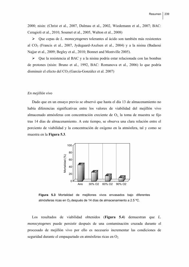
Resumen
239
2000; nisin: (Christ et al., 2007, Dalmau et al., 2002, Wiedemann et al., 2007; BAC:
Ceragioli et al., 2010, Soumet et al., 2005, Walton et al., 2008)
Que cepas de L. monocytogenes tolerantes al ácido son también más resistentes
al CO2 (Francis et al., 2007, Jydegaard-Axelsen et al., 2004) y a la nisina (Badaoui
Najjar et al., 2009; Begley et al., 2010; Bonnet and Montville 2005).
Que la resistencia al BAC y a la nisina podría estar relacionada con las bombas
de protones (nisin: Bruno et al., 1992, BAC: Romanova et al., 2006) lo que podría
disminuir el efecto del CO2 (García-González et al. 2007)
En mejillón vivo
Dado que en un ensayo previo se observó que hasta el día 13 de almacenamiento no
había diferencias significativas entre los valores de viabilidad del mejillón vivo
almacenado atmósferas con concentración creciente de O2, la toma de muestra se fijo
tras 14 días de almacenamiento. A este tiempo, se observa una clara relación entre el
porciento de viabilidad y la concentración de oxígeno en la atmósfera, tal y como se
muestra en la Figura 5.3.
Aire 30% O2 60% O2 90% O20
20
40
60
80
100
Mo
rta
lida
d (
%)
Figura 5.3 Mortalidad de mejillones vivos envasados bajo diferentes
atmósferas ricas en O2 después de 14 días de almacenamiento a 2.5 ºC.
Los resultados de viabilidad obtenidos (Figura 5.4) demuestran que L.
monocytogenes puede persistir después de una contaminación cruzada durante el
procesado de mejillón vivo por ello es necesario incrementar las condiciones de
seguridad durante el empaquetado en atmósferas ricas en O2.

240 Resumen
día0 día5 día8 día12Aire 30%O2 60%O2 90%O2
5
5,5
6
6,5
7
7,5
8
UF
C
día0 día5 día8 día12 día0 día5 día8 día12 día0 día5 día8 día12
Figura 5.4 Viabilidad de mejillones vivos envasados bajo atmósferas
ricas en O2 después de 5, 8 y 13 días de almacenamiento a 2.5 ºC.