Embed Size (px)
Citation preview

Nematology, 2006, Vol. 8(5), 659-669
Bursaphelenchus antoniae sp. n.(Nematoda: Parasitaphelenchidae) associated with
Hylobius sp. from Pinus pinaster in PortugalAna Catarina PENAS 1,!, Kai METGE 2, Manuel MOTA 3 and Vera VALADAS 3
1 Departamento de Protecção das Plantas, Estação Agronómica Nacional (EAN), INIA, Quinta do Marquês, 2784-505Oeiras, Portugal
2 Federal Biological Research Centre for Agriculture and Forestry, Institute for Plant Virology, Microbiology andBiosafety, Messeweg 11, 38104 Braunschweig, Germany
3 NemaLab/ICAM, Departamento de Biologia, Universidade de Évora, 7002-554 Évora, Portugal
Received: 15 August 2005; revised: 4 July 2006Accepted for publication: 5 July 2006
Summary – Bursaphelenchus antoniae sp. n. is described and illustrated. Dauer juveniles were isolated from the body of the large pineweevil, Hylobius sp., collected from maritime pine (Pinus pinaster) stumps, in Portugal. Bursaphelenchus antoniae sp. n. was rearedand maintained in P. pinaster wood segments and on Petri dish cultures of the fungi Botrytis cinerea and Monilinia fructicola. The newspecies is characterised by a relatively small body length of ca 583 µm (females) and 578 µm (males), a lateral field with two incisures,presence of a small vulval flap and a conoid female tail with a rounded or pointed terminus. Males have stout spicules with a disc-likecucullus and seven caudal papillae arranged as a single midventral precloacal papilla, one precloacal pair and two postcloacal pairs. Inthe character of the lateral field, B. antoniae sp. n. comes close to B. abietinus, B. rainulfi and B. hylobianum, whilst spicule charactersplace it within the piniperdae-group sensu Ryss et al. Morphologically, B. antoniae sp. n. is closest to B. hylobianum; the spicules ofthese two species having flattened, wing-like, alae on the distal third of the lamina. Bursaphelenchus antoniae sp. n. is distinguishedfrom B. hylobianum on the arrangement of the caudal papillae (two vs three pairs). ITS-RFLP profiles and the failure to hybridisesupport the separation of the two species. Phylogenetic analysis of the new species, based on the 18S rDNA sequence, supports theinclusion of this new species in the B. hylobianum-group sensu Braasch. Sequence analysis of the 28S rDNA D2/D3 domain did notplace the new species in a definite group.
Keywords – cross-breeding, ITS-RFLP, morphology, morphometrics, phylogeny, sequence analysis, taxonomy.
During intensive annual surveys for the pine wood ne-matode, Bursaphelenchus xylophilus (Steiner & Buhrer,1934) Nickle, 1970, in Portugal, dauer juveniles of a newBursaphelenchus species were found associated with thelarge pine weevil, Hylobius sp. (Coleoptera: Curculion-idae) in maritime pine, Pinus pinaster Aiton. This newspecies was previously identified in Penas et al. (2004) asBursaphelenchus hylobianum (Korenchenko, 1980) Hunt,1993. The close resemblance between B. antoniae sp. n.and B. hylobianum was obvious, both species being foundassociated with the same insect host genus and display-ing similar morphological features. However, further de-tailed studies, i.e., morphological, biometrical and mole-cular (ITS-RFLP pattern and rDNA sequencing analy-
* Corresponding author, e-mail: [email protected]
sis) studies, as well as cross-breeding experiments, haveshown this population to be an undescribed species. Thenew species is herein described as Bursaphelenchus anto-niae sp. n.
Materials and methods
NEMATODE ISOLATION AND CULTURE
Sixty-two large pine weevils were collected from P.pinaster trees from Leiria, North-western Portugal, onOctober, 2000. These insects were placed individuallyin a Syracuse dish in a small amount of water andanalysed under a stereo microscope (Olympus SZX12)
© Koninklijke Brill NV, Leiden, 2006 659Also available online - www.brill.nl/nemy
A.C. Penas et al.
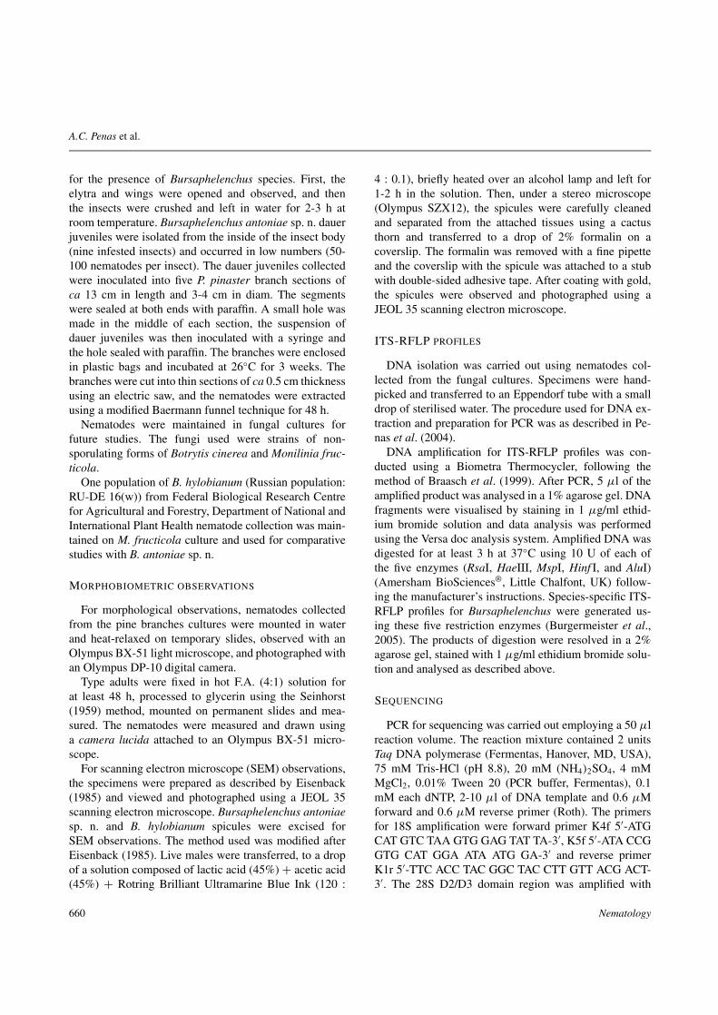
for the presence of Bursaphelenchus species. First, theelytra and wings were opened and observed, and thenthe insects were crushed and left in water for 2-3 h atroom temperature. Bursaphelenchus antoniae sp. n. dauerjuveniles were isolated from the inside of the insect body(nine infested insects) and occurred in low numbers (50-100 nematodes per insect). The dauer juveniles collectedwere inoculated into five P. pinaster branch sections ofca 13 cm in length and 3-4 cm in diam. The segmentswere sealed at both ends with paraffin. A small hole wasmade in the middle of each section, the suspension ofdauer juveniles was then inoculated with a syringe andthe hole sealed with paraffin. The branches were enclosedin plastic bags and incubated at 26"C for 3 weeks. Thebranches were cut into thin sections of ca 0.5 cm thicknessusing an electric saw, and the nematodes were extractedusing a modified Baermann funnel technique for 48 h.
Nematodes were maintained in fungal cultures forfuture studies. The fungi used were strains of non-sporulating forms of Botrytis cinerea and Monilinia fruc-ticola.
One population of B. hylobianum (Russian population:RU-DE 16(w)) from Federal Biological Research Centrefor Agricultural and Forestry, Department of National andInternational Plant Health nematode collection was main-tained on M. fructicola culture and used for comparativestudies with B. antoniae sp. n.
MORPHOBIOMETRIC OBSERVATIONS
For morphological observations, nematodes collectedfrom the pine branches cultures were mounted in waterand heat-relaxed on temporary slides, observed with anOlympus BX-51 light microscope, and photographed withan Olympus DP-10 digital camera.
Type adults were fixed in hot F.A. (4:1) solution forat least 48 h, processed to glycerin using the Seinhorst(1959) method, mounted on permanent slides and mea-sured. The nematodes were measured and drawn usinga camera lucida attached to an Olympus BX-51 micro-scope.
For scanning electron microscope (SEM) observations,the specimens were prepared as described by Eisenback(1985) and viewed and photographed using a JEOL 35scanning electron microscope. Bursaphelenchus antoniaesp. n. and B. hylobianum spicules were excised forSEM observations. The method used was modified afterEisenback (1985). Live males were transferred, to a dropof a solution composed of lactic acid (45%) + acetic acid(45%) + Rotring Brilliant Ultramarine Blue Ink (120 :
4 : 0.1), briefly heated over an alcohol lamp and left for1-2 h in the solution. Then, under a stereo microscope(Olympus SZX12), the spicules were carefully cleanedand separated from the attached tissues using a cactusthorn and transferred to a drop of 2% formalin on acoverslip. The formalin was removed with a fine pipetteand the coverslip with the spicule was attached to a stubwith double-sided adhesive tape. After coating with gold,the spicules were observed and photographed using aJEOL 35 scanning electron microscope.
ITS-RFLP PROFILES
DNA isolation was carried out using nematodes col-lected from the fungal cultures. Specimens were hand-picked and transferred to an Eppendorf tube with a smalldrop of sterilised water. The procedure used for DNA ex-traction and preparation for PCR was as described in Pe-nas et al. (2004).
DNA amplification for ITS-RFLP profiles was con-ducted using a Biometra Thermocycler, following themethod of Braasch et al. (1999). After PCR, 5 µl of theamplified product was analysed in a 1% agarose gel. DNAfragments were visualised by staining in 1 µg/ml ethid-ium bromide solution and data analysis was performedusing the Versa doc analysis system. Amplified DNA wasdigested for at least 3 h at 37"C using 10 U of each ofthe five enzymes (RsaI, HaeIII, MspI, Hinf I, and AluI)(Amersham BioSciences®, Little Chalfont, UK) follow-ing the manufacturer’s instructions. Species-specific ITS-RFLP profiles for Bursaphelenchus were generated us-ing these five restriction enzymes (Burgermeister et al.,2005). The products of digestion were resolved in a 2%agarose gel, stained with 1 µg/ml ethidium bromide solu-tion and analysed as described above.
SEQUENCING
PCR for sequencing was carried out employing a 50 µlreaction volume. The reaction mixture contained 2 unitsTaq DNA polymerase (Fermentas, Hanover, MD, USA),75 mM Tris-HCl (pH 8.8), 20 mM (NH4)2SO4, 4 mMMgCl2, 0.01% Tween 20 (PCR buffer, Fermentas), 0.1mM each dNTP, 2-10 µl of DNA template and 0.6 µMforward and 0.6 µM reverse primer (Roth). The primersfor 18S amplification were forward primer K4f 5#-ATGCAT GTC TAA GTG GAG TAT TA-3#, K5f 5#-ATA CCGGTG CAT GGA ATA ATG GA-3# and reverse primerK1r 5#-TTC ACC TAC GGC TAC CTT GTT ACG ACT-3#. The 28S D2/D3 domain region was amplified with
660 Nematology

Bursaphelenchus antoniae sp. n. from Portugal
forward primer D2A 5#-ACA AGT ACC GTG AGG GAAAGT TG-3# and reverse primer D3Br 5#-TCG GAA GGAACC AGC TAC TA-3#. Sequencing PCR was performedwith a Biometra T1 thermal cycler (Biometra, Göttingen,Germany). The PCR program consisted of an initialdenaturation for 2 min, 30 s at 96"C, 35 cycles with 1min denaturation at 94"C, 1 min annealing at 55"C, 2min extension at 72"C and a final extension for 6 min at72"C. After completion of the PCR, small aliquots of thesamples were separated electrophoretically using a 1.8%agarose gel and 0.5$TBE buffer.
Amplicons were concentrated and desalted using Mi-crocon YM-100 centrifugal filter devices (Millipore, Bil-lerica, MA, USA) to accomplish sequencing productsof good quality. Working steps were performed follow-ing the manufacturer instructions. Additionally, the mem-brane was washed with 50 µl ddH2O. Small aliquots ofeach final sample were applied to 1.8% agarose gel and0.5$TBE buffer to estimate the concentration of desaltedDNA. Gels were stained with ethidium bromide (1 µg/ml)and visualised with a UV transilluminator.
According to the instructions of the sequencing com-pany (MWG Biotech, Ebersberg, Germany), 20 ng/100 bpof PCR amplicon were air dried and sent to the companytogether with primers to use their Value Read Service.Each fragment was sequenced in both directions using theappropriated PCR primers for 18S rDNA and 28S D2/D3domain to obtain overlapping sequences of the forwardand reverse DNA strand.
The sequence data of B. antoniae sp. n. were comparedto sequences of Bursaphelenchus species from differentgroups (abietinus-, eggersi-, fungivorus-, hylobianum-,sexdentati- and xylophilus-group) deposited in the Gen-Bank database under accession numbers AM279709 PT(18S rDNA) and AM279710 PT (28S D2D3). Aphelen-choides besseyi was used as an outgroup. Alignmentswere calculated with ClustalW and phylogenetic treeswere generated by neighbour-joining (NJ) and maximumparsimony (MP) algorithms in Mega 3.1 (Kumar et al.,2004).
CROSS-BREEDING TESTS
Cross-breeding tests between B. antoniae sp. n. and B.hylobianum were carried out under controlled conditions.Reciprocal mating between J4-adult female moults andmales were performed. Under an Olympus SZX12 stereomicroscope J4-adult female moults and adult males werehand-picked individually from fungal cultures under anOlympus SZX12 stereomicroscope. Ten 3 cm diam. Petri
dishes were prepared: five were inoculated with B. cinereaand the others with M. fructicola. Each of these tenPetri dishes was inoculated with 25 J4-adult females and25 males (five dishes with B. antoniae sp. n. !$ B.hylobianum " and five dishes with B. antoniae sp. n. "$B. hylobianum !). As controls, two Petri dish cultures ofM. fructicola were inoculated with 25 specimens of bothsexes of each species. All dishes were incubated at 20"Cand after 3 weeks the nematodes were extracted using amodified Baermann funnel technique and counted.
Bursaphelenchus antoniae* sp. n.=B. hylobianum in Penas et al., 2004
=Bursaphelenchus sp. in Penas et al., 2006(Figs 1-5)
MEASUREMENTS
See Table 1.
DESCRIPTION
Male
Displaying all features of Aphelenchoidoidea accord-ing to Hunt (1993). Body slender, cylindrical. Distalpart of body curved and hook-like ventrally when heat-relaxed. Cuticle with fine annulations. Lateral field withtwo distinct incisures (i.e., one ridge). Lip region high (ca3.1 µm), rounded, offset by constriction. Stylet with smallbasal thickenings. Median bulb elongate-oval, ca 13.8 µmlong and ca 9.1 µm diam. with a length-diam. ratio ofca 1.5. Excretory pore located 1.0-1.5 body diam. poste-rior to median bulb. Hemizonid located 5-6 µm posteriorto excretory pore. Pharyngeal glands dorso-lateral, over-lapping intestine for 2-3 body diam. Testis monorchic,usually anteriorly outstretched, occasionally reflexed; oc-cupying ca half of body length, cells initially arrangedin single row and then in two rows. Spicules paired, ro-bust, rosethorn-shaped, strongly curved; rostrum promi-nent, not sharply pointed; condylus rounded and well-developed, distal end with disc-like cucullus (not alwaysdiscernible). Distal third of dorsal limb of spicules later-ally expanded forming flattened, wing-like, alae. Tail ar-cuate, terminus pointed; bursa usually truncate with pos-terior margin indented in some specimens. Seven cau-dal papillae arranged as a single midventral precloacal
* The species is named in honour of Maria Antónia Bravo forher contributions to Nematology and for her support during thefirst author’s Ph.D. studies.
Vol. 8(5), 2006 661

A.C. Penas et al.
Fig. 1. Bursaphelenchus antoniae sp. n. A: Entire female; B: Entire male; C: Female anterior region; C1: Head; D: Female vulvalregion; E: Female tails; F: Tail of J2; G: Tail of J3; H: Tail of J4 (female); I: Tail of J4 (male); J: Ventral view of male tail showingpapillal disposition; J1: Spicules, ventral view; K: Male tail; L: Variation in male spicule shape.
662 Nematology

Bursaphelenchus antoniae sp. n. from Portugal
Fig. 2. Bursaphelenchus antoniae sp. n. A: Light micrograph (LM) of anterior region; B: Scanning electron micrograph (SEM) of head;C: SEM of lateral field; D: LM of vulval region; E: SEM of vulval region; F: LM of female tail; G: LM of male tail; H: SEM of maletail; I, J: SEM of bursa.
Vol. 8(5), 2006 663

A.C. Penas et al.
Fig. 3. Excised spicules of Bursaphelenchus antoniae sp. n. (A, B) and B. hylobianum (C). Note the flattened wing-like structure in thedistal third of the dorsal limb (arrows).
Fig. 4. ITS-RFLP pattern of Bursaphelenchus antoniae sp. n.
papilla, one precloacal pair and two postcloacal pairs (onepair at 42% of tail length from cloacal aperture and otherpair just before bursa at 60% of tail length from cloacalaperture).
Female
Body slightly ventrally curved when heat-relaxed. Gen-ital tract monoprodelphic, outstretched, cells initiallyarranged in single row and thereafter with two rows. Sper-matheca differentiated, roundish irregular rectangle, filled
with rounded sperm. Quadricollumela visible. Postuterinebranch extending for ca 60% of vulva-anus distance, of-ten containing sperm. Vulva inclined anteriad at ca 45"
to body axis. Vulva with anterior lip slightly extended toform a small, distinct flap. Female tail medium length,conoid, gradually tapering to bluntly rounded or acute ter-minus.
Dauer juvenile
Body 400-440 µm long. Head dome-shaped, lips notdefined, stylet just visible. Median bulb not well definedbut recognisable in most cases. Pharynx and pharyngealglands degenerate. Body filled with granular lipid mate-rial. Tail conical, terminus pointed.
Juvenile stages
Juveniles with conical tail: J3 and J4 (female), tailslightly ventrally curved; J4 male tail also ventrallycurved. Developing gonad visible in posterior region ofJ4 male.
TYPE LOCALITY AND HABITAT
The new species was isolated from inside the bodyof the large pine weevil, Hylobius sp., (Coleoptera:Curculionidae). The insects emerged from P. pinaster(maritime pine) stumps collected from a pine wood atLeiria, north-west Portugal.
TYPE MATERIAL
Holotype male, six female paratypes and five maleparatypes, deposited in the Departamento Protecção dasPlantas, Estação Agronómica Nacional, Oeiras, Portu-gal. Other paratypes: one slide with five males and one
664 Nematology
Bursaphelenchus antoniae sp. n. from Portugal
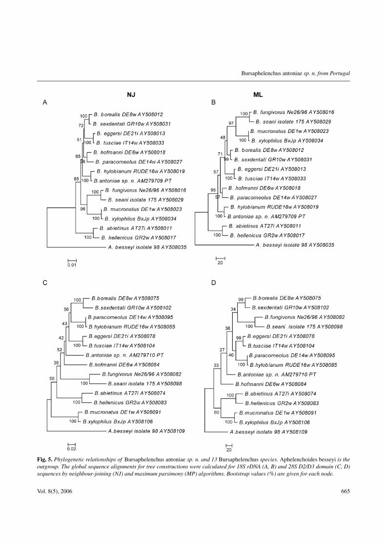
Fig. 5. Phylogenetic relationships of Bursaphelenchus antoniae sp. n. and 13 Bursaphelenchus species. Aphelenchoides besseyi is theoutgroup. The global sequence alignments for tree constructions were calculated for 18S rDNA (A, B) and 28S D2/D3 domain (C, D)sequences by neighbour-joining (NJ) and maximum parsimony (MP) algorithms. Bootstrap values (%) are given for each node.
Vol. 8(5), 2006 665

A.C. Penas et al.
Table 1. Morphometrics of Bursaphelenchus antoniae sp. n. Allmeasurements are in µm and in the form: mean ± standarddeviation (range).
Male Female
Holotype Paratypes Paratypes
n – 20 21L 574 578 ± 64 584 ± 54
(476-660) (512-709)a 39.6 39.8 ± 3.1 36.6 ± 2.6
(34.2-44.0) (32.0-42.1)b 8.2 8.1 ± 0.5 8.5 ± 0.6
(7.2-9.2) (7.7-10.0)c 19.1 18.7 ± 1.6 14.6 ± 0.9
(15.5-21.3) (12.8-16.3)c# 2.3 2.4 ± 0.2 4.5 ± 0.4
(2.1-2.9) (3.9-5.1)V – – 70.8 ± 0.9
(69.1-72.2)Lip region diam. 6 5.9 ± 0.2 6.1 ± 0.4
(5.5-6.0) (5.5-7.0)Lip constriction diam. 5 5.0 ± 0.2 5.2 ± 0.5
(4.5-5.5) (4.5-6.5)Lip region height 3 3.1 ± 0.2 3.1 ± 0.2
(3.0-3.5) (3.0-3.5)Stylet 13 12.1 ± 1.0 12.0 ± 1.0
(11-14) (10-14)Pharynx length 70 71.0 ± 5.7 68.8 ± 2.9
(59-80) (63-74)Median bulb length 15 13.8 ± 0.7 14.2 ± 0.7
(12-15) (13.5-16.0)Median bulb diam. 9 9.1 ± 0.5 9.8 ± 0.8
(8-10) (8-12)Median bulb length / 1.7 1.5 ± 0.1 1.5 ± 0.1
median bulb diam. (1.4-1.6) (1.3-1.8)Body diam. at middle 12 11.8 ± 1.0 12.6 ± 0.7
of median bulb (10-14) (11-14)Body diam. at base 12 12.2 ± 1.0 13.0 ± 0.7
of median bulb (10-14) (12.0-14.5)Distance from 84 81.8 ± 6.4 80.0 ± 6.2
anterior end to (71-91) (71-93)excretory pore
Distance from 89 87.0 ± 5.6 85.6 ± 4.9anterior end (78-95) (75-95)
to hemizonidDistance from 140 143.1 ± 19.3 135.3 ± 17.0
anterior end to (120-177) (116-177)posterior endof pharyngeal glands
Body diam. at end 14 13.8 ± 1.3 14.4 ± 1.0of pharyngeal glands (11-16) (13-17)
Anterior genital 301 343.8 ± 54.5 207.1 ± 26.2branch (228-426) (161-261)
Table 1. (Continued).
Male Female
Holotype Paratypes Paratypes
Posterior genital – – 81.6 ± 9.9branch (64-100)
Body diam. at vulva – – 16.4 ± 1.4(14-20)
Vulva to anus distance – – 130.6 ± 14.2(108-160)
Distance from – – 412.8 ± 39.4anterior end (354-503)to vulva
G1 (%) – – 35.4 ± 2.8(31.4-41.9)
G2 (%) – – 14.0 ± 1.5(12.1-18.2)
Anal/cloacal 13 12.8 ± 1.1 9.0 ± 0.8body diam. (10.5-14.5) (8-11)
Tail 30 30.9 ± 2.0 40.1 ± 2.9(27-34) (34-46)
T 52.4 59.4 ± 5.9 –(47.9-66.0)
Spicule (condylus 18.5 17.9 ± 1.4 –to distal end) (15-20)
Spicule (rostrum 11 10.4 ± 0.8 –to distal end) (9-11.5)
Spicule (curved 17.5 17.3 ± 1.6 –median line) (14-19.5)
Spicule (rostrum 8.5 8.6 ± 1.0 –to condylus) (6.5-10)
Distance from 4 4.2 ± 0.4 –single precloacal (3.5-5)papilla to cloacalaperture
Distance from 15 13.0 ± 1.0 –cloacal aperture to (11-15.5)first pair ofpostcloacal papillae
Distance from 19.5 18.7 ± 1.3 –cloacal aperture to (16-21.5)second pair ofpostcloacal papillae
slide with five females in USDA Nematode Collection,Beltsville, MD, USA; one slide with five males and oneslide with five females in Plant-Pathogen Interactions Di-vision, Rothamsted Research, Harpenden, UK; one slidewith five males and one slide with five females in KyotoUniversity, Environmental Mycoscience Laboratory Col-lection, Kyoto, Japan. All specimens were collected frominoculated P. pinaster branch segments.
666 Nematology

Bursaphelenchus antoniae sp. n. from Portugal
DIAGNOSIS AND RELATIONSHIPS
Bursaphelenchus antoniae sp. n. is characterised by therelatively short body in both sexes, the presence of twolines or incisures in the lateral field and by the robust andstrongly curved spicules. The spicule lamina is angulardistally, the rostrum digitate, and the condylus is rounded.Females have a very small vulval flap formed by a smallextension of the cuticle of the anterior lip, and a conicaltail that gradually tapers to an almost straight or slightlyrecurved, pointed or rounded terminus.
Based on these morphological characters, the newspecies appears close to B. abietinus Braasch & Schmut-zenhofer, 2000, B. rainulfi Braasch & Burgermeister,2002 and B. hylobianum (following Braasch’s (2001)classification).
According to the key to species groups of Ryss etal. (2005), the new species belongs to the piniperdae-group. This group is characterised by the stout andhook-like spicule with dorsal and ventral limbs joinedat the narrowed tip; elongate capitulum, rostrum andcondylus well-developed and separate; rostrum locatedmore anteriorly and condylus not recurved anteriorly;dorsal contour of lamina anteriorly smoothly curved butangular at midpoint; small cucullus present.
Bursaphelenchus antoniae sp. n. has much larger andstouter spicules than B. abietinus (17 vs 13 µm); thenew species has only one precloacal pair and a singleprecloacal caudal papilla while B. abietinus has twoprecloacal pairs.
Comparing with B. rainulfi, B. antoniae sp. n. differsin the position of the excretory pore (in B. rainulfi it islocated in the posterior region of the median bulb) and inspicule shape and length (13 vs 17 µm).
The new species seems closest to B. hylobianum be-cause of spicule shape, both species sharing a character-istic structure (Fig. 3) on the spicules, i.e., a flattened,wing-like, alae laterally expanded in the distal third ofthe dorsal limb. This feature has not been reported forany other Bursaphelenchus species. Furthermore, the newspecies, was found associated with weevils of the genusHylobius (Penas et al., 2004, 2006) as reported for B. hy-lobianum (Korenchenko, 1980). However, despite thesesimilarities B. antoniae sp. n. is distinguishable from B.hylobianum by several morphological characters. Bursa-phelenchus antoniae sp. n. has two postcloacal pairs ofpapillae whereas the original description of B. hylobianumreported three postcloacal pairs, although according toBraasch and Braasch-Bidasak (2002) B. hylobianum hasonly one postcloacal pair. The spicules of B. antoniae sp.
Table 2. ITS-RFLP profiles of Bursaphelenchus antoniae sp. n.
ITS-PCR RsaI HaeIII MspI Hinf I AluIproduct
Frag- 1150 610 790 490 340 790ment sizes 290 360 370 290 340(%bp) 230 250
220
n. resemble B. hylobianum spicules in shape but have ashorter condylus and a well-defined, disc-like cucullus; B.hylobianum was described as having cucullus by Braaschand Braasch-Bidasak (2002), although the original de-scription does not refer to the presence of a cucullus onthe distal tip of the spicules (Korenchenko, 1980).
ITS-RFLP PROFILES
Despite the morphological similarities between B. an-toniae sp. n. and B. hylobianum, the ITS-RFLP pattern ofB. antoniae sp. n. is different from that of B. hylobianum(Braasch & Burgermeister, 2002) (Fig. 4; Table 2). The re-striction fragment pattern obtained for HaeIII was similar,but differed in the other four enzymes used.
SEQUENCE ANALYSIS
The total length of the aligned rDNA for Bursaphe-lenchus species varies from 1636 to 1646 bases in the18S rDNA and 678 to 695 bases in the 28S D2/D3 re-gion. Excluding the outgroup, the global sequence align-ments of the 18S rDNA sequences examined have 1672sites, of which 1448 are conserved, 199 variable and 158parsimony-informative. Sequence alignments of the 28SD2/D3 region have 731 sites, of which 429 are conserved,284 variable and 234 parsimony-informative. The phylo-genetic analysis using NJ and MP methods yielded treeswith different topologies for both rDNA regions (Fig. 5).Based on the 18S rDNA sequences, B. antoniae sp. n. islocated within the hylobianum-group. These results arehighly supported by 100% bootstrap values. The analy-sis of the 28S rDNA D2/D3 domain did not group B. an-toniae sp. n. with species of a specific group, bootstrapvalues being low.
The phylogenetic analysis supports the conclusion fromthe other studies that B. antoniae sp. n. is a new speciesand close to B. hylobianum.
Vol. 8(5), 2006 667
A.C. Penas et al.
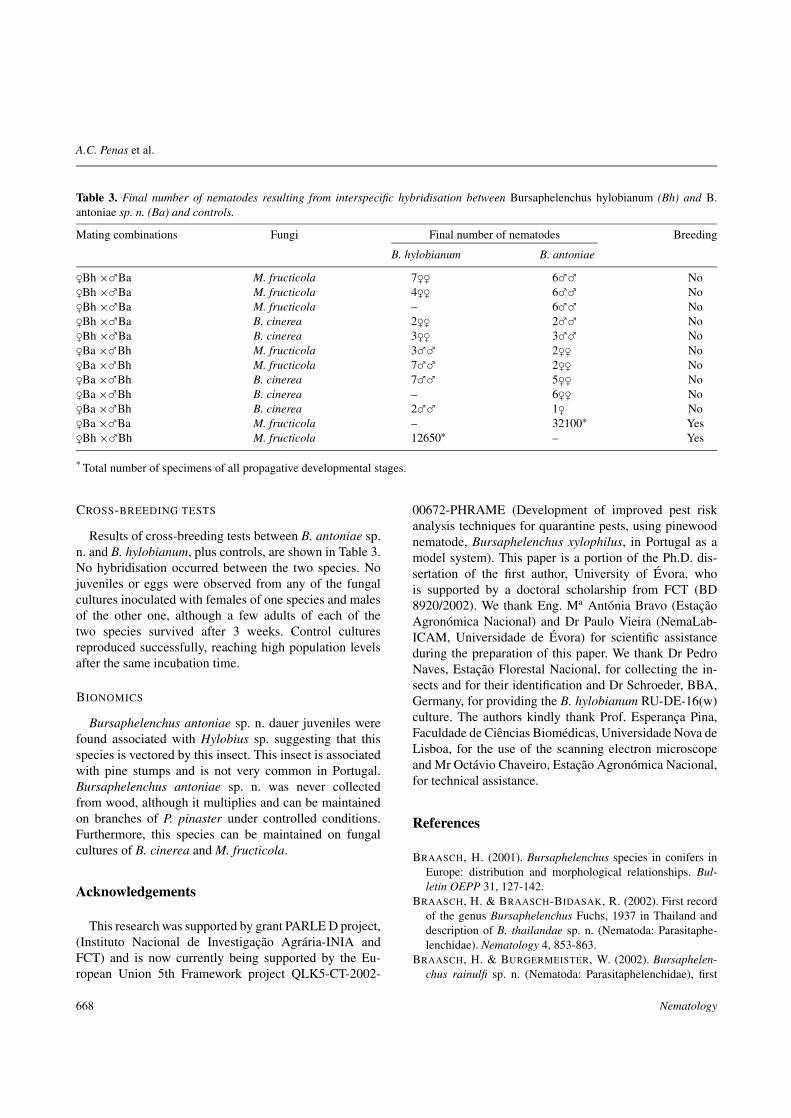
Table 3. Final number of nematodes resulting from interspecific hybridisation between Bursaphelenchus hylobianum (Bh) and B.antoniae sp. n. (Ba) and controls.
Mating combinations Fungi Final number of nematodes Breeding
B. hylobianum B. antoniae
!Bh $"Ba M. fructicola 7!! 6"" No!Bh $"Ba M. fructicola 4!! 6"" No!Bh $"Ba M. fructicola – 6"" No!Bh $"Ba B. cinerea 2!! 2"" No!Bh $"Ba B. cinerea 3!! 3"" No!Ba $"Bh M. fructicola 3"" 2!! No!Ba $"Bh M. fructicola 7"" 2!! No!Ba $"Bh B. cinerea 7"" 5!! No!Ba $"Bh B. cinerea – 6!! No!Ba $"Bh B. cinerea 2"" 1! No!Ba $"Ba M. fructicola – 32100! Yes!Bh $"Bh M. fructicola 12650! – Yes
* Total number of specimens of all propagative developmental stages.
CROSS-BREEDING TESTS
Results of cross-breeding tests between B. antoniae sp.n. and B. hylobianum, plus controls, are shown in Table 3.No hybridisation occurred between the two species. Nojuveniles or eggs were observed from any of the fungalcultures inoculated with females of one species and malesof the other one, although a few adults of each of thetwo species survived after 3 weeks. Control culturesreproduced successfully, reaching high population levelsafter the same incubation time.
BIONOMICS
Bursaphelenchus antoniae sp. n. dauer juveniles werefound associated with Hylobius sp. suggesting that thisspecies is vectored by this insect. This insect is associatedwith pine stumps and is not very common in Portugal.Bursaphelenchus antoniae sp. n. was never collectedfrom wood, although it multiplies and can be maintainedon branches of P. pinaster under controlled conditions.Furthermore, this species can be maintained on fungalcultures of B. cinerea and M. fructicola.
Acknowledgements
This research was supported by grant PARLE D project,(Instituto Nacional de Investigação Agrária-INIA andFCT) and is now currently being supported by the Eu-ropean Union 5th Framework project QLK5-CT-2002-
00672-PHRAME (Development of improved pest riskanalysis techniques for quarantine pests, using pinewoodnematode, Bursaphelenchus xylophilus, in Portugal as amodel system). This paper is a portion of the Ph.D. dis-sertation of the first author, University of Évora, whois supported by a doctoral scholarship from FCT (BD8920/2002). We thank Eng. Ma Antónia Bravo (EstaçãoAgronómica Nacional) and Dr Paulo Vieira (NemaLab-ICAM, Universidade de Évora) for scientific assistanceduring the preparation of this paper. We thank Dr PedroNaves, Estação Florestal Nacional, for collecting the in-sects and for their identification and Dr Schroeder, BBA,Germany, for providing the B. hylobianum RU-DE-16(w)culture. The authors kindly thank Prof. Esperança Pina,Faculdade de Ciências Biomédicas, Universidade Nova deLisboa, for the use of the scanning electron microscopeand Mr Octávio Chaveiro, Estação Agronómica Nacional,for technical assistance.
References
BRAASCH, H. (2001). Bursaphelenchus species in conifers inEurope: distribution and morphological relationships. Bul-letin OEPP 31, 127-142.
BRAASCH, H. & BRAASCH-BIDASAK, R. (2002). First recordof the genus Bursaphelenchus Fuchs, 1937 in Thailand anddescription of B. thailandae sp. n. (Nematoda: Parasitaphe-lenchidae). Nematology 4, 853-863.
BRAASCH, H. & BURGERMEISTER, W. (2002). Bursaphelen-chus rainulfi sp. n. (Nematoda: Parasitaphelenchidae), first
668 Nematology

Bursaphelenchus antoniae sp. n. from Portugal
record of the genus Bursaphelenchus Fuchs, 1937 fromMalaysia. Nematology 4, 971-978.
BRAASCH, H. & SCHMUTZENHOFER, H. (2000). Bursaphe-lenchus abietinus sp. n. (Nematoda, Parasitaphelenchidae).Russian Journal of Nematology 8, 1-6.
BRAASCH, H., METGE, K. & BURGERMEISTER, W. (1999).[Bursaphelenchus species (Nematoda, Parasitaphelenchidae)found in coniferous trees in Germany and their ITS-RFLPpatterns.] Nachrichtenblatt des Deutschen Pflanzenschutzdi-enste 51, 312-320.
BURGERMEISTER, W., METGE, K., BRAASCH, H. & BUCH-BACH, E. (2005). ITS-RFLP patterns for differentiation of26 Bursaphelenchus species (Nematoda: Parasitaphelenchi-dae) and observations on their distribution. Russian Journalof Nematology 13, 29-42.
EISENBACK, J. (1985). Techniques for preparing nematodes forscanning electron microscopy. In: Barker, K.R., Carter, C.C.& Sasser, N.S. (Eds). An advanced treatise on Meloidogyne,Volume II. Raleigh, NC, USA, North Carolina State Univer-sity Graphics, pp. 79-105.
HUNT, D.J. (1993). Aphelenchida, Longidoridae and Tricho-doridae: Their systematics and bionomics. Wallingford, UK,CABI Publishing, 352 pp.
KORENCHENKO, E.A. (1980). [New species of nematodes fromthe family Aphelenchoididae, parasites of stem pests of theDahurian Larch.] Zoologichesky Zhurnal 59, 1768-1780.
KUMAR, S., TAMURA, K. & NEI, M. (2004). MEGA3:Integrated software for Molecular Evolutionary GeneticsAnalysis and sequence alignment. Briefings in Bioinformatics5, 150-163.
PENAS, A.C., CORREIA, P., BRAVO, M.A., MOTA, M. &TENREIRO, R. (2004). Species of Bursaphelenchus Fuchs,1937 (Nematoda: Parasitaphelenchidae) associated with mar-itime pine in Portugal. Nematology 6, 437-453.
PENAS, A.C., BRAVO, M.A., NAVES, P., BONIFÁCIO, L.,SOUSA, E. & MOTA, M. (2006). Species of BursaphelenchusFuchs, 1937 (Nematoda: Parasitaphelenchidae) and othernematode genera associated with insects from Pinus pinasterin Portugal. Annals of Applied Biology 148, 121-131.
RYSS, A., VIEIRA, P., MOTA, M. & KULINICH, O. (2005).A synopsis of the genus Bursaphelenchus Fuchs, 1937(Aphelenchida: Parasitaphelenchidae) with keys to species.Nematology 7, 393-458.
SEINHORST, J.W. (1959). A rapid method for the transfer ofnematodes from fixative to anhydrous glycerin. Nematologica4, 67-69.
Vol. 8(5), 2006 669