Embed Size (px)
Citation preview

7/16/2019 Building Blocks of Human Life - Understanding Mature Cells and Stem Cells (Booklet).pdf
http://slidepdf.com/reader/full/building-blocks-of-human-life-understanding-mature-cells-and-stem-cells-bookletpdf 1/73
Professor John K. Young HOWARD UNIVERSITY
COLLEGE OF MEDICINE
THE BUILDING
BLOCKS OF
HUMAN L IFE:
UNDERSTANDING
M ATURE CELLS AND
STEM CELLS
COURSE GUIDE

7/16/2019 Building Blocks of Human Life - Understanding Mature Cells and Stem Cells (Booklet).pdf
http://slidepdf.com/reader/full/building-blocks-of-human-life-understanding-mature-cells-and-stem-cells-bookletpdf 2/73
The Building Blocks of Human Life:Understanding Mature Cells and Stem Cells
Professor John K. Young
Howard UniversityCollege of Medicine
Recorded Books™ is a trademark of
Recorded Books, LLC. All rights reserved.

7/16/2019 Building Blocks of Human Life - Understanding Mature Cells and Stem Cells (Booklet).pdf
http://slidepdf.com/reader/full/building-blocks-of-human-life-understanding-mature-cells-and-stem-cells-bookletpdf 3/73
The Building Blocks of Human Life:
Understanding Mature Cells and Stem Cells
Professor John K. Young
Executive Producer
John J. Alexander
Executive Editor
Donna F. Carnahan
RECORDING
Producer - David Markowitz
Director - Matthew Cavnar
COURSE GUIDE
Editor - James Gallagher
Contributing Editors -Leonard Likas
Karen Sparrough
Design - Edward White
Lecture content ©2007 by John K. Young
Course guide ©2007 by Recorded Books, LLC
72007 by Recorded Books, LLC
Cover image: © Photo Disc/Clipart.com/Recorded Books, LLC
#UT115 ISBN: 978-1-4281-8578-4
All beliefs and opinions expressed in this audio/video program and accompanying course guide
are those of the author and not of Recorded Books, LLC, or its employees.

7/16/2019 Building Blocks of Human Life - Understanding Mature Cells and Stem Cells (Booklet).pdf
http://slidepdf.com/reader/full/building-blocks-of-human-life-understanding-mature-cells-and-stem-cells-bookletpdf 4/73
3
Course Syllabus
The Building Blocks of Human Life:
Understanding Mature Cells and Stem Cells
About Your Professor...................................................................................................4
Introduction...................................................................................................................5
Lecture 1 Origin of Cell Types...............................................................................6
Lecture 2 Epithelial Cells .....................................................................................11
Lecture 3 Muscle Cells ........................................................................................15
Lecture 4 Cells of Nervous Tissue.......................................................................19
Lecture 5 Connective Tissue Cells ......................................................................24
Lecture 6 Blood Cells and Their Progenitors.......................................................29
Lecture 7 Cells of Cartilage and Bone.................................................................33
Lecture 8 Cells of the Skin...................................................................................38
Lecture 9 Exocrine Cells......................................................................................43
Lecture 10 Endocrine Cells....................................................................................47
Lecture 11 Sensory Cells.......................................................................................53
Lecture 12 Cells of Reproductive Organs..............................................................58
Lecture 13 Extreme Cells ......................................................................................63
Lecture 14 Death and Aging of Cells.....................................................................69

7/16/2019 Building Blocks of Human Life - Understanding Mature Cells and Stem Cells (Booklet).pdf
http://slidepdf.com/reader/full/building-blocks-of-human-life-understanding-mature-cells-and-stem-cells-bookletpdf 5/73
4
John K. Young is a professor of anatomy at Howard University, where he
conducts research on the hypothalamus, the part of the brain that regulates
eating, drinking, and sexual behavior.
In 1998, Professor Young won the Kaiser-Permanente Award for Excellence
in Teaching. He is a member of the American Association of Anatomists, the
Endocrine Society, and the Society for Neuroscience.
Professor Young has published more than forty articles in scientific journals
and is the author of the books Hormones: Molecular Messengers and Cells: Amazing Forms and Functions. Professor Young also coauthored Cell
Biology/Histology Tutorial with R.S. Hakim and Integrated Histology with
Alvin Telser and Kate Baldwin.
About Your Professor
John K. Young P
h o t o c o u r t e s y o f J o h n K . Y o u n g
You will get the most out of this course if you have the following book:
Jonathan Slack’s Essential Developmental Biology , 2nd ed. (London:Blackwell Science, Ltd., 2005).

7/16/2019 Building Blocks of Human Life - Understanding Mature Cells and Stem Cells (Booklet).pdf
http://slidepdf.com/reader/full/building-blocks-of-human-life-understanding-mature-cells-and-stem-cells-bookletpdf 6/73
5
Introduction
Every human is composed of an amazing assortment of cells and tissues
that carry out myriad functions necessary for sustaining life. In this series of
lectures, Professor John K. Young of the Howard University College of
Medicine takes audiences through the microscope on a fascinating journey of
discovery into the world of cells and tissues, where a complex scheme of
activity is taking place all the time, literally just beneath the surface.In clear, concise language, Professor Young explains the basic categories
of cells and tissues and then delves into their specialized functions, whether it
be for muscle cells and nervous tissue or the cells of reproductive organs and
the highly unusual entities known as “extreme” cells. Finally, Professor Young
wraps up the lectures with a topic of universal interest—the death and aging
of cells.
©
P h o t o D i s c / C l i p a r t . c o m / R e c o r d e d B o o k s , L L C
An anatomy model on a background of stem cells (left) and red blood cells (right).

7/16/2019 Building Blocks of Human Life - Understanding Mature Cells and Stem Cells (Booklet).pdf
http://slidepdf.com/reader/full/building-blocks-of-human-life-understanding-mature-cells-and-stem-cells-bookletpdf 7/73
The Four Basic Categories
of Cells
There are four basic categories
of tissues that cells belong to.
Cells of epithelial tissue line hollow
organs or cover surfaces; they alsoform glands.
Cells of muscle tissue contract to
move bones or other organs. A third
category of cells, that of nervous tis-
sue, functions as a communication
and command system for the body.
Finally, cells of the fourth category—
connective tissue cells—strengthen
organs and fight disease. All of these
four types of cells are usually visible
in virtually any portion of the body.
Origin of the Four Tissue Types
All cells originate from the embryo,
which in turn arises from the fertiliza-
tion of an egg cell (oocyte) by a
sperm cell. Oocytes can be extreme-
ly large in many species; in mam-
mals, they have a diameter ten
times greater than that of most cells
and a volume one thousand times
greater. Oocytes mature in the ovary
within hollow structures called ovari-
an follicles, which are composed of
hundreds of accessory cells that sur-
round the oocyte. Oocytes are
released from the ovary when hor-mones from the pituitary gland stim-
ulate the weakening and rupture of
an ovarian follicle. This event is
termed ovulation.
L E C T U R E O N E
The Suggested Readings for this lecture are Alan Marzilli’s Stem
Cell Research and Cloning and Jonathan Slack’s Essential
Developmental Biology.
Lecture 1:
Origin of Cell Types
6
Fig. 1
Epithelial cells of a venule and of a gland.
Fig. 2
Nerve and muscle cells.
Fig. 3
Connective tissue cells are located between
epithelial, muscle, and nerve cells.
©
J o h n K . Y o u n g
©
J o h n K . Y o u n g
©
J o h n K . Y o u n g
Venule
Gland
Neuron
Muscle
Connective
Tissue

7/16/2019 Building Blocks of Human Life - Understanding Mature Cells and Stem Cells (Booklet).pdf
http://slidepdf.com/reader/full/building-blocks-of-human-life-understanding-mature-cells-and-stem-cells-bookletpdf 8/73
Fertilization of the Oocyte by a Sperm Cell and the Development of the
Early Embryo
To prepare for fertilization, an ovulated oocyte must reduce its DNA from two
times the normal amount to one-half the normal amount. Two specialized cell
divisions, called meiotic divisions, accomplish this.
Oocytes divide unequally during meiosis. Some of the chromosomes are
moved into small portions of cytoplasm (polar bodies) that are pinched off
from the egg cell. These degenerate and are discarded.
During fertilization, a sperm fuses with an egg cell and releases its nucleus
into the egg cell cytoplasm. Sperm and egg cell pronuclei fuse together to
merge their chromosomes.
Early Events in Embryogenesis
Some poorly understood components of the egg cell cytoplasm can repro-
gram the DNA of the sperm cell chromosomes. If sperm-specific genes were
not suppressed in this way, the fertilized egg would merely turn into many
new sperm cells.
This amazing reprogramming ability of the oocyte cytoplasm can be used in
reproductive cloning. In cloning, the nucleus is removed from an egg cell and
replaced with a nucleus from a donor cell (for example, a skin cell or a gland
cell). The modified egg is then stimulated mechanically to divide. The resul-
tant embryo is genetically identical to the donor cell.
This procedure has been utilized to produce genetically valuable farm ani-
mals. For example, a sheep named Dolly was one of the first animals to be
cloned. Her donor cell nucleus was from a mammary gland epithelial cell.
Development of the Early Embryo (Zygote)
The dividing oocyte turns into a ball-shaped mass of cells called a morula.
All the cells of the morula initially look alike. Then, however, cells of the
morula begin secreting fluid and form a fluid-filled cavity. The morula is now
called a blastocyst.
7
Fig. 4
A large egg cell (oocyte) sur-
rounded by accessory cells of
a secondary ovarian follicle. ©
J o h n K . Y o u n g

7/16/2019 Building Blocks of Human Life - Understanding Mature Cells and Stem Cells (Booklet).pdf
http://slidepdf.com/reader/full/building-blocks-of-human-life-understanding-mature-cells-and-stem-cells-bookletpdf 9/73
L E C T U R E O N E
At one pole of the blasto-
cyst, cells flatten out to form
a layer called the trophoblast.
Trophoblast cells will later
become the placenta. At the
other pole of the blastocyst,cells remain round and form a
clump called the inner cell
mass. This will become the
baby. Why do the cells of the
blastocyst specialize to form
these two cell types?
Specialization
(Differentiation) of Cells
of the Early Embryo
In frogs, specialized polar molecules form a cloud in the unfertilized egg
called nuage. Later, when the egg cytoplasm is partitioned into daughter
cells, the daughter cells that incorporate nuage are forced to differentiate into
sperm or egg cells of the adult frog. In mammals, a somewhat similar event
occurs: a protein found at one pole of the egg cell (Cdx2 protein) forces the
daughter cells that incorpo-
rate it to become tro-
phoblast cells.
The Cdx2 protein is one
example of a family of many
DNA-binding proteins called
homeotic proteins. These
important proteins turn on
specialized genes in cells
during embryogenesis and
regulate the patterning of
the embryo.
Cells of the inner cell
mass are initially “pluri-
potent” embryonic stem
cells and are capable of
turning into any cell type.
The “pluri-potent” nature of stem cells may eventually permit them to be
used to replace adult cells damaged by disease. Also, they can be used to
generate genetically modified animals. A stem cell can be removed from an
embryo, grown in a dish, and one or more genes in the cell can be altered.Then the stem cell is put back into another growing embryo and will turn into
millions of cells in the adult animal. Some of these cells will be sperm cells. If
a genetically altered sperm cell fertilizes a genetically altered egg, this sec-
ond generation embryo will have all of its cells containing an altered gene.
Embryonic stem cells initially specialize to form three germ cell layers. Each
layer will have a different fate. The ectoderm layer will differentiate into skin
8
Fig. 5
The two portions of a blastocyst of an early embryo.
Fig. 6
The inner cell mass of a blastocyst differentiates into three
fundamental germ layers.
Blastocyst Forms from a Morula
Inner Cell Mass
Trophoblast
(Placenta)
Trilaminar Disc Forms from Inner Cell Mass
Ectoderm
Mesoderm
Endoderm
©J o h n
K.Y o u n
g
©J o h n
K.Y o u n
g

7/16/2019 Building Blocks of Human Life - Understanding Mature Cells and Stem Cells (Booklet).pdf
http://slidepdf.com/reader/full/building-blocks-of-human-life-understanding-mature-cells-and-stem-cells-bookletpdf 10/73
epithelial cells or cells
of the nervous system.
The mesoderm layer
will differentiate into
muscle and connec-
tive tissue cells andcells of the reproduc-
tive system. Finally,
the endoderm layer
will differentiate into
epithelial cells of the
yolk sac (a structure that
will later develop into hollow
gastrointestinal organs) and
into glands associated with
the gastrointestinal system.
Embryonic Stem Cell Differentiation
The pluri-potent state of embryonic stem cells is first maintained by five
DNA- or RNA-binding proteins that affect the activity of at least nine hundred
other genes. Then, the genesis of the germ cell layers from embryonic stem
cells probably depends upon stimulation of the stem cells by various extracel-
lular molecules. For example, a protein called Leukemia Inhibitory Factor
stimulates differentiation into ectoderm or endoderm. Another protein,
Oncostatin, stimulates differentiation into blood cell types, and so forth.
Stem cells could potentially be used to replace nerve or muscle cells dam-
aged by disease. For example, in strokes, heart attacks, Alzheimer’s disease,
or Parkinson’s disease, heart muscle or nerve cells die and are not normally
replaced. Infusions of stem cells may possibly correct these problems.
However, there are ethical problems to be considered if human embryos are
to be disassembled and used for medical purposes. Also, practical problems
remain: stem cells can be grown well in vitro, but when they are infused into
an animal or a human they may not survive, or may turn into unwanted cell
types or into tumors.
9
Fig. 7
The three germ layers turn into specific types of tissues.
The ectoderm forms
epithelia that will become
the skin and the
nervous system.
The mesoderm
forms muscle and
connective tissue.
The endoderm forms
the epithelial lining of the
gastrointestinal tract and
the glands (liver, pancreas)
associated with it. ©
J o h n
K .Y o u n g

7/16/2019 Building Blocks of Human Life - Understanding Mature Cells and Stem Cells (Booklet).pdf
http://slidepdf.com/reader/full/building-blocks-of-human-life-understanding-mature-cells-and-stem-cells-bookletpdf 11/73
L E C T U R E O N E
1. Would you expect fertilized egg cells from frogs or chickens to divide into
an inner cell mass and a trophoblast? Why or why not?
2. Why is it necessary for an egg cell to undergo two meiotic divisions and
reduce the amount of DNA in its nucleus?
Marzilli, Alan. Stem Cell Research and Cloning . New York: Chelsea
House, 2007.
Slack, Jonathan. Essential Developmental Biology . 2nd ed. London:
Blackwell Science, Ltd., 2005.
Deb, K., et al. “Cdx2 Gene Expression and Trophectoderm LineageSpecification in Mouse Embryos.” Science. Vol. 311, pp. 992–994, 2006.
Tanaka, T.S., et al. “Esg1, Expressed Exclusively in Preimplantation Embryos,
Germline, and Embryonic Stem Cells, Is a Putative RNA-binding Protein
with Broad RNA Targets.” Development, Growth, and Differentiation.
Vol. 48, pp. 381–390, 2006.
National Institutes of Health PubMed website — www.pubmed.gov
Probably the most useful website for people interested in biology is that
maintained by the National Institutes of Health in Washington, DC. It contains
links to all the scientific papers published on biology. Type in the name of an
author of a scientific paper and the year it was published, and the website will
find the paper and display a summary of it. This is useful for non-scientists as
well as scientists. The papers cited above can be located at PubMed.
Websites to Visit
Questions
Suggested Reading
FOR GREATER UNDERSTANDING
Articles of Interest
10

7/16/2019 Building Blocks of Human Life - Understanding Mature Cells and Stem Cells (Booklet).pdf
http://slidepdf.com/reader/full/building-blocks-of-human-life-understanding-mature-cells-and-stem-cells-bookletpdf 12/73
The Suggested Reading for this lecture is Jonathan Slack’s Essential Developmental Biology.
Lecture 2:
Epithelial Cells
11
Formation of Epithelial Layers
Epithelial cells form a layer by secreting molecules (for example, type IV col-
lagen) that form a basal lamina. The basal lamina attaches to both the epithe-
lial cells and connective tissue beneath them, allowing the formation of a con-
tinuous layer of epithelial cells. An epithelium can consist of a single layer of
cells of various shapes (simple epithelium) or many layers of cells (stratifiedepithelium).
Simple Epithelia
A simple squamous epithelium is composed of a single layer of flat, or squa-
mous, cells. These cells are found lining the interior of blood vessels and lym-
phatic vessels and constitute an endothelium. The endothelium provides a
smooth, slick surface that blood cells rush by without adhering to it.
Occasionally, deposits of lipid (cholesterol) form beneath the endothelium.
This condition is called atherosclerosis. Lipid deposits irritate endothelial cellsand cause them to secrete Monocyte Chemoattractant Protein (MCP). MCP
recruits monocytes from the bloodstream, which accumulate beneath the
endothelium. This can cause the epithelium to loosen and tear off, exposing
blood proteins to proteins of connective tissue. This initiates a clotting cas-
cade that can provoke a heart attack or stroke.
Cells that are stem cells for
endothelium can be found in the
blood and bone marrow. They
migrate to the heart after damage,multiply, and insert themselves into
existing blood vessels, partly under
the influence of a protein called
fibroblast growth factor-2. Their
ability to help repair blood vessels
might be used to treat heart attacks.
A simple cuboidal epithelium is
composed of a single layer of
square cells with round nuclei. Thistype of epithelium can be found in
the ducts of some salivary glands or
in the thyroid gland.
A simple columnar epithelium is composed of tall cells with oval nuclei and
can be found lining the interior of many organs (for example, in the intestines
or the gall bladder).
Fig. 8
Simple squamous epithelial cells line blood andlymphatic vessels.
©
J o h n K . Y o u n g
Artery
Lymphatic

7/16/2019 Building Blocks of Human Life - Understanding Mature Cells and Stem Cells (Booklet).pdf
http://slidepdf.com/reader/full/building-blocks-of-human-life-understanding-mature-cells-and-stem-cells-bookletpdf 13/73
Why are some cells of simple
epithelia taller than others? This per-
haps is related to the ability of taller
cells to form a more impermeable
barrier. This is dependent upon clus-
ters of membrane proteins (calledclaudin and occludin) that form
ridges or ribbons within cell mem-
branes. When these ridges in one
cell membrane touch those of adja-
cent cell membrane, they fuse to
form a water-tight seal called a tight
junction. Taller cells can make more
tight junctions and can form a more
impermeable epithelium.
Tight junctions are particularly
important for the simple columnar epithelium lining the intestines. These pre-
vent entry into the body of bacteria or of large, non-digested proteins. To
absorb smaller nutrient molecules,
intestinal epithelial cells use mem-
brane pores made up of transporter
proteins that take up glucose, amino
acids, and other nutrients. These
nutrients are transported throughthe epithelial cell and are released
into connective tissue at the basal
surface of the cell.
Stratified Epithelia
Stratified epithelia form a much
more impermeable and rugged bar-
rier than simple epithelia. Stratified
squamous epithelium (for example,
in the esophagus or skin) forms
from round stem cells that continual-
ly divide to replace some of their
daughter cells, which flatten out as
they move toward the top of the
epithelium. These flattened apical
cells die and are ultimately lost.
Stratified cuboidal epithelium and
stratified columnar epithelium are
found in the larger ducts of glands
like salivary glands.
A pseudostratified columnar epitheli-
um is found in portions of the respi-
ratory system. In this epithelium,
round, basal stem cells multiply to
turn into more apical, columnar
Fig. 9
Simple columnar epithelial cells line the gall blad-
der. The small, white, ribbon-like structures above
each cell nucleus represent portions of the Golgi
apparatus, which packages proteins for secretion
from the cell.
©
J o h n K . Y o u n g
Fig. 10
Stratified squamous epithelial cells of
the esophagus.
©
J o h n K . Y o u n g
Fig. 11
On the right, stratified cuboidal epithelial cells
form a duct for glandular aggregations of cells
(left) that form secretory acini.
©
J o h n K . Y o u n g
12
L E C T U R E T W O

7/16/2019 Building Blocks of Human Life - Understanding Mature Cells and Stem Cells (Booklet).pdf
http://slidepdf.com/reader/full/building-blocks-of-human-life-understanding-mature-cells-and-stem-cells-bookletpdf 14/73
cells. These taller apical cells bear
cilia on their upper surfaces. The
cilia on these cells continually beat
in the direction of the oral cavity.
This allows for the transport of
mucus, together with trapped parti-cles and bacteria, out of the respira-
tory system.
Genesis of Stratified Epithelia
Why do epithelia become strati-
fied? This is dependent upon the
presence of a DNA-binding protein
called p63, which activates the
genes needed for stem-cell division.
Without this gene, stem cells do not
divide and a stratified squamous
epithelium becomes transformed into a simple columnar epithelium.
Transitional Epithelium of the Urinary System
An unusual stratified epithelium is present in the urinary system. Unlike in
other stratified epithelium, the uppermost cells are very large and have a
rounded overall shape.
These rounded, uppermost cellshave the remarkable ability to
change their shape and flatten out
to cover more surface area as the
bladder expands and fills with urine.
To do this, the cell membrane of
each cell must enclose the same
cell volume as before, but has to
acquire a much greater surface area
for the flatter cell shape. How is this
accomplished?
The solution to this problem is
that spare cell membrane is stored
within the cytoplasm in the form of
hollow vesicles. When needed,
these vesicles move to cell membrane and fuse with it to add more surface
area to the cell.
Fig. 12
Pseudostratified columnar epithelial cells of the
respiratory system. The rapidly dividing stem cells
are round and basally located, whereas the colum-
nar cells bear cilia on the apical surfaces.
©
J o h n K . Y o u n g
Fig. 13
Transitional epithelial cells of the urinary bladder.
©
J o h n K . Y o u
n g
13
Stem Cells

7/16/2019 Building Blocks of Human Life - Understanding Mature Cells and Stem Cells (Booklet).pdf
http://slidepdf.com/reader/full/building-blocks-of-human-life-understanding-mature-cells-and-stem-cells-bookletpdf 15/73
1. What are the benefits of having a stratified epithelium cover over a surface
instead of a simple epithelium?
2. Why would it be advantageous for blood vessels to be lined with a
simple epithelium?
3. In which type of epithelium would rates of cell division be higher? Which
type of epithelium would be more damaged by radiation that causesbreaks in chromosomes? Which areas of the body would be most dam-
aged by radiation poisoning?
Slack, Jonathan. Essential Developmental Biology . 2nd ed. London:
Blackwell Science, Ltd., 2005.
Telser, A., K. Baldwin, and J.K. Young. Integrated Histology . Philadelphia:
Elsevier, 2007.
Young, John K. Cells: Amazing Forms and Functions. New York: Franklin-
Watts, 1990.
Charo, I.F., and M.B. Taubman. “Chemokines in the Pathogenesis of Vascular Disease.” Circulation Research. Vol. 95, pp. 858–866, 2004.
Daniely, Y., et al. “Critical Role of p63 in the Development of a Normal
Esophageal and Tracheobronchial Epithelium.” American Journal of
Physiology, Cell Physiology . Vol. 287, pp. C171–C181, 2004.
Lu, H., et al. “Combinatorial Protein Therapy of Angiogenic and Arteriogenic
Factors Remarkably Improves Collaterogenesis and Cardiac Function in
Pigs.” Proceedings of the National Academy of Sciences. Vol. 12140–
12145, 2007.Wang, Y., et al. “Evidence for Ischemia Induced Host-derived Bone Marrow
Cell Mobilization into Cardiac Allografts.” Journal of Molecular and Cellular
Cardiology . Vol. 41, pp. 478–487, 2006.
Articles of Interest
Questions
Suggested Reading
FOR GREATER UNDERSTANDING
Other Books of Interest
14
L E C T U R E T W O

7/16/2019 Building Blocks of Human Life - Understanding Mature Cells and Stem Cells (Booklet).pdf
http://slidepdf.com/reader/full/building-blocks-of-human-life-understanding-mature-cells-and-stem-cells-bookletpdf 16/73
15
The Suggested Reading for this lecture is Jonathan Slack’s Essential Developmental Biology.
Lecture 3:
Muscle Cells
Types of Muscle
Three types of muscle exist: smooth muscle, cardiac muscle, and
skeletal muscle.
Smooth Muscle
Smooth muscle is composed of small, spindle-shaped cells with cen-
trally positioned nuclei. Smooth mus-
cle cells originate from the mesoder-
mal layer of the embryo. Smooth
muscle surrounds hollow organs like
blood vessels, bronchioles, gastroin-
testinal organs, the uterus, and so
forth. Thus, smooth muscle, by con-
tracting or relaxing, has a critical rolein regulating blood pressure, the
entry of air into the lungs, digestion,
and the birth of a baby.
Cardiac Muscle
Cardiac muscle is composed of rectangularly shaped cells with centrally
positioned nuclei. Each cell is connected to another cardiac muscle cell end-
to-end via specialized junctions called intercalated discs. These junctions
both maintain the strength of muscle and also permit the passage of electricalimpulses between muscle cells.
Cardiac muscle cells contract
autonomously and rhythmically,
indefinitely throughout the life of our
bodies. Also, they secrete a hor-
mone called atrial natriuretic peptide
(ANP) into the bloodstream when an
increased blood volume stretches
the atria of the heart. ANP stimu-lates the kidneys to excrete more
sodium (natrium in Latin) and water
from the body, thus decreasing
blood volume.
Fig. 14
Smooth muscle cells (arrow) surrounding
an arteriole.
©
J o
h n K . Y o u n g
Fig. 15
Cardiac muscle cells.
©
J o h n K . Y o u n g

7/16/2019 Building Blocks of Human Life - Understanding Mature Cells and Stem Cells (Booklet).pdf
http://slidepdf.com/reader/full/building-blocks-of-human-life-understanding-mature-cells-and-stem-cells-bookletpdf 17/73
Skeletal Muscle
Skeletal muscle is composed of
extremely long cells that are 0.1 mm
wide and 100 mm long! Each cell is
called a muscle fiber, and possesses
five hundred to one thousand nuclei!In the cytoplasm of each fiber, promi-
nent striations or stripes are visible.
Masses of skeletal muscle cells are
attached to bones via tendons.
Skeletal muscle cells originate
from mesoderm-derived cells called
myoblasts. Myoblasts line up end-to-
end and fuse to form a giant cell.
This process requires a proteincalled ADAM (A Disintegrin and
Metalloprotease) that fuses
cell membranes.
The striations crossing the cytoplasm of muscle cells have been known for
eighty years, but their composition and
function have only been worked out
more recently. Under the microscope,
dark-staining A bands and light-staining
I bands can be seen. Also, within thepale I band, a thin dark line (the Z line,
from the German word Zwischen-
scheibe) can be seen. What is the
meaning of these bands?
Proteins Involved in
Muscle Contraction
Thick filaments of myosin occupy
most of the A band, whereas thinner filaments made of the protein actin occupy most of the I band. Contraction
occurs when myosin slides against actin filaments and shortens the cell.
Sliding of myosin and actin forms the basis for the contraction of the two
other types of muscle, as well as contraction of all cells in general. Skeletal
and cardiac muscle are special because myosin and actin have become
enormously more abundant and organized into bands.
A protein called
troponin is associated
with actin filaments.
When it binds calcium,
it permits the sliding of
myosin upon actin.
Fig. 16Skeletal muscle cells are shown at the right and
are innervated by a peripheral nerve shown at
the left.
©
J o h n K . Y o u n g
Fig. 17
Higher magnification view of skeletal muscle
cells, showing the dark A bands and the light-
staining I bands crossing the cytoplasm.
©
J o h n K . Y o u n g
16
L E C T U R E T H R E E
Fig. 18
Diagram of the protein fibers found in skeletal muscle cells.
©
J o h n K . Y o u n g
A Bands
Z Line

7/16/2019 Building Blocks of Human Life - Understanding Mature Cells and Stem Cells (Booklet).pdf
http://slidepdf.com/reader/full/building-blocks-of-human-life-understanding-mature-cells-and-stem-cells-bookletpdf 18/73
17
Other Proteins Found in Skeletal Muscle
Proteins called desmin, titin, and nebulin all regulate the organization of the
A and I bands. Another protein, a-actinin, anchors the actin filaments in place
and forms the Z line.
A protein called dystrophin attaches actin filaments to the cell membrane.
When it is abnormal, as in muscular dystrophy, muscle contractions graduallydamage the cell membrane and the muscle fiber dies.
Responses of Skeletal Muscle to Damage
When a muscle fiber dies, connective tissue cells called macrophages arrive
to clean up the debris. Macrophages secrete molecules that activate muscle
stem cells called satellite cells.
Satellite cells (small cells hidden amongst the muscle fibers) repeat the
process of muscle development begun by myoblasts and fuse to form new
muscle fibers.
Thus, skeletal muscle can recover completely from damage. Curiously,
skeletal muscle fibers regrow, but never grow larger than their appropriate
size. This is because muscle size is regulated by a protein produced by mus-
cle and connective tissue cells called myostatin.
If mice are genetically engineered to lack myostatin, their muscles become
huge and the animals become quite muscle-bound.
Responses of Cardiac Muscle and Smooth Muscle to DamageLoss of blood flow to heart muscle, due to blockages in coronary arteries of
the heart, causes cardiac muscle cell death and a heart attack (myocardial
infarction). Dead cells release cardiac muscle type troponin into the circula-
tion, so a blood test for this protein can diagnose a heart attack. Electrical
impulses cannot flow through dead cells, so that the pattern of electrical
impulses through the heart (electrocardiogram) becomes abnormal and is
also diagnostic for a heart attack.
Heart muscle contains no stem cells, so cardiac muscle cells are not
replaced. This is in marked contrast to smooth muscle. Smooth muscle cellsdivide and regenerate easily.
Enormous amounts of smooth muscle appear in the uterus during pregnancy
and disappear following the birth of the baby.
Regulation of Muscle Contraction
Skeletal muscles contract in response to stimulation by nerves. Smooth
muscle contracts in response to either nerves or to hormones like oxytocin.
Cardiac muscle cells, however, can rhythmically contract in a dish, isolated
from any outside stimulus. Why does this happen?
All muscle cells contract in response to release of calcium from a membra-
nous organelle, the smooth endoplasmic reticulum (sER). In smooth and
skeletal muscle, calcium is not released unless stimulation by a nerve or
hormone occurs. In cardiac muscle, however, the sER rhythmically releases
calcium forty to fifty times a minute, in a regular pattern termed the “calcium
clock.” The reasons for this vital phenomenon are still not entirely understood.

7/16/2019 Building Blocks of Human Life - Understanding Mature Cells and Stem Cells (Booklet).pdf
http://slidepdf.com/reader/full/building-blocks-of-human-life-understanding-mature-cells-and-stem-cells-bookletpdf 19/73
The contraction of skeletal muscle is voluntary and regulated by nerves that
respond to our thoughts. In contrast, the contractions of cardiac and smooth
muscle is involuntary and responds to nerves originating in the autonomic
nervous system. Signals in the autonomic nervous system originate in a brain
region called the hypothalamus. The hypothalamus is sensitive to blood-
borne molecules that signal it when blood pressure, blood sodium, and bloodglucose need adjusting. The hypothalamus, however, is relatively insensitive
to conscious thoughts originating in the cortex. Why would it be advanta-
geous to regulate skeletal muscle contractions differently from contractions of
smooth and cardiac muscle?
Slack, Jonathan. Essential Developmental Biology . 2nd ed. London:
Blackwell Science, Ltd., 2005.
Yamanouchi, K., et al. “Expression of Myostatin Gene in Regenerating
Skeletal Muscle of the Rat and Its Localization.” Biochemical and
Biophysical Research Communications. Vol. 270, pp. 510–516, 2000.
Questions
Suggested Reading
FOR GREATER UNDERSTANDING
Articles of Interest
18
L E C T U R E T H R E E

7/16/2019 Building Blocks of Human Life - Understanding Mature Cells and Stem Cells (Booklet).pdf
http://slidepdf.com/reader/full/building-blocks-of-human-life-understanding-mature-cells-and-stem-cells-bookletpdf 20/73
19
The Suggested Reading for this lecture is Jonathan Slack’s Essential Developmental Biology.
Lecture 4:
Cells of Nervous Tissue
Peripheral Nerves
Nerve cells in the brain and spinal cord send long processes called axons
out to innervate the organs of the body. Large bundles of these axons are
grouped together to form peripheral nerves. A peripheral nerve can contain
motor axons or sensory axons, and axons of either type can vary greatly in
diameter. Moreover, each axon can either be surrounded by a thick insulatinglayer of myelin or lack myelin altogether. Thus, peripheral nerves contain a
great variety of axons, or nerve fibers.
Motor Axons
These axons originate from
nerve cells in the spinal
cord. They stimulate the
function of muscles and
glands. Motor axons end inswellings called myo-neural
junctions in muscle and
synaptic boutons in glands.
Synaptic boutons contain
many vesicles; each vesicle
is filled with molecules
called neurotransmitters.
When an electrical impulse
arrives at a synapse, it stim-ulates calcium channels in the cell membrane to open. Calcium rushes in and
binds to a vesicle protein called synaptotagmin. Calcium-activated synapto-
tagmin interacts with other vesicle proteins that cause
the vesicle to fuse with the pre-synaptic membrane
and release neurotransmitter mole-
cules. These molecules bind to recep-
tors on the post-synaptic membrane
(that is, the cell membrane of a mus-
cle cell or a gland cell) and activatethe post-synaptic cell.
Disorders of Synaptic Function
Botulism toxin (BOTOX) breaks
down synaptic vesicle proteins and
prevents the vesicles from fusing to
the pre-synaptic membrane. This
Fig. 19
Motor axons terminating upon skeletal muscle cells.
©
J o h n K . Y o u n g
Myo-Neural
Junctions
Fig. 20
Diagram of a synapse, showing synaptic vesicles
and their associated proteins, plus a calcium chan-
nel protein in the cell membrane.
©
J o h n K . Y
o u n g
VesiclesCa++
Channel

7/16/2019 Building Blocks of Human Life - Understanding Mature Cells and Stem Cells (Booklet).pdf
http://slidepdf.com/reader/full/building-blocks-of-human-life-understanding-mature-cells-and-stem-cells-bookletpdf 21/73
20
L E C T U R E F O U R
results in paralysis of a muscle innervated by the affected nerves and can be
used to reduce wrinkling of the skin.
Myasthenia gravis is a disease in which the immune system attacks recep-
tors for a neurotransmitter called acetylcholine on the post-synaptic mem-
brane. This can cause gradual muscle weakness.
In the brain, proteins called neuroligins and neurexins anchor the synapse tothe post-synaptic membrane. An inherited abnormality in these and related
proteins is suspected to contribute to autism, a disorder that affects the ability
to communicate and socialize with others.
Another synaptic protein, Fragile X Mental Retardation Protein (FMRP)
controls the remodeling of synapses during the formation of new memories.
Abnormalities in this protein cause the most common form of mental disability,
occurring in one out of four thousand male newborns.
Cells of the Central Nervous System
Billions of neurons inhabit the central nervous system (CNS—the brain and
spinal cord) and the peripher-
al nervous system (ganglia
connected to peripheral
nerves outside of the CNS).
Each cell has an axon that
carries an electrical impulse
(action potential) away from
the region containing thenucleus (the cell body or
soma of a nerve cell).
Neurons also possess long
cell processes (dendrites) that
carry an impulse toward the
cell body. The dendritic “tree”
extending from each neuron
has its own distinctive shape,
depending on where the cellis located in the CNS.
The pattern of dendrites
extending from a neuron
determines a) the distribution
of electrical charge upon the
cell and b) the numbers and
distribution of contacts
(synapses) from other neu-
rons. Thus, the shape of a
neuron greatly influences its
function. Why does each type of neuron have its own pattern of dendrites?
The overall anatomy of a neuron is determined during embryogenesis.
Neurons in the brain and spinal cord originate in the neural tube, which
forms from the ectoderm in the embryo. The fluid within the neural tube con-
tains a gradient of two molecules, retinoic acid and fibroblast growth factor.
Fig. 21
Different types of nerve cells showing different patterns of
dendrites. Axons are not visible in this picture.
©
J o h n K . Y o u n g

7/16/2019 Building Blocks of Human Life - Understanding Mature Cells and Stem Cells (Booklet).pdf
http://slidepdf.com/reader/full/building-blocks-of-human-life-understanding-mature-cells-and-stem-cells-bookletpdf 22/73
Depending upon the concentrations
of these two molecules, developing
neurons in each region synthesize
their own unique pattern of DNA-
binding proteins called homeotic pro-
teins. These determine the positionsof the segments of the brain and
spinal cord and seem to stimulate a
particular pattern of dendritic growth
in each brain region.
Damage and Loss of Neurons
In most parts of the CNS, damaged
neurons die and are never replaced,
because stem cells for neurons are lacking. This is why stroke and other neu-
rological diseases are so serious. It makes sense for adult neurons to never
divide and change their shape. If they did,
they would temporarily lose their connections
with other neurons and would have to re-form
them. This would likely result in a loss of all
memories, which simply are represented by a
series of long-lasting connections—circuits—
between neurons.
However, in restricted CNS regions, calledthe subventricular zone and the hippocampus,
new neurons can form from stem cells.
Stem Cells of the Brain
It is uncertain why neural stem cells are found
in only these two small brain regions. The
presence of stem cells in the hippocampus,
however, may relate to its function. The hip-
pocampus is a site where new memories are
acquired, particularly memories of objects in
three-dimensional space. Nerve cells in this
region must be particularly able to make new
connections and form new memories. This
may be related to the production of new neu-
rons in the hippocampus. Curiously, the hippocampus is the brain region most
affected by Alzheimer’s disease. Why the hippocampus is especially vulnera-
ble to damage in Alzheimer’s disease is uncertain, but this may relate to the
plasticity (changeability) of neuronal anatomy and function in this brain region.
In Parkinson’s disease, neurons in a part of the brain called the substantia
nigra die off rapidly and provoke abnormalities in the control of movement.
Recent studies have shown that infusion of neural stem cells into the sub-
stantia nigra can cure Parkinson’s disease in monkeys. Curiously, most of
these stem cells did not differentiate into neurons, but turned into astrocytes.
These stem-cell-derived astrocytes may have secreted growth factors that
aided the function of neurons.
Fig. 22
The neural tube of an embryo. This is formed by
an infolding of the ectoderm and will give rise to
the brain and spinal cord and all of its cell types.
©
J o h n K . Y o u n g
Fig. 23
A cross section of a rat brain, show-
ing the hippocampus, a fluid-filled
space called the third ventricle, and
the hypothalamus, surrounding the
third ventricle.
©
J o h n K . Y o u n g
21
Hippocampus
Ventricle

7/16/2019 Building Blocks of Human Life - Understanding Mature Cells and Stem Cells (Booklet).pdf
http://slidepdf.com/reader/full/building-blocks-of-human-life-understanding-mature-cells-and-stem-cells-bookletpdf 23/73
Non-neural Cells of the Nervous
System (Glial Cells)
There are three types of accessory
(glial) cells in the brain. The first
type, called an astrocyte, transfers
nutrients from capillaries to neuronsand provides neurons with many
molecules (growth factors, glutamine,
lactate) essential for their function.
A second type of glial cell, called
an oligodendrocyte, possesses
short, stubby process and wraps
them around axons. These form
insulating layers of myelin. In the
peripheral nervous system, thisfunction is performed by similar cells
called Schwann cells.
The third type of glial cell, microglia, comprise cells that have much in com-
mon with monocytes of the blood. They ingest and destroy debris or other
abnormal molecules within the CNS.
They are easily infected, like mono-
cytes, with the AIDS virus, which pro-
vokes brain abnormalities and dementia.
A homeotic protein called Dlx1 acts as
a molecular “switch” during brain devel-
opment and determines whether a stem
cell will develop into a neuron or into a
glial cell.
Fig. 24
A type of glial cell called an astrocyte supplies
neurons with nutrients and growth factors. This
astrocyte is stained to illustrate thin fibers of Glial
Fibrillary Acidic Protein that extend away from the
region of the cell nucleus.
©
J o h n K . Y o u n g
Fig. 25
A type of glial cell called an oligodendrocyte
that provides an insulating layer called myelin
for axons.
©
J o h n K . Y o u n g
22
L E C T U R E F O U R
Astrocyte
Neuron

7/16/2019 Building Blocks of Human Life - Understanding Mature Cells and Stem Cells (Booklet).pdf
http://slidepdf.com/reader/full/building-blocks-of-human-life-understanding-mature-cells-and-stem-cells-bookletpdf 24/73
Some of the few cells in the body that almost never divide or change their
shape are nerve cells and cardiac muscle cells. What functions do these two
cell types have in common that would require this stability in cell shape
and attachments?
Slack, Jonathan. Essential Developmental Biology . 2nd ed. London:
Blackwell Science, Ltd., 2005.
Garber, K. “Autism’s Cause May Reside in Abnormalities at the Synapse.”
Science. Vol. 371, pp. 190–191, 2007.
Li, J., et al. “Reduced Cortical Synaptic Plasticity and GluR1 Expression Associated with Fragile X Mental Retardation Protein Deficiency.” Molecular
and Cellular Neuroscience. Vol. 19, pp. 138–151, 2002.
Maguire, E.A., et al. “Navigation Expertise and the Human Hippocampus:
A Structural Brain Imaging Analysis.” Hippocampus. Vol. 13,
pp. 250–259, 2003.
Petryniak, M.A., et al. “Dlx1 and Dlx2 Control Neuronal Versus
Oligodendroglial Cell Fate Acquisition in the Developing Forebrain.”
Neuron. Vol. 55, pp. 417–453, 2007.
Redmond, D.E, et al. “Behavioral Improvement in a Primate Parkinson’s
Model Is Associated with Multiple Homeostatic Effects of Human Neural
Stem Cells.” Proceedings of the National Academy of Sciences. Vol. 104,
pp. 12175–12180, 2007.
Questions
Suggested Reading
FOR GREATER UNDERSTANDING
Articles of Interest
23

7/16/2019 Building Blocks of Human Life - Understanding Mature Cells and Stem Cells (Booklet).pdf
http://slidepdf.com/reader/full/building-blocks-of-human-life-understanding-mature-cells-and-stem-cells-bookletpdf 25/73
The Suggested Reading for this lecture is Jonathan Slack’s Essential Developmental Biology.
Lecture 5:
Connective Tissue Cells
24
L E C T U R E
F I V E
Connective tissue is easily distinguished from the other three types of tissue
because the cells secrete abundant amounts of extracellular molecules. These
molecules accumulate between the cells and separate them from each other.
Fibroblasts
Fibroblasts are the most abun-dant connective tissue cell. They
are spindle-shaped cells and
secrete proteins into their envi-
ronment that form fibers made of
collagen or elastin. Collagen is
the most abundant protein in the
body and provides the body with
structural strength. Although
seventeen types of collagen are
known, fibroblasts are the most
abundant type, called type I col-
lagen. Elastin makes the extracellular component of connective tissue more
flexible and elastic. Abnormal genes for these extracellular fibers can pro-
voke disorders like Ehlers-Danlos syndrome or Marfan’s syndrome. In these
diseases, connective tissue is very fragile or else fails to stretch and snap
back properly. In Marfan’s syndrome, abnormal elastin can result in
aneurysms (ballooning) of portions of the aorta, which can rupture and
cause serious bleeding.
Another cause of collagen abnormality is a deficiency in vitamin C, which is
a molecule that fibroblasts need to synthesize collagen properly. Scurvy is a
vitamin deficiency disorder in which collagen is weak, resulting in, for exam-
ple, loosening of teeth in their sockets and bleeding gums.
Collagen proteins have the remarkable property of self-assembly. This
allows fibroblasts to secrete a precursor of collagen, called tropocollagen, into
their environment. Later, outside of the cell, tropocollagen molecules are
modified by an enzyme that permits them to spontaneously assemble into
large collagen fibers. Fibroblasts can multiply rapidly in response to injury andform scar tissue.
Fat Cells
A type of cell called a white fat cell is the most common type of fat cell. A
white fat cell possesses a single, huge droplet of intracellular lipid (fat). These
cells release lipids into the bloodstream during periods of fasting, so that mus-
cle and other tissues have a source of energy-rich molecules.
Fig. 26
Fibroblasts of connective tissue, surrounded by extra-
cellular fibers of type I collagen.
©
J o h n K . Y o u n g

7/16/2019 Building Blocks of Human Life - Understanding Mature Cells and Stem Cells (Booklet).pdf
http://slidepdf.com/reader/full/building-blocks-of-human-life-understanding-mature-cells-and-stem-cells-bookletpdf 26/73
Numbers of white fat cells seem set
at birth, so that enlargement of fat
depots occurs through the enlarge-
ment of fat cells but not the birth of
new fat cells.
A protein called Klotho promotes fatcell differentiation; if this protein is
blocked, mice are born with virtually
no fat cells!
In addition to storing lipids, fat cells
also secrete a hormone into the
bloodstream called leptin. As fat cells
shrink during fasting, they secrete
less leptin. This signals an appetite-
regulating portion of the brain, thehypothalamus, that the body is run-
ning out of fuel. The hypothalamus
then stimulates feeding behavior.
Certain strains of rodents produce an
inactive form of leptin or have brain
cells that are insensitive to it. These
rodents overeat and become
extremely obese.
Unlike in these experimental ani-
mals, obesity in humans is most prob-
ably not caused by a simple lack of
leptin. However, scientists are still
exploring whether or not changes in
the sensitivity of the brain to leptin
could result in obesity in humans.
A second type of fat cell, called a
brown fat cell, stores lipids in manytiny droplets and oxidizes lipids ineffi-
ciently. This process generates heat
for non-shivering thermogenesis.
Cells That Migrate into Connective Tissues from the Blood
Lymphocytes originate in bone marrow (B lymphocytes) and may further
mature in the thymus (T lymphocytes). A lymphocyte is a small, round cell
with a dark, round nucleus. Each cell has its own, unique receptor protein on
its surface. There may be one hundred million different types of receptors onone hundred million different types of lymphocytes!
Each receptor binds only to a molecule (a molecule foreign to the body called
an antigen) having a specific shape. When a foreign antigen is “handed off” to
a lymphocyte by an antigen-presenting cell, the cell becomes activated.
Activated T-helper lymphocytes activate other lymphocytes (B-lymphocytes).
B-lymphocytes turn into plasma cells that secrete the specific receptor into
Fig. 27
White fat cells are shown on the right; brown fat
cells are shown on the left.
25
©
J o h n K . Y o u n g
Fig. 28Examples of normal (left) and genetically obese
(right) rats. The hypothalamus of the genetically
obese rat is unresponsive to a hormone called
leptin secreted from fat cells.
©
J o h n K . Y o u n g

7/16/2019 Building Blocks of Human Life - Understanding Mature Cells and Stem Cells (Booklet).pdf
http://slidepdf.com/reader/full/building-blocks-of-human-life-understanding-mature-cells-and-stem-cells-bookletpdf 27/73
the blood as an antigen-specific
Antibody. An antibody binds to this
specific foreign molecule and pro-
tects the body from it.
How are millions of slightly different
receptor proteins for millions of dif-ferent lymphocytes generated? Each
receptor is composed of twelve dif-
ferent protein subunits. Moreover,
each gene for each one of these pro-
tein subunits comes in fifty to one
hundred different varieties.
Ultimately, assembling a receptor
out of all these possible types of
subunits results in many potentiallydifferent shapes of receptor that
can bind many differently shaped
antigen molecules.
The way this receptor diversity is
generated is highly unusual.
Recombination activating proteins
cut and splice the DNA for receptors
and recombine it so that twelve out
of hundreds of genes are selected tocode for the receptor of each lym-
phocyte. The remaining DNA cut out
of the chromosome is discarded by
the cell. This is the only example in
which chromosomes are physically
cut and spliced back together to
direct the function of an adult cell.
T-lymphocytes are easily infected by the HIV virus, which kills the cells. The
death of T-cells compromises the ability of the immune system to attack for-eign antigens. AIDS patients are thus very susceptible to infections.
Mast cells originate in bone marrow. They possess many highly stained
cytoplasmic vesicles (granules). When mast cells bind an antigen, they
release the contents of their granules into their environment. Histamine,
released from granules, binds to receptors on vessel endothelial cells. In
response, the tight junctions between endothelial cells disperse, causing cap-
illaries to become “leaky.” The flow of fluid and cells out of capillaries at an
infected site helps the body destroy the foreign antigen. Mast cell histamine is
a cause of “hay fever” and other allergic reactions. To combat these reac-
tions, drugs that block histamine receptors on blood vessels can be taken.
Macrophages have a pale, bean-shaped nucleus and originate from blood
cells called monocytes. Their function is to ingest (phagocytize) harmful mater-
ial and digest the material in small cytoplasmic vesicles called lysosomes.
Macrophages must be capable of distinguishing harmful, foreign material from
the normal molecules of the body. How do macrophages know when to eat
Fig. 29
Diagram of the interrelationships between an
antigen-presenting cell like a macrophage, a
thymus-derived T lymphocyte, a bone marrow-
derived B lymphocyte, and a plasma cell.
©
J o h n K . Y o u n g
Antibodies
Plasma
CellB-Cell
T-Cell
Antigen-
PresentingCell
26
L E C T U R E
F I V E
Fig. 30
A plasma cell and a lymphocyte.
©
J o h n K . Y o u n g
Lymphocyte
Plasma Cell

7/16/2019 Building Blocks of Human Life - Understanding Mature Cells and Stem Cells (Booklet).pdf
http://slidepdf.com/reader/full/building-blocks-of-human-life-understanding-mature-cells-and-stem-cells-bookletpdf 28/73
something and when to not eat
something?
The answer is that foreign antigens
(for example, proteins of bacteria)
become coated with antibodies
produced by plasma cells. Macro-phages have receptors for antibod-
ies on their cell membranes, so that
an antigen coated with antibodies
will stick to the surface of the
macrophage. This binding acts
as a signal for phagocytosis.
Macrophages also can ingest a new
antigen that has never been detected
by the immune system previously. Macrophages digest the antigen and dis-play fragments of it on their surfaces. This allows them to function as antigen-
presenting cells that activate lymphocytes.
Fig. 31
A mast cell and a macrophage, surrounded
by lymphocytes.
©
J o h n K . Y o u n g
27
Macrophage
Mast Cell
Fig. 32
A macrophage presenting phagocytized antigen
molecules (brown) to a lymphocyte.
©
J o h n K . Y o u n g
Lymphocyte
Macrophage

7/16/2019 Building Blocks of Human Life - Understanding Mature Cells and Stem Cells (Booklet).pdf
http://slidepdf.com/reader/full/building-blocks-of-human-life-understanding-mature-cells-and-stem-cells-bookletpdf 29/73
Connective tissue cells have many very different shapes and sizes. What
function do they all have in common? Are they closely attached to each
other, or are they spaced apart from each other? How does this relate to
their functions?
Slack, Jonathan. Essential Developmental Biology . 2nd ed. London:
Blackwell Science, Ltd., 2005.
Mori, K., et al. “Disruption of Klotho Gene Causes an Abnormal Energy
Homeostasis in Mice.” Biochemical and Biophysical Research
Communications, Vol. 278, pp. 665–670, 2000.
Rahman, N.S., et al. “DNA Cleavage of a Cryptic Recombination Signal
Sequence by RAG1 and RAG2. Implications for Partial Vh Gene
Replacement.” Journal of Biological Chemistry . Vol. 281,
pp. 12370–12380, 2006.
Woods, S.C., and R.J. Seeley. “Adiposity Signals and the Control of Energy
Homeostasis.” Nutrition. Vol. 16, pp. 894–902, 2000.
Questions
Suggested Reading
FOR GREATER UNDERSTANDING
Articles of Interest
28
L E C T U R E
F I V E

7/16/2019 Building Blocks of Human Life - Understanding Mature Cells and Stem Cells (Booklet).pdf
http://slidepdf.com/reader/full/building-blocks-of-human-life-understanding-mature-cells-and-stem-cells-bookletpdf 30/73
The Suggested Reading for this lecture is Jonathan Slack’s Essential Developmental Biology.
Lecture 6:
Blood Cells and Their Progenitors
29
Red Blood Cells
Red blood cells (erythrocytes) account for 99 percent of all the cells in the
blood. Each cell is unusual in that it lacks a cell nucleus and has a cytoplasm
lacking membranous organelles. Almost all of the cell is completely filled with
the oxygen-carrying molecule hemoglobin. Initially, cells produce a fetal form
of hemoglobin and then switch to an adult form.Genetically abnormal hemoglobins can result in premature red blood cell
destruction, as in sickle cell anemia and thalassemia. Recent studies show
that administration of a simple chemical, hydroxyurea, can stimulate blood
cells to switch back to making a fetal form of hemoglobin rather than the
adult form. This can reduce the adverse symptoms of sickle cell anemia by
50 percent.
White Blood Cells (Cells with Nuclei)
Lymphocytes have round nuclei; all other white blood cells have unusuallyirregular nuclei and are called polymorphonuclear leukocytes.
Monocytes are large cells with a bean-shaped nucleus; they migrate from
the blood to connective tissue to become macrophages. Numbers of these
cells greatly increase in response to some infections, for example, by a bac-
teria called Listeria monocytogenes, which causes infectious mononucleosis.
Neutrophils have nuclei with many constrictions and bulges. They function to
phagocytize bacteria.
Eosinophils have cytoplasmic vesicles that are highly stained by a pink, acidicstain called eosin. These vesicles stain with eosin because they contain a pro-
tein called the Major Basic Protein. These and other proteins in the vesicles,
when released, are toxic to parasites and blood-borne protozoa. This is an
important function in much
of the world: two hundred
million people worldwide are
infected by roundworms.
Eosinophils, for uncertain
reasons, are more abundantin tissues of patients suffer-
ing from asthma. Release of
eosinophil proteins can be
toxic to human cells as well
as to parasites and may
damage respiratory epithelia
in asthma.Fig. 33
Red and white cells of the blood.
©
J o h n K . Y o u n g
Eosinophil
Monocyte
Neutrophil

7/16/2019 Building Blocks of Human Life - Understanding Mature Cells and Stem Cells (Booklet).pdf
http://slidepdf.com/reader/full/building-blocks-of-human-life-understanding-mature-cells-and-stem-cells-bookletpdf 31/73
30
L E C T U R E S I X
Basophils have cytoplasmic vesicles that are highly stained by a blue, basic
stain for blood cells. The vesicles contain histamine, like those of mast cells.
Basophils are very infrequent in the blood. When they migrate to connective
tissue, they function much like mast cells.
Platelets are cell fragments that initiate blood clotting when a vessel is
damaged.
Origin of Blood Cells
All blood cells develop in bone marrow from blood stem cells. Red blood cell
development is stimulated by erythropoietin, a hormone produced by fibrob-
last cells in the kidney. These kidney cells react to a lowered oxygen content
of the blood by secreting erythropoietin, which is carried to the bone marrow
to stimulate red blood cell production. Immature precursor cells called ery-
throblasts respond to erythropoietin by accumulating hemoglobin, by destroy-
ing their cytoplasmic organelles, and by discarding their cell nuclei.Synthetic forms of erythropoietin can be used to stimulate red blood cell
production, for example, in cancer patients whose bone marrow has been
damaged by chemotherapy.
White blood cell development is stimulated by proteins called colony-stimu-
lating factors (CSFs), which are secreted by stromal or endothelial cells of
bone marrow. White blood cell precursor cells (myelocytes) have ovoid
nuclei, but after exposure to retinoic acid, the nuclei develop the mature con-
strictions and bulges.
All blood cells develop from a tiny population (0.5 percent of marrow cells) of
hematopoietic stem cells. These cells have an unremarkable appearance, but
can be identified by specific staining procedures.
Platelets arise as fragments of huge cells called megakaryocytes. These
cells differentiate and multiply in response to a hormone secreted by liver
cells called throm-
bopoietin, which has
some similarities
to erythropoietin.Megakaryocytes are
very unusual: they
have a diameter five
to ten times that of
surrounding blood
cells and a huge,
oddly shaped nucleus
that can contain six-
teen times the normalamount of DNA.
Megakaryocytes
attach themselves to
blood vessels in bone
marrow and push a
thin cytoplasmic
Fig. 34
A huge megakaryocyte, surrounded by blood-forming cells of
bone marrow.
©
J o h n K . Y o u n g

7/16/2019 Building Blocks of Human Life - Understanding Mature Cells and Stem Cells (Booklet).pdf
http://slidepdf.com/reader/full/building-blocks-of-human-life-understanding-mature-cells-and-stem-cells-bookletpdf 32/73
process into the lumen of the
vessel. This process then frag-
ments into platelets.
Other Stem Cells of
Bone Marrow
In addition to blood cells, mar-
row contains very small numbers
of other types of stem cells.
These stem cells have proteins
characteristic of muscle cells,
nerve cells, or liver cells.
Such cells are not as flexible as
embryonic stem cells and don’t
seem to have the ability to turninto any cell type. They may migrate from the bone marrow to damaged
organs to replace dead cells.
Fig. 35
A diagram showing how a megakaryocyte extends a
thin process into the lumen of a blood vessel to deliver
platelets into the circulation.
©
J o h n K . Y o u n g
31

7/16/2019 Building Blocks of Human Life - Understanding Mature Cells and Stem Cells (Booklet).pdf
http://slidepdf.com/reader/full/building-blocks-of-human-life-understanding-mature-cells-and-stem-cells-bookletpdf 33/73
Red blood cells of animals like fish and frogs also carry hemoglobin, but
unlike human cells, retain their nuclei. Can you think of a reason why this
might not be a problem for these animals?
Slack, Jonathan. Essential Developmental Biology . 2nd ed. London:
Blackwell Science, Ltd., 2005.
Anderson, N. “Hydroxyurea Therapy: Improving the Lives of Patients with
Sickle Cell Disease.” Pediatric Nursing . Vol. 32, pp. 541–543, 2006.
Kucia, M., et al. “Tissue-Specific Muscle, Neural, and Liver Stem/Progenitor
Cells Reside in the Bone Marrow, Respond to an SDF-1 Gradient and AreMobilized into Peripheral Blood During Stress and Tissue Injury.” Blood
Cells, Molecules, & Diseases. Vol. 32, pp. 52–57, 2004.
Questions
Suggested Reading
FOR GREATER UNDERSTANDING
Articles of Interest
32
L E C T U R E S I X

7/16/2019 Building Blocks of Human Life - Understanding Mature Cells and Stem Cells (Booklet).pdf
http://slidepdf.com/reader/full/building-blocks-of-human-life-understanding-mature-cells-and-stem-cells-bookletpdf 34/73
33
The Suggested Reading for this lecture is Jonathan Slack’s Essential Developmental Biology.
Lecture 7:
Cells of Cartilage and Bone
Features of Cartilage
Like in other connective tissues and in bone, an abundant extracellular
matrix is present in cartilage. Unlike in other connective tissues and bone,
cartilage lacks blood vessels. Also, cartilage resists calcification and is flexi-
ble rather than rigid like bone.
Cartilage Cells
Cartilage cells are called chondrocytes and occupy spaces called lacunae.
Chondrocytes are surrounded by abundant extracellular material called matrix.
Types of Cartilage
The most common type of cartilage, found in ribs and in the joint surfaces of
bone, is called hyaline cartilage. In hyaline cartilage, cells are surrounded by
a clear-appearing matrix. Components of the matrix of hyaline cartilage
include several molecules. Type II collagen is the main protein of cartilageand is produced only by chondrocytes. Chondrocytes also synthesize highly
charged molecules called proteoglycans. These molecules attract and immo-
bilize water molecules in the matrix. Chondrocytes are not close to blood ves-
sels, so they must receive oxygen and nutrients via diffusion from external
capillaries through the watery matrix.
Another secreted protein called chondromodulin functions as an inhibitor of
angiogenesis and accounts for the avascular nature of cartilage. In the pres-
ence of chondromodulin, capillaries are not able to create channels into the
matrix and penetrate cartilage. Also, high concentrations of inorganic phos-phate (pyrophosphate) in the matrix inhibit its calcification.
In elastic cartilage, elastic fibers
are also present in the matrix to
increase the flexibility of the car-
tilage. This type is found in the
outer ear and larynx.
In fibrocartilage, tough fibers of
type I collagen are added to the
matrix to increase strength. This
type is found in intervertebral
discs of the vertebral column.
The fibrocartilage surrounds a
gelatinous, cushiony fluid called
the nucleus pulposus. When the
fibrocartilage ruptures andFig. 36
Hyaline cartilage, containing chondrocyte cells.
©
J o h n K . Y o u n g

7/16/2019 Building Blocks of Human Life - Understanding Mature Cells and Stem Cells (Booklet).pdf
http://slidepdf.com/reader/full/building-blocks-of-human-life-understanding-mature-cells-and-stem-cells-bookletpdf 35/73
allows the material of the nucleus pulposus
to escape, this results in a ruptured disc. The
escaped material can exert pressure on the
spinal cord and can cause serious neurologi-
cal problems.
Disorders of Cartilage
Osteoarthritis is a disease of the cartilagi-
nous surfaces of the joints between bones.
Its exact cause is unknown. The disorder
affects 50 percent of people over sixty-five
years old. In some forms, calcified deposits
occur in the joints. Treatments include
anti-inflammatory agents like aspirin or
joint replacement.
In a mouse model of osteoarthritis, mice
have a mutation in a gene called ank (anky-
losing spondylitis). This gene, when dysfunc-
tional, provokes a decrease in pyrophosphate (a calcification inhibitor) in car-
tilage and can lead to calcifications in joints and fusions of joints between
bones. Perhaps something similar occurs in osteoarthritis in humans.
In rheumatoid arthritis, cells of the immune system attack joint cartilage, for
uncertain reasons. In the beginning, lymphocytes are activated by some
unknown antigen. They stimulate the secretion of a protein called TNF-a,along with digestive enzymes by cells in the joint cavity. Cartilage then
becomes eroded and joint mobility decreases. Drugs that block effects of
TNF seem helpful in treating rheumatoid arthritis.
Features of Bone
Bone forms rigid structural elements pulled on by muscles to allow an erect
posture. It also forms a protective armor around soft tissues like brain and
bone marrow. Finally, bone represents a metabolic reservoir for calcium and
phosphate that can be used to modulate levels of these important elementsin the bloodstream.
Types of Bone
Flat bones of the skull and the bony collars of long bones are formed
by a process called intramembranous ossification. The long bones of the
limbs are formed by replacement of a preexisting cartilage model (endo-
chondral ossification).
Cells within Bone
Osteoblasts differentiate from mesodermal cells and secrete the organic
components of bone matrix. When osteoblasts have secreted so much bone
matrix that they become trapped in lacunae in the bone matrix, they stop
secreting bone matrix and are called osteocytes. Osteocytes can sense
stresses that are applied to bone and stimulate osteoblasts to make more
bone when necessary.
Fig. 37
Fibrocartilage that composes an inter-
vertebral disc.
©
J o h n K . Y o u n g
34
L E C T U R E S E V E N
Nucleus
Pulposus
Fibrocartilage

7/16/2019 Building Blocks of Human Life - Understanding Mature Cells and Stem Cells (Booklet).pdf
http://slidepdf.com/reader/full/building-blocks-of-human-life-understanding-mature-cells-and-stem-cells-bookletpdf 36/73
35
A master gene—a DNA-binding transcription factor called Runx2—controls
dozens of genes specific for osteoblasts. Deletion of Runx2 in mice causes a
complete loss of bone formation and a skeleton formed mainly of cartilage.
Bone Matrix
Bone matrix contains proteins like type I collagen, secreted by osteoblasts. It
also contains crystals of a calcium phosphate compound called hydroxyap-
atite that give bone its strength and rigidity. Calcification of the matrix is pos-
sible because osteoblasts secrete enzymes that destroy calcificationinhibitors like pyrophosphate.
Osteoclasts
Osteoclasts are cells that settle onto bony surfaces and resorb and remodel
bone. They are derived from monocytes that fuse into huge, multinuclear
cells (as many as twenty nuclei). Osteoclasts tightly adhere to the surface of
bone, forming a fluid-filled space beneath them. They secrete acid into this
space to dissolve hydroxyapatite and secrete enzymes to dissolve bone
matrix proteins.
The fusion of monocytes into a giant osteoclast is aided by proteins similar to
those that aid myoblast fusion into muscle cells: ADAM and another protein
called DC-STAMP. These proteins promote the fusion of cell membranes.
Osteoclasts are con-
trolled by parathyroid
hormone, which stimu-
lates bone resorption,
and by another hormone,calcitonin, which
decreases osteoclast
activity. Also, osteoblasts
produce a protein—
osteoprotegerin—that
depresses osteoclast
differentiation.
Fig. 38
Cells that form bone.
©
J o h n K . Y o u n g
Osteoblasts
Osteocytes
Fig. 39
Osteoclasts are huge cells that resorb bone matrix.
©
J o h n K . Y o u n g
Osteoblasts
Osteoclast

7/16/2019 Building Blocks of Human Life - Understanding Mature Cells and Stem Cells (Booklet).pdf
http://slidepdf.com/reader/full/building-blocks-of-human-life-understanding-mature-cells-and-stem-cells-bookletpdf 37/73
36
L E C T U R E S E V E N
Endochondral Ossification
In embryonic long bones, unlike bones of the skull, struts of bone are
assembled within the boundaries of a cartilage model. How is a nonvascular
tissue (cartilage) replaced with highly vascular bone? How is a tissue resis-
tant to calcification replaced by calcified bone? Chondrocytes themselves
overcome these obstacles in several ways.Chondrocytes in the center of the shaft start multiplying and stack up into
long lines leading to the ends of the cartilage model. Then, the enlarged
chondrocytes remaining in the
center of the cartilage enlarge
(hypertrophy) and start secreting
enzymes that aid in the calcifica-
tion of the matrix. The hypertro-
phied chondrocytes become
surrounded by calcified matrixand die, leaving hollow spaces
that permit the entry of blood
vessels and associated bone-
forming osteoblasts.
Multiplying chondrocytes retreat
toward the ends of the bone and
form the growth plates that per-
sist throughout childhood. Enlargement of these car-
tilage growth plates is what allows bone elongation.
Disorders of Growth
Growth hormone stimulates growth plate cartilage;
excessive secretion of growth hormone leads to
gigantism. On the other hand, a protein called fibrob-
last growth factor (FGF) actually inhibits cartilage
growth; an abnormal form of the receptor for FGF is
the cause of the most common form of dwarfism.
Fig. 40
The process of replacement of cartilage by bone in
developing long bones.
©
J o h n K . Y o u n g
Fig. 41
A cartilage growth plate,
showing enlarged chondro-cytes and invading bone cells.
©
J o h n K . Y o u n g
Bone
Cartilage

7/16/2019 Building Blocks of Human Life - Understanding Mature Cells and Stem Cells (Booklet).pdf
http://slidepdf.com/reader/full/building-blocks-of-human-life-understanding-mature-cells-and-stem-cells-bookletpdf 38/73
About half of the human adult population has a stature distinctly shorter than
the other half. Who are we talking about? This growth limitation stems from
an inhibition of cartilage growth in the growth plates. What hormones could
be responsible for this?
Slack, Jonathan. Essential Developmental Biology . 2nd ed. London:
Blackwell Science, Ltd., 2005.
Ho, A.M., M.D. Johnson, and D.M. Kingsley. “Role of the Mouse Ank Gene
in Control of Tissue Calcification and Arthritis.” Science. Vol. 289,
pp. 265–268, 2000.
Shukunami, C., Y. Oshima, and Y. Hiraki. “Chondromodulin-I and
Tenomodulin: A New Class of Tissue-Specific Angiogenesis Inhibitors
Found in Hypovascular Connective Tissues.” Biochemical and Biophysical
Research Communications. Vol. 333, pp. 299–307, 2005.
Questions
Suggested Reading
FOR GREATER UNDERSTANDING
Articles of Interest
37

7/16/2019 Building Blocks of Human Life - Understanding Mature Cells and Stem Cells (Booklet).pdf
http://slidepdf.com/reader/full/building-blocks-of-human-life-understanding-mature-cells-and-stem-cells-bookletpdf 39/73
The Suggested Reading for this lecture is Jonathan Slack’s Essential
Developmental Biology.
Lecture 8:
Cells of the Skin
38
L E C T U R E E I G H T
Skin is a stratified squamous epithelium (epidermis) composed mainly of
cells called keratinocytes. The connective tissue beneath the skin is called
the dermis. Skin cells arise from ectoderm. Ectodermal cells that respond to
fibroblast growth factor (FGF) become nervous tissue cells. Ectoderm cells
that are blocked from responding to FGF form skin cells.
Types of SkinThin skin covers most of the body. Thick skin, on the palms of the hands
and soles of the feet, is thicker and also lacks hair and sebaceous glands;
melanin is also less abundant in thick skin.
Keratinocyte Cells
Keratinocytes are the most
numerous epithelial cells of the
skin. They undergo a process of
maturation that takes place in dis-tinct stages as they migrate
toward the surface of the skin.
Each stage is found in distinct lay-
ers of the skin. In the deepest
layer, the stratum basale, stem
cells divide and begin the process
of differentiation. Above this layer,
in the stratum spinosum, cells
acquire numerous cell junctionscalled desmosomes. Higher up, in
the stratum granulosum, cells
acquire dark-staining granules
containing a protein called profi-
laggrin. This protein helps orga-
nize fibers of keratin into large
bundles. Finally, in the topmost
layer, the stratum corneum, cells destroy all of their organelles, including the
nucleus, and become completely filled with keratin.Why do the keratinocytes undergo these changes? Cell division in the stra-
tum basale is stimulated by molecules from the connective tissue cells of the
underlying dermis. These include keratinocyte growth factor and fibroblast
growth factor. Retinoic acid, also released by connective tissue cells, increas-
es the rate of cell differentiation and decreases mitosis, leading to a thinner
skin. RetinA cream is now available commercially to treat abnormal local
thickenings on the skin.
Fig. 42
Diagram of the stages of keratinocyte develop-
ment in the skin.
©
J o h n K . Y o u n g
S. Corneum
S. Granulosum
S. Spinosum
S. Basale

7/16/2019 Building Blocks of Human Life - Understanding Mature Cells and Stem Cells (Booklet).pdf
http://slidepdf.com/reader/full/building-blocks-of-human-life-understanding-mature-cells-and-stem-cells-bookletpdf 40/73
Melanocyte Cells
Melanocytes migrate into the
skin. They arise in the
embryo from an outgrowth of
the neural tube called the
neural crest. Melanocytes liein the stratum basale but
send long cell processes that
contact many keratinocytes in
different layers. These
processes contain particles
called melanosomes that contain the brown pigment, melanin. Melanosomes
extruded from these cell processes are phagocytized by the keratinocytes,
thus transferring melanin to them. Melanin protects the DNA in keratinocytes
from damage by ultraviolet (UV) light.Recent research has identified why melanocytes in some people are more
active in synthesizing melanin than in others. A newly discovered ion channel
protein called SLC24A5 is present in the membrane surrounding melano-
somes. Genetic variations in the activity of this protein alter the size of
melanosomes and account for the variations in skin pigmentation between
different races.
Sunshine and UV light also provoke more melanin production and cause a
protective darkening of the skin. How does this happen? When keratinocytes
are damaged by sunlight, they release a hormone called MelanocyteStimulating Hormone (MSH). This stimulates the melanocytes to make
more melanin.
Some effects of sunshine on the body are beneficial. UV light that pene-
trates through the skin transforms an inactive form of vitamin D to its active
form. Vitamin D is required for calcium absorption from the intestines.
Vitiligo is a disorder in which some regions of the skin lose their melanin.
This occurs in about 1 percent of the population. Vitiligo is due to an auto-
immune attack upon melanocytes by cells of the immune system. Its causeis unknown.
Langerhans Cells
Langerhans cells are pale-staining cells
that probably originate in bone marrow and
migrate into the skin. Their function is to
serve as antigen-presenting cells of the
stratum spinosum. When they encounter
an invading bacterium or other source of foreign antigens in the skin, they become
activated and migrate back out of the skin
and into vessels in the underlying connec-
tive tissue. They find their way to lymph
nodes, where they present their antigen to
lymphocytes and begin an immune reac-
tion against the antigen.
Fig. 43
A melanocyte.
©
J o h n K . Y o u n g
39
Fig. 44
Langerhans cells are light-staining cells
moving among the pink-staining ker-
atinocytes of the skin. A mitotic figure of a
dividing keratinocyte can be seen in the
stratum basale on the right.
©
J o h n K . Y o u n g
Langerhans
Cells

7/16/2019 Building Blocks of Human Life - Understanding Mature Cells and Stem Cells (Booklet).pdf
http://slidepdf.com/reader/full/building-blocks-of-human-life-understanding-mature-cells-and-stem-cells-bookletpdf 41/73
40
L E C T U R E E I G H T
The migratory life of these cells is particularly remarkable when you consider
that they start out imprisoned amongst keratinocytes of the skin that are tight-
ly attached to each other. How do Langerhans cells escape from the skin?
They appear to secrete powerful enzymes that break up proteins attaching
keratinocytes to one another and also break up the basal lamina beneath the
skin so that the Langerhans cells can escape.Merkel Cells
Merkel cells of the stratum basale seem to function as pressure sensors.
They are pale-staining cells that position themselves next to sensory nerve
endings in the underlying dermis. How they react to pressure, and how they
send signals to nerve endings is still uncertain.
Epidermal Appendages of the Skin: Hair and Glands
Hair follicles represent cylindrical accumulations of specialized keratinocytes
that extend both above the skin and down beneath the epidermis into the der-
mis. The dead cells that derive from hair follicles become the hairs. Small
masses of smooth muscle called arrector pili muscles are attached to hair fol-
licles and can contract to make the hair stand on end.
The mid-portions of hair follicles appear to contain stem cells that are capa-
ble of regenerating all the structures of the skin. Skin burns partly heal
because of the migration of skin stem cells from surviving hair follicles.
Disorders of Hair Growth
In a disorder called alopecia areata, which affects about 2 percent of people
at some time in their lives, an autoimmune attack upon hair follicles can
cause complete hair loss. This can be treated with topical application of
steroid creams that inhibit the function of immune cells.
In male pattern baldness, the growth of hair follicles is inhibited by testos-
terone. This may be an indirect effect of a protein called TGF-b secreted by
dermal fibroblasts in response to testosterone. This condition affects about 50
percent of men after the age of fifty and is more severe in men having a
polyglycine repeat in the sequence of the testosterone receptor. This can betreated by application of a chemical called rogaine (minoxidil), which stimu-
lates the production of growth factors by cells of the dermis.
Sebaceous Glands
These glands are masses of specialized epithelial cells that form close to
hair follicles. Each cell contains many droplets containing an oily substance
that lubricates the hair and skin and helps keep them waterproof. These cells
secrete their lipids in an unusual way: they rupture and commit suicide, and
by doing so release their contents into the hair follicle.

7/16/2019 Building Blocks of Human Life - Understanding Mature Cells and Stem Cells (Booklet).pdf
http://slidepdf.com/reader/full/building-blocks-of-human-life-understanding-mature-cells-and-stem-cells-bookletpdf 42/73
Sweat Glands
The most common form of sweat glands are
called eccrine sweat glands. These are locat-
ed in all regions of the skin and are often
found near hair follicles.
Eccrine sweat glands contain two types of cells. The most abundant cell extracts fluid
from the surrounding connective tissue and
secretes it into the lumen of the gland. This
fluid will flow into a small duct, where other
cells will extract sodium from it to make it
more dilute, and then it will be secreted
onto the surface of the skin for evaporative
cooling of the skin. A second type of cell
secretes a protein called dermicidin that hasantibacterial properties.
Less common glands called apocrine sweat glands are located in the skin
under the armpits and in the anogenital region. They secrete a complex mix
of molecules that become degraded by skin bacteria. The degradation prod-
ucts of these molecules produce odors that may function as pheromones,
chemical signals that are passed from one organism to another. In mice,
such pheromonal signals have a potent effect upon reproductive processes.
Fig. 45
At the top of this picture, a cross
section through a hair follicle and a
dark-staining hair can be seen; at
the bottom, a cross section through
an eccrine sweat gland is visible.
©
J o h n K . Y o u n g
41

7/16/2019 Building Blocks of Human Life - Understanding Mature Cells and Stem Cells (Booklet).pdf
http://slidepdf.com/reader/full/building-blocks-of-human-life-understanding-mature-cells-and-stem-cells-bookletpdf 43/73
Why might it be advantageous for people originating in northern Europe to
have diminished melanin pigment in their skin?
Slack, Jonathan. Essential Developmental Biology . 2nd ed. London:
Blackwell Science, Ltd., 2005.
Oshima, H., et al. “Morphogenesis and Renewal of Hair Follicles from Adult
Multipotent Stem Cells.” Cell . Vol. 104, pp. 233–245, 2001.
Schittek, B., et al. “Dermicidin: A Novel Human Antibiotic Peptide Secreted by
Sweat Glands.” Nature Immunology . Vol. 2, pp. 1133–1137, 2001.
Questions
Suggested Reading
FOR GREATER UNDERSTANDING
Articles of Interest
42
L E C T U R E E I G H T

7/16/2019 Building Blocks of Human Life - Understanding Mature Cells and Stem Cells (Booklet).pdf
http://slidepdf.com/reader/full/building-blocks-of-human-life-understanding-mature-cells-and-stem-cells-bookletpdf 44/73
43
The Suggested Reading for this lecture is Jonathan Slack’s Essential Developmental Biology.
Lecture 9:
Exocrine Cells
Exocrine cells secrete materials into a hollow organ or into ducts, unlike
endocrine glands, which secrete directly into the bloodstream.
Goblet Cells
Some exocrine cells exist in isolation
among other epithelial cells. Gobletcells of the intestinal epithelium are one
example. They are highly polarized cells
filled with secretion granules at the api-
cal portion of the cell. They develop
from conventional, simple columnar
epithelial stem cells. These stem cells
are located at the bottoms of folds in
the epithelium called crypts. Stem cells
of the epithelium divide and differentiate
extremely rapidly: seventy billion cells
are newly formed in the human intestine
each day. They migrate upwards
towards the tips of intestinal villi, die,
and are shed after about three to four
days of life.
Goblet cells develop under the influence of a DNA-binding protein called
Math-1, which causes them to differentiate from stem cells. These cells
secrete one of a large family of so-called mucin proteins that composemucus. Mucin proteins are long chains of amino acids that are decorated by
highly charged carbohydrate molecules. The charges on the carbohydrates
repel each other, forcing the protein into a long, highly charged rod-like con-
figuration. This makes mucus viscous.
Mucus appears to have an essential, protective function in the intestine. If
the gene for goblet-cell-specific mucus is deleted in mice, intestinal cells are
more readily damaged by bacterial and foreign molecules, and respond with
increased cell division. This leads to tumor development in these mice.
Goblet cells of the respiratory system secrete a mucus that traps particlesand bacteria that would otherwise damage the respiratory system.
Other mucus-producing epithelial cells, similar to goblet cells, exist in
the stomach and protect tissues from stomach acids. Stomach acid itself is
a product of epithelial cells of the stomach lining called parietal cells. All
stomach epithelia differentiate into their specialized types because the
embryonic mesoderm surrounding the developing stomach epithelia produce
Fig. 46
At the far left of this view of intestinal epithe-
lium, a pale-staining goblet cell secreting its
mucus into the lumen can be seen.
©
J o h n K . Y o u n g

7/16/2019 Building Blocks of Human Life - Understanding Mature Cells and Stem Cells (Booklet).pdf
http://slidepdf.com/reader/full/building-blocks-of-human-life-understanding-mature-cells-and-stem-cells-bookletpdf 45/73
a stomach-specific DNA binding protein, a
homeotic protein called Barx1. If this protein
is blocked, stomach epithelium becomes
transformed into an abnormal type of
intestinal epithelium.
A final type of exocrine cell found in theintestines is the Paneth cell. Paneth cells con-
tain brightly stained secretory granules and
secrete antibacterial proteins into the lumen of
the intestines.
Other types of exocrine cells are not merely
inserted into a lining epithelium, but form sep-
arate organs of their own.
Exocrine Glands of the BodyMany types of exocrine glands exist in the
body. These include three types of major sali-
vary glands plus many smaller salivary glands
located along the oral cavity, lacrimal glands
of the eye that produce tears, the pancreas that produces digestive enzymes,
the liver, and glands of the reproductive system, including the mammary
glands in females and the prostate gland in males. Except for the liver,
exocrine cells of these organs are organized into secretory acini.
Secretory Acini
Secretory acini contain highly
polarized cells that are clustered
around a lumen. Secreted mole-
cules that accumulate in the
lumen are carried away via a
system of ducts that connect to
the interior of a hollow organ.
Each secretory acinus is sur-rounded by an incomplete layer
formed from cells called myoep-
ithelial cells. These are unusual
cells that contain proteins found
in both smooth muscle cells and
epithelial cells. Their function is
to contract, squeeze the secre-
tory acini, and facilitate the
movement of secretion into ducts.
The development of myoepithelial cells is dependent upon a DNA binding
protein called myocardin-related transcription factor (proteins similar to this
protein also drive the development of smooth and cardiac muscle cells). If a
mammary gland-specific form of MRTF is blocked in developing mice, the
female mice, when adults, cannot expel milk from mammary gland acini and
cannot nurse their young.
Fig. 47
Paneth cells in folds of intestinal
epithelium possess red-stained
secretory vesicles.
©
J o h n K . Y o u n g
Fig. 48
A diagram of an exocrine, secretory acinus, with red-
staining secretory vesicles in secretory epithelial cells
that are surrounded by myoepithelial cells.
44
L E C T U R E N I N E
Goblet Cell
Paneth Cell
Myoepithelial
Cells
© J o h n
K .Y o u n
g

7/16/2019 Building Blocks of Human Life - Understanding Mature Cells and Stem Cells (Booklet).pdf
http://slidepdf.com/reader/full/building-blocks-of-human-life-understanding-mature-cells-and-stem-cells-bookletpdf 46/73
45
Secretion from the secretory cells themselves is stimulated by nerve endings
that contact the cells and release neurotransmitters like acetylcholine or nor-
epinephrine. This provokes the secretory cells to move their secretory vesi-
cles to the plasma membrane, fuse them with it, and release the contents of
the vesicles into the lumen of a duct.
Examples of Exocrine Gland Functions
Salivary glands produce about one liter of fluid a day. Saliva contains
enzymes that initiate the digestion of food. Also, they produce other proteins
(proline-rich protein, statherin) that bind to tooth enamel and which maintain a
high level of calcium in saliva. If salivary glands are damaged, the reduced
flow of saliva has a catastrophic effect upon dental health and results in dra-
matic increases in dental cavities and tooth loss.
Lacrimal glands produce tears that lubricate the surface of the eye and
protect against bacteria. Once again, damage to an eyelid or to the nervesinnervating a lacrimal gland provokes a drying out of the cornea and
potential blindness.
The pancreas, when stimulated just after a meal, can secrete 10 ml/min of
fluid rich in enzymes. These digestive enzymes break up proteins, fats, car-
bohydrates, DNA, and RNA into small, easily absorbed molecules. Powerful
enzymes like pancreatic trypsin have the potential to destroy the proteins of
the pancreas itself. It does not do this because it is secreted in an inactive
form, called trypsinogen. When trypsinogen enters the intestinal lumen via
the pancreatic duct, it is cleaved by an intestinal protein called enteropepti-dase and converted into its active form. Also, pancreatic cells secrete a
trypsin inhibitor that also protects them from trypsin. If these defenses fail to
work, a form of pancreatic damage called pancreatitis can result. This affects
perhaps one out of one thousand adults in the general population.
Exocrine Functions of the Liver
Liver cells are not organized into classical acini, but still perform an exocrine
function by secreting bile. Bile fluid contains a complicated mixture of mole-
cules. One component, bile salt molecules, are secreted into the intestinallumen and act as detergent-like molecules that break up droplets of oil into
smaller, more digestible droplets. Other components of bile are essentially
waste products that are potentially harmful and must be secreted from the
body. The pigment bilirubin, which is derived from the hemoglobin of dead
red blood cells, is one component of bile. Bile is also the only route of excre-
tion for excess atoms of copper. In a rare genetic disorder called Wilson’s
disease, the transporter protein that moves copper from liver cells into bile is
inactive, so that levels of copper build up in organs like the brain. This causes
damage to a brain region called the corpus striatum and provokes symptomslike difficulty in moving and walking. Also, a diagnostic feature of Wilson’s dis-
ease is a buildup of copper in the iris of the eye, which forms a bright green
ring in the iris.

7/16/2019 Building Blocks of Human Life - Understanding Mature Cells and Stem Cells (Booklet).pdf
http://slidepdf.com/reader/full/building-blocks-of-human-life-understanding-mature-cells-and-stem-cells-bookletpdf 47/73
Exocrine cells are highly polarized; that is, they concentrate secretory vesi-
cles at one pole of the cell (nearest the lumen of an acinus) and they main-
tain a cell nucleus and other organelles at the other pole of the cell (nearest
the surrounding connective tissue). Why would this be advantageous for
exocrine cells? What would be the possible consequences if cells secreted
enzymes in the wrong direction (that is, toward the connective tissue instead
of into a lumen)?
Slack, Jonathan. Essential Developmental Biology . 2nd ed. London:
Blackwell Science, Ltd., 2005.
Dontu, G., et al. “Role of Notch Signaling in Cell-Fate Determination of
Human Mammary Stem/Progenitor Cells.” Breast Cancer Research.
Vol. 6, pp. R605–R615, 2004.
Drenth, J.P.H., et al. “Mutations in Serine Protease Inhibitor Kazal Type 1
Are Strongly Associated with Chronic Pancreatitis.” Gut . Vol. 50,
pp. 687–692, 2002.
Giannakis, M., et al. “Molecular Properties of Adult Mouse Gastric and
Intestinal Epithelial Progenitors in Their Niches.” Journal of Biological
Chemistry . Vol. 81, pp. 11292–11300, 2006.Kim, B.P., et al. “The Stomach Mesenchymal Transcription Factor Barx1
Specifies Gastric Epithelial Identity Through Inhibition of Transient Wnt
Signaling.” Development and Cell . Vol. 8, pp. 611–622, 2005.
Li, S., et al. “Requirement of a Myocardin-related Transcription Factor for
Development of Mammary Myoepithelial Cells.” Molecular and Cellular
Biology . Vol. 26, pp. 5797–5808, 2006.
Shroyer, N.F., et al. “Intestine-specific Ablation of Mouse Atonal Homolog 1
(Math1) Reveals a Role in Cellular Homeostasis.” Gastroenterology .Vol. 132, pp. 2478–2488, 2007.
Velcich, A., et al. “Colorectal Cancer in Mice Genetically Deficient in the
Mucin Muc2.” Science. Vol. 295, pp. 1726–1729, 2002.
Questions
Suggested Reading
FOR GREATER UNDERSTANDING
Articles of Interest
46
L E C T U R E N I N E

7/16/2019 Building Blocks of Human Life - Understanding Mature Cells and Stem Cells (Booklet).pdf
http://slidepdf.com/reader/full/building-blocks-of-human-life-understanding-mature-cells-and-stem-cells-bookletpdf 48/73
The Suggested Reading for this lecture is Jonathan Slack’s Essential Developmental Biology.
Lecture 10:
Endocrine Cells
47
Endocrine cells secrete molecules (hormones) into the bloodstream to affect
target organs very distant from themselves. This broad definition of an
endocrine cell would include almost all cell types. Cells that secrete hor-
mones include fat cells (leptin), kidney fibroblasts (erythropoietin), and car-
diac muscle cells (atrial natriuretic peptide). However, these cells all have
functions in addition to secreting hormones. This lecture focuses on cells thatsecrete hormones and do
nothing else.
The two basic types of hor-
mones (protein hormones
and steroid hormones) are
secreted by two basically
different types of hormone-
secreting cells.
Cells that secrete proteinhormones manufacture the
proteins in a membranous
organelle called the rough
endoplasmic reticulum (rER).
The proteins are sent to the
Golgi apparatus for packag-
ing, and then are stored in
numerous secretion vesicles
(granules) that are placed throughout the cytoplasm.
Cells that secrete steroid hormones store cholesterol in numerous lipid
droplets and then modify the cholesterol in a membranous organelle called
the smooth endoplasmic reticulum and also in mitochondria, which also act
as energy-producing organelles for cells. After passing through these two
organelles, the final modified form of cholesterol—a steroid hormone—some-
how is transported out of the cell.
Mechanisms of Action of Hormones
After secretion into the bloodstream, hormones bind to other proteins, calledhormone receptor proteins, in target cells. Protein hormones attach to recep-
tor proteins located in the cell membranes of cells. These receptors activate
intracellular enzymes called kinases that add phosphate molecules to other
proteins and directly affect their function.
Steroid hormones attach to receptors in the nucleus of the cell that can
bind to DNA and activate specific genes. The steroid hormone estrogen, for
Lipid
Droplet
rER
Fig. 49
Diagrams of cells that secrete protein hormones or
steroid hormones.
©J o h n
K .Y o u n
g

7/16/2019 Building Blocks of Human Life - Understanding Mature Cells and Stem Cells (Booklet).pdf
http://slidepdf.com/reader/full/building-blocks-of-human-life-understanding-mature-cells-and-stem-cells-bookletpdf 49/73
48
L E C T U R E T E N
example, modulates the activity of at least two hundred thirty different genes
in the DNA of target cells.
Single Endocrine Cells in Epithelia
In the epithelium of the gastrointestinal tract, isolated endocrine cells can be
found, much like the isolated exocrine cells (goblet cells) that are also pre-sent there. These cells are called enteroendocrine cells, and unlike goblet
cells, they secrete molecules into the connective tissue capillaries beneath
the epithelium rather than into the lumen of the intestines. Enteroendocrine
cells respond to the presence of food in the intestine by secreting protein hor-
mones like cholecystokinin and secretin. These hormones are carried in the
blood to the gall bladder and pancreas and signal that it is time to release bile
and enzymes into the intestinal lumen to aid digestion.
A recently discovered product of enteroendocrine cells is called Ghrelin.
Ghrelin stimulates appetite and provokes an increase in concentrations of blood glucose. When the stomach is operated on in gastric bypass surgeries,
it responds by secreting much less ghrelin. A lack of ghrelin immediately
depresses appetite and reduces blood glucose levels. This is one reason for
the beneficial effects of gastric bypass surgery in obesity.
Cells of the Thyroid Gland
The thyroid gland is a peculiar
endocrine organ because it
appears to have originated as anexocrine gland. In ancient fishes,
cells in a structure called an
endostyle developed the ability
to store atoms of iodine and
secrete an iodine- and protein-
rich fluid into the pharynx of the
fish. This was helpful in fish that
had left the oceans for fresh
water and needed to maintain a
source of iodine atoms. Later in
evolution, the thyroid gland lost
its connecting duct to the phar-
ynx and instead, its cells secret-
ed an iodinated protein into hol-
low spaces called thyroid follicles.
The development of thyroid follicle cells is stimulated by two DNA-binding
proteins (homeotic proteins) called TTF-1 and PAX-8.
To manufacture a hormone out of the fluid in each thyroid follicle, the simplecuboidal epithelial cells surrounding each follicle (follicle cells) reabsorb a
large protein called thyroglobulin from this fluid. Then they digest this protein
into small fragments. Most of the amino acids in these fragments are reused
for other purposes, but a few fragments form molecules of tetra-iodo-thryro-
nine, or thyroxine. Thyroxine is secreted from the cells by a transporter pro-
tein in the cell membrane.
Fig. 50
Simple cuboidal epithelial cells surround fluid-filled
follicles in the thyroid gland. Lighter-staining parafollic-
ular cells lie between thyroid follicles.
© J
o h n K . Y o u n g

7/16/2019 Building Blocks of Human Life - Understanding Mature Cells and Stem Cells (Booklet).pdf
http://slidepdf.com/reader/full/building-blocks-of-human-life-understanding-mature-cells-and-stem-cells-bookletpdf 50/73
49
Thyroxine acts upon cells in much the same way as steroid hormones, by
binding to a thyroid hormone receptor protein that affects the function of
DNA. It stimulates the metabolic rate of cells and enhances the maturation of
nerve cells in the developing brain. If a developing baby makes too little thy-
roxine, this results in a condition called cretinism, in which the development
of the brain is impaired.If an adult makes too much thyroxine, this results in a condition called hyper-
thyroidism. In hyperthyroidism, an enlarged thyroid gland is detectable as a
swelling in the neck called a goiter . Also, the elevated metabolic rate of cells
causes a depletion of body fat and a rapid heartbeat. Curiously, excessive
blood levels of thyroxine particularly affect the tissues behind the eyeball,
causing them to enlarge and forcing the eyeball forward in its socket.
Cells near the follicle cells, called parafollicular cells, secrete a hormone
called calcitonin that reduces blood calcium levels by inhibiting the activity
of osteoclasts.
Cells of the Parathyroid Gland
Cells in the nearby parathyroid gland called chief cells secreted a protein
hormone called parathyroid hormone (PTH). This stimulates bone resorption
by osteoclasts. Cells secrete this hormone when a special calcium-sensitive
protein on the cell membrane senses that blood levels of calcium have fallen
too low and need to be replenished from bone.
The parathyroid is not the only source of this hormone. A similar molecule
can be secreted by bone marrow cells. When bone marrow cells become
cancerous in a condition called multiple myeloma, they secrete too much of
this PTH-like hormone and damage the skeleton. So-called malignant hyper-
calcemia can be present in 20 percent of all cancer patients.
Cells of the Adrenal Gland
Cells of the outermost cortex of
the adrenal gland secrete steroid
hormones like cortisol. These
hormones provoke a release of glucose from the liver into the
bloodstream and suppress
inflammation of damaged tissue.
This can be temporarily helpful
in times of danger or injury.
Adrenal cortical cells possess
many pale-staining lipid droplets
in the cytoplasm.
Cells of the innermost adrenal
medulla secrete small peptide
molecules made out of short
chains of amino acids. One
example is enkephalin, a peptide that blocks the sensing of pain by neurons
that respond to tissue injury. Other molecules secreted by cells of the adrenal
medulla include norepinephrine and epinephrine, substances that are used by
Fig. 51
Cells of the adrenal cortex are filled with light-staining
lipid droplets.
©
J o h n K . Y o u n g

7/16/2019 Building Blocks of Human Life - Understanding Mature Cells and Stem Cells (Booklet).pdf
http://slidepdf.com/reader/full/building-blocks-of-human-life-understanding-mature-cells-and-stem-cells-bookletpdf 51/73
50
L E C T U R E T E N
neurons in the CNS as neurotransmitters. These molecules are also helpful in
adjusting bodily functions during dangerous or stressful situations.
Cells of the adrenal medulla originate in the neural crest portion of the
embryo and migrate into the developing adrenal gland. Their neural character
explains why they make neurotransmitter molecules.
Cells of the
Endocrine Pancreas
Endocrine cells of the pancreas
account for only about 4 percent
of the total number of cells in the
pancreas. They are gathered
together into clusters called Islets
of Langerhans. They contain at
least four cell types, but twotypes of cells are most abundant
and important. These are beta
cells that secrete a protein hor-
mone called insulin (85 percent
of islet cells) and alpha cells that
secrete a protein hormone called
glucagon (5 to 10 percent of
islet cells).
Insulin is important because itstimulates the uptake of glucose
from the blood by muscle and fat
cells. Insulin accomplishes this by stimulating the insertion of a glucose trans-
porting protein called GLUT-4 into the cell membranes of target cells. An
impaired effect of insulin on cells is the main cause of diabetes mellitus.
It is important that insulin-secreting beta cells only secrete insulin when
blood levels of glucose are high (that is, after a sugar-rich meal). Otherwise,
insulin could cause a fall in blood glucose (hypoglycemia) and brain dysfunc-
tion. Beta cell function is regulated by a special type of glucose transporter present in the membranes of beta cells. This transporter, termed a GLUT-2
transporter, only reacts to high levels of blood glucose but is unresponsive to
low levels. Thus, beta cells become activated to release insulin only when
GLUT-2 transporters sense that blood glucose is too high.
Cells of the Pituitary Gland
Cells in one portion of the pituitary gland, the anterior lobe, synthesize a
wide variety of protein hormones. These include prolactin, which stimulates
milk synthesis by mammary glands, and growth hormone (GH), which stimu-lates growth. GH itself does not seem to directly stimulate growth; instead, it
provokes the liver and other tissues to secrete another hormone called
insulin-like growth factor that provokes the growth of bones and muscle.
Other pituitary hormones include adrenocorticotrophic hormone (ACTH),
which stimulates the adrenal cortex, thyroid stimulating hormone (TSH),
Fig. 52
A pancreatic islet of Langerhans, composed of
endocrine cells and surrounded by darker-stainingexocrine acinar cells of the pancreas.
©
J o h n K . Y o u n g

7/16/2019 Building Blocks of Human Life - Understanding Mature Cells and Stem Cells (Booklet).pdf
http://slidepdf.com/reader/full/building-blocks-of-human-life-understanding-mature-cells-and-stem-cells-bookletpdf 52/73
51
which stimulates the thryoid gland, and b-endorphin, which impairs the sen-
sation of pain. Two additional hormones stimulate the gonads, and are called
gonadotropins. These are follicle stimulating hormone (FSH) and luteinizing
hormone (LH), and modulate the secretion of sex hormones by the testes
and ovaries. Anterior pituitary cells originate from an upgrowth of the oral
ectoderm in the embryo that is stimulated by DNA-binding homeotic proteinscalled PTx1 and LHx3.
The posterior lobe of the pituitary, unlike the anterior lobe, is composed
mainly of axons that originate from nerve cells located above the pituitary
gland in a brain region called the hypothalamus. These neurons synthesize
small peptide hormones, oxytocyin and vasopressin, and transport them
down axons for storage in the posterior pituitary. Oxytocin regulates the con-
traction of smooth muscle cells of the uterus or myoepithelial cells of the
mammary glands. Vasopressin regulates the contraction of smooth muscle
cells of blood vessels.

7/16/2019 Building Blocks of Human Life - Understanding Mature Cells and Stem Cells (Booklet).pdf
http://slidepdf.com/reader/full/building-blocks-of-human-life-understanding-mature-cells-and-stem-cells-bookletpdf 53/73
What general type of cells is directly affected by anterior pituitary hormones?
What cell process is regulated by anterior pituitary hormones? How do these
questions relate to the common appellation of the anterior pituitary as a “mas-
ter gland”?
Slack, Jonathan. Essential Developmental Biology . 2nd ed. London:
Blackwell Science, Ltd., 2005.
Young, J. K. Hormones: Molecular Messengers. New York: Franklin-
Watts, 1994.
Bourdeau, V., et al. “Genome-wide Identification of High-Affinity Estrogen
Response Elements in Human and Mouse.” Molecular Endocrinology .
Vol. 18, pp. 1411–1427, 2004.
Date, Y., et al. “Ghrelin, a Novel Growth Hormone-Releasing Acylated
Peptide Is Synthesized in a Distinct Endocrine Cell Type in the
Gastrointestinal Tracts of Rats and Humans.” Endocrinology . Vol. 141,
pp. 4255–4261, 2000.Wickremesekera, K., et al. “Loss of Insulin Resistance After Roux-en-Y
Gastric Bypass Surgery: A Time Course Study.” Obesity Surgery . Vol. 15,
pp. 474–481, 2005.
Questions
Suggested Reading
FOR GREATER UNDERSTANDING
Articles of Interest
Other Books of Interest
52
L E C T U R E T E N

7/16/2019 Building Blocks of Human Life - Understanding Mature Cells and Stem Cells (Booklet).pdf
http://slidepdf.com/reader/full/building-blocks-of-human-life-understanding-mature-cells-and-stem-cells-bookletpdf 54/73
The Suggested Reading for this lecture is Jonathan Slack’s Essential Developmental Biology.
Lecture 11:
Sensory Cells
53
Sensory Nerve Endings
Axons that provide sensory innervation to the skin and dermis have no spe-
cialized endings like the synapses of motor neurons, so until recently it has
been difficult to determine how they function. For example, bare nerve end-
ings have long been known to respond to painful stimuli, but why do they
respond to painful stimuli and not to the normal cellular environment or toheat, cold, or pressure? We now know that such endings have receptors that
bind a molecule that is released only from damaged, living cells: ATP, the
energy-rich molecule released from mitochondria. Also, such cells possess a
specialized sodium channel in the axonal membrane called Na(v)1.7. If these
sensory proteins are inactivated in mice or in people, the ability to sense
painful stimuli vanishes.
Other sensory nerve endings possess molecules that enable them to
respond to cold or to heat. These molecules were discovered through the
use of food flavorings. Chili peppers, for example, contain a molecule calledcapsaicin that induces a sensation of heat in nerve endings. Other foods
contain menthol, a molecule that mimics the sensation of cold. We now
know that these flavorings bind to specialized, temperature-sensitive calcium
channels in nerves that allow them to sense temperature. Since the food fla-
vorings that helped identify them are all related to the chemical in vanilla
beans, the corresponding neuronal receptors are called transient receptor
potential vanilloid receptors.
Olfactory CellsThe olfactory epithelium lining the upper portions of the nasal cavity resem-
bles the pseudostratified columnar epithelium seen in the rest of the respira-
tory system. However, hidden among the basal stem cells and tall columnar
cells are numerous small olfactory nerve cells that resemble the surrounding
epithelial cells. They extend ten to twenty nonmovable cilia onto the surface
of the epithelium. Each cilium has receptor proteins that bind odorants.
Human cells can make about four hundred different olfactory receptor pro-
teins, whereas mice make at least one thousand two hundred different pro-
teins. When an olfactory receptor protein binds an airborne molecule, theneuron sends a signal into the nervous system and the odor is identified.
Olfactory epithelium is unusual, since the basal stem cells can produce new
neurons. This is one of the few regions of the body where new neurons are
born. Also, the use of cilia as sensors seemed unusual until recently. Now it
is known that many CNS neurons possess single, nonmoving cilia that seem
to function as sensors. In addition, single cilia that are found in cells of kidney

7/16/2019 Building Blocks of Human Life - Understanding Mature Cells and Stem Cells (Booklet).pdf
http://slidepdf.com/reader/full/building-blocks-of-human-life-understanding-mature-cells-and-stem-cells-bookletpdf 55/73
54
L E C T U R E E L E V E N
tubules seem to sense urine flow. If these sensory cilia don’t function proper-
ly, kidney cells react by multiplying rapidly and forming large, fluid-filled cysts.
This is the cause of a common kidney disorder, polycystic kidney disease. So
the sensory function of certain types of cilia is becoming better recognized.
In many mammals, an additional olfactory structure is present in the palate
called the vomeronasal organ (VNO). Mice make an additional one hundredeighty-seven olfactory receptor proteins for cells in this structure. Humans
have far fewer of these proteins, and the vomeronasal organ of humans is
very small and perhaps poorly functional. In mice, this organ mediates the per-
ception of pheromones and has a dramatic influence upon sexual behavior. If
a protein specific for this organ is made nonfunctional, female mice acquire
the patterns of sexual behavior normally expressed only by male mice.
Taste Buds
Taste buds are onion-shapedstructures that are composed of
specialized epithelial cells orient-
ed perpendicular to the orienta-
tion of most of the cells in the
epithelium. Taste buds contain
basally located stem cells and
more apical taste cells. Each
taste cell is in contact with the
fluid in the mouth via a so-calledtaste pore at the top of each
taste bud.
Taste buds develop in response
to some unknown substance
secreted by nerve endings. If
nerves to the tongue are severed
in developing or even adult animals, taste buds disappear and the taste bud
epithelium reverts to its previous undifferentiated appearance.
Humans can taste five basic taste modalities: sweet, bitter, sour, salty, andan additional savory flavor that takes its name from the Japanese word
umami . These molecules bind to specific receptor proteins present on the
apical cell membranes of taste cells.
Sweet tastes are elicited by molecules like sugars and alcohols. These sub-
stances bind to cell membrane proteins called T1Rs. Three types of these
sweet-sensitive receptors exist; they are found in about 30 percent of all taste
bud cells.
Bitter tastes are elicited by a wide range of substances like strychnine or cyanide. Many of these are produced by plants and are poisonous. These
substances bind to T2R proteins. Twenty-four types of these proteins have
been found in humans; they can identify many toxins that may be present
in food.
Fig. 53
Taste buds within the epithelium of the tongue.
©
J o h n K . Y o u n g

7/16/2019 Building Blocks of Human Life - Understanding Mature Cells and Stem Cells (Booklet).pdf
http://slidepdf.com/reader/full/building-blocks-of-human-life-understanding-mature-cells-and-stem-cells-bookletpdf 56/73
55
Sour tastes are related to high concentrations of acid (hydrogen ions) in a
food. These ions are detected by a newly discovered protein called PKD2L1.
This protein is one of a family of proteins that are also found in sensory cilia
in kidney cells. Salty tastes seem detected by some type of protein that forms
a sodium channel in the cell membrane.
Several candidate proteins have been suggested to act as sensors for savory tastes. They include proteins called TiR3 protein and the metabotropic
glutamate receptor 4.
Visual Photoreceptors
Human eyes possess two types of retinal neurons that respond to light: rods
(responsive to dim light) and cones (responsive to colors in bright light). Each
eye possesses about
seven million cones and
one hundred millionrods. Photoreceptor
neurons are peculiar
because instead of
possessing dendrites,
they have only a single,
highly modified cilium.
The space within this
cilium is filled by a thou-
sand flattened, membra-nous discs. The modi-
fied cilium of rods is
cylindrical in overall
shape, whereas in
cones, it is conical. This
portion of a photorecep-
tor neuron is called the
outer segment.
Rhodopsin is a molecule found in the membranes of the membranous discsof photoreceptors. It is composed of a doughnut-shaped protein called opsin
and a smaller molecule called retinal. When light strikes rhodopsin, it causes
retinal to dissociate from opsin. This triggers an electrical signal in the neuron
that light has been detected. In cone cells, three types of opsin are made;
each one responds best either to blue, red, or green light. This explains color
vision. Many nonprimate mammals have only a few cones and have poor
color vision.
Why do these membranous discs form? Recent studies have shown that if
the synthesis of rhodopsin is blocked in embryonic mice, rod and cone cells
fail to form discs and even the entire outer segment. This suggests that
rhodopsin itself stimulates the assembly of these discs and the development
of the bizarre cilium that forms the outer segment of the cell.
Fig. 54
Diagrams of a rod photoreceptor cell of the retina, showing
the location of rhodopsin on the membranous discs of the rod
outer segment.
©
J o h n K . Y o u n gRhodopsin
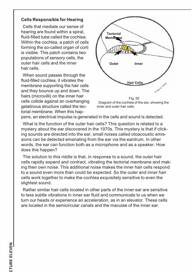
7/16/2019 Building Blocks of Human Life - Understanding Mature Cells and Stem Cells (Booklet).pdf
http://slidepdf.com/reader/full/building-blocks-of-human-life-understanding-mature-cells-and-stem-cells-bookletpdf 57/73
56
L E C T U R E E L E V E N
Cells Responsible for Hearing
Cells that mediate our sense of
hearing are found within a spiral,
fluid-filled tube called the cochlea.
Within the cochlea, a patch of cells
forming the so-called organ of cortiis visible. This patch contains two
populations of sensory cells, the
outer hair cells and the inner
hair cells.
When sound passes through the
fluid-filled cochlea, it vibrates the
membrane supporting the hair cells
and they bounce up and down. The
hairs (microvilli) on the inner hair cells collide against an overhanging
gelatinous structure called the tec-
torial membrane. When this hap-
pens, an electrical impulse is generated in the cells and sound is detected.
What is the function of the outer hair cells? This question is related to a
mystery about the ear discovered in the 1970s. This mystery is that if click-
ing sounds are directed into the ear, small noises called otoacoustic emis-
sions can be detected emanating from the ear via the eardrum. In other
words, the ear can function both as a microphone and as a speaker. Howdoes this happen?
The solution to this riddle is that, in response to a sound, the outer hair
cells rapidly expand and contract, vibrating the tectorial membrane and mak-
ing their own noise. This additional noise makes the inner hair cells respond
to a sound even more than could be expected. So the outer and inner hair
cells work together to make the cochlea exquisitely sensitive to even the
slightest sound.
Rather similar hair cells located in other parts of the inner ear are sensitiveto less subtle vibrations in inner ear fluid and communicate to us when we
turn our heads or experience an acceleration, as in an elevator. These cells
are located in the semicircular canals and the maculae of the inner ear.
Fig. 55
Diagram of the cochlea of the ear, showing the
inner and outer hair cells.
Tectorial
Membrane
Outer Inner
Hair Cells
© J o h n
K .Y o u n g

7/16/2019 Building Blocks of Human Life - Understanding Mature Cells and Stem Cells (Booklet).pdf
http://slidepdf.com/reader/full/building-blocks-of-human-life-understanding-mature-cells-and-stem-cells-bookletpdf 58/73
Which of the five senses would you consider the most vital for humans?
Would the same statement be true for rodents like rats or mice, which are
nocturnal and live in dark nests? Which sense, based upon the number of
genes, would be most important to rodents?
Slack, Jonathan. Essential Developmental Biology . 2nd ed. London:
Blackwell Science, Ltd., 2005.
Chuang, J.-Z., et al. “SARA-Regulated Vesicular Targeting Underlies
Formation of the Light-Sensing Organelle in Mammalian Rods.” Cell .
Vol. 130, pp. 535–547, 2007.
Cox, J.J., et al. “An SCN9A Channelopathy Causes Congenital Inability to
Experience Pain.” Nature. Vol. 444, pp. 894–898, 2006.
Grus, W.E., et al. “Dramatic Variation of the Vomeronasal Pheromone
Receptor Gene Repertoire Among Five Orders of Placental and Marsupial
Mammals.” Proceedings of the National Academy of Sciences. Vol. 102,
pp. 5767–5772, 2005.
Huang, A.L., et al. “The Cells and Logic for Mammalian Sour Taste Detection.”
Nature. Vol. 442, pp. 934–938, 2006.
Keller, A., and L.B. Vosshall. “Influence of Odorant Receptor Repertoire on
Odor Perception in Humans and Fruit Flies.” Proceedings of the National
Academy of Sciences. Vol. 104, pp. 5614–5619, 2007.
Kimchi, T., et al. “A Functional Circuit Underlying Male Sexual Behavior in the
Female Mouse Brain.” Nature, Vol. 448, pp. 1009–1014, 2007.
Sugimoto, K., and Y. Ninomiya. “Introductory Remarks on Umami Research:
Candidate Receptors and Signal Transduction Mechanisms on Umami.”
Chemical Senses. Vol. 30, pp. 121–127, 2005.
Questions
Suggested Reading
FOR GREATER UNDERSTANDING
Articles of Interest
57

7/16/2019 Building Blocks of Human Life - Understanding Mature Cells and Stem Cells (Booklet).pdf
http://slidepdf.com/reader/full/building-blocks-of-human-life-understanding-mature-cells-and-stem-cells-bookletpdf 59/73
The Suggested Reading for this lecture is Jonathan Slack’s Essential Developmental Biology.
Lecture 12:
Cells of Reproductive Organs
Germ Cells
Germ cells of the ovaries and testes do not actually originate in these
organs, but migrate from their birthplace in the embryonic yolk sac. At this
point, they are called primordial germ cells, and have not “decided” whether
to differentiate into sperm or egg cells. They follow a gradient of a protein
called stromal cell derived factor until they migrate to a mesodermal structurecalled the gonadal ridge. If the gonadal ridge is in a female, it will become the
ovary and it produces retinoic acid, which stimulates the germ cells to form
oocytes. If the gonadal ridge is in a male, it produces an enzyme that
destroys retinoic acid and forms the testis. In the absence of retinoic acid,
germ cells eventually become sperm cells.
Female Reproductive Cells of
the Ovaries
When oocytes burrow into a devel-oping ovary, they somehow induce
the formation of one or more layers
of cells around them that form ovari-
an follicles. Initially, the follicles are
small, but a few of them enlarge to
first form primary follicles and then,
eventually, large secondary follicles
that become filled with fluid pro-
duced by the surrounding cells. Outof the five hundred thousand
oocytes present in the ovaries of a
baby girl, only a few will ultimately
enlarge and develop large, fluid-
filled secondary follicles that are
capable of ovulation.
The cells in close contact with the oocyte are called granulosa cells. They
supply nutrients to the oocyte and also appear to regulate the enormous
growth of the oocyte. Granulosa cells synthesize a growth factor called StemCell Factor; this binds to receptors on oocytes and stimulate their growth.
Granulosa cells also send some sort of unknown molecule into the cyto-
plasm of the oocyte via tiny pores in the cell membrane called gap junctions.
In this way, the granulosa cells restrain the completion of meiosis in egg
cells until ovulation.
58
L E C T U R E T W E L V E
Fig. 56
A primary ovarian follicle, with a huge oocyte
surrounded by granulosa cells.
©
J o h n K . Y o u n g

7/16/2019 Building Blocks of Human Life - Understanding Mature Cells and Stem Cells (Booklet).pdf
http://slidepdf.com/reader/full/building-blocks-of-human-life-understanding-mature-cells-and-stem-cells-bookletpdf 60/73
59
Cells just outside the granulosa cell layer are called theca cells; their main
function is to synthesize the precursors of the female sex hormone, estrogen.
Estrogen has diverse effects upon the body, from restraining bone growth, to
encouraging fat deposition in portions of the body, and to stimulating brain
regions that regulate sexual behavior and appetite.
Ovulation is provoked by a massive release of gonadotropins (FSH, LH) intothe bloodstream from the pituitary. FSH and LH cause the granulosa cells to
release their inhibition upon the oocyte, and also to loosen their connections
with each other. The result is that the oocyte completes the first meiotic divi-
sion and also is released from the follicle, which splits open and releases the
oocyte from the ovary. The oocyte will travel down the oviduct to the uterus
and may be fertilized by a sperm on its journey.
The surge of gonadotropins that takes place at ovulation also has a trans-
forming effect upon the granulosa and theca cells that remain in the ovary.
They multiply and form a large structure called a corpus luteum that secretesanother hormone, progesterone, into the bloodstream. Progesterone prepares
the uterine lining for a possible pregnancy and also suppresses the further
release of LH and FSH from the pituitary gland, so that another ovulatory
surge does not occur prematurely. Artificial, synthetic versions of these two
sex hormones are used in birth control pills to prevent ovulation.
Most of the oocytes in the ovary do not undergo this process. Instead, they
slowly degenerate, a process called atresia. Recent studies from several labs
suggest that stem cells for oocytes persist in the bone marrow of adults and
may migrate into the ovaries to replace degenerating oocytes. This revolu-tionary proposal requires additional investigation.
Male Reproductive Cells of the Testis
Germ cells of the testis mature in fluid-filled, tubular structures called semi-
niferous tubules. The walls of the tubules are formed by Sertoli cells, epithe-
lial cells with pale, oval nuclei and highly folded cell membranes. Germ cells
become nestled within the folded cell membranes of the Sertoli cells, which
nourish them and stimulate them to divide. Like ovarian granulosa cells,
Sertoli cells also produce the protein stem cell factor (SCF), which stimulatesthe maturation of male germ cells as well as female germ cells. An impaired
function of SCF is thought to contribute to male infertility. On the other hand,
mutations in SCF that increase its activity contribute to the development of
testicular cancer, which is one of the most common types of cancer in young
males.
As the germ cells mature, they move toward the lumen of the seminiferous
tubules and pass through several developmental stages with different
names. Spermatogonia are the stem cells of the testis, and they divide very
energetically to constantly produce new sperm cells. The testis can produce
one hundred million new sperm each day. Some of the daughter cells of
spermatogonia start differentiating and begin the process of meiosis. These
cells are called spermatocytes. After a spermatocyte has completed two
meiotic divisions, it becomes a small, round cell called a spermatid. All of
these cell types are visible mixed among the Sertoli cells in a given seminif-
erous tubule.

7/16/2019 Building Blocks of Human Life - Understanding Mature Cells and Stem Cells (Booklet).pdf
http://slidepdf.com/reader/full/building-blocks-of-human-life-understanding-mature-cells-and-stem-cells-bookletpdf 61/73
60
L E C T U R E T W E L V E
Once spermatids have formed, they must begin a complicated process of
specialization to transform themselves from plain, round cells to small, spe-
cialized, torpedo-shaped sperm. Mature sperm cells have a complicatedanatomy. At the front of the cell is a membranous compartment called the
acrosome. This contains hydrolytic enzymes that will later be used to pene-
trate barriers surrounding the ovulated oocyte. The cell nucleus of the sperm
is also unusual: it is very streamlined, to promote ease in movement through
fluid, and has only one-fourtieth the volume of a normal cell nucleus. Finally,
sperm cells grow a long tail, or flagellum, that propels them through fluids.
Where do these specialized structures come from?
The acrosome arises from vesicles that bud off of the Golgi apparatus, a
structure that normally packages proteins for transport to the cell membraneand secretion. In spermatids,
however, these vesicles are sent
the wrong way and wind up
attached to the cell nucleus.
The cell nucleus of a spermatid
undergoes remarkable changes
itself. In order to pack the DNA of
the nucleus into a much smaller
volume, the chromosomes arecompletely restructured.
Normally, the DNA in a chromo-
some is associated with small
particles (core particles) com-
posed of proteins called his-
tones. Because of their large
Fig. 57
A seminiferous tubule within the testis.
©
J o h n K . Y o u n g
Sertoli Cells
Spermatids
Leydig
Cells
Fig. 58
Diagram of a sperm cell. The acrosome is
colored green.
Mitochondria
Axoneme
Outer
Dense
Fibers
© J o h n
K .Y o u n g

7/16/2019 Building Blocks of Human Life - Understanding Mature Cells and Stem Cells (Booklet).pdf
http://slidepdf.com/reader/full/building-blocks-of-human-life-understanding-mature-cells-and-stem-cells-bookletpdf 62/73
61
size, histones are removed from the DNA of sperm cells and replaced with a
much smaller molecule called protamine that now permits the DNA to be
crammed into a small volume. Also, the entire nucleus of a spermatid is
pushed through a narrow diameter cytoplasmic ring that squeezes it into a
longer, narrow shape.
The flagellum of a sperm cell originates from a structure called a centriole. Inspermatids, this structure embeds itself in the nuclear envelope and grows
enormously into a long flagellum. It attracts mitochondria to itself to provide
energy for flagellar movement. The center of the flagellum is called an
axoneme, composed of structures called microtubules. In other cells, an
axoneme forms the interior of cilia, which have the ability to generate move-
ment by sliding against each other.
Finally, spermatids strengthen the flagellum by enormously overproducing a
protein that is present in small traces around the centrioles of most cells. This
protein aggregates into nine strong fibers called outer dense fibers that addstrength to the flagellum.
Although some of the molecules involved in these transformations have
been identified, it is still unclear how they are stimulated and coordinated to
produce the final product.
Testosterone-Producing Cells
Cells outside of the seminiferous tubules called Leydig cells are responsible
for the production of the steroid hormone, testosterone. Like other steroid-
producing cells, they contain many pale-staining lipid droplets. They also pro-duce another hormone related to insulin called Insulin-3. When this hormone
is secreted during development, it causes ligaments to exert tension upon the
testis and move it from a position within the abdominal cavity to a final resting
place within the scrotal sac. If this hormone is given to developing female
mice, the ovaries in these mice will move in the same direction as if they
were testes and will come to rest near the tail region of the mouse.

7/16/2019 Building Blocks of Human Life - Understanding Mature Cells and Stem Cells (Booklet).pdf
http://slidepdf.com/reader/full/building-blocks-of-human-life-understanding-mature-cells-and-stem-cells-bookletpdf 63/73
1. Why is it necessary for spermatocytes and oocytes to undergo meiotic divi-
sions that reduce the amount of DNA in each cell?
2. Sex hormones alter the anatomy of many regions of the body, such as
muscles, bones, the skin, and fat tissue. This is partly to provide sex differ-
ences in appearance that promote reproductive behavior. What other
organ also adjusts its function in response to sex hormones?
Slack, Jonathan. Essential Developmental Biology . 2nd ed. London:
Blackwell Science, Ltd., 2005.
Adham, I.M., et al. “The Overexpression of the Insl3 in Female MiceCauses Descent of the Ovaries.” Molecular Endocrinology . Vol. 16,
pp. 244–252, 2002.
Bowles, J., et al. “Reinoid Signaling Determines Germ Cell Fate in Mice.”
Science. Vol. 312, pp. 596–600, 2006.
Klinger, F.G., and M. De Felice. “In Vitro Development of Growing Oocytes
from Fetal Mouse Oocytes: Stage-Specific Regulation by Stem Cell Factor
and Granulosa Cells.” Developmental Biology . Vol. 244, pp. 85–95, 2002.
Matzuk, M.M., et al. “Intercellular Communication in the Mammalian Ovary:Oocytes Carry the Conversation.” Science. Vol. 296, pp. 2178–2181, 2002.
Tilly, J.L., and J. Johnson. “Recent Arguments Against Germ Cell Renewal in
the Adult Human Ovary.” Cell Cycle. Vol. 6, pp. 879–883, 2007.
Questions
Suggested Reading
FOR GREATER UNDERSTANDING
Articles of Interest
62
L E C T U R E T W E L V E

7/16/2019 Building Blocks of Human Life - Understanding Mature Cells and Stem Cells (Booklet).pdf
http://slidepdf.com/reader/full/building-blocks-of-human-life-understanding-mature-cells-and-stem-cells-bookletpdf 64/73
63
The Suggested Reading for this lecture is Jonathan Slack’s Essential Developmental Biology.
Lecture 13:
Extreme Cells
Many of the cells described in preceding lectures have unusual features, like
a large size, an oddly shaped nucleus, or peculiar overall features. The cells
in this lecture, however, go beyond the conventional parameters that define
the lives of most cells of the body.
Lens CellsThe extremely long cells that form the lens of the eye are remarkable in
many ways: they lack nuclei and all membranous organelles and are perfectly
transparent to light, but still remain alive. Where do these cells come from,
and how can they function with these features?
The lens of the eye forms when the
developing eye bulges outward from
the developing brain, forming an
optic cup that touches the overlying
ectoderm. As it does so, it induces
the overlying ectoderm to fold in
upon itself to form a hollow lens
vesicle, composed of simple
cuboidal epithelial cells. The lens
vesicle becomes sealed to the ante-
rior opening of the optic cup. Then,
the posterior cells of the lens vesicle
multiply, elongate, and become
packed together to form the lens.
Why do these events occur? A
DNA-binding homeotic protein
called Pax6 initiates the develop-
ment of the optic cup; when Pax6 is
abnormal, eyes fail to form. The optic cup then secretes factors (for exam-
ple, fibroblast growth factor) that stimulate the lens cells to multiply and
elongate. They also synthesize proteins called crystallins that fill the cells
and take the place of all organelles. The extremely regular packing of the
crystallin proteins and the lack of organelles seem to explain why these cells
are transparent.
Since the cells no longer have mitochondria, they cannot oxidize nutrients to
produce ATP. Also, the lens has no blood vessels, so cells deep within it
have no direct access to nutrients. How do they survive? Nutrient molecules
are absorbed from the fluid within the eyeball and passed from one cell to the
other via small pores in the cell membrane called gap junctions.
Fig. 59
Diagrams of lens cells (yellow) surrounded by the
optic cup in the embryo (top) and in the fully devel-
oped lens of the eye (bottom).
© J o h n
K.Y o u n
g

7/16/2019 Building Blocks of Human Life - Understanding Mature Cells and Stem Cells (Booklet).pdf
http://slidepdf.com/reader/full/building-blocks-of-human-life-understanding-mature-cells-and-stem-cells-bookletpdf 65/73
64
L E C T U R E T H I R T E E N
Lens cells also have no protein-synthesizing machinery, so they cannot
repair themselves and must last the life of the individual. This poses addition-
al problems. Exposure to UV light damages the crystallins and causes a
gradual fogging of the lens called cataract. Surgery to replace the natural
lens with plastic lenses is the most common surgery in the United States for
people sixty-five and older. Another source of crystallin pathology is the pres-ence of high levels of blood glucose, as in diabetics. Glucose can be metabo-
lized to another sugar, sorbitol, by lens cells and may accumulate in the lens;
also, glucose can directly attach to crystallin proteins and disorder them.
These factors account for high rates of cataract formation in diabetics.
Thymic Nurse Cells
Peculiar epithelial cells found just
beneath the surface of the thymus have
the amazing ability to engulf and com-
pletely enclose within their cytoplasm
many developing lymphocytes (twenty
to two hundred per nurse cell) as well
as occasional macrophages. These
cells seem to be involved in controlling
the maturation and selection of lympho-
cytes in the thymus.
Each day, about fifty precursor cells
leave the bone marrow and enter thethymus. Each cell then divides twenty
times over the next two weeks, gener-
ating about fifty million new T-lympho-
cytes. However, only about one million of these cells will ever leave the thy-
mus, because about 95 percent of developing T-cells are destroyed before
they can reach the bloodstream. The cells that are destroyed (by macro-
phages) are the ones that have receptors that react with normal cell proteins
that are native to the body and are not foreign antigens. If this process of
T-cell selection failed to occur, the immune system would attack our
own tissues.
How are the “bad” lymphocytes identified and separated from the “good”
ones? The epithelial cells that make up the framework of the thymus have a
crucial role in this process, and thymic nurse cells belong to this family of
epithelial cells.
Another related type of epithelial cell in the innermost medulla of the thymus
also has a remarkable function. These medullary epithelial cells can express
at least four hundred different proteins on their cell membranes that were pre-
viously thought to be restricted to completely different organs. For example,cells in the thymic medulla display proteins otherwise found only on muscle
cells, kidney cells, ovarian cells, and other such cells. These proteins act as
“bait” to attract self-reactive T-lymphocytes. If the T-cells bind to muscle pro-
teins on the medullary epithelial cells, for example, they are killed before they
can escape the thymus and actually damage muscle.
Fig. 60
Diagram of a thymic nurse cell that hasengulfed seven lymphocytes (green).
©J o h n
K .
Y o u n g

7/16/2019 Building Blocks of Human Life - Understanding Mature Cells and Stem Cells (Booklet).pdf
http://slidepdf.com/reader/full/building-blocks-of-human-life-understanding-mature-cells-and-stem-cells-bookletpdf 66/73
65
How do these epithelial cells form? A DNA-binding factor called FOXN1 is
first expressed in both the skin and a portion of the embryo destined to
become the thymus. This homeotic protein is required for skin to form hairs
and for the thymus to form epithelial cells. Epithelial cells of the thymus
mature and have the ability to attract T-cell precursors from the bone marrow.
A genetically abnormal mouse called the nude mouse lacks this DNA-bindingprotein, and has a skin that cannot grow hairs and a thymus that cannot
attract lymphocytes. Consequently, any type of foreign tissue can be trans-
planted onto this mouse and won’t be rejected by the immune system,
because the thymus doesn’t function.
Cerebellar Purkinje Neurons
Purkinje cells of the cerebellar cortex derive their name from a Czech scien-
tist, Johannes Purkinje, who first decribed them. They have a striking and
unusually complex “tree” of dendritic processes: the initial dendrite from each
neuron branches dozens of times and produces a thin mass of dendritic
processes that resemble fan coral in the sea. Some dendritic trees of
Purkinje cells can have as many as eight hundred individual segments.
The dendritic tree of a Purkinje cell is oriented perpendicular to dozens of
axons, called parallel fibers, that pass through it, much like telephone wires
passing through multiple crossbeams on old-fashioned telephone poles.
Thousands of synapses from parallel fibers make contact with the dendritic
tree of a single Purkinje cell; all of these input signals are integrated into a
single output signal that leaves the Purkinje cell via the basal axon. No other
Fig. 61
Diagram of a Purkinje neuron in the cerebellum (left) and a photo-
graph of a Purkinje neuron, together with a few parallel fibers that
innervate its extensive dendritic tree (right).
©
J o h n K . Y o u n g
Parallel Fibers

7/16/2019 Building Blocks of Human Life - Understanding Mature Cells and Stem Cells (Booklet).pdf
http://slidepdf.com/reader/full/building-blocks-of-human-life-understanding-mature-cells-and-stem-cells-bookletpdf 67/73
66
L E C T U R E T H I R T E E N
neuron in the CNS has an anatomy that even comes close to matching the
complexity of Purkinje cells. These neurons are critical for the function of the
cerebellum, which regulates the movements of the arms, legs, and body in
three-dimensional space.
What stimulates their development? Curiously, a DNA-binding protein called
Pancreas Transcription Factor is responsible for the initial formation of thecerebellum; without this protein, mice are born lacking both a pancreas and a
cerebellum! As Purkinje cells develop, they acquire synapses from parallel
fibers. Activation of these synapses is what appears to stimulate the repeated
branching of the dendrites.
Podocytes
Podocytes are epithelial cells of the kidney that wrap
around capillaries within structures called renal corpus-
cles. They begin life as simple squamous epithelialcells, but eventually acquire a shape that makes it
hard to classify them as any particular kind
of epithelium.
Podocytes derive their name from the
Latin word for foot ( ped ), because of all the
cell processes, termed foot processes,
which they throw around capillaries. Fluid
escaping from openings in the capillary
endothelium passes through tiny slitsbetween foot processes called filtration
slits. This fluid is collected as an early,
unmodified form of urine. This fluid will
pass through many kidney tubules, and on its way will have many small,
vital molecules like sugars and amino acids reabsorbed from the urine. The
podocytes begin the job by preventing large molecules and cells from escap-
ing the capillaries into the urine.
How do podocytes acquire such a peculiar shape? Many proteins have been
identified that guide the complex process of kidney development. One proteinin particular, called podocalyxin, seems especially important for podocyte
development. This protein adjusts the attachment of podocytes to capillaries
and modifies cytoskeletal proteins that regulate cell shape. If this protein is
inactivated during development, the podocytes still settle upon capillaries, but
they retain a more conventional, square shape and never produce foot
processes. This prevents the proper filtration of the urine and is an alteration
incompatible with complete embryonic development and postnatal survival.
Gomori-positive AstrocytesGomori-positive (GP) astrocytes derive their name from the fact that they
possess large granules in their cytoplasms that stain strongly after applying a
so-called Gomori staining procedure to nervous tissue. These astrocytes are
abundant in only a few brain regions, including the region of the hypothala-
mus close to the third ventricle and the hippocampus.
Fig. 62Diagram of a podocyte (green) wrapping its
extensive cell processes around a capillary.
©J o h n
K .Y o u n
g

7/16/2019 Building Blocks of Human Life - Understanding Mature Cells and Stem Cells (Booklet).pdf
http://slidepdf.com/reader/full/building-blocks-of-human-life-understanding-mature-cells-and-stem-cells-bookletpdf 68/73
The identity of the cytoplasmic gran-
ules in GP astrocytes was long debat-
ed, but now it is known that they repre-
sent the iron- and copper-rich rem-
nants of destroyed mitochondria.
These organelles oxidize nutrients toprovide energy for the cell. They may
become damaged by this oxidative
activity. All cells in the body destroy
their organelles to some extent; most
organelles are continually being turned
over by being digested within lyso-
somes every two to five days.
However, GP astrocytes have unusu-
ally prominent deposits of nondigested
debris derived from mitochondria. Why
does this high rate of mitochondrial
destruction take place?
One clue is that these astrocytes are
richly endowed with a special form of
glucose transporter protein called
Glut2. This glucose transporter is also
present in pancreatic islet cells, and
permits them to secrete insulin onlywhen blood glucose levels are high. In
the hypothalamus, this protein permits
astrocytes to function as glucose sensors, detecting when blood glucose lev-
els become too high or too low. Astrocytes then modulate the function of
nearby neurons, which regulate functions such as the control of appetite and
the reflex control of insulin from the pancreas. Abnormalities in this brain
region may therefore be important in the development of abnormalities such
as obesity or diabetes mellitus.
The reasons why GP astrocytes destroy their own mitochondria are still notclear. It may be that mitochondria become damaged when they oxidize large
amounts of glucose or fats, which are avidly taken up by GP astrocytes. In
any event, chronic damage to mitochondria in the hypothalamus cannot be
expected to have beneficial effects upon the function of this brain region.
Fig. 63
The hypothalamus (top) in the brain region
contains abundant Gomori-positive astrocytes
(dark, iron-rich spots) and a high-magnification
view of Gomori-positive astrocytes (bottom),
with their dark-staining granules near the
cell nucleus.
©
J o h n K . Y o u n g
67
Neuron
Gomori and Astrocyte
Hypothalamus

7/16/2019 Building Blocks of Human Life - Understanding Mature Cells and Stem Cells (Booklet).pdf
http://slidepdf.com/reader/full/building-blocks-of-human-life-understanding-mature-cells-and-stem-cells-bookletpdf 69/73
In addition to lens cells, two other types of cells in the body, described in pre-
ceding lectures, have the ability to destroy their nuclei and fill themselves with
large amounts of specific proteins. Which other two cells can also do this?
Slack, Jonathan. Essential Developmental Biology . 2nd ed. London:
Blackwell Science, Ltd., 2005.
Call, M.K., and M.W. Grogg. “Eye on Regeneration.” Anatomical Record . Vol.
287B, pp. 42–48, 2005.
Gotter, J., et al. “Medullary Epithelial Cells of the Human Thymus Express
a Highly Diverse Selection of Tissue-Specific Genes Colocalized inChromosomal Clusters.” Journal of Experimental Medicine. Vol. 199,
pp. 155–166, 2004.
Marty N, et. al. “Regulation of Glucagon Secretion by Glucose Transporter
Type 2 (Glut2) and Astrocyte-Dependent Glucose Sensors.” Journal of
Clinical Investigation. Vol. 115, pp. 3545–3550, 2005.
Pascual, M., et al. “Cerebellar GABAergic Progenitors Adopt an External
Granule Cell-like Phenotype in the Absence of Ptf1a Transcription Factor
Expression.” Proceedings of the National Academy of Sciences. Vol. 104,
pp. 5193–5198, 2007.
Robinson, M.L. “An Essential Role for FGF Receptor Signaling in Lens
Development.” Seminars in Cell and Developmental Biology . Vol. 17,
pp. 726–740, 2006.
Webb, O., et al. “The Identification of Thymic Nurse Cells in Vivo and the
Role of Cytoskeletal Proteins in Thymocyte Internalization.” Cell
Immunology . Vol. 228, pp. 119–129, 2004.
Young, J.K., and J.C. McKenzie. “GLUT2 Immunoreactivity in Gomori-
Positive Astrocytes of the Hypothalamus.” Journal of Histochemistry and Cytochemistry . Vol. 52, pp. 1519–1524, 2004.
Questions
Suggested Reading
FOR GREATER UNDERSTANDING
Articles of Interest
68
L E C T U R E T H I R T E E N

7/16/2019 Building Blocks of Human Life - Understanding Mature Cells and Stem Cells (Booklet).pdf
http://slidepdf.com/reader/full/building-blocks-of-human-life-understanding-mature-cells-and-stem-cells-bookletpdf 70/73
69
The Suggested Reading for this lecture is Jonathan Slack’s Essential Developmental Biology.
Lecture 14:
Death and Aging of Cells
Death by Macrophage
The lifespan and death of red blood cells (erythrocytes) have been exten-
sively studied by scientists interested in the longevity of cells. They have
found that red blood cells circulate in the bloodstream for about one hundred
twenty days. As they age, they accumulate abnormalities in their cell mem-brane and other proteins that they cannot repair, since they no longer pos-
sess a cell nucleus or other organelles that could replace damaged mole-
cules. One such abnormality is that the surface of the cell membrane begins
to contain molecules of a lipid, phosphatidyl serine, that is normally only
found on the inner, and not the outer, surface of the cell membrane.
Macrophages have receptors for this type of lipid, and when they encounter
a red blood cell with this lipid freely exposed on their surfaces, they ingest
and destroy the red blood cell. However, even old red blood cells are safe
when circulating within blood vessels, where they are not threatened byconnective tissue macrophages. Unfortunately for the red blood cell, this
period of safety ends when red blood cells enter the peculiar blood vessels
of the spleen.
In most tissues, red blood cells travel
through arteries, to capillaries, and to
veins that carry them back to the heart in
an uninterrupted pathway. In the spleen,
however, capillaries do not connect direct-
ly to veins. Instead, red blood cells gushout of openings in the ends of capillaries
and form masses within the connective tis-
sue of the spleen. To reenter the general
circulation, red blood cells must traverse
the connective tissue, locate a thin-walled
vessel called a sinusoid, and push their
way through the sinusoid endothelial cells
to get into the lumen of the vessel. To
accomplish this, they must run the gaunt-let of macrophages that cluster around
vessels, lying in wait for old red blood
cells. Those red blood cells that don’t
survive this process end their lives as
brown-staining granules within the
cytoplasm of macrophages.
Fig. 64
Aging red blood cells in the spleen must
bypass macrophages to enter a blood ves-
sel and return to the general circulation.
©
J o h n K . Y o u n
g
Macrophages
Red Blood Cells

7/16/2019 Building Blocks of Human Life - Understanding Mature Cells and Stem Cells (Booklet).pdf
http://slidepdf.com/reader/full/building-blocks-of-human-life-understanding-mature-cells-and-stem-cells-bookletpdf 71/73
70
L E C T U R E F O U R T E E N
Why aren’t other long-lived cells also destroyed by macrophages? For
example, nerve cells and lens cells of the eye can live for eighty years
and surely also accumulate abnormal lipid in their membranes that should
attract macrophages.
The answer to this question is that the lens and brain cannot be penetrated
by macrophages. The lens lacks blood vessels, and the brain possesses spe-cial blood vessels that are tightly sealed and not “leaky.” This prevents
macrophages or their precursors, monocytes, from gaining access to aging
cells in these areas and destroying them.
Death by Suicide
Many cells in the body are not destined for a long life, but commit suicide
(programmed cell death) in a number of conditions. For example, growing
nerve cells in the neural tube of the embryo are constantly sending axons into
other regions of the body to innervate structures. When they contact a mus-cle or a gland and make a synapse, they are rewarded by the target cells,
which make growth factors that preserve the life of the neuron. However, if
the neuron fails to find a proper target, it is doomed.
Eventually, the nerve cells that fail to make connections self-destruct, in a
process called apoptosis. This word originates from the Greek words for top
(apo) and for falling away from ( ptosis), and is an analogy for the fact that
dead leaves self-destruct and fall away from the tops of trees. It is estimated
that one-half of the neurons in the developing CNS undergo apoptosis. So
the anatomy of the CNS is modified by widespread cell death.
Mechanism of Programmed Cell Death
If a cell like a neuron fails to react to growth factors, or experiences starva-
tion or has other metabolic problems, the energy-producing organelles called
mitochondria seem to be the first structures affected. Mitochondrial mem-
branes become leaky and allow the escape into the cytoplasm of a protein
called cytochrome C.
Cytochrome C activates caspases. Caspases are a family of fifteen enzymes
in the cell that can destroy proteins, organelles, and DNA. These proteins areextremely dangerous to cells and are present only as inactive precursors that
must be activated by cytochrome C. After organelle destruction, apoptotic
cells also display phosphatidyl serine on their cell membranes, so that
macrophages are recruited to ingest the debris.
Death by Perforation
Cells infected by invading viruses must also be eliminated, in a way that
avoids rupture of the cell and dissemination of the infectious virus into other
cells. This is accomplished by so-called cytotoxic T lymphocytes, which iden-tify an infected cell by the presence of viral proteins on the cell membrane.
These T lymphocytes secrete proteins called perforins that create tiny pores,
or perforations, in the membrane of the infected cell. Then the T lymphocyte
secretes enzymes into the infected cell that activate apoptotic caspases. The
cell responds by dying. This once again attracts the attention of
macrophages, which ingest cell fragments and eliminate all traces of the
infected cell.

7/16/2019 Building Blocks of Human Life - Understanding Mature Cells and Stem Cells (Booklet).pdf
http://slidepdf.com/reader/full/building-blocks-of-human-life-understanding-mature-cells-and-stem-cells-bookletpdf 72/73
71
Death by Sunshine
DNA in a cell’s chromosomes can easily be damaged by ultraviolet light.
This is particularly true of skin cells, but can also apply to cells just beneath
the skin in connective tissue or blood vessels. It is important that cells with
damaged DNA stop multiplying and possibly be eliminated; otherwise, some
cells will turn into harmful, rogue cells, like cancer cells, that can damage thebody. How is DNA damage detected, and how are mutant cells destroyed?
The first answers to this puzzle came from study of a rare genetic disorder
called ataxia telangiectasia (AT). People suffering from this disorder show
extreme cell damage after exposure to sunlight and have rates of cancer
development sixteen times higher than normal! In 1995, the abnormal pro-
teins in this disease were identified; one of them is called the ATM protein.
The ATM protein and related proteins associate with chromosomes and
detect breaks in the DNA. They then activate another protein called p53.
The p53 protein is a DNA-binding protein that regulates the function of at
least eighty-five other genes on chromosomes. Some of these genes code for
the caspases that initiate apoptosis. Other genes regulate cell division and
DNA repair enzymes. When p53 is activated, the cell responds by stopping
DNA duplication and by trying to repair DNA damage. If these attempts fail,
the mechanisms for apoptosis are activated and the cell commits suicide.
A failure of p53 to act can have serious consequences. About 50 percent of
all cancerous cells in the body are not detected and destroyed because they
have an abnormal p53 protein.Death at the Ends of Chromosomes
Some cells in the body, such as stem cells, germ cells, or cancer cells,
seem to have the ability to keep dividing indefinitely and live forever. Most
cells, like fibroblasts, however, seem to stop dividing after about fifty divisions
and begin to age. This limit to lifespan seems related to the end pieces of
chromosomes, called telomeres.
Telomeres are stretches of DNA that cap each end of a chromosome and
prevent it from unraveling. Telomeric DNA, unlike the rest of DNA, can’teasily be duplicated by normal cell enzymes, since these enzymes require
an unbroken stretch of DNA without an end portion. Thus, with each cell
division, the telomere of a chromosome becomes shorter, so that finally
there is nothing left to the telomere and the cell cannot divide any longer.
Also, a damaged telomere activates proteins like p53 that respond to
damaged DNA; this can provoke apoptosis.
Stem cells, cancer cells, and germ cells get around this problem by making
a special enzyme called telomerase that can rebuild the telomeres of chromo-
somes. If this enzyme is experimentally suppressed in mice, the lifespan of amouse can be reduced by 50 percent! Increasing the activity of telomerase in
more cells could possibly have a strong anti-aging effect and could increase
lifespan; however, it could also have the unwanted effect of allowing more
cancer cells to grow and multiply.

7/16/2019 Building Blocks of Human Life - Understanding Mature Cells and Stem Cells (Booklet).pdf
http://slidepdf.com/reader/full/building-blocks-of-human-life-understanding-mature-cells-and-stem-cells-bookletpdf 73/73
1. What happens to red blood cells when they enter the blood vessels of
the spleen?
2. Why aren’t nerve cells and lens cells destroyed by macrophages?
Slack, Jonathan. Essential Developmental Biology . 2nd ed. London:
Blackwell Science, Ltd., 2005.
Lodish, H.F., et al. Molecular Cell Biology . New York: W.H. Freeman, 2007.
Collado, M., et al. “Cellular Senescence in Cancer and Aging.” Cell . Vol. 130,
pp. 223–233, 2007.
Fadok, V.A., et al. “Loss of Phospholipid Asymmetry and Surface Exposure of
Phophatidylserine Is Required for Phagocytosis of Apoptotic Cells by
Macrophages and Fibroblasts.” Journal of Biological Chemistry .
Vol. 276, pp. 1071–1077, 2001.
Fernandes, N., et al. “DNA Damage-Induced Association of ATM with Its
Target Proteins Requires a Protein Interaction Domain in the N Terminus of
of ATM.” Journal of Biological Chemistry . Vol. 280, pp. 15158–15164, 2005.
Horvath, M.M., et al. “Divergent Evolution of Human p53 Binding Sites:
Cell Cycle vs. Apoptosis.” PLoS Genetics. Vol. 3, pp. 1284–1291, 2007.
Questions
Suggested Reading
FOR GREATER UNDERSTANDING
Articles of Interest
Other Books of Interest


