Embed Size (px)
Citation preview
“BUBBLES IN THE CHEST”THE PEDIATRIC
SPECTRUM OF AIR CONTAINING PULMONARY
LESIONSMaria d' Almeida, MD • Katie Lopez, MD • Julieta Oneto, MD • Ricardo Restrepo, MD
Miami Children’s Hospital, Miami, Florida
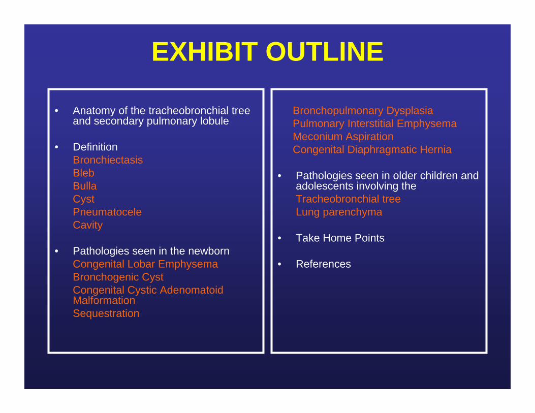
EXHIBIT OUTLINE
• Anatomy of the tracheobronchial tree and secondary pulmonary lobule
• Definition BronchiectasisBlebBullaCystPneumatoceleCavity
• Pathologies seen in the newbornCongenital Lobar EmphysemaBronchogenic CystCongenital Cystic Adenomatoid MalformationSequestration
Bronchopulmonary DysplasiaPulmonary Interstitial EmphysemaMeconium Aspiration Congenital Diaphragmatic Hernia
• Pathologies seen in older children and adolescents involving theTracheobronchial treeLung parenchyma
• Take Home Points
• References

ANATOMY OF THE TRACHEOBRONCHIAL TREE
appost
antRUL
lat
medRML
ab
lb pb
mbRLL
ap/p
ant
supinf
lb
pb am
LUL
LINGULA
LLL
Bronchus – Cartilage is seen in the wall
Bronchiole – Absence of cartilage in the wall
Respiratory bronchiole – Alveoli are seen in the wall
Lobular bronchiole – Supplies secondary pulmonary lobule
Distal trachea

ANATOMY OF THE SECONDARY PULMONARY LOBULE
Secondary pulmonary lobule – Functional pulmonary unit appearing as an irregular polyhedron separated from each other by thin fibrous interlobular septa.
Acinus – Functionally most important subunit of lung consisting of all parenchymal tissue distal to one terminal bronchiole.
Lobular artery
Pulmonary vein
Lobular bronchioleAcinus
Terminal bronchiole
Interlobular septa
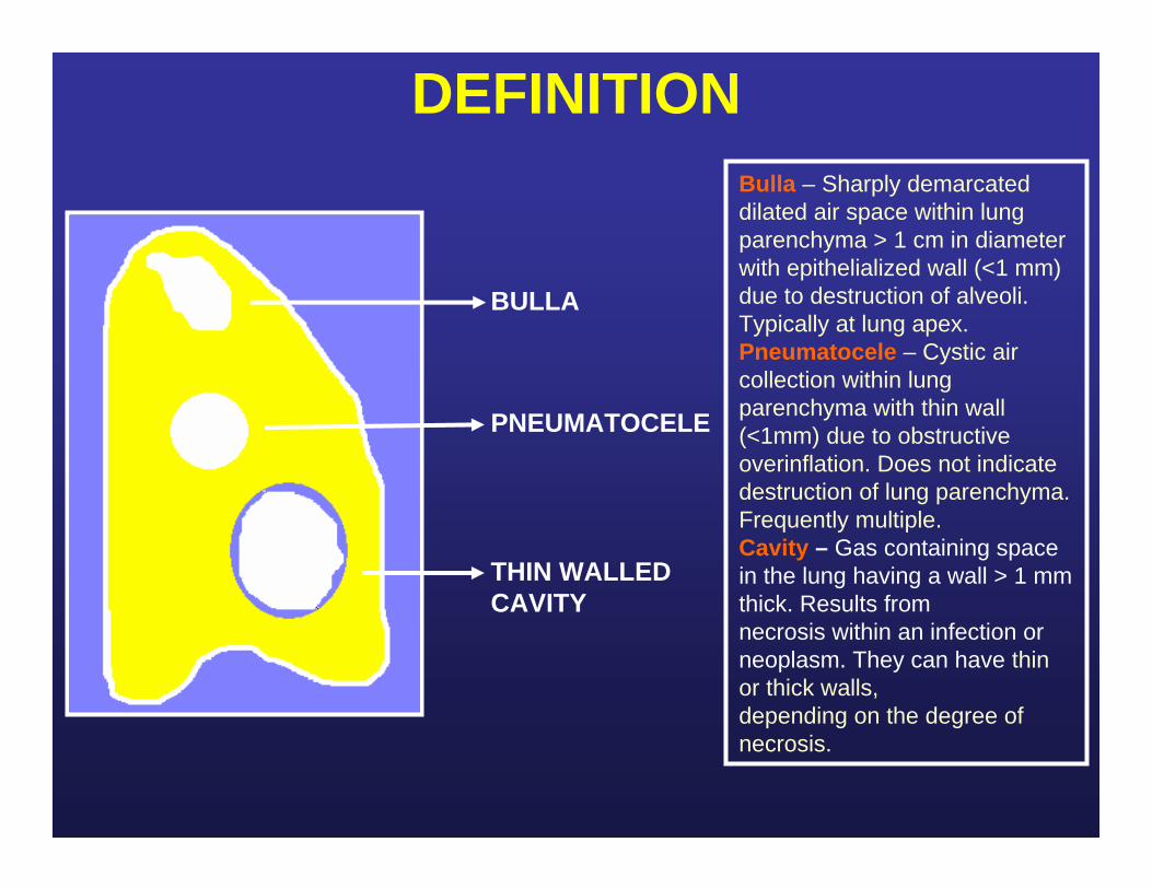
DEFINITION
BULLA
PNEUMATOCELE
THIN WALLED CAVITY
Bulla – Sharply demarcated dilated air space within lung parenchyma > 1 cm in diameter with epithelialized wall (<1 mm) due to destruction of alveoli. Typically at lung apex.Pneumatocele – Cystic air collection within lung parenchyma with thin wall (<1mm) due to obstructive overinflation. Does not indicatedestruction of lung parenchyma. Frequently multiple.Cavity – Gas containing space in the lung having a wall > 1 mm thick. Results fromnecrosis within an infection orneoplasm. They can have thin or thick walls,depending on the degree of necrosis.
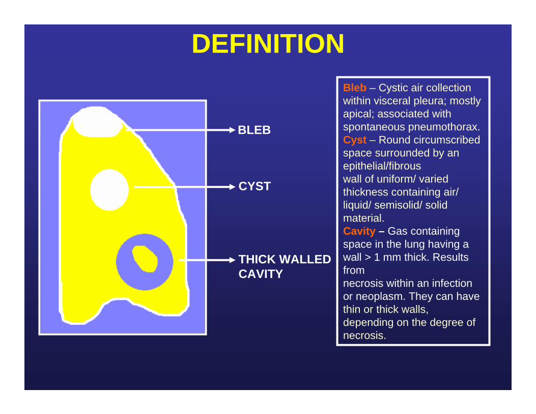
DEFINITION
BLEB
CYST
THICK WALLED CAVITY
Bleb – Cystic air collection within visceral pleura; mostly apical; associated with spontaneous pneumothorax.Cyst – Round circumscribed space surrounded by an epithelial/fibrouswall of uniform/ varied thickness containing air/ liquid/ semisolid/ solid material.Cavity – Gas containing space in the lung having a wall > 1 mm thick. Results fromnecrosis within an infection or neoplasm. They can have thin or thick walls,depending on the degree of necrosis.

DEFINITIONBronchiectasis – Localized mostly irreversible dilatation of the bronchioften with thickening of the bronchial wall. Cylindrical/Tubular/Fusiform – Mildly and uniformly dilated bronchi(least severe type). Varicose – Moderately dilated and beaded bronchi (rare). Saccular/Cystic – Marked cystic dilatation (most severe type).
A. Axial CT scan demonstrates tubular bronchiectasis in the right middle lobe (arrow). B. Histology in a different patient demonstrates tubular dilatation of the bronchi (arrows). C. Gross specimen in a different patient depicts dilated bronchi (arrow).
A B C

NEWBORN PATHOLOGIES
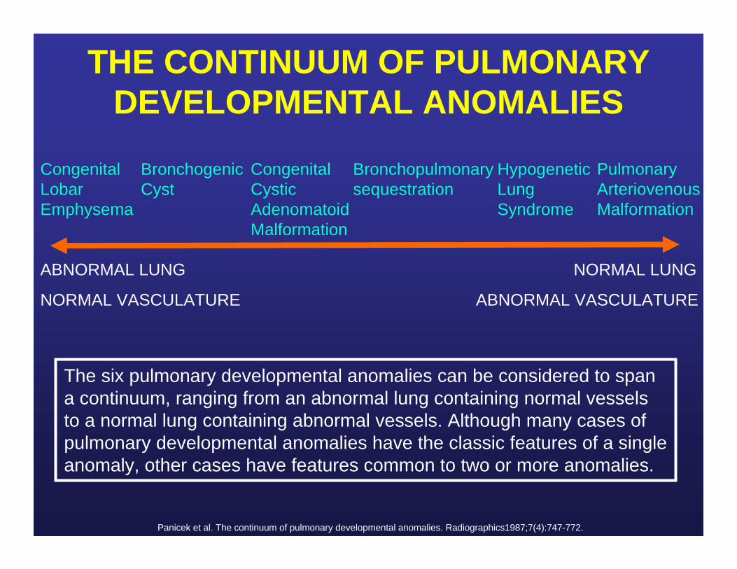
THE CONTINUUM OF PULMONARY DEVELOPMENTAL ANOMALIES
ABNORMAL LUNG
NORMAL VASCULATURE
NORMAL LUNG
ABNORMAL VASCULATURE
Congenital Lobar Emphysema
Bronchogenic Cyst
Congenital Cystic Adenomatoid Malformation
Bronchopulmonary sequestration
Hypogenetic Lung Syndrome
Pulmonary Arteriovenous Malformation
The six pulmonary developmental anomalies can be considered to span a continuum, ranging from an abnormal lung containing normal vessels to a normal lung containing abnormal vessels. Although many cases of pulmonary developmental anomalies have the classic features of a single anomaly, other cases have features common to two or more anomalies.
Panicek et al. The continuum of pulmonary developmental anomalies. Radiographics1987;7(4):747-772.

CONGENITAL LOBAR EMPHYSEMA
A. Chest radiograph shows focal hyperlucency in the right upper lobe (arrow). B. Axial CT demonstrates focal hyperlucency in the right upper lobe (arrow).
Most patients present in the neonatal period with respiratory distress.
There is a lobar predilection, with the most common site being the left upper lobe (43%), followed by the right middle lobe (32%) and right lower lobe (20%).
Hyperlucent and hyperexpanded lobe is seen on chest radiograph. On CT, air is in the alveoli with the interstitial septa and bronchovascular bundles seen at the periphery of the lucency.
A B

BRONCHOGENIC CYSTSecondary to abnormal budding of the tracheobronchial tree during development.
Seen in the mediastinum and lung parenchyma with equal frequency.
They do not contain air until they become infected.
May appear as well-defined soft tissue attenuation or cystic air-fluid containing masses.
Axial CT demonstrates an air containing intraparenchymal bronchogenic cyst in the right middle lobe (arrow).
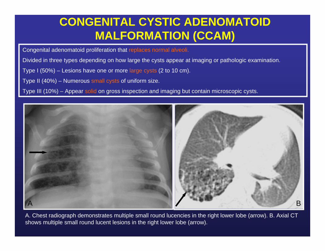
CONGENITAL CYSTIC ADENOMATOID MALFORMATION (CCAM)
A. Chest radiograph demonstrates multiple small round lucencies in the right lower lobe (arrow). B. Axial CT shows multiple small round lucent lesions in the right lower lobe (arrow).
Congenital adenomatoid proliferation that replaces normal alveoli.
Divided in three types depending on how large the cysts appear at imaging or pathologic examination.
Type I (50%) – Lesions have one or more large cysts (2 to 10 cm).
Type II (40%) – Numerous small cysts of uniform size.
Type III (10%) – Appear solid on gross inspection and imaging but contain microscopic cysts.
A B

SEQUESTRATION
A. Axial CT angiogram demonstrates marked enhancement of right lower lobe abnormal pulmonary tissue. B. CT shows large area of parenchymal lucency adjacent to the area of sequestration. C. Reconstructed coronal image demonstrates abnormal arterial supply (arrow) from the aorta.
Congenital area of abnormal pulmonary tissue without a normal connection to the bronchial tree.
Anomalous arterial supply to the abnormal lung via a systemic artery arising from the aorta.
Most commonly presents with a history of recurrent pneumonia.
It appears as a radiopaque mass in the neonatal period. Air may be introduced after infection has occurred.
Can be intralobar (more common) or extralobar (separate pleura and can be associated with other abnormalities).
A
B
C
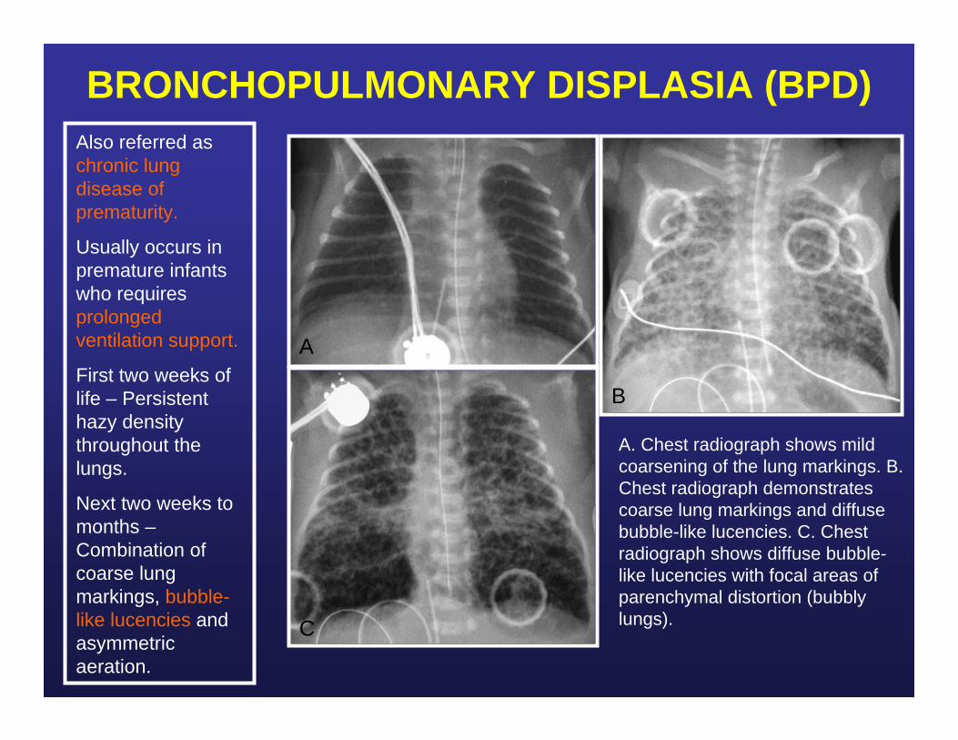
BRONCHOPULMONARY DISPLASIA (BPD)
A. Chest radiograph shows mild coarsening of the lung markings. B. Chest radiograph demonstrates coarse lung markings and diffuse bubble-like lucencies. C. Chest radiograph shows diffuse bubble-like lucencies with focal areas of parenchymal distortion (bubbly lungs).
Also referred as chronic lung disease of prematurity.
Usually occurs in premature infants who requires prolonged ventilation support.
First two weeks of life – Persistent hazy density throughout the lungs.
Next two weeks to months –Combination of coarse lung markings, bubble-like lucencies and asymmetric aeration.
A
B
C

CHRONIC LUNG DISEASE - BPDA. Chest radiograph demonstrates hypoventilation of the lungs, bilateral parenchymal lucencies and coarsening of the lung markings. B and C. Axial CT images show focal areas of hyperlucency (arrow) associated with parenchymal distortion (arrowhead) bilaterally.A
B C

BPD IN AN OLDER PATIENT
A. Chest radiograph demonstrates hyperlucency of the right lung (arrow). B. Axial CT reveals focal hyperaeration (arrow) and pseudo fissure formation (arrowhead).
Over years in children who survive, many of the radiographic findings of bronchopulmonary dysplasia decrease in prominence, and only hyperaeration may be present.
Clinically, many children with severe BPD during infancy may have problems such as exercise intolerance, predisposition to infection or asthma.
A B

AIR LEAK PHENOMENA – PULMONARY INTERSTITIAL EMPHYSEMA
Mechanical ventilation is an important risk factor contributing to air leak in premature infants treated for lung disease.
Rupture at bronchioloalveolar junctions permits passage of gas into the perivascular and peribronchial spaces, a condition that has been termed pulmonary interstitial emphysema.
Appears on radiographs as bubble-like or linear lucencies which can be focal or diffuse.
Chest radiograph shows bubble-like lucencies (arrow) within the left lung.
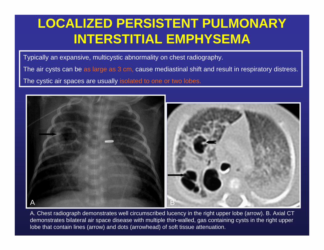
LOCALIZED PERSISTENT PULMONARY INTERSTITIAL EMPHYSEMA
A. Chest radiograph demonstrates well circumscribed lucency in the right upper lobe (arrow). B. Axial CT demonstrates bilateral air space disease with multiple thin-walled, gas containing cysts in the right upper lobe that contain lines (arrow) and dots (arrowhead) of soft tissue attenuation.
Typically an expansive, multicystic abnormality on chest radiography.
The air cysts can be as large as 3 cm, cause mediastinal shift and result in respiratory distress.
The cystic air spaces are usually isolated to one or two lobes.
A B
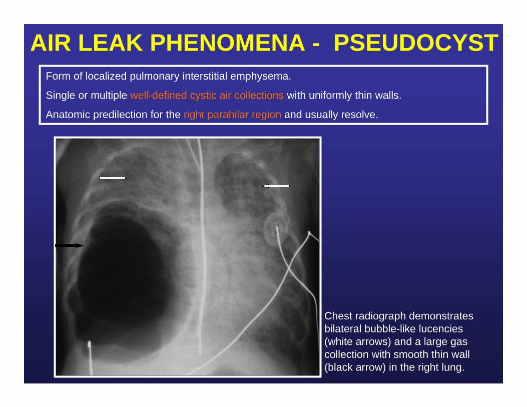
AIR LEAK PHENOMENA - PSEUDOCYSTForm of localized pulmonary interstitial emphysema.
Single or multiple well-defined cystic air collections with uniformly thin walls.
Anatomic predilection for the right parahilar region and usually resolve.
Chest radiograph demonstrates bilateral bubble-like lucencies(white arrows) and a large gas collection with smooth thin wall (black arrow) in the right lung.

AIR LEAK PHENOMENA – PNEUMOMEDIASTINUM (“SPINNAKER SAIL SIGN”)
Chest radiograph demonstrates extensive pneumomediastinum with “continuous diaphragm sign”(arrowhead) and air outlining the thymus with superior displacement (“spinnaker sail sign”) (arrow).
Mediastinal air may elevate the lobes of the thymus ("angel wing" or "spinnaker sail sign"), track within the extrapleural space and outline the inferior aspect of the heart ("continuous diaphragm sign"), and dissect into the soft tissues of the neck or chest wall.
Spinnaker sail

MECONIUM ASPIRATIONOccurs secondary to intrapartum or intrauterine aspiration of meconium.
The aspirated meconium causes obstruction of small airways secondary to its tenacious nature as well as a chemical pneumonitis.
Radiographic findings include areas of hyperinflation alternating with areas of atelectasis. Pleural effusion can be present. Pneumothorax is present in 20 to 40% of cases.
Pneumothorax in supine newborns tends to collect anteriorly, simulating a cystic pulmonary mass.
A. Chest radiograph shows large lung volumes and coarse bilateral perihilar markings. Right pneumothorax (arrow) is also seen. B. Chest radiograph demonstrates interval worsening of right pneumothorax which is loculated anteriorly simulating a round cystic mass (arrow).
A B
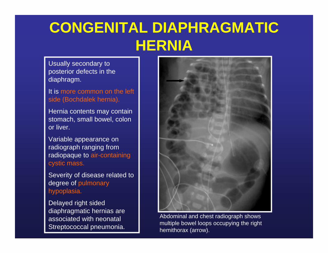
CONGENITAL DIAPHRAGMATIC HERNIA
Abdominal and chest radiograph shows multiple bowel loops occupying the right hemithorax (arrow).
Usually secondary to posterior defects in the diaphragm.
It is more common on the left side (Bochdalek hernia).
Hernia contents may contain stomach, small bowel, colon or liver.
Variable appearance on radiograph ranging from radiopaque to air-containing cystic mass.
Severity of disease related to degree of pulmonary hypoplasia.
Delayed right sided diaphragmatic hernias are associated with neonatal Streptococcal pneumonia.

OLDER CHILDREN AND ADOLESCENTS PATHOLOGIES
TRACHEOBRONCHIAL TREE
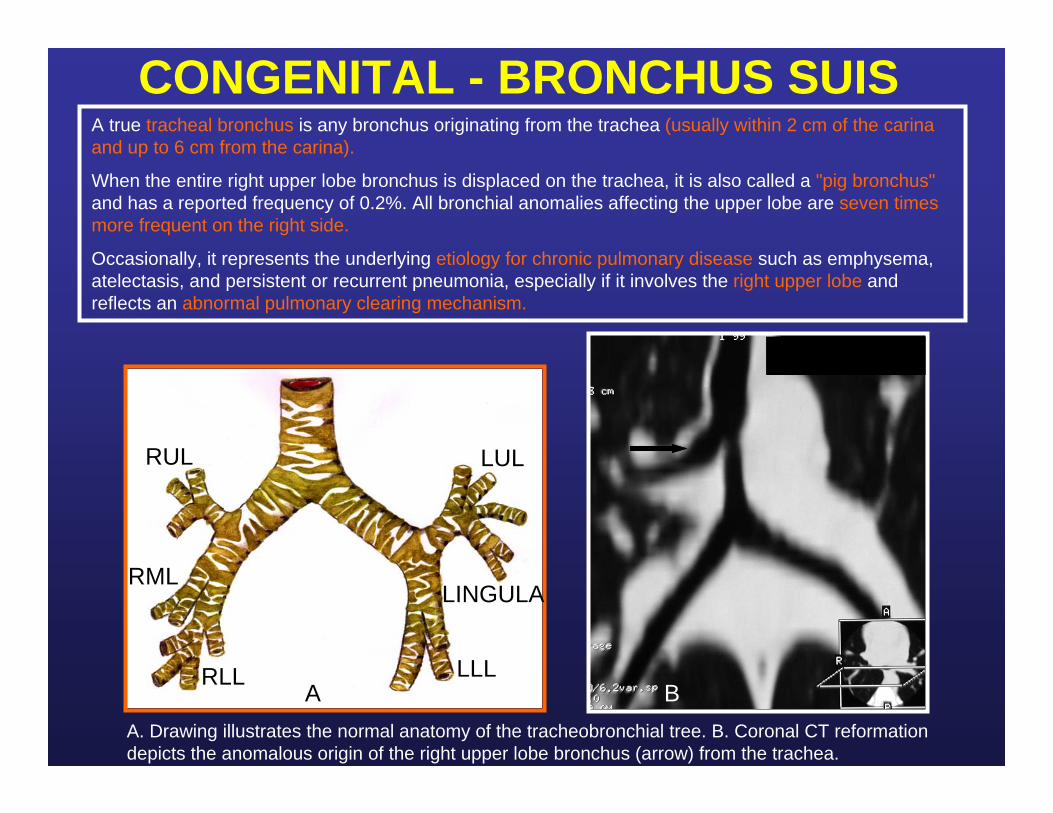
CONGENITAL - BRONCHUS SUIS
aaaa
A. Drawing illustrates the normal anatomy of the tracheobronchial tree. B. Coronal CT reformation depicts the anomalous origin of the right upper lobe bronchus (arrow) from the trachea.
A true tracheal bronchus is any bronchus originating from the trachea (usually within 2 cm of the carina and up to 6 cm from the carina).
When the entire right upper lobe bronchus is displaced on the trachea, it is also called a "pig bronchus"and has a reported frequency of 0.2%. All bronchial anomalies affecting the upper lobe are seven times more frequent on the right side.
Occasionally, it represents the underlying etiology for chronic pulmonary disease such as emphysema, atelectasis, and persistent or recurrent pneumonia, especially if it involves the right upper lobe and reflects an abnormal pulmonary clearing mechanism.
B
RUL
RML
RLL
LUL
LINGULA
LLLA
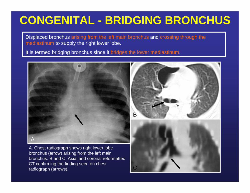
CONGENITAL - BRIDGING BRONCHUS
A. Chest radiograph shows right lower lobe bronchus (arrow) arising from the left main bronchus. B and C. Axial and coronal reformatted CT confirming the finding seen on chest radiograph (arrows).
Displaced bronchus arising from the left main bronchus and crossing through the mediastinum to supply the right lower lobe.
It is termed bridging bronchus since it bridges the lower mediastinum.
A
B
C
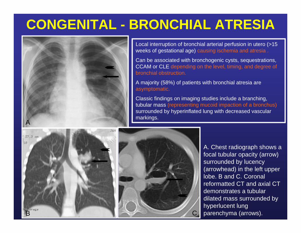
CONGENITAL - BRONCHIAL ATRESIA
A. Chest radiograph shows a focal tubular opacity (arrow) surrounded by lucency (arrowhead) in the left upper lobe. B and C. Coronal reformatted CT and axial CT demonstrates a tubular dilated mass surrounded by hyperlucent lung parenchyma (arrows).
A
B C
Local interruption of bronchial arterial perfusion in utero (>15weeks of gestational age) causing ischemia and atresia .
Can be associated with bronchogenic cysts, sequestrations, CCAM or CLE depending on the level, timing, and degree of bronchial obstruction.
A majority (58%) of patients with bronchial atresia are asymptomatic.
Classic findings on imaging studies include a branching, tubular mass (representing mucoid impaction of a bronchus)surrounded by hyperinflated lung with decreased vascular markings.

INFECTIOUS - PAPILLOMATOSIS
A. Chest radiograph shows bilateral ill defined parenchymal opacities and air fluid level in the right upper lobe (arrows). B. CT demonstrates multiple cystic lesions (arrows) with and infected lesion with air fluid level in the right upper lobe (arrowhead).
A B
Disease of childhood seen in children born from mothers with active human papilloma virusinfection at the time of delivery.
CT demonstrates nodularity of the tracheal mucosa and nodular parenchymal lesions that can be either solid or cavitated, and tend to get infected (presenting with air/fluid levels).
Patients with papillomatosis may also present with bronchiectasis.

MECHANICAL – FOREIGN BODY ASPIRATION
A. Chest radiograph demonstrates hyperlucency and hyperinflation of the left lung. B. Chest radiograph obtained with patient in expiration shows accentuation of the air trapping in the left lung.
When foreign bodies are aspirated, the bronchus is the most common site for lodgment.
The majority (97%) of bronchial foreign bodies are nonradiopaque.
The most important radiographic feature is a lack of change in lung volumedemonstrated at different phases of the respiratory cycle.
EXPIRATIONA B

MECHANICAL – ENDOBRONCHIAL CARCINOID
A and B. Axial CT images demonstrate a focal endobronchial lesion in the left upper lobe bronchus (arrow) with associated hyperlucency of the left upper lobe consistent with air-trapping (arrowhead) with post obstructive pneumonitis.
Endobronchial tumors are rare in patients under 20 years of age.
Greater than 90% of this tumors are malignant.
Most common endobronchial tumor is carcinoid.
Children nearly always present with complaints related to airway obstruction.
The endobronchial lesion can cause check valve phenomenon and air-trapping.
A B
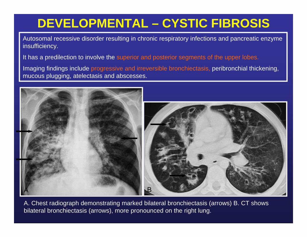
DEVELOPMENTAL – CYSTIC FIBROSIS
A. Chest radiograph demonstrating marked bilateral bronchiectasis (arrows) B. CT shows bilateral bronchiectasis (arrows), more pronounced on the right lung.
A B
Autosomal recessive disorder resulting in chronic respiratory infections and pancreatic enzyme insufficiency.
It has a predilection to involve the superior and posterior segments of the upper lobes.
Imaging findings include progressive and irreversible bronchiectasis, peribronchial thickening, mucous plugging, atelectasis and abscesses.

DEVELOPMENTAL – SWEYER JAMES
EXPIRATION
A. Chest radiograph shows hyperlucency and volume loss of the right lung. B and C. Axial CT demonstrates patchy areas of air trapping (arrows) on expiration. INSPIRATION
A
B
C
Secondary to acute viral bronchiolitis in infancy/early childhood preventing normal development of lung.
Imaging findings include hyperlucency of one lung with diminished pulmonary vessels as well as smallhemithorax and air trapping during expiration.
LUNG PARENCHYMA
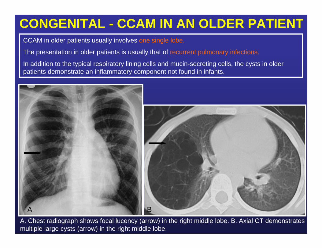
CONGENITAL - CCAM IN AN OLDER PATIENT
A. Chest radiograph shows focal lucency (arrow) in the right middle lobe. B. Axial CT demonstrates multiple large cysts (arrow) in the right middle lobe.
BA
CCAM in older patients usually involves one single lobe.
The presentation in older patients is usually that of recurrent pulmonary infections.
In addition to the typical respiratory lining cells and mucin-secreting cells, the cysts in older patients demonstrate an inflammatory component not found in infants.

CONGENITAL – BRONCHOGENIC CYST
A. Chest radiograph demonstrates a cavitary lesion in the left upper lobe (arrow) which contains air-fluid level (arrowhead). B. Axial CT shows air fluid level (arrowhead) within a round cystic lesion consistent with an infected bronchogenic cyst.
Secondary to abnormal budding of the tracheobronchial tree during development.
Seen in the mediastinum and lung parenchyma with equal frequency.
They do not contain air until they become infected.
May appear as well-defined soft tissue attenuation or cystic air-fluid containing masses.
A B

INFECTION – NECROTIZING PNEUMONIA
A and B. Axial CT with lung and mediastinal windows show large consolidation in the right upper lobe containing air bronchograms (arrows) and focal areas of cavitary necrosis (arrowheads). Additionally, there is a small parapneumonic effusion (curved arrow).
Most common suppurative complication of pneumonia.
More commonly seen as a complication of S. pneumoniae infection.
CT findings include loss of normal lung architecture, decreased parenchymal enhancement,loss of lung parenchymal margin, and multiple thin- walled cavities containing air or fluid and lacking an enhancing border.
A B
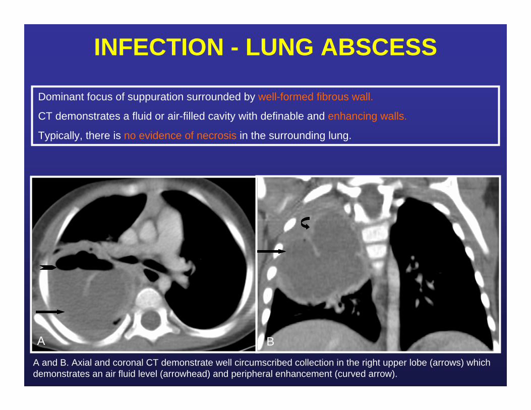
INFECTION - LUNG ABSCESS
A and B. Axial and coronal CT demonstrate well circumscribed collection in the right upper lobe (arrows) which demonstrates an air fluid level (arrowhead) and peripheral enhancement (curved arrow).
Dominant focus of suppuration surrounded by well-formed fibrous wall.
CT demonstrates a fluid or air-filled cavity with definable and enhancing walls.
Typically, there is no evidence of necrosis in the surrounding lung.
A B

INFECTION – ROUND PNEUMONIA
A and B. Frontal and lateral chest radiograph demonstrates a round opacity in the superior segment of the right lower lobe (arrows), containing few air bronchograms (arrowhead).
There is propensity for pneumonia to appear “round” in younger patients (<8 years old).
Most often due to Streptococcal pneumoniae.
This pattern is thought to be related to poor development of pathways of collateral air drift, channels of Lambert, pores of Kohn and incomplete fissures.
They are usually in the bases of the lungs, touching the pleural surface or a fissure.
A B

INFECTION – TUBERCULOSIS (TB)Children with primary tuberculosis can present with pulmonary consolidationwithin any lobe.
It is often associated with hilar lymphadenopathyand pleural effusion.
Lung consolidation with hilar lymphadenopathy in a non acutely ill child should be highly suspicious for tuberculosis.
A. Axial CT shows low density mediastinal lymph nodes (arrows). B. Chest radiograph shows right upper lobe and left lower lobe ill-defined opacities (arrows). C. Axial CT shows air space disease (arrow) with pneumatocele (arrowhead) in the left lower lobe.
A
B
C

INFECTION – CONGENITAL MILIARY TUBERCULOSIS
A. Chest radiograph demonstrates a diffuse micronodular pattern and a focal area of cavitation in the right lung (arrow). B. Axial CT shows micronodularity (arrow) and a cavitary lesion in the right upper lobe (arrowhead).
Congenital TB typically presents in the early weeks of life with poor feeding and failure to gain weight. Upper respiratory symptoms and progressive hepatosplenomegaly may appear later.
Progressive respiratory distress, apnea, jaundice and abdominal distension are frequent hallmarks of the disease.
In miliary TB, imaging studies demonstrate diffusely scattered discrete 1-2 mm lung nodules.
BA
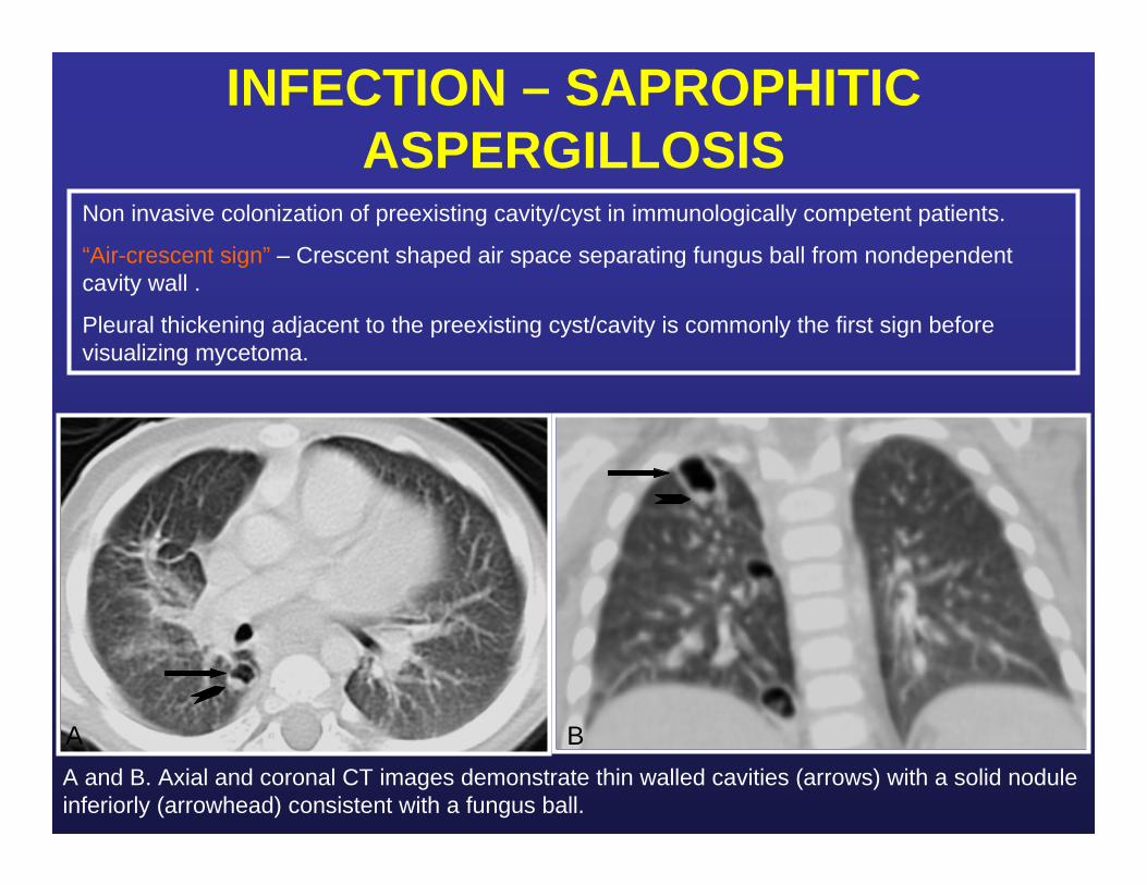
INFECTION – SAPROPHITIC ASPERGILLOSIS
A and B. Axial and coronal CT images demonstrate thin walled cavities (arrows) with a solid nodule inferiorly (arrowhead) consistent with a fungus ball.
BA
Non invasive colonization of preexisting cavity/cyst in immunologically competent patients.
“Air-crescent sign” – Crescent shaped air space separating fungus ball from nondependent cavity wall .
Pleural thickening adjacent to the preexisting cyst/cavity is commonly the first sign before visualizing mycetoma.

INFECTION – SEPTIC EMBOLI
A. Axial CT demonstrates multiple cavitary lesions (arrow) in the lungs. B. Axial CT through the level of the lower pelvis shows a distended right common femoral vein containing a central hypodensity (arrow) consistent with thrombosis. There is a right hip effusion consistent with septic arthritis (curved arrow).
Lodgment of an infected thrombus in a pulmonary artery.
Predisposing factors include immunodeficiency, congenital heart disease and dermal infections.
There is a predilection for the lung bases.
CT findings include multiple peripheral parenchymal nodules +/- cavitation.
Cavitation occur in approximately 50% of the cases, especially with staphylococcal emboli.
BA
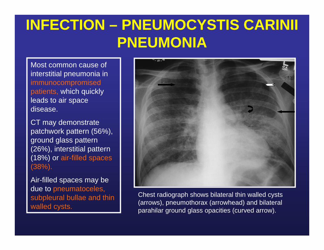
INFECTION – PNEUMOCYSTIS CARINII PNEUMONIA
Chest radiograph shows bilateral thin walled cysts (arrows), pneumothorax (arrowhead) and bilateral parahilar ground glass opacities (curved arrow).
Most common cause of interstitial pneumonia in immunocompromised patients, which quickly leads to air space disease.
CT may demonstrate patchwork pattern (56%), ground glass pattern (26%), interstitial pattern (18%) or air-filled spaces (38%).
Air-filled spaces may be due to pneumatoceles, subpleural bullae and thin walled cysts.

INFECTION - PNEUMATOCELE
A. Chest radiograph demonstrates a large consolidation (arrow) in the right lung. B. Follow-up chest radiograph shows interval resolution of consolidation with development of a pneumatocele (arrow). C. CT demonstrates a pneumatocele in the right upper lobe (arrow).
FOLLOW-UP CHEST RADIOGRAPHBA
C

TRAUMA - PNEUMATOCELE
Axial CT in a different patient shows bilateral pneumothoraces (arrows), parenchymal consolidations, pneumatoceles (arrowhead) and subcutaneous emphysema (white arrow).
Coronal reformatted CT demonstrates an ellipsoid lucent lesion in the right upper lobe (arrow), bilateral pneumothoraces (curved arrows), chest tubes and consolidation in the left lower lobe. Note the vertebral body fracture at the midthoracic spine (arrowhead).
Jet ski accident
Gunshot wound
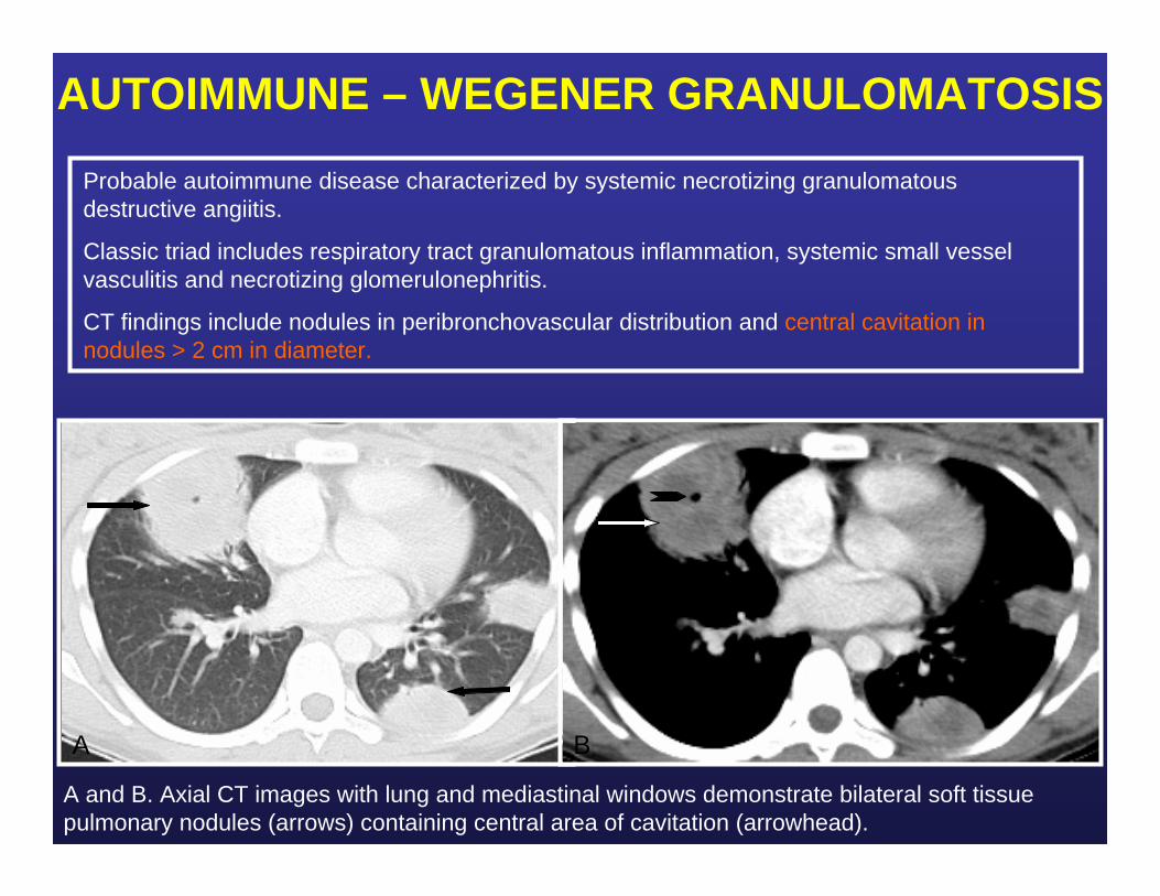
AUTOIMMUNE – WEGENER GRANULOMATOSIS
A and B. Axial CT images with lung and mediastinal windows demonstrate bilateral soft tissue pulmonary nodules (arrows) containing central area of cavitation (arrowhead).
Probable autoimmune disease characterized by systemic necrotizing granulomatous destructive angiitis.
Classic triad includes respiratory tract granulomatous inflammation, systemic small vessel vasculitis and necrotizing glomerulonephritis.
CT findings include nodules in peribronchovascular distribution and central cavitation in nodules > 2 cm in diameter.
A B
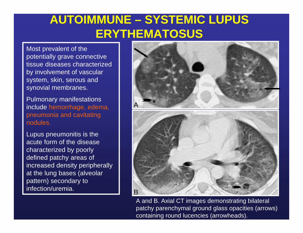
AUTOIMMUNE – SYSTEMIC LUPUS ERYTHEMATOSUS
A and B. Axial CT images demonstrating bilateral patchy parenchymal ground glass opacities (arrows) containing round lucencies (arrowheads).
Most prevalent of the potentially grave connective tissue diseases characterized by involvement of vascular system, skin, serous and synovial membranes.
Pulmonary manifestations include hemorrhage, edema, pneumonia and cavitating nodules.
Lupus pneumonitis is the acute form of the disease characterized by poorly defined patchy areas of increased density peripherally at the lung bases (alveolar pattern) secondary to infection/uremia.
A
B
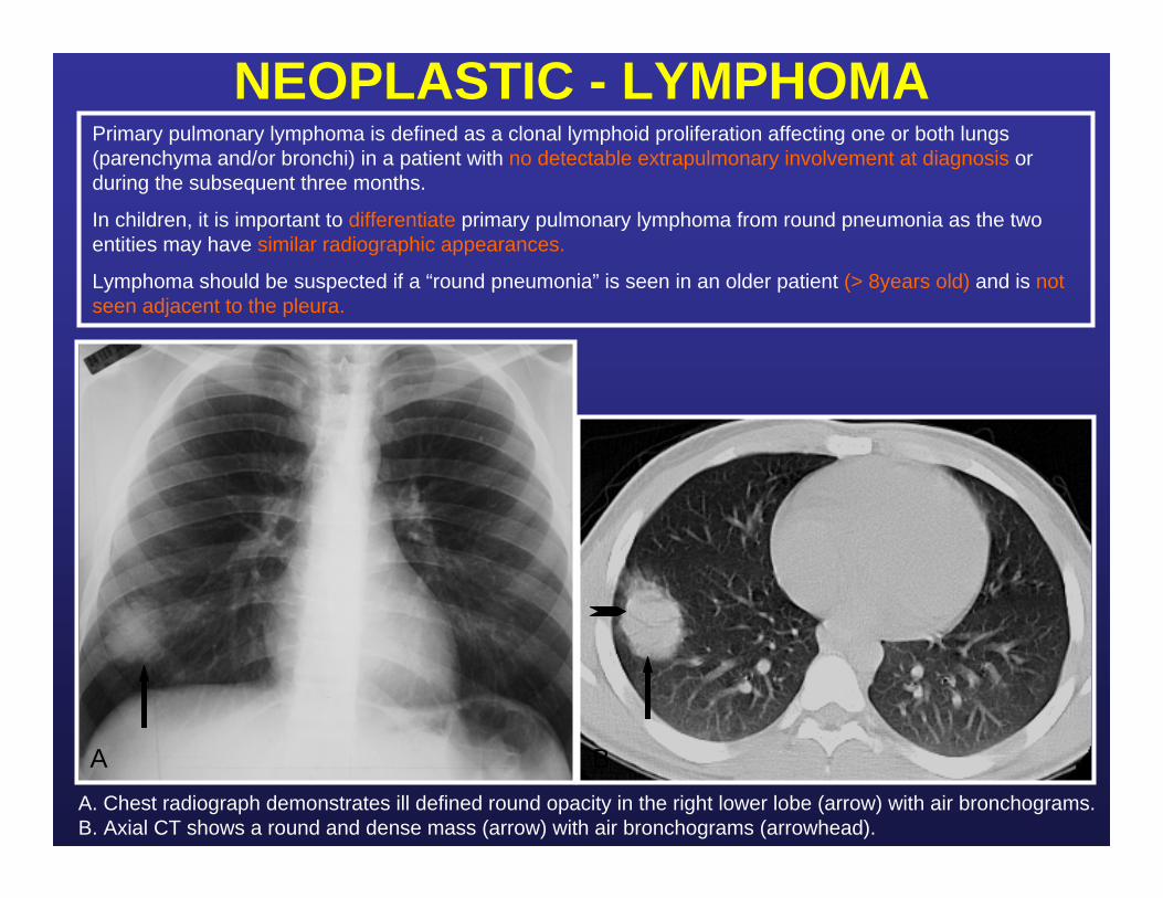
NEOPLASTIC - LYMPHOMA
A. Chest radiograph demonstrates ill defined round opacity in the right lower lobe (arrow) with air bronchograms. B. Axial CT shows a round and dense mass (arrow) with air bronchograms (arrowhead).
A B
Primary pulmonary lymphoma is defined as a clonal lymphoid proliferation affecting one or both lungs (parenchyma and/or bronchi) in a patient with no detectable extrapulmonary involvement at diagnosis or during the subsequent three months.
In children, it is important to differentiate primary pulmonary lymphoma from round pneumonia as the two entities may have similar radiographic appearances.
Lymphoma should be suspected if a “round pneumonia” is seen in an older patient (> 8years old) and is not seen adjacent to the pleura.

NEOPLASTIC – EOSINOPHILIC GRANULOMA
A. Chest radiograph demonstrates increased lung volumes and multiple round lucencies bilaterally. B. Axial CT shows multiple bilateral thin walled cystic lesions (arrow).
Abnormal clonal proliferation of Langerhans cells resulting in granulomatous infiltration of the lungs, bone, skin, lymph nodes, liver, spleen, brain, kidneys and endocrine glands.
The disease has an upper lobe predominance. It appears as multiple nodules which progress to cavitation, thick walled cysts and thin walled cysts.
Increased lung volume and spontaneous pneumothoraces may also be seen.
A B
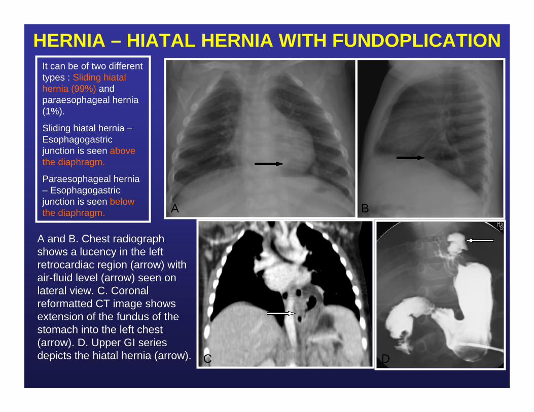
HERNIA – HIATAL HERNIA WITH FUNDOPLICATION
A and B. Chest radiograph shows a lucency in the left retrocardiac region (arrow) with air-fluid level (arrow) seen on lateral view. C. Coronal reformatted CT image shows extension of the fundus of the stomach into the left chest (arrow). D. Upper GI series depicts the hiatal hernia (arrow).
A B
C D
It can be of two different types : Sliding hiatal hernia (99%) and paraesophageal hernia (1%).
Sliding hiatal hernia –Esophagogastric junction is seen above the diaphragm.
Paraesophageal hernia – Esophagogastric junction is seen below the diaphragm.
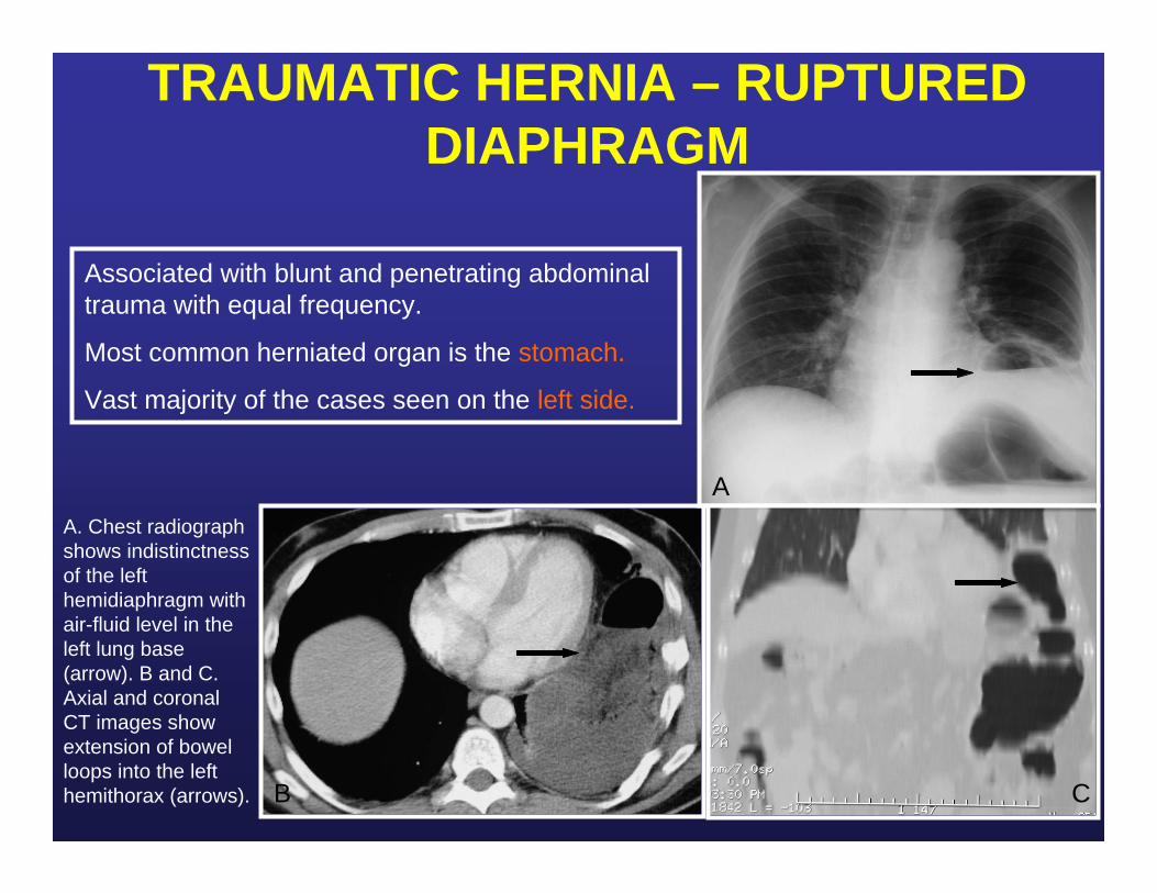
TRAUMATIC HERNIA – RUPTURED DIAPHRAGM
A. Chest radiograph shows indistinctness of the left hemidiaphragm with air-fluid level in the left lung base (arrow). B and C. Axial and coronal CT images showextension of bowel loops into the left hemithorax (arrows).
Associated with blunt and penetrating abdominal trauma with equal frequency.
Most common herniated organ is the stomach.
Vast majority of the cases seen on the left side.
C
A
B

SYNDROME – MARFAN SYNDROME
A and B. Coronal and axial CT images demonstrate a small right apical bleb (arrows) and pneumothorax (arrowhead) in the left lung apex from a ruptured bleb.
A
B
It results from abnormal cross-linking of collagen fibers.
Disease that affects multiple systems including musculoskeletal (arachnodactyly and ligamentous laxity), cardiovascular (annuloaortic ectasia and dissection), pulmonary (cystic lung disease and recurrent pneumothorax) and gastrointestinal (recurrent biliary obstruction).

SYNDROME – CHRONIC GRANULOMATOUS DISEASE
A. Chest radiograph demonstrates multiple bilateral ill defined opacities, more prominent on the left lung (arrow). B. Axial CT shows bilateral ill defined pulmonary nodules (arrow) with areas of cavitation (arrowhead).
Immunodeficiency disorder resulting in purulent infections and granuloma formation primarily involving lymph nodes, skin and lungs.
Chest manifestations include chronic pneumonia, hilar lymphadenopathy and pleural/pericardial effusions.
A B

SYNDROME – KARTAGENER SYNDROME
A. Chest radiograph shows dextrocardia (arrow), tubular branching dilated bronchi in the right lung base (arrowhead) and the stomach bubble on the right side of the abdomen (curved arrow). B. Axial CT demonstrates right lower lobe tubular bronchiectasis (arrow). C. Coronal CT reconstruction shows dilated bronchi (arrows) in the right lung base.
Also known as immotile cilia syndrome.
Triad consists of situs inversus,nasal polyposis with chronic sinusitis and bronchiectasis.
Deafness and infertility are also associated with the disease.
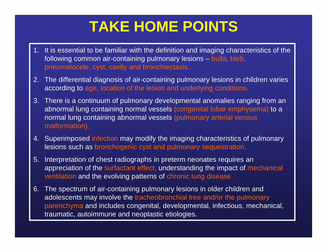
TAKE HOME POINTS1. It is essential to be familiar with the definition and imaging characteristics of the
following common air-containing pulmonary lesions – bulla, bleb, pneumatocele, cyst, cavity and bronchiectasis.
2. The differential diagnosis of air-containing pulmonary lesions in children varies according to age, location of the lesion and underlying conditions.
3. There is a continuum of pulmonary developmental anomalies ranging from an abnormal lung containing normal vessels (congenital lobar emphysema) to a normal lung containing abnormal vessels (pulmonary arterial venous malformation).
4. Superimposed infection may modify the imaging characteristics of pulmonarylesions such as bronchogenic cyst and pulmonary sequestration.
5. Interpretation of chest radiographs in preterm neonates requires an appreciation of the surfactant effect, understanding the impact of mechanical ventilation and the evolving patterns of chronic lung disease.
6. The spectrum of air-containing pulmonary lesions in older children and adolescents may involve the tracheobronchial tree and/or the pulmonary parenchyma and includes congenital, developmental, infectious, mechanical,traumatic, autoimmune and neoplastic etiologies.

REFERENCES1. Donnely LF (2001) Fundamentals of pediatric radiology.1st edition, Philadelphia, PA: Saunders
2. Agron GA, Courtney SE, Stocker JT, Markowitz RI. From the archives of the AFIP - Lung disease in premature neonates: Radiologic-pathologic correlation. Radiographics 2005;25:1047-1073.
3. Howling SJ, Northway WH, Hansell DM, Moss RB, Ward S, Müller NL. Pulmonary Sequelae of Bronchopulmonary Dysplasia Survivors. AJR 2000; 174:1323-1326.
4. Oppenheim C, Mamou-Mani T, Sayegh N, de Blic J, Scheinmann P, Lallemand D. Bronchopulmonary dysplasia: Value of CT in identifying pulmonary sequelae. AJR 1994;163:169-172.
5. Kothari NA, Kramer S. Bronchial diseases and lung aeration in children. Journal of thoracic imaging 16:207-223.
6. Ng CSH, Wan S, Lee TW, Yim APC. Cystic lesions of the lung: a forgotten menace. European Respiratory Journal; 25 (4) : 749.
7. Aquino SL, Schechter MS, Chiles C, Ablin DS, Chipps B, Webb WR. High resolution inspiratory and expiratory CT in older children and adults with bronchopulmonary dysplasia. AJR 1999;173:963-967.
8. Donnely LF, Frush DP. Localized radiolucent chest lesions in neonates:Causes and differentiation.AJR 1999;172:1651-1658.
9. Panicek et al. The continuum of pulmonary developmental anomalies. Radiographics 1987;7(4):747-772.
10. Williams DW, Merten DF, Effmann EL, Scatliff JH. Ventilator-induced pulmonary pseudocysts in preterm neonates. AJR 1988;150:885-887.