Embed Size (px)
Citation preview

BB(PH
I
taHthipetna
Biology of Blood and Marrow Transplantation 13:749-759 (2007)� 2007 American Society for Blood and Marrow Transplantation1083-8791/07/1307-0001$32.00/0doi:10.1016/j.bbmt.2007.05.001
ronchiolitis Obliterans Syndrome (BOS),ronchiolitis Obliterans Organizing PneumoniaBOOP), and Other Late-Onset Noninfectiousulmonary Complications following Allogeneicematopoietic Stem Cell Transplantation
Satoshi Yoshihara,1 Gregory Yanik,2 Kenneth R. Cooke,2 Shin Mineishi1
1Division of Hematology, Department of Internal Medicine, Hyogo College of Medicine, Hyogo, Japan; and2Blood and Marrow Transplant Program, University of Michigan, Ann Arbor, Michigan
Correspondence and reprint requests: Shin Mineishi, MD, Blood and Marrow Transplant Program, Universityof Michigan, 5303 Cancer Center, 1500 East Medical Center Drive, Ann Arbor, MI 48109 (email:[email protected]).
Received January 31, 2007; accepted May 1, 2007
ABSTRACTPulmonary dysfunction is a significant complication following allogeneic hematopoietic stem cell transplan-tation (HSCT), and is associated with significant morbidity and mortality. Effective antimicrobial prophylaxisand treatment strategies have increased the incidence of noninfectious lung injury, which can occur in the earlyposttransplant period or in the months and years that follow. Late-onset noninfectious pulmonary complica-tions are frequently encountered, but diagnostic criteria and terminology for these disorders can be confusingand therapeutic approaches are suboptimal. As a consequence, inaccurate diagnosis of these conditions mayhamper the appropriate data collection, enrollment into clinical trials, and appropriate patient care. Thepurpose of this review is to clarify the pathogenesis and diagnostic criteria of representative conditions, suchas bronchiolitis obliterans syndrome and bronchiolitis obliterans organizing pneumonia, and to discuss theappropriate diagnostic strategies and treatment options.© 2007 American Society for Blood and Marrow Transplantation
KEY WORDSBronchiolitis obliterans syndrome (BOS) ● Bronchiolitis obliterans organizing pneumonia (BOOP)● Late-onset noninfectious pulmonary complications ● Allogeneic ● Pathogenesis ● Diagnostic
criteriapch(Hnpapiop(
NTRODUCTION
Allogeneic hematopoietic bone marrow transplan-ation (allo-HSCT) is a curative treatment option for
variety of malignant and nonmalignant disorders.owever, a number of complications associated with
he occurrence of either acute or chronic graft versusost disease (aGVHD, cGVHD) have limited the util-
ty of this procedure. In particular, pulmonary com-lications occur in 25%-50% of allo-HSCT recipi-nts, and can account for approximately 50% ofransplant related deaths [1-6]. In nearly 50% of cases,o infectious organisms are identified in the lungs of
ffected individuals. Both an acute and a subacute pattern of lung injury have been recognized in thisontext (Figure 1). Noninfectious, acute lung injuryas been defined as idiopathic pneumonia syndromeIPS). IPS occurs with in the first 120 days after
SCT and typically has a rapidly progressing fulmi-ant course resulting in death in 60% to 80% ofatients. By contrast, subacute lung injury is associ-ted with a later onset, typically �6 months posttrans-lant, and the clinical course tends to be more insid-ous and protracted (Figure 1). This review will focusn 2 common subacute or late-onset pulmonary com-lications, namely bronchiolitis obliterans syndromeBOS), and bronchiolitis obliterans with organizing
neumonia (BOOP).749

B
pfiBrcwiaconBefScfph2t
P
bttilbhacclnet
kmtBptccvplatptlbctga(
flrmHtrrBrpdm
I
tvnattoI[prprfi
FB
S. Yoshihara et al.750
OS
“Bronchiolitis obliterans” describes a histologicattern of small airway inflammation that includesbrogenic deposition in small airways or bronchioles.ronchioles consist of terminal bronchioles and respi-
atory bronchioles, and serve as the bridge betweenonducting airways and alveoli. BO is synonymousith the term “constrictive bronchiolitis,” and is man-
fested clinically by the presence of airflow obstructionnd recognized histologically by submucosal bron-hiolar fibrosis, along with luminal narrowing andbliteration. BOS is a clinical term defined by pulmo-ary function changes rather than histology [7-10].OS is defined as an irreversible decline in forcedxpiratory volume in 1 second (FEV1) of at least 20%rom baseline, and is graded using the Internationalociety for Heart and Lung Transplantation (ISHLT)riteria [11]. Although BO and BOS may include dif-erent patient cohorts, indicated by the fact that someatients with positive histologic findings of BO do notave pulmonary function changes seen in BOS, theseentities are believed to greatly overlap, and the terms
end to be used interchangeably [9,11].
ATHOGENESIS
The pathogenesis of BOS after HSCT has noteen well defined. However, the heterogeneous his-opathologic findings and clinical course suggest thathe development of BOS is a multifactorial processnvolving both alloimmunologic and nonalloimmuno-ogic reactions. Because the occurrence of BOS haseen closely associated with cGVHD, it has beenypothesized that BOS is mediated, at least in part, bylloimmunologic injury to host bronchiolar epithelialells [12]. Unlike IPS, elevations of pro-inflammatoryytokine levels within the serum and broncho-alveolaravage (BAL) compartment of affected subjects haveot been well documented in humans or animal mod-ls [13-21]. Murine models have suggested, however,
igure 1. Schematic diagram of the time of occurrence of IPS,OOP, and BOS.
hat lymphocytes and associated chemokines and cyto- H
ines, including tumor necrosis factor alpha (TNF�),ay be important contributors to the development of
he chronic inflammatory process that characterizesOS after lung transplantation [13,14,22,23]. Theathogenesis of disease is believed to primarily involvehe interplay between immune effector cells (mono-ytes, lymphocytes, neutrophils) that have been re-ruited to the lung and cells resident to the pulmonaryascular endothelium and interstitium. This complexrocess results in the loss of type I pulmonary epithe-ial cells, proliferation of type II cells, the recruitmentnd proliferation of endothelial cells, and the deposi-ion of the extracellular matrix. In response to thisattern of injury, cytokines released from these effec-or cells and lung cells (macrophages, alveolar epithe-ial, and vascular endothelial cells) can stimulate fibro-last proliferation and increase the synthesis ofollagen and extracellular matrix proteins. Ultimately,his leads to enhanced deposition of collagen andranulation tissue in and around bronchial structures,nd eventually complete obliteration of small airwaysFigure 2) [12].
Clinical data suggest that nonalloimmunologic in-ammatory conditions such as viral infections, recur-ent aspiration, and conditioning chemoradiotherapyay also play a role in the pathogenesis of BOS afterSCT. A recent report indicated that respiratory
ract infections with some community respiratory vi-uses (parainfluenza virus and respiratory syncytial vi-us) were associated with late airflow decline [15].OS after autologous transplantation and decreasedisk of BOS after reduced intensity conditioning sup-ort a contribution to tissue damage from chemora-iotherapy that is part of HSCT conditioning regi-ens [16,17], as has been observed with IPS [18].
NCIDENCE AND RISK FACTORS
Because of the lack of defined diagnostic criteria,he reported incidence of BOS following allo-HSCTaries widely from 1.7% to 26% [8,19-21,24]. Asoted above, the development of BOS in both pedi-tric and adult populations is closely associated withhe occurrence of cGVHD. Other potential risk fac-ors that have been identified include the use of meth-trexate for aGVH prophylaxis [8], decreased serumgG levels [19], the prior occurrence of acute GVHD25], older recipient age, older donor age [26], lowerretransplant FEV1/forced vital capacity (FVC) ratio,espiratory viral infections within the first 100 daysosttransplant [20], busulfan-based conditioningegimen, peripheral blood stem cell transplantation,emale donor to male recipient, a prior episode ofnterstitial pneumonitis [24], and the intensity of
SCT conditioning [17].

C
yHpwBpatccp
D
P
d(s(onts
pcwi7
csf
daCwt
tructio
T
S
PL
P
R
P
Pulmonary Complications following Allogeneic HSCT 751
LINICAL PRESENTATION
The onset of BOS varies from 3 months to �10ears, with a median onset approximately 1 year post-SCT [10,21,24,27,28]. Common symptoms include
rogressive dyspnea, nonproductive cough, andheezing. Unlike BOOP, fever is usually absent inOS. Many patients remain asymptomatic for longeriods, despite having evidence of moderate to severeirway obstruction on pulmonary function tests. Inhis context, and because �33% of patients withGVHD develop signs of airflow obstruction [20],lose observation should be a part of the follow-uplans in these patients.
IAGNOSIS
ulmonary Function Testing (PFT)
Airflow obstruction is the hallmark of BOS. Aecrease in forced expiratory volume in 1 secondFEV1) and diminished FEV1/FVC ratio are ob-erved. Total lung capacity is not usually affectedTable 1). Lung diffusion capacity for carbon mon-xide (DLCO) is often decreased; however, this is aonspecific finding that may be seen in many pa-ients following HSCT irrespective of airflow ob-truction [29].
For patients undergoing allogeneic lung trans-lantation, ISHLT diagnostic criteria for BOS in-lude a �20% decline in FEV1 values when comparedith the postoperative baseline [30]. Recently, the
ntroduction of forced expiratory flow rates (FEF25-
Figure 2. Histology of BO. Characterized by concentric obs
5) has been added to these diagnostic criteria [31]. In
omparison to FEV1, the FEF25-75 may be a moreensitive indicator of early airflow obstruction in BOSollowing allogeneic lung transplant [32].
In several studies, airflow obstruction has beenefined as a decrease of the FEV1/FVC ratio to �0.7nd FEV1 to �80% of the predicted value [21,27].hien et al. [20] recently proposed new criteria, inhich patients were defined as having airflow obstruc-
ion if the annualized rate of predicted FEV1 decline
n of the small airways and relative sparing of alveolar space.
able 1. Comparison of Clinical Presentations of BOS and BOOP
BOS BOOP
ymptom Progressive dyspnea FeverNon productive cough Nonproductive coughWheezing Dyspnea (usually
mild)hysical exam. Wheezing Ralesab data Non specific Elevated level of CRP
Increased neutrophilFT Obstructive lung disease Restrictive lung
diseaseFEV1/FVC Decreased NormalTLC Normal DecreasedDLCO Decreased Decreased
adiologyCT scan Air trapping (expiration
phase)Consolidation
Mosaic perfusion Ground glass opacityBronchiectasis NodulesBronchial wall thickeningCentrilobular nodules
FT indicates pulmonary function test; BOS, bronchiolitis obliter-ans syndrome; BOOP, bronchiolitis obliterans organizing pneu-
moria.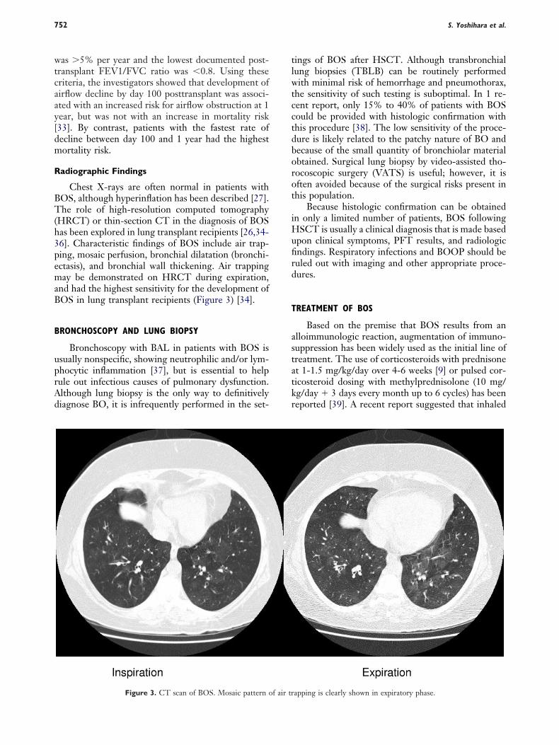
wtcaay[dm
R
BT(h3pemaB
B
uprAd
tlwtcctdborot
iHufird
T
astatkr
S. Yoshihara et al.752
as �5% per year and the lowest documented post-ransplant FEV1/FVC ratio was �0.8. Using theseriteria, the investigators showed that development ofirflow decline by day 100 posttransplant was associ-ted with an increased risk for airflow obstruction at 1ear, but was not with an increase in mortality risk33]. By contrast, patients with the fastest rate ofecline between day 100 and 1 year had the highestortality risk.
adiographic Findings
Chest X-rays are often normal in patients withOS, although hyperinflation has been described [27].he role of high-resolution computed tomography
HRCT) or thin-section CT in the diagnosis of BOSas been explored in lung transplant recipients [26,34-6]. Characteristic findings of BOS include air trap-ing, mosaic perfusion, bronchial dilatation (bronchi-ctasis), and bronchial wall thickening. Air trappingay be demonstrated on HRCT during expiration,
nd had the highest sensitivity for the development ofOS in lung transplant recipients (Figure 3) [34].
RONCHOSCOPY AND LUNG BIOPSY
Bronchoscopy with BAL in patients with BOS issually nonspecific, showing neutrophilic and/or lym-hocytic inflammation [37], but is essential to helpule out infectious causes of pulmonary dysfunction.lthough lung biopsy is the only way to definitivelyiagnose BO, it is infrequently performed in the set-
Figure 3. CT scan of BOS. Mosaic pattern of air tr
ings of BOS after HSCT. Although transbronchialung biopsies (TBLB) can be routinely performedith minimal risk of hemorrhage and pneumothorax,
he sensitivity of such testing is suboptimal. In 1 re-ent report, only 15% to 40% of patients with BOSould be provided with histologic confirmation withhis procedure [38]. The low sensitivity of the proce-ure is likely related to the patchy nature of BO andecause of the small quantity of bronchiolar materialbtained. Surgical lung biopsy by video-assisted tho-ocoscopic surgery (VATS) is useful; however, it isften avoided because of the surgical risks present inhis population.
Because histologic confirmation can be obtainedn only a limited number of patients, BOS following
SCT is usually a clinical diagnosis that is made basedpon clinical symptoms, PFT results, and radiologicndings. Respiratory infections and BOOP should beuled out with imaging and other appropriate proce-ures.
REATMENT OF BOS
Based on the premise that BOS results from anlloimmunologic reaction, augmentation of immuno-uppression has been widely used as the initial line ofreatment. The use of corticosteroids with prednisonet 1-1.5 mg/kg/day over 4-6 weeks [9] or pulsed cor-icosteroid dosing with methylprednisolone (10 mg/g/day � 3 days every month up to 6 cycles) has beeneported [39]. A recent report suggested that inhaled
apping is clearly shown in expiratory phase.
ca[Hb[mwo[iacGtrmpttisowacivpbbtatptt[
P
ppimimrtrcfimb
B
lfifitltilscinB
P
HieiTf[rtoaa
I
dehHicwht(Hu
C
foH
Pulmonary Complications following Allogeneic HSCT 753
yclosporine may be effective both in the preventionnd treatment of BOS after lung transplantation40,41], but this strategy has not been investigated in
SCT recipients. The role of azithromycin has alsoeen explored in the setting of lung transplantation42,43]. Recently, the efficacy of azithromycin (500
g every day for 3days, followed by 250 mg 3 times aeek for 12 weeks) has been reported in the treatmentf a small number of patients with BOS after HSCT44]. Azithromycin has been shown to have aniti-nflammatory effects, particularly with respect to IL-8nd airway neutrophilia, and these properties mayontribute to beneficial effects observed in BOS [45].iven the potential role of inflammatory cytokines,
he use of TNF� neutralizing agents has also beeneported, and may represent a novel therapy for theanagement of BOS. Yanik et al. [46] reported the
otential efficacy of etanercept, human TNF� neu-ralizing agent, in the treatment of IPS and have foundhat this molecule may be useful for subacute lungnjury as well [47]. Larger prospective trials in eachetting are ongoing. Fullmer et al. [48] reported a casef BOS following HSCT in a pediatric patient thatas successfully treated with infliximab, a monoclonal
ntibody for human TNF�. Other interventions in-luding the long-term administration of intravenousmmunoglobulin has failed to show the effect on pre-enting the development of BOS [49]. Extracorporealhotochemotherapy (ECP), which has been shown toe a promising treatment for cGVHD [50,51], haseen used with some success for BOS following lungransplantation [52]. However, only a few case reportsre available that examine the effectiveness of thisherapy for BOS after HSCT [53,54]. Because of theoor response to traditional therapies, lung transplan-ation has been increasingly reported as a possibleherapeutic option for end-stage BOS after HSCT55-60].
ROGNOSIS
Despite treatment with augmented immunosup-ression, the prognosis for patients with BOS remainsoor. Improvement of lung function once significant
mpairment has occurred can be obtained in only ainority of the patients. Mortality remains high, vary-
ng from 14% to 100% (mean 61%) [61]. In theajority of cases, death is attributed to progressive
espiratory failure and opportunistic pulmonary infec-ions. Clark et al. [27] reported that a 3-year mortalityate of 65% in the patients with BOS accompanyingGVHD. Rapid decline in FEV1 [27,33], resistance torst-line therapy [21], earlier onset (earlier than 6onths or 200 days) [17,21] are all factors reported to
e associated with a worse prognosis. t
OOP
BOOP is a disorder involving bronchioles, alveo-ar ducts, and alveoli, the lumen of which becomelled with buds of granulation tissue consisting ofbroblasts and an associated matrix of loose connec-ive tissue. The bronchiolitis in BOOP is of the pro-iferative type, and generally includes mild inflamma-ion of the bronchiolar walls. In contrast to BO, theres no prominent bronchiolar wall fibrosis or bronchio-ar distortion [62]. The intraluminal lesions and con-equent organizing pneumonia are the most signifi-ant histopathologic findings in BOOP. Recently,diopathic BOOP has been termed cryptogenic orga-izing pneumonia (COP) to avoid confusion with theO nomenclature [63].
ATHOGENESIS
Although the pathogenesis of BOOP followingSCT is poorly understood, involvement of an allo-
mmunologic reaction has been again been consid-red. In animal studies, BOOP develops followingnfection with reovirus, and a significant role for
cells and Th1-derived cytokines, including inter-eron-�, was implicated in the development of disease64]. A case reported following syngeneic bone mar-ow transplantation suggests that BOOP is not alwayshe result of an allogeneic immune response [65]. Inther non-HSCT settings BOOP has been seen inssociation with infection, drugs, radiation therapy,nd a number of connective tissue disorders [62].
NCIDENCE AND RISK FACTORS
Patients with BOOP following HSCT have beenescribed in several reports [62,65-73]. Freudenbergert al. [74] recently reported a case-control study ofistologic BOOP. Of 5340 patients who received allo-SCT, 49 cases (0.9%) of histologic BOOP were
dentified. An association between aGVHD andGVHD and the subsequent development of BOOPas noted. Patients with BOOP were more likely toave acute cutaneous GVHD and cGVHD involvinghe gut and oral cavity. In other reports, 3 of 1791.6%) patients receiving matched sibling donor
SCT [72] and 4 of 39 (10.3%) patients who receivednrelated donor HSCT developed BOOP [75].
LINICAL PRESENTATION
The onset of BOOP has been reported to varyrom 5 to �2800 days posttransplant, with a medianccurrence by day 108 post-HSCT [74] FollowingSCT, BOOP has a similar clinical presentation to
hat seen with idiopathic BOOP (a.k.a. COP). Fever,

nnbljp
D
P
todt
R
comfiBpcpGacmrp95
vrwct
B
pBwnbBTVoT
T
tmB1m[fsfsf5mm
a2dtAgoi
O
ra[efiP
S. Yoshihara et al.754
onproductive cough, and dyspnea are all typicallyoted. On physical examination, rales are common,ut wheezing is generally absent. Elevations in serum
evels of C-reactive protein have been seen in con-unction with a moderate leukocytosis and neutro-hilia.
IAGNOSIS
FT
PFTs typically reveal a mild to moderate restric-ive defect, the degree of which depends on the extentf the pulmonary inflammation. DLCO is commonlyecreased, and in contrast with BOS, airflow obstruc-ion is usually absent.
adiology
The radiologic findings in idiopathic BOOP in-lude diffuse, “fluffy” consolidations, ground glasspacity, and nodules (Figure 4). Consolidation is oftenigratory [76,77]. Lee et al. [78] reported the CTndings in 43 patients with biopsy-proven idiopathicOOP. The most common pattern was consolidation,resent alone or as part of a mixed pattern in 79% ofases. The consolidation had a predominantly sub-leural and/or peribronchovascular distribution.round-glass opacity and nodules were seen in 60%
nd 30%, respectively. Of note, BOOP in immuno-ompromised patients (following HSCT, with leuke-ia, or myelodysplastic syndrome) showed different
adiographic patterns from that in immunocompetentatients. Less areas of consolidation (45% versus1%), greater ground-glass involvement (73% versus6%) and more frequent pulmonary nodules (55%
cFigure 4. CT scan of BOOP. Consolidation/nodules are shown.
ersus 22%) are seen in this group. Dodd et al. [79]ecently reported the HRCT findings of 4 patientsith BOOP following HSCT. All patients showed
onsolidation and significant ground-glass opacifica-ions.
ronchoscopy and Lung Biopsy
Bronchoscopy and BAL are useful in ruling outulmonary infection and establishing the diagnosis ofOOP (Figure 5). BAL fluid reveals lymphocytosis,ith a decreased CD4/CD8 ratio. Despite the useful-ess of BAL, histologic confirmation is still believed toe necessary for a diagnosis of BOOP. Unlike BO,OOP usually can be sufficiently diagnosed withBLB by demonstrating organizing pneumonia.ATS is required in the cases with atypical featuresr in whom the diagnosis cannot be made withBLB [80].
REATMENT AND PROGNOSIS
Although there is no standardized treatment pro-ocol for BOOP after HSCT, corticosteroid is theainstay of therapy. In the treatment of idiopathicOOP, prolonged treatment (prednisone 1 mg/kg for-3 months, 40 mg for 3 months, followed by 10-20g for a total of 1 year) was proposed to avoid relapses
81]. The rate of relapse in idiopathic BOOP variesrom 9% to 58%, although relapses may not neces-arily affect long-term outcome [80]. Based on thisact, the use of lower corticosteroid dosages with ahorter duration of treatment (prednisone 0.75 mg/kgor 4 weeks, 0.5 mg/kg for 4 weeks, 10 mg for 6 weeks,mg for 6 weeks) has been proposed [82]. The role ofacrolides has also been explored also in the treat-ent in BOOP [73].
Freudenberger et al. [74] reported that BOOPfter HSCT resolved in 57% and remained stable in1% of cases. BOOP progressed in 22% of patientsespite corticosteroids, with the majority of these pa-ients (16% of total) dying from respiratory failure.lthough the overall prognosis of idiopathic BOOP isood, with mortality rates 5% to 15%, the prognosisf BOOP after HSCT still remains to be defined, ands potentially lower [69,75].
THER COMPLICATIONS
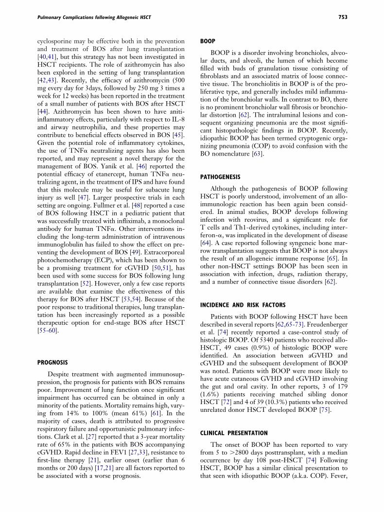
Pulmonary veno-occlusive disease (PVOD) is aare complication observed following both auto- andllo-HSCT that results in pulmonary hypertension83-88]. The pathologic hallmark of PVOD is thextensive and diffuse occlusion of pulmonary veins bybrous tissue [89]. In addition to HSCT recipients,VOD has been reported in patients who received
hemotherapy [90] and in patients with an associated
vntPTramPdipsmsda
Phatrwt
rtatcapt
r space
Pulmonary Complications following Allogeneic HSCT 755
iral illness [91]. Autoimmune destruction of pulmo-ary venules has not been commonly observed inhose patients, and only a minority of patients withVOD have associated autoimmune disorders [92].hese findings suggest that PVOD after HSCT is a
esult of tissue injury from HSCT conditioningnd/or infection. The onset of PVOD varies from aonth to a year after HSCT. Most patients withVOD present with nonspecific complaints such asyspnea on exertion and fatigue. The physical exam-
nation and radiologic findings are consistent withulmonary hypertension. Right heart catheterizationhows elevated pulmonary artery pressure with nor-al pulmonary artery wedge pressure. The triad of
evere pulmonary hypertension, radiographic evi-ence of pulmonary edema, and a normal pulmonary
Figure 5. Histology of BOOP. Alveola
rtery occlusion pressure is thought to be diagnostic of P
VOD. However, many patients with PVOD do notave this triad. Surgical lung biopsy is the only way fordefinitive diagnosis, and thus should be considered
o confirm the clinical suspicion of PVOD [89]. Mosteported cases of PVOD after HSCT were treatedith high-dose corticosteroids, with minimal effec-
iveness, and the prognosis of PVOD remains poor.Pulmonary cytolytic thrombi (PCT) is a recently
ecognized rare noninfectious pulmonary complica-ion seen almost exclusively in pediatric patients postllo-HSCT [93-97]. PCT appears as pulmonaryhrombi in small to medium distal pulmonary vessels,onsisting predominantly of monocytes of both donornd recipient origin, and associated with hemorrhagiculmonary infarction [94,97]. The onset ranges be-ween 8 and 343 days (median 72 days) after HSCT.
is occupied by organizing pneumonia.
CT presents with fever, cough, chest pain, respira-

tsvopomni
pHphmamgrPnr
S
nHdi2tctsomfci
aifiwtescdic
A
o
tuLoA
R
1
1
1
1
1
1
S. Yoshihara et al.756
ory distress. Chest CT scans may reveal multiplemall peripheral or subpleural nodules and opacities ofarying size. The majority of cases had active aGVHDr cGVHD. Woodard et al. [93] reported that 9 of 13atients with PCT were alive with a median follow-upf 1.5 years. Several patients showed clinical improve-ent and radiologic resolution of their pulmonary
odules after immunosuppressive therapy was admin-stered.
As noted earlier, acute noninfectious interstitialneumonitis (IP) occurring within 120 days afterSCT is termed IPS. In the majority of cases, affected
atients also exhibit signs of aGVHD. In contrast, itas been reported that patients develop progressive IPuch later (11 months to 8 years) after HSCT in
ssociation with severe cGVHD, especially scleroder-atous skin GVHD [75,98,99]. This late onset pro-
ressive IP presents with dyspnea, and cough and PFTesults commonly show a severe restrictive pattern.neumothorax may occur secondary to severe pulmo-ary fibrosis. The prognosis is poor because of respi-atory failure despite augmented immunosuppression.
UMMARY
Both BOS and BOOP are potential “late” pulmo-ary complications that may occur following allo-SCT. Importantly, BOS and BOOP each represents
istinct clinical entity, and should not to be usednterchangeably, although such usage is common. The
disorders are clearly distinct with respect to his-opathologic findings, radiographic and functionalharacteristics, and most importantly, response toherapy. BOOP following allo-HSCT is responsive toteroids (and in other settings may resolve spontane-usly), whereas BOS is not. Although diagnosticethods and treatment strategies for pulmonary in-
ections have improved significantly over the last de-ade, options in the treatment of noninfectious lungnjury remain limited.
Because the onset and progression of both BOSnd BOOP can be insidious, pulmonary function test-ng should be performed every 3 to 4 months for therst 2 years posttransplant, particularly in patientsith cGVHD. A significant drop in pulmonary func-
ion should prompt a workup in which careful physicalxamination, radiographic analysis, and BAL are con-idered. The appropriate use of defined diagnosticriteria for each condition, in combination with earlyiagnosis and well-designed prospective clinical trials,
s required to make progress in outcomes in theseonditions.
CKNOWLEDGMENTSDr. Cooke is an Amy Strelzer-Manasevit Scholar
f the National Marrow Donor Program, a Fellow of
he Robert Wood Johnson Harold Amos Medical Fac-lty Development Program, a Clinical Scholar of theeukemia and Lymphoma Society, and the recipientf a Clinical Scientist in Translational Researchward from the Burroughs Wellcome Fund.
EFERENCES
1. Clark JG, Madtes DK, Martin TR, Hackman RC, Farrand AL,Crawford SW. Idiopathic pneumonia after bone marrow trans-plantation: cytokine activation and lipopolysaccharide amplifi-cation in the bronchoalveolar compartment. Crit Care Med.1999;27:1800-1806.
2. Crawford SW, Hackman RC. Clinical course of idiopathicpneumonia after bone marrow transplantation. Am Rev RespirDis. 1993;147:1393-1400.
3. Weiner RS, Bortin MM, Gale RP, et al. Interstitial pneumo-nitis after bone marrow transplantation. Assessment of riskfactors. Ann Intern Med. 1986;104:168-175.
4. Quabeck K. The lung as a critical organ in marrow transplan-tation. Bone Marrow Transplant. 1994;14(Suppl 4):S19-S28.
5. Crawford SW, Longton G, Storb R. Acute graft-versus-hostdisease and the risks for idiopathic pneumonia after marrowtransplantation for severe aplastic anemia. Bone Marrow Trans-plant. 1993;12:225-231.
6. Kantrow SP, Hackman RC, Boeckh M, Myerson D, CrawfordSW. Idiopathic pneumonia syndrome: changing spectrum oflung injury after marrow transplantation. Transplantation. 1997;63:1079-1086.
7. Ralph DD, Springmeyer SC, Sullivan KM, Hackman RC,Storb R, Thomas ED. Rapidly progressive air-flow obstructionin marrow transplant recipients. Possible association betweenobliterative bronchiolitis and chronic graft-versus-host disease.Am Rev Respir Dis. 1984;129:641-644.
8. Clark JG, Schwartz DA, Flournoy N, Sullivan KM, CrawfordSW, Thomas ED. Risk factors for airflow obstruction in recip-ients of bone marrow transplants. Ann Intern Med. 1987;107:648-656.
9. Crawford SW, Clark JG. Bronchiolitis associated with bonemarrow transplantation. Clin Chest Med. 1993;14:741-749.
0. Curtis DJ, Smale A, Thien F, Schwarer AP, Szer J. Chronicairflow obstruction in long-term survivors of allogeneic bonemarrow transplantation. Bone Marrow Transplant. 1995;16:169-173.
1. Chan CK, Hyland RH, Hutcheon MA, et al. Small-airwaysdisease in recipients of allogeneic bone marrow transplants. Ananalysis of 11 cases and a review of the literature. Medicine(Baltimore). 1987;66:327-340.
2. Cooke KR, Ferrara JLM. The pathophysiology of lung Injuryafter hematopoietic stem cell transplantation. In: James LM,Ferrra KRC, Deeg HJ, eds. Graft-vs-Host Disease. 3rd editionNew York: Marcel-Dekker; 2005:329-367.
3. Belperio JA, Keane MP, Burdick MD, et al. Critical role for thechemokine MCP-1/CCR2 in the pathogenesis of bronchiolitisobliterans syndrome. J Clin Invest. 2001;108:547-556.
4. Aris RM, Walsh S, Chalermskulrat W, Hathwar V, NeuringerIP. Growth factor upregulation during obliterative bronchioli-tis in the mouse model. Am J Respir Crit Care Med. 2002;166:417-422.
5. Erard V, Chien JW, Kim HW, et al. Airflow decline after
myeloablative allogeneic hematopoietic cell transplantation: the
1
1
1
1
2
2
2
2
2
2
2
2
2
2
3
3
3
3
3
3
3
3
3
3
4
4
4
4
4
4
4
4
4
4
Pulmonary Complications following Allogeneic HSCT 757
role of community respiratory viruses. J Infect Dis. 2006;193:1619-1625.
6. Paz HL, Crilley P, Patchefsky A, Schiffman RL, Brodsky I.Bronchiolitis obliterans after autologous bone marrow trans-plantation. Chest. 1992;101:775-778.
7. Yoshihara S, Tateishi U, Ando T, et al. Lower incidence ofBronchiolitis obliterans in allogeneic hematopoietic stem celltransplantation with reduced-intensity conditioning comparedwith myeloablative conditioning. Bone Marrow Transplant.2005;35:1195-1200.
8. Fukuda T, Hackman RC, Guthrie KA, et al. Risks and out-comes of idiopathic pneumonia syndrome after nonmyeloabla-tive and conventional conditioning regimens for allogeneichematopoietic stem cell transplantation. Blood. 2003;102:2777-2785.
9. Holland HK, Wingard JR, Beschorner WE, Saral R, SantosGW. Bronchiolitis obliterans in bone marrow transplantationand its relationship to chronic graft-v-host disease and lowserum IgG. Blood. 1988;72:621-627.
0. Chien JW, Martin PJ, Gooley TA, et al. Airflow obstructionafter myeloablative allogeneic hematopoietic stem cell trans-plantation. Am J Respir Crit Care Med. 2003;168:208-214.
1. Dudek AZ, Mahaseth H, DeFor TE, Weisdorf DJ. Bronchi-olitis obliterans in chronic graft-versus-host disease: analysis ofrisk factors and treatment outcomes. Biol Blood Marrow Trans-plant. 2003;9:657-666.
2. Boehler A, Bai XH, Liu M, et al. Upregulation of T-helper 1cytokines and chemokine expression in post-transplant airwayobliteration. Am J Respir Crit Care Med. 1999;159:1910-1917.
3. Neuringer IP, Walsh SP, Mannon RB, Gabriel S, Aris RM.Enhanced T cell cytokine gene expression in mouse airwayobliterative bronchiolitis. Transplantation. 2000;69:399-405.
4. Santo Tomas LH, Loberiza FR, Jr., Klein JP, et al. Risk factorsfor bronchiolitis obliterans in allogeneic hematopoietic stem-cell transplantation for leukemia. Chest. 2005;128:153-161.
5. Schwarer AP, Hughes JM, Trotman-Dickenson B, Krausz T,Goldman JM. A chronic pulmonary syndrome associated withgraft-versus-host disease after allogeneic marrow transplanta-tion. Transplantation. 1992;54:1002-1008.
6. Worthy SA, Park CS, Kim JS, Muller NL. Bronchiolitis oblit-erans after lung transplantation: high-resolution CT findings in15 patients. AJR Am J Roentgenol. 1997;169:673-677.
7. Clark JG, Crawford SW, Madtes DK, Sullivan KM. Obstruc-tive lung disease after allogeneic marrow transplantation. Clin-ical presentation and course. Ann Intern Med. 1989;111:368-376.
8. Philit F, Wiesendanger T, Archimbaud E, Mornex JF, Brune J,Cordier JF. Post-transplant obstructive lung disease (“bronchi-olitis obliterans”): a clinical comparative study of bone marrowand lung transplant patients. Eur Respir J. 1995;8:551-558.
9. Marras TK, Chan CK, Lipton JH, Messner HA, Szalai JP,Laupacis A. Long-term pulmonary function abnormalities andsurvival after allogeneic marrow transplantation. Bone MarrowTransplant. 2004;33:509-517.
0. Cooper JD, Billingham M, Egan T, et al. A working formula-tion for the standardization of nomenclature and for clinicalstaging of chronic dysfunction in lung allografts. InternationalSociety for Heart and Lung Transplantation. J Heart LungTransplant. 1993;12:713-716.
1. Estenne M, Maurer JR, Boehler A, et al. Bronchiolitis obliter-ans syndrome 2001: an update of the diagnostic criteria. J Heart
Lung Transplant. 2002;21:297-310.2. Reynaud-Gaubert M, Thomas P, Badier M, Cau P, GiudicelliR, Fuentes P. Early detection of airway involvement in oblit-erative bronchiolitis after lung transplantation. Functional andbronchoalveolar lavage cell findings. Am J Respir Crit Care Med.2000;161:1924-1929.
3. Chien JW, Martin PJ, Flowers ME, Nichols WG, Clark JG.Implications of early airflow decline after myeloablative alloge-neic stem cell transplantation. Bone Marrow Transplant. 2004;33:759-764.
4. Leung AN, Fisher K, Valentine V, et al. Bronchiolitis obliter-ans after lung transplantation: detection using expiratoryHRCT. Chest. 1998;113:365-370.
5. Bankier AA, Van Muylem A, Knoop C, Estenne M, GevenoisPA. Bronchiolitis obliterans syndrome in heart-lung transplantrecipients: diagnosis with expiratory CT. Radiology. 2001;218:533-539.
6. Siegel MJ, Bhalla S, Gutierrez FR, Hildebolt C, Sweet S.Post-lung transplantation bronchiolitis obliterans syndrome:usefulness of expiratory thin-section CT for diagnosis. Radiol-ogy. 2001;220:455-462.
7. St John RC, Gadek JE, Tutschka PJ, Kapoor N, Dorinsky PM.Analysis of airflow obstruction by bronchoalveolar lavage fol-lowing bone marrow transplantation. Implications for patho-genesis and treatment. Chest. 1990;98:600-607.
8. Chan A, Allen R. Bronchiolitis obliterans: an update. Curr OpinPulm Med. 2004;10:133-141.
9. Ratjen F, Rjabko O, Kremens B. High-dose corticosteroid ther-apy for bronchiolitis obliterans after bone marrow transplantationin children. Bone Marrow Transplant. 2005;36:135-138.
0. Iacono AT, Corcoran TE, Griffith BP, et al. Aerosol cyclo-sporin therapy in lung transplant recipients with bronchiolitisobliterans. Eur Respir J. 2004;23:384-390.
1. Iacono AT, Johnson BA, Grgurich WF, et al. A randomizedtrial of inhaled cyclosporine in lung-transplant recipients.N Engl J Med. 2006;354:141-150.
2. Gerhardt SG, McDyer JF, Girgis RE, Conte JV, Yang SC,Orens JB. Maintenance azithromycin therapy for bronchiolitisobliterans syndrome: results of a pilot study. Am J Respir CritCare Med. 2003;168:121-125.
3. Verleden GM, Dupont LJ. Azithromycin therapy for patientswith bronchiolitis obliterans syndrome after lung transplanta-tion. Transplantation. 2004;77:1465-1467.
4. Khalid M, Al Saghir A, Saleemi S, et al. Azithromycin inbronchiolitis obliterans complicating bone marrow transplan-tation: a preliminary study. Eur Respir J. 2005;25:490-493.
5. Verleden GM, Vanaudenaerde BM, Dupont LJ, Van Raem-donck DE. Azithromycin reduces airway neutrophilia and in-terleukin-8 in patients with bronchiolitis obliterans syndrome.Am J Respir Crit Care Med. 2006;174:566-570.
6. Yanik G, Hellerstedt B, Custer J, et al. Etanercept (Enbrel)administration for idiopathic pneumonia syndrome after allo-geneic hematopoietic stem cell transplantation. Biol Blood Mar-row Transplant. 2002;8:395-400.
7. Yanik G, Uberti J, Ferrara JL, et al. Etanercept for sub-acutelung injury following allogeneic stem cell transplantation. Blood.2003;102:(Suppl):471a.
8. Fullmer JJ, Fan LL, Dishop MK, Rodgers C, Krance R. Suc-cessful treatment of bronchiolitis obliterans in a bone marrowtransplant patient with tumor necrosis factor-alpha blockade.Pediatrics. 2005;116:767-770.
9. Sullivan KM, Storek J, Kopecky KJ, et al. A controlled trial of
long-term administration of intravenous immunoglobulin to
5
5
5
5
5
5
5
5
5
5
6
6
6
6
6
6
6
6
6
6
7
7
7
7
7
7
7
7
7
7
8
8
8
S. Yoshihara et al.758
prevent late infection and chronic graft-vs.-host disease aftermarrow transplantation: clinical outcome and effect onsubsequent immune recovery. Biol Blood Marrow Transplant.1996;2:44-53.
0. Couriel DR, Hosing C, Saliba R, et al. Extracorporeal photo-chemotherapy for the treatment of steroid-resistant chronicGVHD. Blood. 2006;107:3074-3080.
1. Greinix HT, Volc-Platzer B, Rabitsch W, et al. Successful useof extracorporeal photochemotherapy in the treatment of se-vere acute and chronic graft-versus-host disease. Blood. 1998;92:3098-3104.
2. O’Hagan AR, Stillwell PC, Arroliga A, Koo A. Photopheresis inthe treatment of refractory bronchiolitis obliterans complicat-ing lung transplantation. Chest. 1999;115:1459-1462.
3. Ilhan O, Arat M, Arslan O, et al. Extracorporeal photoimmu-notherapy for the treatment of steroid refractory progressivechronic graft-versus-host disease. Transfus Apher Sci. 2004;30:185-187.
4. Oyan B, Koc Y, Emri S, Kansu E. Improvement of chronicpulmonary graft-vs-host disease manifesting as bronchiolitisobliterans organizing pneumonia following extracorporeal pho-topheresis. Med Oncol. 2006;23:125-129.
5. Calhoon JH, Levine S, Anzueto A, Bryan CL, Trinkle JK. Lungtransplantation in a patient with a prior bone marrow trans-plant. Chest. 1992;102:948.
6. Gascoigne A, Corris P. Lung transplants in patients with priorbone marrow transplants. Chest. 1994;105:327.
7. Boas SR, Noyes BE, Kurland G, Armitage J, Orenstein D.Pediatric lung transplantation for graft-versus-host disease fol-lowing bone marrow transplantation. Chest. 1994;105:1584-1586.
8. Rabitsch W, Deviatko E, Keil F, et al. Successful lung trans-plantation for bronchiolitis obliterans after allogeneic marrowtransplantation. Transplantation. 2001;71:1341-1343.
9. Heath JA, Kurland G, Spray TL, et al. Lung transplantationafter allogeneic marrow transplantation in pediatric patients:the Memorial Sloan-Kettering experience. Transplantation.2001;72:1986-1990.
0. Sano Y, Date H, Nagahiro I, Aoe M, Shimizu N. Living-donorlobar lung transplantation for bronchiolitis obliterans afterbone marrow transplantation. Ann Thorac Surg. 2005;79:1051-1052.
1. Afessa B, Litzow MR, Tefferi A. Bronchiolitis obliterans andother late onset non-infectious pulmonary complications inhematopoietic stem cell transplantation. Bone Marrow Trans-plant. 2001;28:425-434.
2. Cordier JF. Bronchiolitis obliterans organizing pneumonia. Se-min Respir Crit Care Med. 2000;21:135-146.
3. American Thoracic Society/European Respiratory Society In-ternational Multidisciplinary Consensus Classification of theIdiopathic Interstitial Pneumonias. This joint statement of theAmerican Thoracic Society (ATS), and the European Respira-tory Society (ERS) was adopted by the ATS board of directors,June 2001 and by the ERS Executive Committee, June 2001.Am J Respir Crit Care Med. 2002;165:277-304.
4. Majeski EI, Paintlia MK, Lopez AD, Harley RA, London SD,London L. Respiratory reovirus 1/L induction of intraluminalfibrosis, a model of bronchiolitis obliterans organizing pneu-monia, is dependent on T lymphocytes. Am J Pathol. 2003;163:1467-1479.
5. Kanda Y, Takahashi T, Imai Y, et al. Bronchiolitis obliterans
organizing pneumonia after syngeneic bone marrow transplanta-tion for acute lymphoblastic leukemia. Bone Marrow Transplant.1997;19:1251-1253.
6. Thirman MJ, Devine SM, O’Toole K, et al. Bronchiolitisobliterans organizing pneumonia as a complication of alloge-neic bone marrow transplantation. Bone Marrow Transplant.1992;10:307-311.
7. Mathew P, Bozeman P, Krance RA, Brenner MK, Heslop HE.Bronchiolitis obliterans organizing pneumonia (BOOP) in chil-dren after allogeneic bone marrow transplantation. Bone Mar-row Transplant. 1994;13:221-223.
8. Yousem SA. The histological spectrum of pulmonary graft-versus-host disease in bone marrow transplant recipients. HumPathol. 1995;26:668-675.
9. Alasaly K, Muller N, Ostrow DN, Champion P, FitzGeraldJM. Cryptogenic organizing pneumonia. A report of 25 casesand a review of the literature. Medicine (Baltimore). 1995;74:201-211.
0. Kleinau I, Perez-Canto A, Schmid HJ, et al. Bronchiolitisobliterans organizing pneumonia and chronic graft-versus-hostdisease in a child after allogeneic bone marrow transplantation.Bone Marrow Transplant. 1997;19:841-844.
1. Baron FA, Hermanne JP, Dowlati A, et al. Bronchiolitis oblit-erans organizing pneumonia and ulcerative colitis after alloge-neic bone marrow transplantation. Bone Marrow Transplant.1998;21:951-954.
2. Palmas A, Tefferi A, Myers JL, et al. Late-onset noninfectiouspulmonary complications after allogeneic bone marrow trans-plantation. Br J Haematol. 1998;100:680-687.
3. Ishii T, Manabe A, Ebihara Y, et al. Improvement in bronchi-olitis obliterans organizing pneumonia in a child after alloge-neic bone marrow transplantation by a combination of oralprednisolone and low dose erythromycin. Bone Marrow Trans-plant. 2000;26:907-910.
4. Freudenberger TD, Madtes DK, Curtis JR, Cummings P,Storer BE, Hackman RC. Association between acute andchronic graft-versus-host disease and bronchiolitis obliteransorganizing pneumonia in recipients of hematopoietic stem celltransplants. Blood. 2003;102:3822-3828.
5. Patriarca F, Skert C, Sperotto A, et al. Incidence, outcome, andrisk factors of late-onset noninfectious pulmonary complica-tions after unrelated donor stem cell transplantation. Bone Mar-row Transplant. 2004;33:751-758.
6. Miyagawa Y, Nagata N, Shigematsu N. Clinicopathologicalstudy of migratory lung infiltrates. Thorax. 1991;46:233-238.
7. Izumi T, Kitaichi M, Nishimura K, Nagai S. Bronchiolitisobliterans organizing pneumonia. Clinical features and differ-ential diagnosis. Chest. 1992;102:715-719.
8. Lee KS, Kullnig P, Hartman TE, Muller NL. Cryptogenicorganizing pneumonia: CT findings in 43 patients. AJR Am JRoentgenol. 1994;162:543-546.
9. Dodd JD, Muller NL. Bronchiolitis obliterans organizingpneumonia after bone marrow transplantation: high-resolutioncomputed tomography findings in 4 patients. J Comput AssistTomogr. 2005;29:540-543.
0. Wells AU. Cryptogenic organizing pneumonia. Semin RespirCrit Care Med. 2001;22:449-460.
1. Epler GR. Bronchiolitis obliterans organizing pneumonia. Se-min Respir Infect. 1995;10:65-77.
2. Lazor R, Vandevenne A, Pelletier A, Leclerc P, Court-FortuneI, Cordier JF. Cryptogenic organizing pneumonia. Character-
istics of relapses in a series of 48 patients. The Groupe d’Etudes
8
8
8
8
8
8
8
9
9
9
9
9
9
9
9
9
9
Pulmonary Complications following Allogeneic HSCT 759
et de Recherche sur les Maladles “Orphelines” Pulmonaires(GERM“O”P). Am J Respir Crit Care Med 2000;162:571-577.
3. Troussard X, Bernaudin JF, Cordonnier C, et al. Pulmonaryveno-occlusive disease after bone marrow transplantation. Tho-rax. 1984;39:956-957.
4. Hackman RC, Madtes DK, Petersen FB, Clark JG. Pulmonaryvenoocclusive disease following bone marrow transplantation.Transplantation. 1989;47:989-992.
5. Kuga T, Kohda K, Hirayama Y, et al. Pulmonary veno-occlu-sive disease accompanied by microangiopathic hemolytic ane-mia 1 year after a second bone marrow transplantation for acutelymphoblastic leukemia. Int J Hematol. 1996;64:143-150.
6. Williams LM, Fussell S, Veith RW, Nelson S, Mason CM.Pulmonary veno-occlusive disease in an adult following bonemarrow transplantation. Case report and review of the litera-ture. Chest. 1996;109:1388-1391.
7. Trobaugh-Lotrario AD, Greffe B, Deterding R, Deutsch G,Quinones R. Pulmonary veno-occlusive disease after autolo-gous bone marrow transplant in a child with stage IV neuro-blastoma: case report and literature review. J Pediatr HematolOncol. 2003;25:405-409.
8. Mukai M, Kondo M, Bohgaki T, Notoya A, Kohno M. Pul-monary veno-occlusive disease following allogeneic peripheralblood stem cell transplantation for chronic myeloid leukaemia.Br J Haematol. 2003;123:1.
9. Mandel J, Mark EJ, Hales CA. Pulmonary veno-occlusive dis-ease. Am J Respir Crit Care Med. 2000;162:1964-1973.
0. Doll DC, Yarbro JW. Vascular toxicity associated with anti-
neoplastic agents. Semin Oncol. 1992;19:580-596.1. McDonnell PJ, Summer WR, Hutchins GM. Pulmonary veno-occlusive disease. Morphological changes suggesting a viralcause. JAMA. 1981;246:667-671.
2. Sanderson JE, Spiro SG, Hendry AT, Turner-Warwick M. Acase of pulmonary veno-occlusive disease respondong to treat-ment with azathioprine. Thorax. 1977;32:140-148.
3. Woodard JP, Gulbahce E, Shreve M, et al. Pulmonary cytolyticthrombi: a newly recognized complication of stem cell trans-plantation. Bone Marrow Transplant. 2000;25:293-300.
4. Gulbahce HE, Manivel JC, Jessurun J. Pulmonary cytolyticthrombi: a previously unrecognized complication of bone mar-row transplantation. Am J Surg Pathol. 2000;24:1147-1152.
5. Morales IJ, Anderson PM, Tazelaar HD, Wylam ME. Pulmonarycytolytic thrombi: unusual complication of hematopoietic stemcell transplantation. J Pediatr Hematol Oncol. 2003;25:89-92.
6. Gulbahce HE, Pambuccian SE, Jessurun J, et al. Pulmonarynodular lesions in bone marrow transplant recipients: impact ofhistologic diagnosis on patient management and prognosis.Am J Clin Pathol. 2004;121:205-210.
7. Peters A, Manivel JC, Dolan M, Gulbahce HE, Baker KS,Verneris MR. Pulmonary cytolytic thrombi after allogeneichematopoietic cell transplantation: a further histologic descrip-tion. Biol Blood Marrow Transplant. 2005;11:484-485.
8. Griese M, Rampf U, Hofmann D, Fuhrer M, Reinhardt D,Bender-Gotze C. Pulmonary complications after bone marrowtransplantation in children: twenty-four years of experience in asingle pediatric center. Pediatr Pulmonol. 2000;30:393-401.
9. Wolff D, Reichenberger F, Steiner B, et al. Progressive inter-stitial fibrosis of the lung in sclerodermoid chronic graft-versus-
host disease. Bone Marrow Transplant. 2002;29:357-360.