Embed Size (px)
Citation preview

Brief UltraRapid Communication
Exosomes From Human CD34� Stem Cells Mediate TheirProangiogenic Paracrine Activity
Susmita Sahoo, Ekaterina Klychko, Tina Thorne, Sol Misener, Kathryn M. Schultz, Meredith Millay,Aiko Ito, Ting Liu, Christine Kamide, Hemant Agarwal, Harris Perlman, Gangjian Qin,
Raj Kishore, Douglas W. Losordo
Rationale: Transplantation of human CD34� stem cells to ischemic tissues has been associated with reducedangina, improved exercise time, and reduced amputation rates in phase 2 clinical trials and has been shown toinduce neovascularization in preclinical models. Previous studies have suggested that paracrine factors secretedby these proangiogenic cells are responsible, at least in part, for the angiogenic effects induced by CD34�
cell transplantation.Objective: Our objective was to investigate the mechanism of CD34� stem cell–induced proangiogenic paracrine
effects and to examine if exosomes, a component of paracrine secretion, are involved.Methods and Results: Exosomes collected from the conditioned media of mobilized human CD34� cells had the
characteristic size (40 to 90 nm; determined by dynamic light scattering), cup-shaped morphology (electronmicroscopy), expressed exosome-marker proteins CD63, phosphatidylserine (flow cytometry) and TSG101(immunoblotting), besides expressing CD34� cell lineage marker protein, CD34. In vitro, CD34� exosomesreplicated the angiogenic activity of CD34� cells by increasing endothelial cell viability, proliferation, and tubeformation on Matrigel. In vivo, the CD34� exosomes stimulated angiogenesis in Matrigel plug and corneal assays.Interestingly, exosomes from CD34� cells but not from CD34� cell–depleted mononuclear cells had angiogenicactivity.
Conclusions: Our data demonstrate that human CD34� cells secrete exosomes that have independent angiogenicactivity both in vitro and in vivo. CD34� exosomes may represent a significant component of the paracrine effectof progenitor cell transplantation for therapeutic angiogenesis. (Circ Res. 2011;109:00-00.)
Key Words: CD34� cells � paracrine factor � exosomes � angiogenesis
Clinical studies have provided evidence that locally trans-planted autologous CD34� stem cells reduce angina and
improve exercise capacity in patients with refractory angina1
and lower amputation rates in patients with critical limbischemia.2 Preclinical studies indicate that the benefit ofhuman CD34� cell transplantation after ischemic injuryoccurs through increases in neovascularization3; however, themechanisms of new blood vessel formation have not beencompletely characterized. Incorporation of CD34� cells intothe growing vasculature has been documented in multiplestudies4; however, the magnitude of structural contribution oftransplanted cells has typically seemed modest comparedwith the significant overall physiological impact. This dis-
crepancy has led to the assumption that paracrine factorssecreted by CD34� cells contribute significantly to thetherapeutic angiogenesis induced by the cells.5
Exosomes, a component of paracrine secretion, are extra-cellular, membrane-bound nano-vessicles that originate intra-cellularly in multivesicular bodies (MVBs) and are secretedout when the MVBs fuse with the plasma membrane.6 Theyoften carry proteins, RNAs, and/or microRNAs and mediatesome aspects of cell-to-cell signaling.6 We investigated thepotential role of exosomes in CD34� cell–induced neovas-cularization by determining whether CD34� stem cells se-crete exosomes, and, if so, whether these exosomes caninduce angiogenic activity in the absence of CD34� cells.
Original received July 21, 2011; revision received July 29, 2011; accepted August 1, 2011. In July 2011, the average time from submission to firstdecision for all original research papers submitted to Circulation Research was 13.5 days.
Brief UltraRapid Communications (BURCs) are designed to be a format for manuscripts that are of outstanding interest to the readership, reportdefinitive observations, but have a relatively narrow scope. Less comprehensive than Regular Articles but still scientifically rigorous, BURCs presentseminal findings that have the potential to open up new avenues of research. A decision on BURCs is rendered within 7 days of submission.
From the Feinberg Cardiovascular Research Institute, Northwestern University, Chicago, IL (S.S., E.K., T.T., S.M., K.M.S., M.M., A.I., T.L., C.K.,G.Q., R.K., D.W.L.); Feinberg School of Medicine, Northwestern University, Chicago, IL (H.A., H.P.); and the Program in Cardiovascular RegenerativeMedicine, Northwestern Memorial Hospital, Chicago, IL (D.W.L.).
This manuscript was sent to Ali J. Marian, Consulting Editor, for review by expert referees, editorial decision, and final disposition.Correspondence to Douglas W. Losordo, MD, Feinberg Cardiovascular Research Institute, Northwestern University, Program in Cardiovascular
Regenerative Medicine, Northwestern Memorial Hospital, Tarry 14-725, 303 East Chicago Ave, Chicago, IL 60611. E-mail [email protected]© 2011 American Heart Association, Inc.
Circulation Research is available at http://circres.ahajournals.org DOI: 10.1161/CIRCRESAHA.111.253286
1
by guest on July 17, 2018http://circres.ahajournals.org/
Dow
nloaded from
by guest on July 17, 2018http://circres.ahajournals.org/
Dow
nloaded from
by guest on July 17, 2018http://circres.ahajournals.org/
Dow
nloaded from
by guest on July 17, 2018http://circres.ahajournals.org/
Dow
nloaded from
by guest on July 17, 2018http://circres.ahajournals.org/
Dow
nloaded from
by guest on July 17, 2018http://circres.ahajournals.org/
Dow
nloaded from
by guest on July 17, 2018http://circres.ahajournals.org/
Dow
nloaded from
by guest on July 17, 2018http://circres.ahajournals.org/
Dow
nloaded from
by guest on July 17, 2018http://circres.ahajournals.org/
Dow
nloaded from
by guest on July 17, 2018http://circres.ahajournals.org/
Dow
nloaded from
MethodsAll experimental protocols were approved by the NorthwesternUniversity Animal Care and Use Committee. Both CD34� cells3 andCD34� cell–depleted mononuclear cells (MNCs)3 were cultured,and exosomes from the conditioned media were obtained as de-scribed previously.7 Electron microscopy, dynamic light scattering(DLS), flow cytometry, and immunoblotting analyses were per-formed according to established protocols.7 The angiogenic activityof cultured human umbilical vein endothelial cells (HUVECs) wasevaluated by means of the Matrigel tube-formation assay, prolifera-tion was evaluated through 5-bromo-2-deoxyuridine incorporation,and viability was assessed by means of the MTS assay. In vivoangiogenesis was evaluated in nude (nu/J) mice by means of theMatrigel plug and corneal angiogenesis assays. Detailed methods areprovided in the Online Supplemental Methods. Quantified results arepresented as mean�SD; comparisons between groups were evalu-ated with the Student t test; and P�0.05 was considered significant.
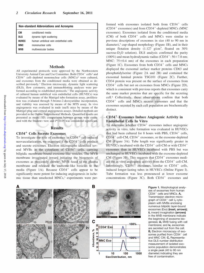
ResultsCD34� Cells Secrete ExosomesTo investigate the role of exosomes in CD34� cell-inducedneovascularization, we examined if the CD34� cells produceand secrete exosomes. Electron micrographs identified sev-eral MVBs in the cytoplasm of CD34� cells, carryingbilipidic membrane-bound exosome-like vesicles. The MVBmembrane invaginated inward initiating the biogenesis ofexosomes as previously shown6; MVB fused to the plasmamembrane and released the exosome-like vesicles to themedia (Figure 1A). Because CD34� cells appear to besignificantly more potent for inducing angiogenesis in ische-mic tissue than unselected MNCs,3 experiments were per-
formed with exosomes isolated both from CD34� cells(CD34� exosomes) and from CD34�-depleted MNCs (MNCexosomes). Exosomes isolated from the conditioned media(CM) of both CD34� cells and MNCs were similar toprevious descriptions of exosomes in size (40 to 90 nm indiameter),7 cup-shaped morphology (Figure 1B), and in theirunique flotation density (1.127 g/cm3, floated on 30%sucrose-D2O solution). DLS analysis confirmed the purity(100%) and mean hydrodynamic radius (CD34�: 50�7.8 nm;MNC: 75�0.4 nm) of the exosomes in each preparation(Figure 1C). Exosomes from both CD34� cells and MNCsdisplayed the exosomal surface marker proteins CD63 andphosphatidylserine (Figure 2A and 2B) and contained theexosomal luminal protein TSG101 (Figure 2C). Further,CD34 protein was present on the surface of exosomes fromCD34� cells but not on exosomes from MNCs (Figure 2D),which is consistent with previous reports that exosomes carrythe same marker proteins that are specific for the secretingcell.6 Collectively, these observations confirm that bothCD34� cells and MNCs secrete exosomes and that theexosomes secreted by each cell population are biochemicallydistinct.
CD34� Exosomes Induce Angiogenic Activity inEndothelial Cells In VitroTo determine whether CD34� exosomes induce angiogenicactivity in vitro, tube formation was evaluated in HUVECsthat had been cultured for 8 hours with PBS, CD34� cells,CD34� cell-CM, CD34� exosomes, or the exosome-depletedCM (Figure 3A). Tube length was significantly greater inHUVECs incubated with the CD34� cell-CM or with CD34�
exosomes than in HUVECs incubated with PBS but wasunchanged in HUVECs incubated with the exosome-depletedCM (Figure 3B). This suggests that CD34� exosomes medi-ate the in vitro angiogenic activity from the CD34� cell-CM.Interestingly, CD34� exosomes, similar to CD34� cells,induced longer-lasting tubes in HUVECs (Online Figure I).Tube formation was less pronounced at lower exosomeconcentrations (Figure 3C). Both CD34� exosomes and
Non-standard Abbreviations and Acronyms
CM conditioned media
DLS dynamic light scattering
HUVEC human umbilical vein endothelial cells
MNC mononuclear cells
MVB multivesicular bodies
Figure 1. Morphological analy-ses of exosomes from humanCD34� cells and MNCs. A,Transmission electron micro-graph of CD34� cell: i, cyto-plasm with MVBs enclosingnumerous bilipidic layer-boundexosomes (Exo) (inset, arrows);ii, inward invagination (arrows)in the MVB membrane indicatethe beginning of exosome bio-genesis; iii, MVB fusing with cellmembrane; and iv, exosomesare secreted out from the cell.B, Electron microscopy of exo-somes purified from CD34� celland MNC CM. C, Representa-tive DLS number distributionmeasurement of isolated exo-some population demonstratesa single peak (40 to 90 nmdiameter) indicating they arefree of contamination.
2 Circulation Research September 16, 2011
by guest on July 17, 2018http://circres.ahajournals.org/
Dow
nloaded from

CD34� cells significantly enhanced HUVEC viability (Fig-ure 3D) and proliferation (Figure 3E). Thus, most of the invitro angiogenic activity associated with CD34� cells appearsto be mediated by exosomes. HUVECs incubated with MNCsor MNC exosomes did not differ significantly from saline-treated cells in any functional parameter (Figure 3D and 3Eand Online Figures I and II). The superior efficacy of CD34�
exosomes compared with MNC exosomes is consistent withprior in vivo studies documenting the enhanced angiogenicactivity of CD34� cells versus MNC for therapeutic angio-genesis.3 Although the mechanisms that mediate the en-hanced potency of CD34� cells versus MNC have not beencompletely clarified, preliminary data show that the proan-giogenic microRNAs 126 and 130a8 are highly expressed inCD34� exosomes compared with MNC (Online Figure III).
CD34� Exosomes Induce Angiogenesis In VivoThe angiogenic potency of CD34� exosomes was evaluatedin vivo by performing the Matrigel-plug and corneal angio-genesis assays in mice. Both CD34� cells and CD34�
exosomes induced the formation of vessel-like endothelial
structures (Figure 4A) and significantly increased the propor-tion of endothelial cells (Figure 4B) in the Matrigel plug. Inthe corneal angiogenesis assay, pellets containing CD34�
exosomes but not MNC exosomes were associated withsignificantly greater vessel growth (Figure 4C and 4D); theeffect of CD34� cells on corneal angiogenesis could not beevaluated because the pellets could not be prepared withviable cells.
DiscussionCD34� cells have been shown to form a structural componentof the neovasculature in ischemic tissue4 and secrete para-crine factors that also stimulate neovascularization.5 Wedemonstrate that a significant component of the proangio-genic paracrine activity associated with CD34� cells ismediated by exosomes. The exosomes secreted by CD34�
cells were morphologically similar in size and shape toexosomes described in previous reports, carried known exo-somal protein markers, and potently induced angiogenicactivity both in vitro and in vivo.
Figure 2. Biochemical analy-ses of exosomes from humanCD34�cells and MNCs. Repre-sentative flow cytometry dot-plots showing detection ofexosomal surface proteins: A,CD63; B, annexin V bound toexposed phosphatidylserine;C, immunoblot showing exo-somal luminal protein TSG101;and D, flow cytometry dot-blotanalysis for CD34 surface pro-tein. The isolated exosomeswere conjugated to 4-�mLatex beads and stained for allflow cytometry analyses, Con-trol represents nonspecificantibody binding to the beads;numbers inside the box repre-sents the percentage of posi-tive beads.
Sahoo et al Exosomes Mediate Paracrine Effects of CD34� Cells 3
by guest on July 17, 2018http://circres.ahajournals.org/
Dow
nloaded from

The cell culture medium was supplemented with growthfactors and may have contained soluble proteins secreteddirectly from the cells, which could, in principle, havecontributed to the angiogenic effects associated with CD34�
exosomes. However, the MNC exosomes were derived fromMNCs cultured with the same growth factors, and theexosome-depleted conditioned media would have containedboth the supplemental growth factors and any secretedsoluble proteins. Because none of these treatments stimulatedangiogenic activity, our findings indicate that the CD34�
exosomes are the key paracrine component of CD34� cell–induced vessel growth.
Exosomes can stimulate both receptor-mediated and geneticmechanisms by transferring proteins, RNA, or microRNA di-rectly into the cytoplasm of target cells.9 We have presented datademonstrating that CD34� exosomes are enriched with proan-giogenic microRNAs; the extent to which these microRNAs aretransferred and induce any molecular changes in the recipientcells will be clarified in ongoing studies. Indeed, the repertoire ofspecific molecules transported by CD34� exosomes remains tobe fully characterized, but they are likely to be more stable thanmolecules secreted directly into the extracellular matrix becausethe exosomal membrane protects the contents of the exosomefrom degradation.6,9 Furthermore, the exosomes used in our
Figure 3. In vitro assays. A, HUVECs(2.5�104) were treated with PBS, 2.0�104
CD34� cells, or with conditioned media(CM), exosomes (Exo), or exosome-depleted CM from 2.0�104 CD34� cellsand plated on Matrigel. B, Tube lengthwas measured 8 hours later andexpressed as percentage of saline-treatedHUVECs (n�6 to 9). C, Representativedose-response of CD34� exosome tubeformation, evaluated in HUVECs incu-bated with exosomes from 1.5�105
CD34� cells and serially diluted withsaline to the indicated ratios: D, viability,and E, proliferation of HUVECs (1�104) inresponse to PBS, 2.5�103 cells, or exo-somes from 2.5�103 cells, measured 20hours later and expressed as percentageof PBS-treated HUVECs (n�3 to 6).*P�0.001 versus PBS, †P�0.05 versusExo-depleted CM, ‡P�0.05 versus MNCsor MNC exosomes.
Figure 4. In vivo assays. A and B, Matrigel plug assay: Matrigel containing PBS, 5�105 CD34� cells, or exosomes from 5�105 CD34�
cells was subcutaneously injected into mice, and the plugs were harvested 7 days later. A, Sections from the plug were stained withisolectin to identify endothelial cells (brown) and vessel-like endothelial structures (arrows). B, The plug was digested, and CD31� en-dothelial cells were quantified by flow cytometry (n�3 to 6). C and D, Corneal angiogenesis assay: Pellets containing PBS or exosomesfrom 1�106 MNCs or CD34� cells were implanted in the corneas of mice; corneas were harvested 7 days later, stained with fluores-cently labeled lectin identifying vascular structures (C), and the extent of vessel growth was quantified (D) (n�4). *P�0.05 versus PBS,‡P�0.01 versus MNC exosomes.
4 Circulation Research September 16, 2011
by guest on July 17, 2018http://circres.ahajournals.org/
Dow
nloaded from

investigation were sufficiently durable to remain intact andbiologically active throughout the isolation procedure, whichsuggests that the functional radius of CD34� exosomes couldextend beyond the immediate vicinity of the secreting cell. Theobservation that in some of the in vitro and in vivo assays theexosomes from CD34� cells appeared more potent than the cellsthemselves is interesting and might also be a byproduct of thedurability of the exosome in culture providing the ability todeliver a high dose of exosomes through collection from culturemedium in which exosomes are secreted over a period of time.
In summary, our observations demonstrate for the first timethat adult human CD34� stem cells secrete exosomes and thatthese exosomes induce angiogenic activity in isolated endo-thelial cells and in murine models of vessel growth. Thus, thebenefit of CD34� cell therapy on functional recovery afterischemic injury could be induced primarily through theexosome-mediated transfer of angiogenic factors to surround-ing cells. Novel therapies designed to exploit this previouslyunidentified mechanism of paracrine signaling may enhancerecovery from ischemic disease or injury.
AcknowledgmentsWe thank Baxter Healthcare for providing the CD34� cells, Dr C.S.Thaxton for providing the DLS machine, J. Marvin for assistancewith flow cytometry measurements, and L. Renolds for assistancewith electron microscopy.
Sources of FundingThis work was supported by grants from the National Institutes ofHealth: 2R01HL053354, 5R01HL095874, 5R01HL077428, and1P01HL108795.
DisclosuresNone.
References1. Losordo DW, Henry TD, Davidson C, Lee JS, Costa MA, Bass T, Men-
delsohn F, Fortuin FD, Pepine CJ, Traverse JH, Amrani D, Ewenstein BM,Riedel N, Story K, Barker K, Povsic TJ, Harrington RA, Schatz RA.Intramyocardial, autologous CD34� cell therapy for refractory angina.Circ Res. 2011;109:428–436.
2. Losordo D. Randomized double-blind, placebo controlled trial ofautologous cd34� cell therapy for critical limb ischemia: 1-year results.Circulation. 2010;122:A16920.
3. Kawamoto A, Iwasaki H, Kusano K, Murayama T, Oyamada A, Silver M,Hulbert C, Gavin M, Hanley A, Ma H, Kearney M, Zak V, Asahara T,Losordo DW. Cd34-positive cells exhibit increased potency and safety fortherapeutic neovascularization after myocardial infarction compared withtotal mononuclear cells. Circulation. 2006;114:2163–2169.
4. Asahara T, Murohara T, Sullivan A, Silver M, van der Zee R, Li T,Witzenbichler B, Schatteman G, Isner JM. Isolation of putative progenitorendothelial cells for angiogenesis. Science. 1997;275:964–967.
5. Kumar AH, Caplice NM. Clinical potential of adult vascular progenitorcells. Arterioscler Thromb Vasc Biol. 2010;30:1080–1087.
6. Chaput N, Thery C. Exosomes: immune properties and potential clinicalimplementations. Semin Immunopathol. 2010 [Epub ahead of print].
7. Thery C, Amigorena S, Raposo G, Clayton A. Isolation and character-ization of exosomes from cell culture supernatants and biological fluids.Curr Protoc Cell Biol. 2006;3:22.
8. Zhang Q, Kandic I, Kutryk MJ. Dysregulation of angiogenesis-relatedmicroRNAs in endothelial progenitor cells from patients with coronaryartery disease. Biochem Biophys Res Commun. 2011;405:42–46.
9. Valadi H, Ekstrom K, Bossios A, Sjostrand M, Lee JJ, Lotvall JO.Exosome-mediated transfer of MRNAs and microRNAs is a novelmechanism of genetic exchange between cells. Nat Cell Biol.2007;9:654–659.
Novelty and Significance
What Is Known?
● CD34� cells have been shown to stimulate therapeutic neovasculariza-tion in preclinical studies and in phase I and II human clinical trials.
● The potency of CD34� cells is greater than unselected mononuclear cells.● The mechanisms by which CD34� cells induce neovascularization
appear to include both direct participation in vessel formation andundefined “paracrine” effects.
● Exosomes are small, membrane-bound vesicles secreted from vari-ous cells that contain protein and nucleic acids and are increas-ingly being shown to mediate cell-to-cell signaling.
What New Information Does This Article Contribute?
● CD34� cells secrete exosomes that independently induce angiogen-esis in vitro and in vivo.
● The proangiogenic activity of CD34� exosomes is significantlygreater than CD34-depleted mononuclear cell exosomes.
● The exosomes of CD34� cells contain higher levels of proangiogenicmicroRNAs.
The clinical potential of CD34� cells for therapeutic neovascu-larization of ischemic tissue is being evaluated in a series ofcompleted and ongoing clinical trials. Hence, the mechanismsby which CD34� cells mediate these effects are of highscientific and clinical importance. Although paracrine effectshave been assumed to be responsible for a significant proportionof the effects of endothelial progenitor cell–based therapies ingeneral, the precise nature of the paracrine phenomena has notbeen defined. Our data show that CD34� cells secrete exo-somes that appear to be responsible for much if not most oftheir paracrine activity. Specifically, the conditioned mediumfrom CD34� cells exerts proangiogenic effects that are abol-ished when the exosomes are removed, whereas the exosomesalone, without any of the soluble material from the conditionedmedium, exhibit the full potency of the conditioned medium.Complete characterization of the exosome content of endothelialprogenitor cells could provide new insights permitting enhance-ment of their therapeutic potency.
Sahoo et al Exosomes Mediate Paracrine Effects of CD34� Cells 5
by guest on July 17, 2018http://circres.ahajournals.org/
Dow
nloaded from

Raj Kishore and Douglas W. LosordoMillay, Aiko Ito, Ting Liu, Christine Kamide, Hemant Agarwal, Harris Perlman, Gangjian Qin, Susmita Sahoo, Ekaterina Klychko, Tina Thorne, Sol Misener, Kathryn M. Schultz, Meredith
Activity Stem Cells Mediate Their Proangiogenic Paracrine+Exosomes From Human CD34
Print ISSN: 0009-7330. Online ISSN: 1524-4571 Copyright © 2011 American Heart Association, Inc. All rights reserved.is published by the American Heart Association, 7272 Greenville Avenue, Dallas, TX 75231Circulation Research
published online August 11, 2011;Circ Res.
http://circres.ahajournals.org/content/early/2011/08/09/CIRCRESAHA.111.253286World Wide Web at:
The online version of this article, along with updated information and services, is located on the
http://circres.ahajournals.org/content/suppl/2011/08/11/CIRCRESAHA.111.253286.DC1Data Supplement (unedited) at:
http://circres.ahajournals.org//subscriptions/
is online at: Circulation Research Information about subscribing to Subscriptions:
http://www.lww.com/reprints Information about reprints can be found online at: Reprints:
document. Permissions and Rights Question and Answer about this process is available in the
located, click Request Permissions in the middle column of the Web page under Services. Further informationEditorial Office. Once the online version of the published article for which permission is being requested is
can be obtained via RightsLink, a service of the Copyright Clearance Center, not theCirculation Researchin Requests for permissions to reproduce figures, tables, or portions of articles originally publishedPermissions:
by guest on July 17, 2018http://circres.ahajournals.org/
Dow
nloaded from

SUPPLEMENTAL MATERIAL
Exosomes from Human CD34+ Stem Cells Mediate their Pro-angiogenic Paracrine Activity
Susmita Sahoo1; Ekaterina Klychko1; Tina Thorne1; Sol Misener1; Kathryn M. Schultz1; Meredith Millay1; Aiko Ito1; Ting Liu1; Christine Kamide1; Hemant Agrawal2; Harris Perlman2; Gangjian Qin1; Raj Kishore1; Douglas W. Losordo1,3
1Feinberg Cardiovascular Research Institute, Northwestern University, Chicago, IL
2Feinberg School of Medicine, Northwestern University, Chicago, IL
3Program in Cardiovascular Regenerative Medicine, Northwestern Memorial Hospital, Chicago, IL

0
200
400
600
800
*†
*†
*†
PBS CD34+
CellsCD34+
CMCD34+
Exo
CD34+ CMExo-Depleted
MNCs MNCCM
MNCExo
MNC CMExo-Depleted
Tu
be
Le
ng
th,
%
Supplemental Figure 1. In vitro matrigel tube formation assay at 24h. HUVECs (2.5x10⁴) were
treated with PBS, with 2.0x10⁴ cells, or with the conditioned media (CM), exosomes (Exo), or exosome-depleted conditioned media from 2.0x10⁴ cells, plated on Matrigel, tube length was measured 24 hourslater and expressed as a percentage of PBS-treated HUVECs; n=3-6. *P<0.005 versus PBS, †P<0.05versus MNCs or MNC exosomes.
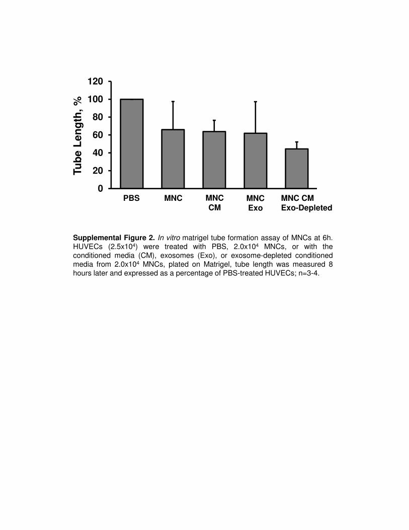
Supplemental Figure 2. In vitro matrigel tube formation assay of MNCs at 6h.HUVECs (2.5x10⁴) were treated with PBS, 2.0x10⁴ MNCs, or with theconditioned media (CM), exosomes (Exo), or exosome-depleted conditionedmedia from 2.0x10⁴ MNCs, plated on Matrigel, tube length was measured 8hours later and expressed as a percentage of PBS-treated HUVECs; n=3-4.
0
20
40
60
80
100
120
PBS MNC MNCCM
MNCExo
MNC CMExo-Depleted
Tu
be
Le
ng
th,
%

miRNA 126 miRNA 130a
Re
lati
ve
Ex
pre
ss
ion
, N
orm
ali
ze
d t
o U
6
0
50
100
150
200900
950
CD34+ Cell
CD34+ Exo
MNC
MNC Exo
Supplemental Figure 3. Pro-Angiogenic miRNA’s are highly expressed in CD34+cell exosomes. Total RNA was isolated from the CD34+ cells, CD34+ depleted MNCand their respective exosomes using Qiagen miRNEASY isolation kit; miRNAexpression from equal amounts of total RNA was measured by qRT-PCR Taqmanassays, data was normalized to the expression of small nuclear RNA U6; miRNA130a, n=2; miRNA 126, n=6.
Supplemental Methods Cells and culture
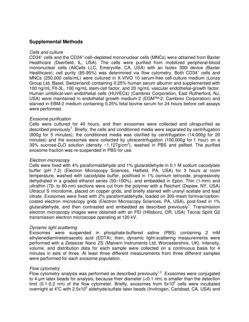
CD34+ cells and the CD34+-cell–depleted mononuclear cells (MNCs) were obtained from Baxter Healthcare (Deerfield, IL, USA). The cells were purified from mobilized peripheral-blood mononuclear cells (AllCells LLC, Emeryville, CA, USA) with an Isolex 300i device (Baxter Healthcare); cell purity (85-95%) was determined via flow cytometry. Both CD34+ cells and MNCs (250,000 cells/mL) were cultured in X-VIVO 10 serum-free cell-culture medium (Lonza Group Ltd, Basel, Switzerland) containing 0.25% human serum albumin and supplemented with 100 ng/mL Flt-3L, 100 ng/mL stem-cell factor, and 20 ng/mL vascular endothelial-growth factor. Human umbilical-vein endothelial cells (HUVECs) (Cambrex Corporation, East Rutherford, NJ, USA) were maintained in endothelial growth medium-2 (EGM™-2; Cambrex Corporation) and starved in EBM-2 medium containing 0.25% fetal bovine serum for 24 hours before cell assays were performed.
Exosome purification
Cells were cultured for 40 hours, and then exosomes were collected and ultrapurified as
described previously1. Briefly, the cells and conditioned media were separated by centrifugation
(800g for 5 minutes); the conditioned media was clarified by centrifugation (14,000g for 20 minutes) and the exosomes were collected by ultracentrifugation (100,000g for 1 hour) on a 30% sucrose-D2O solution (density ~1.127g/cm3), washed in PBS and pellted. The purified exosome fraction was re-suspended in PBS for use.
Electron microscopy
Cells were fixed with 4% paraformaldehyde and 1% glutaraldehyde in 0.1 M sodium cacodylate buffer (pH 7.2) (Electron Microscopy Sciences, Hatfield, PA, USA) for 3 hours at room temperature, washed with cacodylate buffer, postfixed in 1% osmium tetroxide, progressively dehydrated in a graded ethanol series (50–100%), and embedded in Epon. Thin (1-mm) and ultrathin (70- to 80-nm) sections were cut from the polymer with a Reichert (Depew, NY, USA) Ultracut S microtome, placed on copper grids, and briefly stained with uranyl acetate and lead citrate. Exosomes were fixed with 2% paraformaldehyde, loaded on 300-mesh formvar/carbon-coated electron microscopy grids (Electron Microscopy Sciences, PA, USA), post-fixed in 1%
glutaraldehyde, and then contrasted and embedded as described previously1. Transmission
electron microscopy images were obtained with an FEI (Hillsboro, OR, USA) Tecnai Spirit G2 transmission electron microscope operating at 120 kV. Dynamic light scattering Exosomes were suspended in phosphate-buffered saline (PBS) containing 2 mM ethylenediaminetetraacetic acid (EDTA); then, dynamic light-scattering measurements were performed with a Zetasizer Nano ZS (Malvern Instruments Ltd, Worcestershire, UK). Intensity, volume, and distribution data for each sample were collected on a continuous basis for 4 minutes in sets of three. At least three different measurements from three different samples were performed for each exosome population.
Flow cytometry
Flow cytometry analysis was performed as described previously1,2. Exosomes were conjugated to 4-µm latex beads for analysis, because their diameter (<0.1 nm) is smaller than the detection limit (0.1-0.2 nm) of the flow cytometer. Briefly, exosomes from 5x10⁶ cells were incubated overnight at 4oC with 2.5x10⁵ aldehyde/sulfate latex beads (Invitrogen, Carlsbad, CA, USA) and

then blocked with 100 mM glycine for 30 minutes at room temperature to saturate any free binding sites that remained on the beads. To detect the presence of CD63 and CD34, the
exosome-coated beads were resuspended in 500 µL PBS containing 0.5% human serum
albumin (HSA) and 2 mM EDTA; then, 100 µL of the beads were incubated with fluorescein-isothiocyanate (FITC)–conjugated anti-CD63 or FITC-conjugated anti-CD34 antibodies (Beckman Coulter, Inc., Brea, CA, USA) for 30 minutes at 4°C. For phosphatidylserine detection, the beads were resuspended in 100 µL of Annexin-VFLUOS labeling solution (Annexin-V-FLUOS Staining Kit, F. Hoffmann-La Roche Ltd, Basel, Switerland) and incubated for 10 minutes at 25°C. Non-specific signaling was inhibited by the addition of FcR blocking reagent (Miltenyi Biotec Inc., Auburn, CA, USA); the threshold for negative staining was obtained by incubating exosome-free, glycine-blocked beads with each antibody; and additional experiments were performed with identical concentrations of control IgG antibodies to correct for non-specific binding. Flow cytometry data were acquired on a BD LSRII (BD Franklin Lakes, NJ USA) flow cytometer and analyzed with FlowJo software (Tree Star, Ashland, OR, USA). In-vitro Matrigel tube formation assay
HUVECs (2.5x10⁴, serum-starved overnight) were seeded with PBS, with 2.0x10⁴ CD34+ cells or MNCs, or with the conditioned media, exosomes, or exosome-depleted conditioned media from 2.0x10⁴ CD34+ cells or MNCs into 48-well plates that had been coated with 150 µL of growth-factor–reduced Matrigel™ (BD). Tube formation was examined by phase-contrast microscopy 6-8 hours, or, 24 hours later. Each condition in each experiment was assessed in duplicates, and tube length was measured as the mean summed length of capillary-like structures in 2 wells, per high-power fields (HPFs, 2.5x) per well. 6-9 experiments were performed for each condition.
In vitro proliferation and viability assays
Cell proliferation was evaluated via 5-bromo-2-deoxyuridine (BrdU) incorporation. Serum-starved HUVECs (1x10⁴) were incubated with 10 µM BrdU and 2.0x10⁴ CD34+ cells, 2.0x10⁴ MNCs, or exosomes from 2.0x10⁴ CD34+ cells or MNCs for 24 hours, and then washed and fixed with 4% paraformaldehyde at 4 °C. Ten minutes later, the HUVECs were washed in PBS with 1% TritonX100 for 5 minutes, incubated on ice in 1N HCl for 10 minutes, incubated at room temperature in 2N HCl for 10 minutes, and incubated at 37°C for 20 minutes. The HCl was neutralized via three 5-minute washes with borate buffer (0.1M), and then the HUVECs were washed in PBS with 1% TritonX100 at room temperature for 3 minutes, blocked with 5% normal goat serum and 1% Triton X in PBS for 1 hour, and incubated overnight with immunofluorescent sheep anti-BrdU antibodies (Abcam Inc., Cambridge, MA, USA); nuclei were counterstained with DAPI. Cells were viewed at 10x magnification, and BrdU+ cells were counted in 10 HPFs per well, 2 wells per condition. Cell viability was evaluated via the MTS assay. HUVECs (1x10⁴ cells/well) were seeded on 96-well flat-bottomed plates and incubated with 2.0x10⁴ CD34+ cells or MNCs, or with exosomes from 2.0x10⁴ CD34+ cells or MNCs, for 20 hours at 37°C; then, the MTS assay reagent (Promega Corporation, Madison, WI, USA) was added to the wells, and HUVECs were incubated for 3 hours at 37oC. Viability was evaluated by measuring absorbance at 490-nm wavelength with a 96-well ELISA plate reader (spectrophometer SpectraMaxPlus; Molecular Devices, Sunnyvale, CA, USA) in at least 6 wells per experiment, 3-7 experiments per condition.
Western blotting
Cells or purified exosomes were lysed with 0.1M Tris, 0.3 M NaCl, 0.1% SDS, 0.5% sodium deoxycholate, and 1% Triton X-100 in a cocktail of antiproteases (Sigma-Aldrich Corporation,

St. Louis, MO, USA); then, the nuclei and membranes were cleared by centrifugation (15,000g for 10 minutes). Protein extracts were separated on an 8% SDS-PAGE gel, blotted on Immobilon (Millipore, Billerica, MA, USA) with TSG101 (4A10; Abcam Inc.), and visualized with enhanced chemoluminescence substrate (Thermo Fisher Scientific, Rockford, IL, USA). Images were acquired with a Chemidoc XRS (Kodak, Rochester, NY, USA).
In-vivo Matrigel-plug assay
Ice-cold Matrigel (0.5 mL/plug; BD) was mixed with heparin (1 mg/mL) and with PBS, 0.5x10⁶ CD34+ cells, or exosomes from 0.5x10⁶ CD34+ cells and then subcutaneously injected into the flanks of 6- to 8-week-old male nude mice (Nu/J; The Jackson Labortory, Bar Harbor, ME, USA); mice were anesthetized with inhaled isoflurane (2-4%) before injection. Two weeks later, the plug was excised and washed with PBS. To visualize vessel-like endothelial structures, the plug was fixed in methanol and sectioned; then, endothelial cells were stained with biotinylated isolectin B4 (Vector Laboratories Inc, Burlingame, CA, USA), and nuclei were stained with hematoxylin. Images were acquired with an Olympus Vanox bright microscope. For flow-cytometry analysis of endothelial-cell migration, the plug was digested with 0.1% collagenase/dispase (F. Hoffmann-La Roche), 10 mm MgCl2, and 200 units/mL DNase I (F. Hoffmann-La Roche) in 10% fetal calf serum/PBS for 1 hour at 37oC. After digestion, cells were dispersed 4-5 times with a 21g needle, passed through a 70-mm filter (BD), and stained with phycoerythrin-conjugated rat anti-mouse-CD31 antibodies (BD); control assessments were performed with phycoerythrin-conjugated rat immunoglobulin G2a isotype (Invitrogen). Flow cytometry data were acquired on a FACScan (BD) flow cytometer and analyzed with FlowJo software (Tree Star).
Mouse corneal angiogenesis assay
Pellets were prepared and implanted in the corneas of 6- to 8-week-old male nude mice (Nu/J;
The Jackson Laboratory) as described previously.3 Briefly, 5 mg sucrose octasulfate-aluminum
complex (Sigma-Aldrich Corporation) and 10 µL of 12% hydron in ethanol was mixed and partially dried; then, exosomes from 5x10⁵ CD34+ cells or MNCs were added, the mixture was pelleted on a 400-µm nylon mesh (Sefar America Inc., Depew, NY, USA), and the pellets were dried for 5-10 minutes. Pellets were implanted in the corneas of mice that had been anesthetized via intraperitoneal injection of 125 mg/kg Avertin. One week after implantation, the mice were intravenously injected with 50 µL of fluorescein-conjugated BS1-Lectin I (Vector Laboratories) and sacrificed 15 minutes later. Eyes were harvested and fixed with 1% paraformaldehyde; then, the corneas were excised and mounted; angiogenesis was evaluated via BS1-Lectin I fluorescence and quantified with ImageJ software. MicroRNA quantification
Total RNA from the CD34+ cells, CD34-depleted MNCs and their respective exosomes were extracted using the miRNeasy Mini Kit (Qiagen) according to the manufacturer’s protocol including a DNase step. RNA concentrations were verified on the NanoDrop Spectrophotometer (NanoDrop) and the quality of total RNA was assessed using Agilent 2100 Bioanalyzer Pico Chips (Agilent). Equal amount of RNA (5ng) was reverse transcribed using the Taqman MicroRNA Reverse Transcription Kit (Appled Biosystems) using a specific miRNA primer to generate cDNA for use with individual Taqman MicroRNA Assays (Appled Biosystems). Real-time Reactions were performed in triplicate on a 7500FAST Real-Time PCR system (Applied Biosystems). Ct values were averaged and normalized to RNU6B. Relative expression was determined by the ddCt comparative threshold method.

Supplememental References: 1. Thery C, Amigorena S, Raposo G, Clayton A. Isolation and characterization of
exosomes from cell culture supernatants and biological fluids. Curr Protoc Cell Biol. 2006;Chapter 3:Unit 3 22
2. Ostrowski M, Carmo NB, Krumeich S, Fanget I, Raposo G, Savina A, Moita CF, Schauer K, Hume AN, Freitas RP, Goud B, Benaroch P, Hacohen N, Fukuda M, Desnos C, Seabra MC, Darchen F, Amigorena S, Moita LF, Thery C. Rab27a and Rab27b control different steps of the exosome secretion pathway. Nat Cell Biol. 2010;12:19-30; sup pp 11-13
3. Rogers MS, Birsner AE, D'Amato RJ. The mouse cornea micropocket angiogenesis assay. Nat Protoc. 2007;2:2545-2550


