Embed Size (px)
Citation preview

“Breathing through a straw”
Carrie Nicholas
Keelyjo Hindhaugh
Dorothy Millar

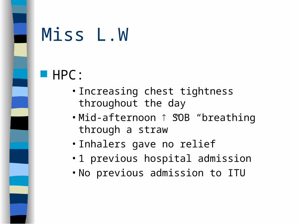
Miss L.W
33 year old female, admitted SGH 28/07/03, 19:15 PC:
• Increasing shortness of breath
HPC: • 1/52 productive cough, white sputum
• rhintis
• Amoxicillin started by GP
Miss L.W
HPC:• Increasing chest tightness throughout the day
• Mid-afternoon SOB “breathing through a straw”
• Inhalers gave no relief
• 1 previous hospital admission
• No previous admission to ITU

Miss L.W
PMH:• Asthma
• Hayfever
• Nasal polyps
Drug history• Seretide 250
• Salbutamol
• Flixonase
ALLERGIES: Aspirin Acute Asthma

Miss L.W
Family history• Mother
• Grandmother
Social history• Lives with mother
• Non-smoker, no-one in house smokes
• Occasional alcohol
• Pets: Dogs, 4 Cats, Bird Garden
Systems enquiry unremarkable

A & E Management
RR 30 Unable to complete sentences Use of accessory muscles Diffuse wheeze, no crackles/consolidation
– High dose O2 (4L)
– 5mg Salbutamol nebulized
– 500mg Atrovent nebulized
– 40mg Prednisolone (oral)
– CXR to exclude pneumothorax

Examination
General:• Apyrexial
• Overweight
Respiratory• RR 25/min, no use of accessory muscles
• chest clear, no crepitations, trachea central
• PEFR: Pre-nebs - 190
Post-nebs - 350
Normal - 400

Examination
Cardiovascular• Pulse: 112 regular• Blood pressure: 140/70• JVP not elevated• Heart sounds normal. No murmurs• No oedema
Abdominal• Soft and non-tender• No organomegaly• No masses• Bowel sounds present

Investigations
Bloods: Na: 142 Hb: 13.9 Neut: 20.1
K: 4.5 WBC: 24.6 Lymph: 1.8
Urea: 4.9 Plt: 345
Creat: 73 MCV: 82
CRP: 29.2
ECG: Sinus Tachycardia CXR: Poor expansion. No pneumothorax, no
consolidation, no effusion

Investigations - ABG’s
Admission +4hrs +8hrs +12hrspH 7.472 7.52 7.46 7.46PCO2 3.90 4.06 4.39 4.3PO2 8.87 7.91 7.54 9.32O2 Sats 95.2% 92.8% 90.5% 93.0%Base XS -2.0 -2.4 -0.6HCO3
- 23.5 23.0 22.8

Management
Regular nebulisers ABG’s Daily predisolone 40mg PO Monitoring clinical picture

Hospital Course
• Improvement of PEFR back to 400
• Afebrile
• Occasional wheeze
• Asthma nurse specialist
• No morning dip
• Converted to inhalers
• Steroid dose tapered
• GP to continue care
• Discharged on same medication as admission

Peak Flow Chart

Asthma
5-8% of the population Three characteristics:
– Airflow limitation; usually reversible– Airway hyper-responsiveness– Inflammation of the bronchi
Can occur at any time in life, although most commonly develops during childhood.
Prevalence is increasing Highest prevalence in affluent, westernized
populations

Pathogenesis
Bronchial inflammation (eosinophils, neutrophils, T lymphocytes, mast cells)
Environmental factors Genetic predisposition
Bronchial hypereactivity + Trigger factors
OedemaBronchoconstrictionMucous production
Airway narrowing and symptoms

TriggersEnvironmental Exposure to allergen
Genetic Factors
Occupational sensitizers:Isocyanates, Colophony fumes
Atmospheric Pollution:Sulphur Dioxide, Ozone, Diesel exhaust
Drugs (oral/topical):NSAIDS, Beta Blockers
Viral Infections:Rhinovirus, Parainfluenza, RSV
Cold air
Emotion
Irritant dusts, vapour and fumes:Perfume, cigarette smoke
Dermatophagoides pteronyssinus, grass pollen, pets

Aspirin Induced Asthma
Triad consisting of:– Asthma– ASA insensitivity– Nasal Polyps
5-30% asthmatics sensitive to aspirin Sensitivity develops between 30-50yrs Non allergenic mechanisms involved

Aspirin induced asthma; pathophysiology: Defect in the oxidative metabolism of
arachidonic acid Causes cysteinyl leukotreine formation Potent inflammatory mediator Produces
– bronchoconstriction– mucous secretion– airway oedema

Clinical presentation
Wheezing, nasal symptoms Facial flushing, angioedema, GI symptoms Tend to have more severe asthma Nasal polyposis often more troubling

Diagnosis
From hx of analgesic use Definitive diagnosis - pulmonary function
monitoring following ASA challenge Also urinary leukotrienes

Treatment Usual guidelines for asthma therapy NSAID avoidance COX-2 inhibitors safe to use Address sinus & nasal disease, aggressive use of
nasal steroids: – retard polyp growth and promote regression
Leukotriene modifiers:– better PEFR in AM, improved symptoms, decreased use
of rescue medications

ACUTE ASTHMA MANAGEMENT

Uncontrolled features
Nocturnal symptoms interrupting sleep - cough, dyspnoea
Worsening cough Increased use of beta-agonists Decreased efficacy of rescue
medication

Features of severe asthma
Peak flow <50% predicted or best achieved by Pt
Tachypnoea (>25 breaths/min) Tachycardia (>110 beats/min) Unable to complete full sentences

Features of potentially fatal asthma Peak flow <33% predicted or best
achieved by Pt Silent chest on auscultation Bradycardia Hypotension Cyanosis / Hypoxia

Monitoring
Measure ABG on admission Pulse oximetry to monitor Pts oxygen
sats Record peak flow on initial assessment,
before and after bronchodilator Rx, & again after at least 1-2 hrs

Oxygen
Most Pts will have low arterial oxygen tension so give high concentration of oxygen at flow rate of 6l/min
Ventimask should not be used

Bronchodilators
Start asap via oxygen-driven nebuliser, e.g. 2.5mg salbutamol
If no improvement, repeat this dose at 15 min intervals
Nebulised ipratropium bromide (250mcg) helps in ~30% Pts
IV bronchodilators only indicated in Pts who fail to respond to repeated nebulised Rx

Corticosteroids Hydrocortisone 200mg IV or prednisolone
30-60mg orally as soon as initial assessment made
If hydrocortisone given, same dose should be repeated every 6hrs for 12hrs then 100mg 6hr’ly
Whichever steroid given initially, after 2 days all Pts should be taking 20-30mg oral prednisolone daily for 14-21 days

Magnesium
Pts with severe asthma who respond poorly to nebulised bronchodilators, give iv Mg at dose of 2g (8mmol) in 250ml of NaCl 0.9% over 1hr

Hydration
Pts tend to become under-hydrated because of decreased fluid intake & extra loss through hyperventilation
This may increase the tenaciousness of the bronchial secretions
Give iv fluids Monitor electrolytes, particularly K

Aminophylline
Only rarely given in acute asthma because difficult to use & has limited efficacy
Administration limited to Pts in whom all other Rx’s failed and Pt continues to deteriorate and intubation is imminent
Therapeutic monitoring essential

Inpatient management
Progressive improvement in morning peak flow should be seen before discharge
Transfer from nebulised to aerosol Rx 24hrs after admission & start on inhaled steroids
Check inhaler technique

Discharge
Discharge on inhaled &/or oral steroids F/U after 2-3wks Home peak flow monitoring

MANAGEMENT OF CHRONIC ASTHMA

Step 1: Occasional relief bronchodilators Inhaled short-acting beta agonist (up to
once daily)

Step 2: Regular inhaled preventer therapy
As Step 1 PLUS Regular standard dose inhaled
corticosteroid
OR Regular cromoglycate or nedocromil

Step 3: High-dose inhaled corticosteroids or standard-dose corticosteroids + long-acting inhaled beta agonist
As Step 2 PLUS Regular inhaled long-acting beta
agonist - salmeterol 50mcg 2x daily or (>18yrs) formoterol 12 mcg 2x daily

Step 4: High-dose inhaled corticosteroids + regular bronchodilators
Inhaled short-acting beta agonist as required
WITH Regular high-dose inhaled
corticosteroid

PLUS sequential therapeutic trial of 1 or more of:
• Inhaled long-acting beta agonist• Modified-release oral theophylline• Inhaled ipratropium or, in adults, oxitropium• Modified-release oral beta agonist• High-dose inhaled bronchodilators• Cromoglicate or nedocromil

Step 5: Regular corticosteroid tablets
As Step 4 PLUS Regular prednisolone tablets (as single
daily dose)

Stepping down
Review Rx every 3-6 mths If control achieved stepwise reduction may
be possible If Rx started recently at Step 4 or 5 reduction
may take place after short interval Other Pts, 1-3 mth or longer stabilising
period required before slow stepwise reduction undertaken