Embed Size (px)
Citation preview

BREAST TUMORS by
DR. SAGIRA CHIMTHANAWALA
Prof & Head, Dept of Hom Gynecology & Obstetrics
THE NATIONAL ACADEMY OF HOMOEOPATHY INDIA
SHAAD HOSPITAL COMPLEX & RESEARCH CENTRE,
NEAR ITWARI RAILWAY STATION NAGPUR-‐440002, INDIA www. homeoacad.org

Dept of Homoeopathic Gynecology & Obstetrics
THE NATIONAL ACADEMY OF
HOMOEOPATHY,INDIA Shaad Homoeopathic
Hospital Near Itwari Railway Station, NAGPUR
INDIA
Breast : Anatomy & Physiology � Modified sweat gland. � Present in pectoral region in mammals & human body.
� Functional during lactation � Fully develops during puberty � Rudimentary in males


Mammary gland consist of • Complex network of branching ducts i.e. tubes or channels • Lobules • Breast ducts • Surrounding fatty tissues • Suspensory ligaments • Connective tissue fibers • Blood vessels & lymphatic

• Each Breasts has 15 - 20 lobes of glandular tissue, arranged like the petals of a daisy. • The lobes are further divided into smaller lobules that produce milk during pregnancy & breast feeding. • Small ducts conduct the milk to a reservoir that lies just beneath the nipple. • Supporting this network is a deeper layer of connective tissue called stroma. • Each breast also contains blood vessels and vessels that carry lymphà small bean-shaped organs called lymph nodes, clusters found-- • under the arm, • above the collarbone • in the chest & many other parts of the body.

TYPES OF BREAST MASSES Benign or Malignant 85% are benign esp. in women under age 40. Benign causes of breast lumps: * Breast infections (abscess) * Fibrocystic breasts-‐ Lumpy masses * Fibroadenoma-‐ age grp 16-‐30, small masses * Fat necrosis (Fat tissue destruction)

Breast lumps • Breast abscess • Cyst • Cancer • Fat necrosis (oil cyst) • Radial scar • Fibroadenoma (mouse in breast) • Breast lipoma • Fibroma • Haematoma • Fibroadenolipoma • Lymphoma • Milk tumor • Fibroadenosis

Phyllodes tumor
Calcified oil cyst
Breast cancer
Fibroadenolipoma

CA BREAST – PREDISPOSTION • Family H/O of breast cancer • A/F Humiliation, Indignation • Stress - Job, Family, Environment • Trauma (Bellis per) / Hematoma (Kent 881) • Surgery or Excision of Benign Tumor (medical & cosmetic ) • Infertility • Multipara (Loss of Vital Fluids) • Repeated Mastitis • Vaccination • Secondary to Ca Cervix / Endometrium • Early onset of menstrual cycles. • Late menopause. • First pregnancy at older age

EXCITING FACTORS -‐ Any acute condition which triggers the malignancy * Stress * Shock – physical or mental * Mechanical interventions * Mastectomy / Tubectomy / LSCS MAINTAINING FACTORS * Suppression of emotions * Repression of sexual desire (sepia, conium, staph) FUNDAMENTAL CAUSES Miasm – Psoro-‐sycotic – any 1 organic miasm

Symptoms Depends on underlying cause of lump 1)Painless / painful lump firm or hard in consistency with irregular borders 2)Nipple – pulling inwards, swelling, itching, color changes 3)Nipple discharge – bloody / straw colored 4)Skin changes – dimpling or orange peeled color, redness, visible veins, ulceration, etc 5)Wt loss 6)Bone pain

Age dependant lumps • New born infants – enlarged breast tissues beneath nipples stimulated by mother’s hormone. (These disappear within few months of birth). • Beginning at early age of 8 yrs, girls may develop tender lumps beneath one/both nipples. These lumps are breast buds & are one of the earliest sign of the beginning of puberty. Note – Hormonal changes just prior to menstruation may give a lumpy or granular feeling to the breast tissues.

Investigations " Haemogram " Breast USG " Breast biopsy " FNAC " Mammogram " MRI

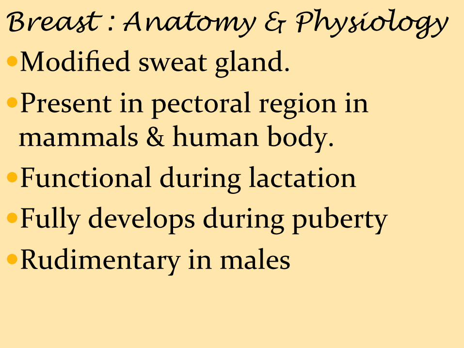
BREAST ABSCESS Def: A painful collection of pus that forms in the breast. Aetiopathogenesis: Infected by bacteria (commonly staphylococcus aureus) Bacteria enter through a crack in the breast / on nipple Mastitis Invades the fatty tissue of breast Swelling or pressure on the milk duct Causes abscess filled with pus
Breast abscess

Common causes are • Skin of the nipples can crack. Usually a result of the baby’s gums pinching the nipple • Staphylococcus aureus usually get into the breast through cracks or abrasions in the nipple. • Greater Risk -‐ Females with Diabetes, or who have under gone Nipple piercing or Lumpectomies or breast irradiation, or are using corticosteroids, or have silicone breast implants or are smokers
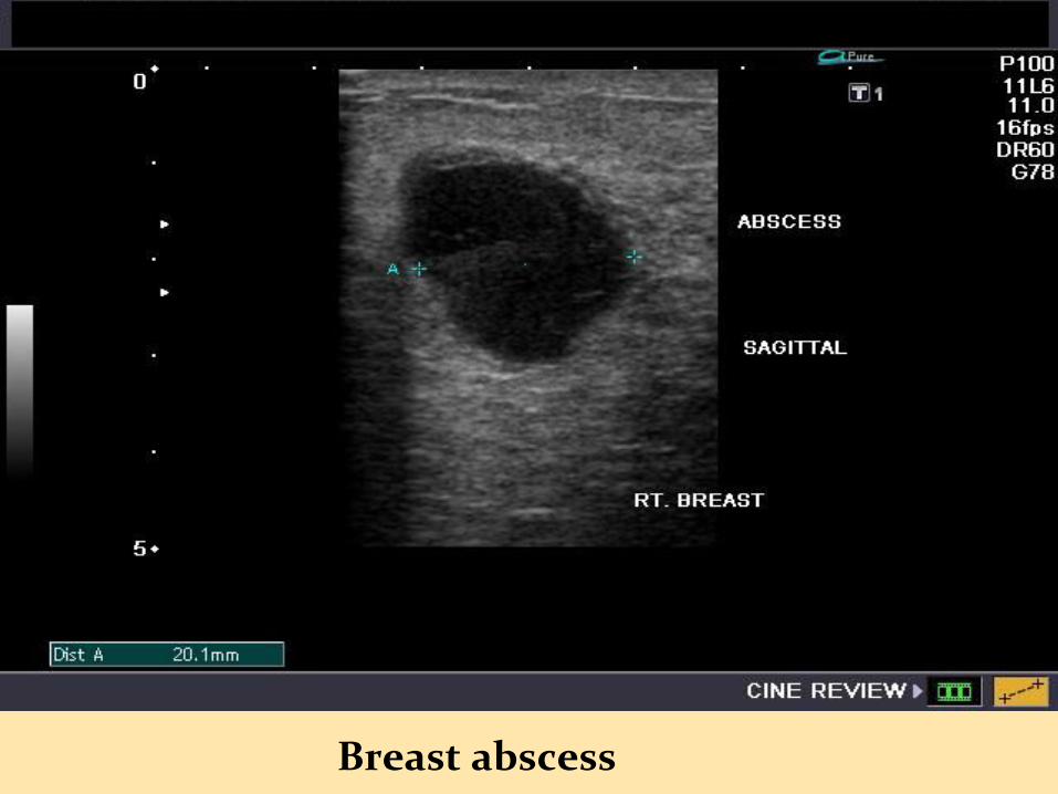
Sign and symptoms • Breast pain • Breast Lump, • Breast enlargement on one side only, • Swelling, tenderness, redness, and heat in breast tissue. • Nipple discharge (may contain pus), • Nipple sensation changes, • Itching, • Tender and/or enlarged lymph nodes in armpit on the same side, • Fever.

Breast abscess -‐ Appearance

Diagnosis In women who are not breast-‐feeding, testing may include: Mammography It is an X-‐ray picture of the Breasts, it is used to detect tumors and cysts, and to identify benign (non-‐cancerous) and malignant (cancerous) growths.

Complications • Fistula • Gangrene • Cellulitis • Mastitis • Spread of infection leads to sepsis • Extra mammary skin infection, etc

Prevention 1. Continue Breast feed. Infected milk will not harm the baby. Bacteria in the infected breast milk are killed in baby’s Stomach. 2. If breastfeeding is too painful -‐use a breast pump to empty the milk from the infected breast. 3. Massaging the affected breast may also increase milk flow.

Fibroadenoma • Most common benign tumor of breast • Proliferation of stromal + glandular elements • Seen in first 3 decades of life • Do not change during the menstrual cycle and regress at menopause Symptoms Usually single & rarely several lumps that may be in 1 or both breasts Lumps are Easily moveable under the skin, Firm, Painless & have a Rubbery consistency with smooth, well-‐defined borders. Grow in size during pregnancy or if Pt on Hormone replacement therapy


Investigations: After a physical examination, one or both of the following tests are usually done: Ø Breast ultrasound Ø Mammogram Complications: Breast cancer

Breast cancer Ø Second-‐leading cause of Ca deaths in women Ø Can also occur in men Ø The cells divide more rapidly than healthy cells do and may metastasize through the breast, to lymph nodes or to other parts of the body
Types Ductal Ca - starts in the tubes (ducts) that move
milk from the breast to the nipple. Lobular Ca - starts in lobules that produce milk

Breast cancer

Sign And Symptoms: Ø Painless Lump or thickening in the breast Ø Spontaneous clear or bloody discharge from the nipple associated with a breast lump
Ø Retraction or indentation of the nipple Ø Change in the size or contours of the breast Ø Any flattening or indentation of the skin over the breast
Ø Redness or pitting of the skin over breast, like the skin of an orange

Screening & Diagnosis Ø Self breast examination beginning at age 20

Investigations Computer-aided detection (CAD) Digital mammography Magnetic resonance imaging (MRI) Breast ultrasound (ultrasonography)

MANAGEMENT Preventive – Carcinocin Therapeutic – Curative : In Predisposed state Growth – less than 2cm, movable, tender, No constitutional symptoms, Menses unaltered {Disease in functional state – lots of sensation, modalities and or concomitants: Calc carb, Lyco, Thyroidinum, etc}
Difficult to Cure – Growth bigger in size, firm to hard,
Fixed and with constitutional symptoms {Disease is still in the hold of the constitution and the Body is vital enough to revert the pathology. Graph, Phytolacca, Cistus can etc}

MANAGEMENT Incurable – Pathology Advanced, stony hard, Ulcerated
Fixed with metastasis with gross constitutional symptoms
{Disease has enveloped the entire constitution: Thuja, Iod, Conium, Carbo Anamalis, Asterius, etc}

RUBRICS For Breast Tumours: Tumors Mammae (1346) Nodule / Lumps (835, 838) Ulcer (882) Swelling (881) Distension (829) Induration / hardness (834) Discoloration Mammae (829) Schirrous / Epithelioma (824) Emaciation (829) Hypertrophy (835) Fistulous Opening (832) Nipple – aphthae (823), atrophy (824), Bleeding(824),
Cracks (828), Deformed (828), Discharge (829), Dryness (829), Excoriation (831), Ulcer (882)

RUBRICS For Metastasis: Ca Axilla: Nodes (824), Induration (835), Exostosis (832), Ca Sternum (824), Pain (845)