Embed Size (px)
Citation preview

Breast cancer pathology andmolecular biology
Tatjana Ivković-KapiclOncology Institute of Vojvodina
Department of Pathology

Conflict of Interest
• I have no financial relationships to disclose

Topics
• Pathological features of breast carcinoma• Standard prognostic and predictive factors
of invasive breast carcinoma• Molecular classification of breast
carcinoma• Molecular markers of invasive breast
carcinoma
Invasive breast carcinoma• BC is a heterogeneous disease • Tumours with similar morphology show variable
behaviour, outcome and response to therapy

Why do we need a classification?Aim 1: Diagnosis
Aim 2: Prognosis
Aim 3: Prediction
Prediction is difficult, especially about the futureNiels Bohr, 1885-1962

Summary of prognostic and predictivefactors for invasive breast cancer
Prognostic PredictivePatient age √√Nodal status √√√Tumor size √√√Lymphovascular invasion √√Histological grade √√ √√Histologic type √ √Steroid receptors √ √√√Her2/neu √ √√√
Eifel P, Axelson JA, Costa J, Crowley J, Curran WJJr, Deshler A, et al. National Institutes of HealthConsensus Development Conference Statement:adjuvant therapy for breast cancer, November 1–3,2000. J Natl Cancer Inst. 2001;93(13):979–89.

Nodal status
For women with equivalent lymph node status, tumor size was associated with
increased lethality, such that each milimeter of tumor diameter was associated with an
additional 1% chance of death
For women with tumors of equivalent size, lethality increased with increasing number of positive lymph nodes, such that
there was an extra 6% chance of death associated with each positive lymph node
The Effect of Tumor Size and Lymph Node Status on Breast Carcinoma Lethality
Cancer 2003;98:2133-43.

Nodal status

Lymph node involvement
• pN1MACROMETASTASISsize >2 mm
• pN1micMICROMETASTASISsize >0.2 mm and <2 mm >200 cells in one LN section
• pN0pN0(i-) pN0(i+) ISOLATED TUMOR CELLS (ITCs) single cells and clusters <0.2 mm, even in H/E-stained slides pN0(mol-) and pN0(mol+)
AJCC 2010

SEER micrometastasis study
Chen SL et al Ann Surg Oncol. 2007, 12:3378-84
209,720 patients (SEER) 1992-2003 pN0 pN1mi (0.3-2 mm) pN1 (>2 mm)
•N1mi significant at multivariate analysis (p<0.0001) vs N0 (HR1.35) vs N1 (HR 0.82)

Sentinel lymph node(SLN) biopsy
• 1st LN draining tumor bed 1st site of local mets • Pathologically negative SN have been shown to
predict negative axillary status with a 98% degree of accuracy
• Standard method in breast cancer patients cN0
Rao R, Euhus D, Mayo HG, Balch C. Axillary nodeinterventions in breast cancer: a systematic review.JAMA. 2013;310(13):1385–94.Thompson AM. New standards of care in the managementof the axilla. Curr Opin Oncol. 2012;24(6):605–11.Zarebczan Dull B, Neuman HB. Management of theaxilla. Surg Clin North Am. 2013;93(2):429–44.Noguchi M, Morioka E, Ohno Y, Noguchi M,Nakano Y, Kosaka T. The changing role of axillarylymph node dissection for breast cancer. BreastCancer. 2013;20(1):41–6.Giuliano AE, Hunt KK, Ballman KV, Beitsch PD,Whitworth PW, Blumencranz PW, et al. Axillary dissectionno axillary dissection in women with invasivebreast cancer and sentinel node metastasis: arandomized clinical trial. JAMA. 2011;305(6):569–75.
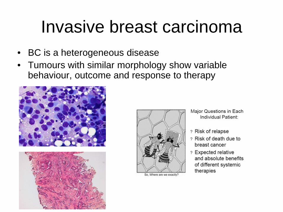
Tumor size
Multiple invasive carcinomassize of the largest is used for T-staging
Invasive carcinoma with surroundingintraductal carcinoma

Tumor grade
• Different grading systems • Nottingham combined histologic grade (the
Elston-Ellis modification of the Scarff-Bloom-Richardson grading system)
• Subjectivity• Adherence to strict criteria is necessary for
reproducibility so that grading can be used as a prognostic marker
Ellis IO, Galea M, Broughton N, Locker A, BlameyRW, Elston CW. Pathological prognostic factors inbreast cancer. II, Histological type. Relationshipwith survival in a large study with long-term followup.Histopathology. 1992;20:479–89.

a HPF high-power field
Breast cancer grade scoring Nottingham combined histologic grade (the Elston-Ellis modification of the Scarff-Bloom-Richardson grading system)
Elston CW and Ellis IO The Breast, Churchill Livingstone 1998

Histologic grade and survival
Elston CW and Ellis IO The Breast, Churchill Livingstone 1998

Histologic type
Hstologic appearance
Gross Features
• Invasive ductal carcinoma of no special type -75%

20 Histological types: morphology matters!
• The identification of special histologic types enables further refinement of the prediction of clinical outcome
Lakhani SR, Ellis IO, Schnitt SJ, Tan PH, vandeVijver MJ, editors. World Health Organization classification of tumors. Pathology and genetics oftumors of the breast and female genital organs. 4thed. Lyon: IARC Press; 2012.56. Ellis IO, Galea M, Broughton N, Locker A, BlameyRW, Elston CW. Pathological prognostic factors inbreast cancer. II, Histological type. Relationshipwith survival in a large study with long-term followup.Histopathology. 1992;20:479–89.
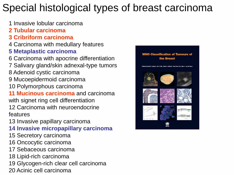
1 Invasive lobular carcinoma2 Tubular carcinoma3 Cribriform carcinoma4 Carcinoma with medullary features5 Metaplastic carcinoma6 Carcinoma with apocrine differentiation7 Salivary gland/skin adnexal-type tumors8 Adenoid cystic carcinoma9 Mucoepidermoid carcinoma10 Polymorphous carcinoma11 Mucinous carcinoma and carcinoma with signet ring cell differentiation12 Carcinoma with neuroendocrine features13 Invasive papillary carcinoma14 Invasive micropapillary carcinoma15 Secretory carcinoma16 Oncocytic carcinoma17 Sebaceous carcinoma18 Lipid-rich carcinoma19 Glycogen-rich clear cell carcinoma20 Acinic cell carcinoma
Special histological types of breast carcinoma

+Tubular carcinoma
Mucinous carcinoma Metaplastic carcinoma
Invasive lobular carcinoma
Strict diagnostic criteria must be used to
ensure the accuracy of diagnosis and, consequently,
the prediction of outcome
Invasive micropapillary carcinoma

Invasive lobularcarcinoma
• bilateral and multifocal• older patients• larger in size• positive for steroid receptors
and negative for Her2/neu• E-cadherin negative

Hormone Receptors• Weak prognostic factors• Predictive factors of the response to
hormonal therapy• Evaluation of ER and PR - a mandatory
component of the pathologic evaluation of breast carcinomas

Hormone Receptors• IHC evaluation - standard of practice • Most guidelines recommend reporting both the
proportion of positively stained nuclei and the intensity of nuclear staining

The interlaboratory variance in ER and PR data is as high as 30 %

• Clinical data indicate that ER positivity as low as 1 % can identify patients who would benefit from hormonal therapy
Harvey JM, Clark GM, Osborne CK, Allred DC.Predicting response to adjuvant endocrine therapy inbreast cancer. J Clin Oncol. 2000;17:1474–81.Rhodes A, Jasani B, Barnes DM, Bobrow LG, MillerKD. Reliability of immunohistochemical demonstrationof oestrogen receptors in routine practice:interlaboratory variance in the sensitivity of detectionand evaluation of scoring systems. J Clin Pathol.2000;53:125–30.Umemura S, Itoh J, Itoh H, Serizawa A, Saito Y,Suzuki Y, et al. Immunohistochemical evaluation ofhormone receptors in breast cancer: which scoringsystem is suitable for highly sensitive procedures?Appl Immunohistochem Mol Morphol. 2004;12:8–13.Leake R, Barnes D, Pinder S, Ellis I, Anderson L,Anderson T, et al. Immunohistochemical detection ofsteroid receptors in breast cancer: a working protocolon behalf of the UK Receptor Group, UK NEQAS,the Scottish Breast Cancer Pathology Group, and theReceptor and Biomarker Study Group of theEORTC. J Clin Pathol. 2000;53:634–5.
ER


Her2/Neu
• Positive in 15–25 %• Poor prognostic factor• Predictive factor of the response
to anti-HER2 therapy
• Her2 testing-IHC-ISH (FISH, CISH, SISH)

IHC scoring: semi-quantitative interpretation of HER2 expression
IHC 0 (negative)
IHC 2+ (equivocal)
IHC 1+ (negative)
IHC 3+ (positive)

HER2 ISH
Wolff AC, Hammond ME, Hicks DG, Dowsett M,McShane LM, Allison KH, et al. Recommendationsfor Human Epidermal Growth Factor Receptor 2Testing in Breast Cancer: American Society ofClinical Oncology/College of American PathologistsClinical Practice Guideline Update. J Clin Oncol.2013;31(31):3997–4013.
>6 gene copies per nucleus, or a ISH gene ratio (ratio of Her2/neu gene signals to
chromosome 17 signals) ≥2
<4 Her2/neu gene copies per nucleus, or a ISH gene ratio <2.0

Her2/neu testing
-All primary invasive breast cancers-All metastasis-All recurrences

Tumor proliferation: Ki67

• 17 of the 18 studies that included more than 200 patients showed statistically significant association between Ki67 and prognosis providing compelling evidence for a biological relationship
• but the cut-offs to distinguish “Ki67 high” from “Ki67 low” varied from 1% to 28.6%, thereby severely limiting its clinical utility
DowsettM et al; JNCI 2011

Ki-67• Limits of procedure– Quantification– Interpretation– Tumor heterogeneity– Tissue fixation
• Artefacts• Staining
– Reproducibility

Ki67 staining: intratumoral heterogeity
– Cut points arbitrary• Various cut points suggested• Still under debate• May vary depending on topic (prognostic or predictive)– For adjuvant treatment choice• Cut points from 5 - 34%• Most frequently 10 – 20%• St.Gallen 2013
– 20% (Panel decision)• Proliferation rates are a
continuum and are not bimodal
• Clinical Limits

St Gallen 2017“…when is traditional pathology (stage, grade, LVI, ER/PR/HER2) not informative enough?”
Traditionalclinicopathological
parameters
Prognosis of patients with
breast carcinoma
Biology?

Prognosis
• High risk: Chemotherapy• Low risk: No chemotherapy• However, clinically indeterminate groups
such as LN-/ER+/ HER2- tumours: Additional prognostic tests are needed(Multigene Prognostic Assays)

Microarray-based gene expression analysis
Perou et al In 2000>1700 genes
Each row is a geneEach column is a sampleGreen: <medianBlack: =medianRed: >medianRt panel: cell linesLeft panel: tissue
Dendrogram: similarities inthe expression patterns


Molecular Subtypes and Prognosis
Sorlie T et al, PNAS 2001

Clinicopathologic surrogate definition
• Luminal A-likeER+, HER2-, Ki67 low, PgR highLow-risk molecular signature (if available)
• Luminal B-likeHER2-negative:
ER+, HER2- and either Ki67 high or PgR lowHigh-risk molecular signature (if available)
HER2-positive:ER-positive, HER2-positive, any Ki67, any PgR
• HER2-positive (non-luminal):HER2+, ER and PgR absent
• Basal-like/Triple-negativeER and PgR absent, HER2-negative
Annals of Oncology 26: v8–v30, 2015

Basal like carcinomas
• Cluster genes characteristically expressed in normal breast basal/myoepithelial cells
• IHC: The basal type of tumors frequently does not express ER, PR, and HER2/neu but also expresses basal cytokeratins 5/6 and 17
• They tend to recur during the first 3 years after diagnosis, and currently there are no specific targeted therapies for them
• Strong association between basal-like carcinomas and BRCA1 mutations carriers

PATHOLOGIC FEATURES OF BASAL-LIKE TUMORS
• High-histologic grade, NOS (75%-100%)
Rakha EA 2006, Foulkes 2004, Kim MJ 2006
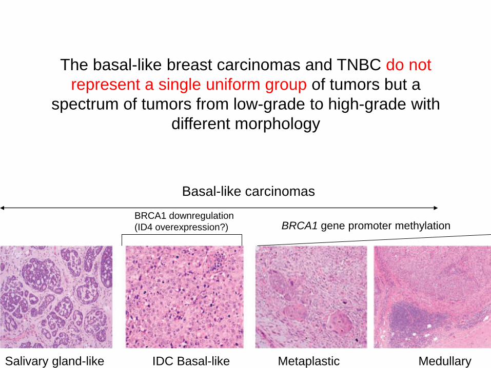
Basal-like carcinomas
Salivary gland-like IDC Basal-like Metaplastic Medullary
BRCA1 downregulation(ID4 overexpression?) BRCA1 gene promoter methylation
The basal-like breast carcinomas and TNBC do not represent a single uniform group of tumors but a
spectrum of tumors from low-grade to high-grade with different morphology

TN is not a synonym for basal-like phenotype!

• There is heterogeneity within the molecular subtypes: EVEN THE SUBTYPES HAVE SUBTYPES
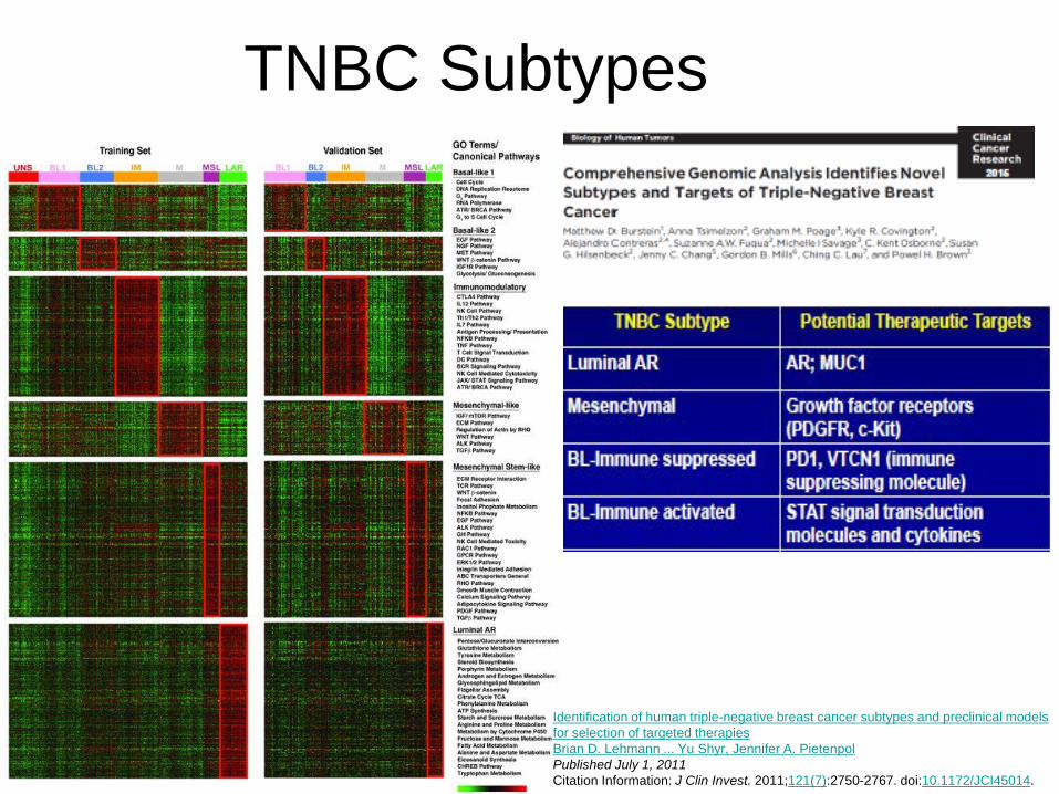
TNBC Subtypes
Identification of human triple-negative breast cancer subtypes and preclinical models for selection of targeted therapiesBrian D. Lehmann ... Yu Shyr, Jennifer A. PietenpolPublished July 1, 2011Citation Information: J Clin Invest. 2011;121(7):2750-2767. doi:10.1172/JCI45014.

Prognostic multigene signatures
• Microarray and RT-PCR based assays- 21 gene signature (Oncotype Dx)- 70 gene signature (MammaPrint)- 76 gene signature (Rotterdam)- 50 genes: Risk of Recurrence (ROR) score (Prosigna)- 12 genes (Endopredict) & Epclin- 5 genes (Molecular grade index)- 2 gene ratio (H/I™)- 97 gene: Genomic grade index (MapQuant Dx)- 14 genes (BreastOncPx)- 14 gene signature (Celera Metastasis Score™)

Multigene signatures• IHC and ISH based assays- 4 gene signature (IHC4; ER, PR, HER2 and Ki67)- 5 gene signature (Mammostrat)- 9 gene signature (Mammostrat Plus; 5 + ER, PR, HER2
and Ki67)- 5 gene signature (ProEx™ Br)- 3 gene signature (eXagenBC™ )
• Signatures based on a biological process- Wound-response signature (442 genes)- Immune signatures (14 genes)- Invasiveness Gene Signature (186 genes)

ASCO guideline recommendation
• In addition to ER, PR and HER2, there is sufficient evidence of clinical utility for the biomarker assays [Oncotype DX, EndoPredict, PAM50, Breast Cancer Index, and urokinase plasminogen activator and plasminogen activator inhibitor type 1 in HR+/HER2- Ln-. groups and can be used.
• These assays should not be used to guide treatmentdecision in LN+, HER2+ or triple negative cancer(No other molecular test (including ki67) should be usedto direct treatment decision)

Oncotype DX™ 21-Gene RecurrenceScore (RS) Assay
• Based on the expression levels of 21 genes, a recurrence score (RS) is generated

• The test is specifically applied to HR+ breast cancers with 0–3 positive nodes that are to be treated with hormonal therapy
• The general consensus is that hormonal therapy without systemic chemotherapy is sufficient for patients with a low RS.
Gnant M, Harbeck N, Thomssen C. St. Gallen 2011:summary of the consensus discussion. Breast Care(Basel). 2011;6:136–41.111. van’t Veer LJ, Dai H, van de Vijver MJ, He YD, HartAA, Mao M, et al. Gene expression profi ling predictsclinical outcome of breast cancer. Nature.2002;415(6871):530–6.

MammaPrint assay
• 70-gene expression assay developed by TheNetherlands Cancer Institute
• It is prognostic for early distant recurrence within the first 5 year after diagnosis and predictive for chemoresponse in poor prognostic patients

Prosigna test• PAM50-based assay offered by
NanoString Technologies (Seattle,WA)• Based on the expression levels of 50
genes and clinical variables, a risk of recurrence (ROR) score is generated that correlates to one of the four molecular subtypes (lum A, lum B, HER2-enriched,and basal-like)

Multigene Prognostic Tests: Unresolved Issues
Is this approach really better than using a combination of clinical and pathologic factors supplemented by appropriate
biomarkers detected by IHC (e.g., ER, PR, HER2 and Ki67)?
Molecular Testing in the Management of Patients with Breast Cancer Current Status and Future Directions. Stuart J. Schnitt, M.D., 2016.

Take Home Messages• The accurate diagnosis of breast cancer is a critical
prerequisite to the therapy decision-making process• Most of the prognostic factors currently used in clinical
practice are based on pathologic evaluation of the primary tumor and lymph nodes ( the LN status are more and more detroned)
• ER, PR, and HER2 testing using ASCO/CAP guidelines remain the most important ancillary tests in the management of patients with breast cancer

Take Home Messages• Among patients with ER+/HER2- (“luminal”) disease,
multigene prognostic tests are of value in further defining risk of recurrence and potential benefit from chemotherapy in addition to endocrine therapy
• Ki67 is not highly predictive for utilisation of adjuvant chemotherapy
• New technologies and genomewide approaches have the potential to identify additional prognostic and predictive markers for invasive breast cancer
• The role of the pathologist has changed from that of descriptive pathology of Virchow, to an important team player in the age of personalised medicine.


![Tissue-Type Plasminogen Activator-Mediated Activation of ... · TISSUE PLASMINOGEN ACTIVATOR IN STREPTOCOCCAL BINDING 197 sodium phosphate, 0.14 Msodium chloride [pH 7.4]) con- taining0.02%(wt/vol)](https://img.dokumen.tips/doc/110x75/5f46a6d9df5f79688c496b2a/tissue-type-plasminogen-activator-mediated-activation-of-tissue-plasminogen.jpg)